-RHYTHM PRACTICE- By Dr.moanes Msc.cardiology Assistant Lecturer of Cardiology Al Azhar University. OBHG Education Subcommittee
|
|
|
- Bridget Potter
- 6 years ago
- Views:
Transcription
1 -RHYTHM PRACTICE- By Dr.moanes Msc.cardiology Assistant Lecturer of Cardiology Al Azhar University

2 The Normal Conduction System
3 Sinus Node Normal Sinus Rhythm (NSR) Sinus Bradycardia Sinus Tachycardia Sinus Arrhythmia Sinus Arrest/Pause Sinoatrial Exit Block
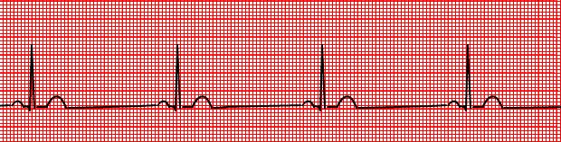
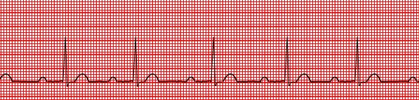
4 Normal Sinus Rhythm (NSR) Rate: beats/min Rhythm: regular P waves: uniform, + (upright) in lead II, one precedes each QRS complex PR interval: seconds and constant QRS duration: < 0.12 sec
5 Normal Sinus Rhythm (NSR)
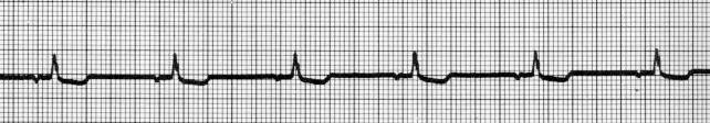
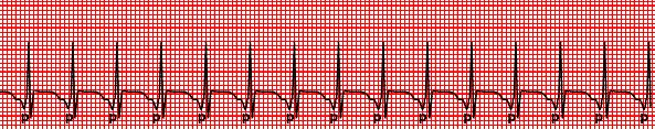
6 Sinus Bradycardia Rate: less than 60 beats/min Rhythm: regular P waves: uniform in appearance, upright and one precedes each QRS complex PR interval: sec and constant QRS duration: < 0.12 sec
7 Sinus Bradycardia
8 Sinus Tachycardia Rate: greater than beats/min Rhythm: regular P waves: uniform, upright, one precedes each QRS PR interval: sec and constant QRS duration: < 0.12 sec
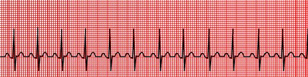
9 Sinus Tachycardia
10 Sinus Arrhythmia Rate: beats/min Rhythm: irregular P waves: uniform in appearance, upright, one precedes each QRS PR interval: sec and constant QRS duration: < 0.12 sec
11 Sinus Arrhythmia
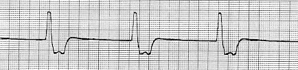
12 Sinus Arrest/Pause Rate: usually normal but varies due to the arrest Rhythm: irregular; the arrest is of undetermined length P waves: uniform in appearance, upright, one precedes each QRS PR interval: sec and constant QRS duration: < 0.12 sec
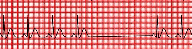
13 Sinus Arrest/Pause
14 Sinoatrial Exit Block Rate: varies due to the pause Rhythm: irregular; each pause is the same as the distance between two other P-P intervals P waves: uniform in appearance, upright, one precedes each QRS PR interval: sec and constant QRS duration: < 0.12 sec

15 Sinoatrial Exit Block
16 Atrial Dysrhythmias Premature Atrial Complexes (PACs) Wandering Atrial Pacemaker (WAP) Supraventricular Tachycardia (SVT) Wolff-Parkinson-White Syndrome (WPW) Atrial Flutter Atrial Fibrillation (A-Fib)
17 Premature Atrial Complexes (PACs) Rate: depends on underlying rhythm Rhythm: regular with premature beats P waves: premature (earlier than expected) and may differ in shape from sinus P waves PR interval: sec and constant QRS duration: < 0.12 sec Note: Ectopic complex
")
18 Premature Atrial Complexes (PACs)
19 Wandering Atrial Pacemaker (WAP) Rate: usually beats/min; if greater than 100 beats/min, it s termed multifocal atrial tachycardia Rhythm: may be normal or irregular due to shift in ectopic atrial locations P waves: size, shape and direction change (3 or more different P waves) PR interval: may be normal or variable QRS duration: < 0.12 sec

20 Wandering Atrial Pacemaker (WAP)
21 Supraventricular Tachycardia (SVT/PSVT) Rate: beats/min Rhythm: regular P waves: not present PR interval: not measurable QRS duration: < 0.12 sec Note: PSVT is an SVT that starts/ends suddenly
22 Supraventricular Tachycardia (SVT)
23 Wolff-Parkinson-White Syndrome (WPW) Rate: usually beats/min (can be associated with runs/episodes of A-Fib) Rhythm: regular P waves: uniform in appearance, upright, one precedes each QRS PR interval: sec and constant QRS duration: usually > 0.12 sec; slurred upstroke of the QRS complex (delta wave)
24 Wolff-Parkinson-White Syndrome (WPW)
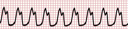
25 Atrial Flutter Rate: atrial rate beats/min; ventricular rate will not usually exceed 180 beats/min Rhythm: atrial regular; ventricular regular or irregular P waves: saw-toothed flutter waves PR interval: not measurable QRS duration: < 0.12 sec

26 Atrial Flutter
27 Atrial Fibrillation (A-fib) Rate: variable Rhythm: irregularly irregular P waves: no identifiable P waves PR interval: not measurable QRS duration: < 0.12 sec

28 Atrial Fibrillation (A-fib)
29 Junctional Rhythms Premature Junctional Complex (PJC) Junctional Rhythm Accelerated Junctional Rhythm Junctional Tachycardia
30 Premature Junctional Complexes (PJC) Rate: usually normal but depends on the underlying rhythm Rhythm: regular with premature beats P waves: may occur before, during or after the QRS (inverted if visible) PR interval: if P visible, < or = to 0.12 sec QRS duration: < 0.12 sec Note: Ectopic complex

31 Premature Junctional Complexes (PJC)
32 Junctional Rhythm Rate: beats/min Rhythm: regular P waves: not present or may occur before, during or after the QRS (inverted if present) PR interval: not measurable, but if present usually < or = to 0.12 sec QRS duration: < 0.12 sec
33 Junctional Rhythm
34 Junctional Rhythm
35 Accelerated Junctional Rhythm Rate: beats/min Rhythm: regular P waves: not present or may occur before, during or after the QRS (inverted if present; may be notched or S waved) PR interval: not measurable, but if present usually < or = to 0.12 sec QRS duration: < 0.12 sec
36 Accelerated Junctional Rhythm
37 Junctional Tachycardia Rate: beats/min Rhythm: regular P waves: not present or may occur before, during or after the QRS (inverted if present) PR interval: not measurable, but if present usually < or = to 0.12 sec QRS duration: < 0.12 sec
38 Junctional Tachycardia
39 Ventricular Rhythms Premature Ventricular Complexes (PVC's) Idioventricular Rhythm Ventricular Tachycardia (VT) Torsades de Pointes (TdeP) Ventricular Fibrillation (VF) Asystole
40 Premature Ventricular Complexes (PVC's) Rate: depends on underlying rhythm Rhythm: regular with premature beats P waves: absent PR interval: none QRS duration: > 0.12 sec, wide and bizarre! Note: Ectopic complex
 Note Complexes not")
41 Premature Ventricular Complexes (PVC's) Note Complexes not Contractions
42 PVC s Uniformed/Multiformed Couplets/Salvos/Runs Bigeminy/Trigeminy/Quadrageminy
43 Uniformed PVC s
44 R on T Phenomena
45 Multiformed PVC s
46 PVC Couplets
47 PVC Salvos and Runs
48 Bigeminy PVC s
49 Trigeminy PVC s
50 Quadrageminy PVC s
51 Ventricular Escape Beats Rate: depends on underlying rhythm Rhythm: regular with late beats; occurs after the next expected sinus beat P waves: absent PR interval: none QRS duration: > 0.12 sec. wide and bizarre Note: Ectopic complex
52 Ventricular Escape Beats
53 Idioventricular Rhythm Rate: beats/min Rhythm: regular P waves: absent PR interval: none QRS duration: > 0.12 sec Note: Wide and Slow, (Oh No!) it s Idio
54 Idioventricular Rhythm
55 Ventricular Tachycardia (VT) Rate: beats/min Rhythm: regular P waves: absent PR interval: none QRS duration: > 0.12 sec. often difficult to differentiate between QRS and T wave Note: Monomorphic - same shape and amplitude

56 Ventricular Tachycardia (VT)
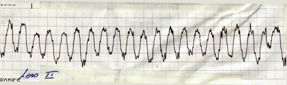
57 V Tach
58 Torsades de Pointes (TdeP) Rate: beats/min Rhythm: regular or irregular P waves: none PR interval: none QRS duration: > 0.12 sec. gradual alteration in amplitude and direction of the QRS complexes
59 Torsades de Pointes (TdeP)
60 Ventricular Fibrillation (VF) Rate: CNO as no discernible complexes Rhythm: rapid and chaotic P waves: none PR interval: none QRS duration: none Note: Fine vs. coarse?
61 Ventricular Fibrillation (VF)

62 Ventricular Fibrillation (VF)
63 Asystole (Cardiac Standstill) The Mother of all Bradycardias Rob Theriault Rate: none Rhythm: none P waves: none PR interval: not measurable QRS duration: absent

64 Asystole (Cardiac Standstill)

65 Asystole The Mother of all Bradycardias
66 Atrioventricular (AV) Blocks First-degree AV block Second-degree AV block, type I (Wenckebach or Mobitz I) Second-degree AV block, type II (Mobitz II) Third-degree AV block (Complete AV Block)
67 First-degree AV block Rate: depends on underlying rhythm (usually regular) Rhythm: regular P waves: normal in size and shape PR interval: > 0.20 sec but constant QRS duration: < 0.12 sec
68 First-degree AV block
69 2 nd Degree AV block Type I (Wenckebach/Mobitz Type I) Rate: atrial rate > ventricular rate Rhythm: atrial reg.; ventricular irreg. P waves: normal in size and shape PR interval: lengthens with each cycle until a P appears with no QRS complex QRS duration: < 0.12 sec (Periodically dropped QRS complex!)

70 2 nd Degree AV block Type I (Wenckebach/Mobitz Type I)
71 2 nd Degree AV block Type II (Mobitz Type II) Rate: atrial rate > the ventricular rate Rhythm: atrial reg.; ventricular irreg. P waves: normal in size and shape PR interval: within normal limits or slightly prolonged but constant QRS duration: < 0.12 sec (Periodically dropped QRS complex!)
72 2 nd Degree AV block Type II (Mobitz Type II)
73 3 rd Degree AV block (Complete AV Block) Rate: atrial rate is > ventricular rate Rhythm: atrial reg.; ventricular reg. (P waves plot through) P waves: normal in size and shape PR interval: no relationship (independent) QRS duration: narrow or wide depending on location of pacemaker
74 3 rd Degree AV block (Complete AV Block)

75 3 rd Degree AV block
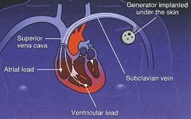
76 Cardiac Pacemakers Definition Delivers artificial stimulus to heart Causes depolarization and contraction Uses Bradyarrhythmias Asystole Tachyarrhythmias (overdrive pacing)
77 Cardiac Pacemakers Fixed Fires at constant rate Can discharge on T-wave Very rare Demand Senses patient s rhythm Fires only if no activity sensed after preset interval (escape interval)
78 Cardiac Pacemakers
79 Cardiac Pacemakers Demand Pacemaker Types Atrial Fires atria Atria fire ventricles Requires intact AV conduction Ventricular Fires ventricles
80 Cardiac Pacemakers Demand Pacemaker Types Atrial Synchronous Senses atria Fires ventricles AV Sequential Two electrodes Fires atria/ventricles in sequence
81 Cardiac Pacemakers Special Considerations Pacemaker does NOT affect treatment of cardiac arrest Do NOT defibrillate directly over pacemaker generator Pacemakers may keep AEDs from advising shock
82 Atrial Pacemaker (Single Chamber) pacemaker Capture?
")
83 Ventricular Pacemaker (Single Chamber) pacemaker
84 Dual Paced Rhythm pacemaker
85 Pulseless Electrical Activity (PEA) The absence of a detectable pulse and blood pressure Presence of electrical activity of the heart as evidenced by ECG rhythm, but not VF or VT + = 0/0 mmhg


86 ONTARIO BASE HOSPITAL GROUP
Basic Dysrhythmia Interpretation
 Basic Dysrhythmia Interpretation Objectives 2 To understand the Basic ECG To understand the meaning of Dysrhythmia To describe the normal heart conduction system. To describe the normal impulse pathways.
2017 BDKA Review. Regularity Rate P waves PRI QRS Interpretation. Regularity Rate P waves PRI QRS Interpretation 1/1/2017
 1. 2017 BDKA Review 2. 3. 4. Interpretation 5. QT 6. 7. 8. 9. 10. QT 11. 12. 13. 14. 15. 16. 17. 18. QT 19. 20. QT 21. 22. QT 23. 24. Where are pacer spikes? Before the P wave or before the QRS complex?
Arrhythmia Study Guide 3 Junctional and Ventricular Rhythms
 Arrhythmia Study Guide 3 Junctional and Ventricular Rhythms JUNCTIONAL RHYTHMS The AV Junction (Bundle of His and surrounding cells) only acts as pacemaker of the heart when the SA Node is not firing normally
Course Objectives. Proper Lead Placements. Review the ECG print paper. Review the mechanics of the Myocardium. Review basics of ECG Rhythms
 ECG Interpretations Course Objectives Proper Lead Placements Review the ECG print paper Review the mechanics of the Myocardium Review basics of ECG Rhythms How Leads Work The ECG Leads we use are Bipolar
Rate: The atrial and ventricular rates are equal; heart rate is greater than 100 bpm (usually between bpm).
.") Sinus Bradycardia Regularity: The R-R intervals are constant; the rhythm is regular. Rate: The atrial and ventricular rates are equal; heart rate is less than 60 bpm. P wave: There is a uniform P wave
Sinus Bradycardia Regularity: The R-R intervals are constant; the rhythm is regular. Rate: The atrial and ventricular rates are equal; heart rate is less than 60 bpm. P wave: There is a uniform P wave
ABCs of ECGs. Shelby L. Durler
 ABCs of ECGs Shelby L. Durler Objectives Review the A&P of the cardiac conduction system Placement and obtaining 4-lead and 12-lead ECGs Overview of the basics of ECG rhythm interpretation Intrinsic
CRC 431 ECG Basics. Bill Pruitt, MBA, RRT, CPFT, AE-C
 CRC 431 ECG Basics Bill Pruitt, MBA, RRT, CPFT, AE-C Resources White s 5 th ed. Ch 6 Electrocardiography Einthoven s Triangle Chest leads and limb leads Egan s 10 th ed. Ch 17 Interpreting the Electrocardiogram
CORONARY ARTERIES. LAD Anterior wall of the left vent Lateral wall of left vent Anterior 2/3 of interventricluar septum R & L bundle branches
 CORONARY ARTERIES RCA Right atrium Right ventricle SA node 55% AV node 90% Posterior wall of left ventricle in 90% Posterior third of interventricular septum 90% LAD Anterior wall of the left vent Lateral
PATIENT WITH ARRHYTHMIA IN DENTIST S OFFICE. Małgorzata Kurpesa, MD., PhD. Chair&Department of Cardiology
 PATIENT WITH ARRHYTHMIA IN DENTIST S OFFICE Małgorzata Kurpesa, MD., PhD. Chair&Department of Cardiology Medical University of Łódź The heart is made up of four chambers Left Atrium Right Atrium Left Ventricle
I have no conflicts of interest relative to this lecture.
 PATHWAYS AROUND THE PITFALLS OF EKG RHYTHM INTERPRETATION Donald D. Brown, MD April 4, 2019 I have no conflicts of interest relative to this lecture. OBJECTIVES FOR PATHWAYS AROUND PITFALLS OF EKG RHYTHM
Rhythm ECG Characteristics Example. Normal Sinus Rhythm (NSR)
") Normal Sinus Rhythm (NSR) Rate: 60-100 per minute Rhythm: R- R = P waves: Upright, similar P-R: 0.12-0.20 second & consistent P:qRs: 1P:1qRs Sinus Tachycardia Exercise Hypovolemia Medications Fever Substances
Normal Sinus Rhythm (NSR) Rate: 60-100 per minute Rhythm: R- R = P waves: Upright, similar P-R: 0.12-0.20 second & consistent P:qRs: 1P:1qRs Sinus Tachycardia Exercise Hypovolemia Medications Fever Substances
TEST BANK FOR ECGS MADE EASY 5TH EDITION BY AEHLERT
 Link download full: http://testbankair.com/download/test-bank-for-ecgs-made-easy-5thedition-by-aehlert/ TEST BANK FOR ECGS MADE EASY 5TH EDITION BY AEHLERT Chapter 5 TRUE/FALSE 1. The AV junction consists
EKG Intermediate Tips, tricks, tools
 Birmingham Regional Emergency Medical Services System 2018 ALCTE Summer Conference EKG Intermediate Tips, tricks, tools Brian Gober, MAT, ATC, NRP, CSCS Education Services Manager ECC Training Center Coordinator
Cardiac Arrhythmias in Sleep
 Cardiac Arrhythmias in Sleep Only 53 Slides! Gauresh H Kashyap, MD, FACP, FCCP, FAASM 1 2 Cardiac Arrhythmias in Sleep Out of 400 Patients with OSA, 48% had some Arrhythmias 20% - 2 PVCs/min 7% - Bradycardia
Basic EKG Interpretation. Nirja Parikh, PT, DPT
 Basic EKG Interpretation Nirja Parikh, PT, DPT Electrocardiogram (EKG) Using surface electrodes record the electrical activity of the heart electrical activity (usually) correlates to mechanical function
UNDERSTANDING YOUR ECG: A REVIEW
 UNDERSTANDING YOUR ECG: A REVIEW Health professionals use the electrocardiograph (ECG) rhythm strip to systematically analyse the cardiac rhythm. Before the systematic process of ECG analysis is described
Electrocardiography for Healthcare Professionals
 Electrocardiography for Healthcare Professionals Chapter 7: Junctional Dysrhythmias 2012 The Companies, Inc. All rights reserved. Learning Outcomes 7.1 Describe the various junctional dysrhythmias 7.2
8/20/2012. Learning Outcomes (Cont d)
") 1 2 3 4 Electrocardiography for Healthcare Professionals Chapter 7: Junctional Dysrhythmias Learning Outcomes 7.1 Describe the various junctional dysrhythmias 7.2 Identify premature junctional complexes
1 2 3 4 Electrocardiography for Healthcare Professionals Chapter 7: Junctional Dysrhythmias Learning Outcomes 7.1 Describe the various junctional dysrhythmias 7.2 Identify premature junctional complexes
COURSE DESCRIPTION. Rev 2.0 7/2013. Page 1 of 26
 COURSE DESCRIPTION Ventricular dysrhythmias (arrhythmias) are unique and potentially dangerous cardiac rhythms. They are often associated with Code Blue calls, and life and death situations. The only two
Cardiac Arrhythmia How to approach นพ.พ น จ แกวส วรรณะ หน วยโรคห วใจและหลอดเล อด
 Cardiac Arrhythmia How to approach นพ.พ น จ แกวส วรรณะ หน วยโรคห วใจและหลอดเล อด EKG paper is a grid where time is measured along the horizontal axis. Each small square is 1 mm in length and represents
EKG Rhythm Interpretation Exam
 as EKG Rhythm Interpretation Exam Name: Date: ID# Unit Assume each strip is a 6 second strip. Passing is 80%. 1. Identify the following rhythm: a. Asystole b. Ventricular fibrillation c. Atrial fibrillation
Dr.Binoy Skaria 13/07/15
 Dr.Binoy Skaria binoyskaria@hotmail.com binoy.skaria@heartofengland.nhs.uk 13/07/15 Acknowledgement Medtronic, Google images & Elsevier for slides Natalie Ryan, Events Manager, HEFT- for organising the
EKG Competency for Agency
 EKG Competency for Agency Name: Date: Agency: 1. The upper chambers of the heart are known as the: a. Atria b. Ventricles c. Mitral Valve d. Aortic Valve 2. The lower chambers of the heart are known as
ECG Interpretation. Introduction to Cardiac Telemetry. Michael Peters, RN, CCRN, CFRN CALSTAR Air Medical Services
 ECG Interpretation Introduction to Cardiac Telemetry Michael Peters, RN, CCRN, CFRN CALSTAR Air Medical Services Disclosures Nothing to disclose Objectives Describe the electrical conduction pathway in
Cardiology Flash Cards
 Cardiology Flash Cards EKG in a nut shell www.brain101.info Conduction System www.brain101.info 2 Analyzing EKG Step by step Steps in Analyzing ECG'S 1. Rhythm: - Regular _ Sinus, Junctional or Ventricular.
ACLS Study Guide for Precourse Self-Assessment
 20 rhythm strips on Precourse Self-Assessment with the following matching choices: Agonal Rhythm/Asystole Atrial Fibrillation Flutter Ventricular Fibrillation Monomorphic Ventricular Normal Sinus Rhythm
BEDSIDE ECG INTERPRETATION
 BEDSIDE ECG INTERPRETATION Presented by: Ryan Dean, RN, MSN, CCRN, CCNS, CFRN Flight Nurse 2017 Based on presentations originally by Gennifer DePaoli, RN Objectives Hospital policies Electrical conduction
Electrocardiography for Healthcare Professionals
 Electrocardiography for Healthcare Professionals Chapter 9: Ventricular Dysrhythmias 2012 The Companies, Inc. All rights reserved. Learning Outcomes 9.1 Describe the various ventricular dysrhythmias 9.2
2) Heart Arrhythmias 2 - Dr. Abdullah Sharif
 Heart Arrhythmias 2 - Dr. Abdullah Sharif") 2) Heart Arrhythmias 2 - Dr. Abdullah Sharif Rhythms from the Sinus Node Sinus Tachycardia: HR > 100 b/m Causes: o Withdrawal of vagal tone & Sympathetic stimulation (exercise, fight or flight) o Fever
2) Heart Arrhythmias 2 - Dr. Abdullah Sharif Rhythms from the Sinus Node Sinus Tachycardia: HR > 100 b/m Causes: o Withdrawal of vagal tone & Sympathetic stimulation (exercise, fight or flight) o Fever
ECG Interpretation Made Easy
 ECG Interpretation Made Easy Dr. A Tageldien Abdellah, MSc MD EBSC Lecturer of Cardiology- Hull University Hull York Medical School 2007-2008 ECG Interpretation Made Easy Synopsis Benefits Objectives Process
Please check your answers with correct statements in answer pages after the ECG cases.
 ECG Cases ECG Case 1 Springer International Publishing AG, part of Springer Nature 2018 S. Okutucu, A. Oto, Interpreting ECGs in Clinical Practice, In Clinical Practice, https://doi.org/10.1007/978-3-319-90557-0
Review Packet EKG Competency This packet is a review of the information you will need to know for the proctored EKG competency test.
 Review Packet EKG Competency 2015 This packet is a review of the information you will need to know for the proctored EKG competency test. Normal Sinus Rhythm Rhythm: Regular Ventricular Rate: 60-100 bpm
EKG Abnormalities. Adapted from:
 EKG Abnormalities Adapted from: http://www.bem.fi/book/19/19.htm Some key terms: Arrhythmia-an abnormal rhythm or sequence of events in the EKG Flutter-rapid depolarizations (and therefore contractions)
4/14/15 HTEC 91. Topics for Today. Guess That Rhythm. Premature Ventricular Contractions (PVCs) Ventricular Rhythms
 Ventricular Rhythms") 4/14/15 Topics for Today HTEC 91 Medical Office Diagnostic Tests Week 5 Ventricular Rhythms PVCs: Premature Ventricular Contractions VT: Ventricular Tachycardia VF: Ventricular Fibrillation Asystole Study
4/14/15 Topics for Today HTEC 91 Medical Office Diagnostic Tests Week 5 Ventricular Rhythms PVCs: Premature Ventricular Contractions VT: Ventricular Tachycardia VF: Ventricular Fibrillation Asystole Study
Cardiac Arrhythmias & Drugs used in Advanced Life Support and Cardiac Emergencies
 Cardiac Arrhythmias & Drugs used in Advanced Life Support and Cardiac Emergencies CNHE Ballarat Health Services Valid from 1 st March 2016 to 31 st June 2018 1 Supraventricular Tachycardia (SVT) An atrial
Step by step approach to EKG rhythm interpretation:
 Sinus Rhythms Normal sinus arrhythmia Small, slow variation of the R-R interval i.e. variation of the normal sinus heart rate with respiration, etc. Sinus Tachycardia Defined as sinus rhythm with a rate
a lecture series by SWESEMJR
 Arrhythmias Automaticity- the ability to depolarize spontaneously. Pacemakers: Sinoatrial node: 70 bpm AV-nodal area: 40 bpm His-Purkinje: 20-40 bpm it Mechanisms of arrhythmias 1. Increased automaticity
Paramedic Rounds. Tachyarrhythmia's. Sean Sutton Dallas Wood
 Paramedic Rounds Tachyarrhythmia's Sean Sutton Dallas Wood Objectives At the end of this session, the paramedic will be able to: State the key components of the cardiac conduction pathway, along with the
ECG Interpretation and Clinical Significance
 Confirming Pages ECG Interpretation and Clinical Significance Chapter Outline 5.1 5.2 5.3 5.4 5.5 5.6 5.7 5.8 5.9 5.10 Learning Outcomes Key Terms Introduction (p. 114) Identifying the Components of the
PACIFIC MEDICAL TRAINING Arrhythmia Interpretation
 PACIFIC MEDICAL TRAINING Arrhythmia Interpretation Introduction Activity Summary Target Audience Educational Objectives Nursing Educational Objective Faculty Physician Continuing Medical Education Nursing
Arrhythmic Complications of MI. Teferi Mitiku, MD Assistant Clinical Professor of Medicine University of California Irvine
 Arrhythmic Complications of MI Teferi Mitiku, MD Assistant Clinical Professor of Medicine University of California Irvine Objectives Brief overview -Pathophysiology of Arrhythmia ECG review of typical
How To Think About Rhythms and Conduction
 How To Think About Rhythms and Conduction Frank Yanowitz, MD Professor of Medicine (Cardiology) University of Utah School of Medicine Medical Director, IHC ECG Services Intermountain Healthcare http://ecg.utah.edu
Chapter 28, Part 1 Cardiology. Cardiac Physiology. Cardiovascular Anatomy
 Chapter 28, Part 1 Cardiology Part 1: Cardiovascular Anatomy & Physiology, ECG Monitoring, and Dysrhythmia Analysis 1 2 Cardiovascular Anatomy Coronary Circulation Cardiac Physiology The cardiac cycle
Basic ECG Interpretation Module Notebook
 Basic ECG Interpretation Module Notebook ECG_Notebook_04.27.05 Page 1 of 142 Basic ECG Interpretation Table of Contents Module Objectives... 3 Module Outline... 6 Lesson I... 6 Lesson II... 8 Lesson III...
ECG Success Exercises in ECG Interpretation
 ECG Success Exercises in ECG Interpretation Shirley A. Jones, MS Ed, MHA, EMT-P Emergency Medical Services Educator Arrhythmia Instructor Riverview Hospital Noblesville, Indiana Basic Life Support Instructor
Arrhythmia Management Joshua M. Cooper, MD, FHRS, FACC
 Arrhythmia Management Joshua M. Cooper, MD, FHRS, FACC Professor of Medicine Director of Cardiac Electrophysiology Temple University Health System Plumbing Electrical System Bradyarrhythmias Sinus Node
Electrocardiography for Healthcare Professionals
 Electrocardiography for Healthcare Professionals Kathryn A. Booth Thomas O Brien Chapter 5: Rhythm Strip Interpretation and Sinus Rhythms Learning Outcomes 5.1 Explain the process of evaluating ECG tracings
SIMPLY ECGs. Dr William Dooley
 SIMPLY ECGs Dr William Dooley Content Basic ECG interpretation pattern Some common (examined) abnormalities Presenting ECGs in context Setting up an ECG Setting up an ECG 1 V1-4 th Right intercostal space
CORONARY ARTERIES HEART
 CARDIAC/ECG MODULE THE HEART CORONARY ARTERIES FIBRILLATING HEART CORONARY ARTERIES HEART PRACTICE RHYTHMS PRACTICE RHYTHMS ELECTRICAL CONDUCTION SA Node (60 100) Primary pacemaker AV Node (40 60) ***Creates
HR: 50 bpm (Sinus) PR: 280 ms QRS: 120 ms QT: 490 ms Axis: -70. Sinus bradycardia with one ventricular escape (*)
 PR: 280 ms QRS: 120 ms QT: 490 ms Axis: -70. Sinus bradycardia with one ventricular escape (*)") 1? HR: 50 bpm (Sinus) PR: 280 ms QRS: 120 ms QT: 490 ms Axis: -70 1 Sinus P waves? 2 sinus cycles The pause (2 sinus cycles) suggests that the sinus fired (?) but did not conduct to the atria (i.e., missing
1? HR: 50 bpm (Sinus) PR: 280 ms QRS: 120 ms QT: 490 ms Axis: -70 1 Sinus P waves? 2 sinus cycles The pause (2 sinus cycles) suggests that the sinus fired (?) but did not conduct to the atria (i.e., missing
Anesthesia Assistants Review Course
 American Association of Oral and Maxillofacial Surgeons Anesthesia Assistants Review Course Four Seasons Las Vegas February 24-25, 2018 Las Vegas, Nevada Anesthesia Assistants Review Course EKG Lecture
3. AV Block 1. First-degree AV block 1. Delay in AV node 2. Long PR interval 3. QRS complex follows each P wave 4. Benign, no tx
 1. Rhythms & arrhythmias SA nodal rhythms Sinus rhythm Sinus tachycardia Sinus bradycardia Sinus arrhythmia Sick sinus syndrome SA block Sinus arrest AV blocks First-degree Second-degree Mobitz Type I
5AB Dysrhythmia Interpretation tation and Management Review Please complete and return by:
 1 5AB Dysrhythmia Interpretation tation and Management Review 2014 RN Please complete and return by: Overview 2 This review begins with a discussion of the physiology of cardiac conduction, and then covers
Paroxysmal Supraventricular Tachycardia PSVT.
 Atrial Tachycardia; is the name for an arrhythmia caused by a disorder of the impulse generation in the atrium or the AV node. An area in the atrium sends out rapid signals, which are faster than those
ECG Interpretation Cat Williams, DVM DACVIM (Cardiology)
") ECG Interpretation Cat Williams, DVM DACVIM (Cardiology) Providing the best quality care and service for the patient, the client, and the referring veterinarian. GOAL: Reduce Anxiety about ECGs Back to
ECG Interpretation Cat Williams, DVM DACVIM (Cardiology) Providing the best quality care and service for the patient, the client, and the referring veterinarian. GOAL: Reduce Anxiety about ECGs Back to
Emergency Medical Training Services Emergency Medical Technician Paramedic Program Outlines Outline Topic: WPW Revised: 11/2013
 Emergency Medical Training Services Emergency Medical Technician Paramedic Program Outlines Outline Topic: WPW Revised: 11/2013 Wolff-Parkinson-White syndrome (WPW) is a syndrome of pre-excitation of the
Chapter 16: Arrhythmias and Conduction Disturbances
 Complete the following. Chapter 16: Arrhythmias and Conduction Disturbances 1. Cardiac arrhythmias result from abnormal impulse, abnormal impulse, or both mechanisms together. 2. is the ability of certain
Module 1: Introduction to ECG & Normal ECG
 Module 1: Introduction to ECG & Normal ECG Importance of Correct anatomical positions Measurements & Morphologies ONLY accurate if Precise anatomical positions adhered to Standardised techniques are used
Conduction disorders
 Conduction disorders L.V. Bogun, N.I. Yabluchansky, F.M. Abdueva, O.Y. Bichkova, A.N. Fomich, P.A. Garkavyi, A.L. Kulik, N.V. Lysenko, N.V. Makienko, L.A. Martimyanova, I.V. Soldatenko, E.E. Tomina Department
a lecture series by SWESEMJR
 Electrolyte disturbances Hypokalaemia Decreased extracellular potassium increases excitability in the myocardial cells and consequently the effect of very severe hypokalaemia is ventricular arrhythmia.
Junctional Premature Contraction (JPC)
") Where s the PAC? Junctional Premature Contraction (JPC) A junctional premature contraction (JPC) is a beat that originates prematurely in the AV node. It can occur sporadically or in a grouped pattern.
Where s the PAC? Junctional Premature Contraction (JPC) A junctional premature contraction (JPC) is a beat that originates prematurely in the AV node. It can occur sporadically or in a grouped pattern.
EHRA Accreditation Exam - Sample MCQs Cardiac Pacing and ICDs
 EHRA Accreditation Exam - Sample MCQs Cardiac Pacing and ICDs Dear EHRA Member, Dear Colleague, As you know, the EHRA Accreditation Process is becoming increasingly recognised as an important step for
Answer: The electrical current has difficulty traveling down the normal conduction pathway.
 1 Electrocardiography for Healthcare Professionals Chapter 8: Heart Block Dysrhythmias 2 3 4 5 6 Learning Outcomes 8.1 Describe the various heartblock dysrhythmias 8.2 Identify first degree heartblock
Supraventricular Arrhythmias. Reading Assignment. Chapter 5 (p17-30)
") Supraventricular Arrhythmias Reading Assignment Chapter 5 (p17-30) The Supraventricular Rhythms In Our Lives Site of Origin Single Events Slow Rates Intermediate Rates Fast Rates (>100 bpm) Sinus Sinus
Supraventricular Arrhythmias Reading Assignment Chapter 5 (p17-30) The Supraventricular Rhythms In Our Lives Site of Origin Single Events Slow Rates Intermediate Rates Fast Rates (>100 bpm) Sinus Sinus
FLB s What Are Those Funny-Looking Beats?
 FLB s What Are Those Funny-Looking Beats? Reading Assignment (pages 27-45 in Outline ) The 5-Step Method ECG #: Mearurements: Rhythm (s): Conduction: Waveform: Interpretation: A= V= PR= QRS= QT= Axis=
Lake EMS Basic EKG Review: Atrial Rhythms. The Lake EMS Quality Development Team
 Lake EMS Basic EKG Review: Atrial Rhythms The Lake EMS Quality Development Team This program is the Intellectual Property of Lake Emergency Medical Services Use of this program is limited to training and
HTEC 91. Performing ECGs: Procedure. Normal Sinus Rhythm (NSR) Topic for Today: Sinus Rhythms. Characteristics of NSR. Conduction Pathway
 Topic for Today: Sinus Rhythms. Characteristics of NSR. Conduction Pathway") HTEC 91 Medical Office Diagnostic Tests Week 3 Performing ECGs: Procedure o ECG protocol: you may NOT do ECG if you have not signed up! If you are signed up and the room is occupied with people who did
HTEC 91 Medical Office Diagnostic Tests Week 3 Performing ECGs: Procedure o ECG protocol: you may NOT do ECG if you have not signed up! If you are signed up and the room is occupied with people who did
CARDIAC ARRHYTHMIAS IN NEONATE
 Introduction: CARDIAC ARRHYTHMIAS IN NEONATE DR. PANKAJ SAKHUJA Neonatologist Average heart rate in a healthy newborn is 120-160 beats/min and the range is from 80-230 beats/min. A normal (sinus) rhythm
12/2/2016. Basic Cardiac Anatomy. Blood Flow Through the Heart. Fun Fact.. Layers of the Heart. Layers of the Heart
 Basic Cardiac Anatomy Blood Flow Through the Heart Septum 1. Blood enters right atrium via inferior & superior vena cava 2. Right atrium contracts, sending blood through the tricuspid valve and into the
Chapter 9. Learning Objectives. Learning Objectives 9/11/2012. Cardiac Arrhythmias. Define electrical therapy
 Chapter 9 Cardiac Arrhythmias Learning Objectives Define electrical therapy Explain why electrical therapy is preferred initial therapy over drug administration for cardiac arrest and some arrhythmias
ECG ABNORMALITIES D R. T AM A R A AL Q U D AH
 ECG ABNORMALITIES D R. T AM A R A AL Q U D AH When we interpret an ECG we compare it instantaneously with the normal ECG and normal variants stored in our memory; these memories are stored visually in
Electrocardiography for Healthcare Professionals
 Electrocardiography for Healthcare Professionals Kathryn A. Booth Thomas O Brien Chapter 10: Pacemaker Rhythms and Bundle Branch Block Learning Outcomes 10.1 Describe the various pacemaker rhythms. 10.2
SIMPLY ECGs. Dr William Dooley
 SIMPLY ECGs Dr William Dooley 1 No anatomy just interpretation 2 Setting up an ECG 3 Setting up an ECG 1 V1-4 th Right intercostal space at sternal border 2 V2-4 th Left intercostal space at sternal border
ECG Interactive Session
 SA HEART 2018 PRE-CONGRESS WORKSHOP 4 OCTOBER 2018 ECG Interactive Session Ashley Chin Cardiologist/Electrophysiologist University of Cape Town Groote Schuur Hospital ECG 1 Which ONE of the following is
Pathologic ECG. Adelina Vlad, MD PhD
 Pathologic ECG Adelina Vlad, MD PhD Basic Interpretation of the ECG 1) Evaluate calibration 2) Calculate rate 3) Determine rhythm 4) Determine QRS axis 5) Measure intervals 6) Analyze the morphology and
Miscellaneous Stuff Keep reading the Outline
 Miscellaneous Stuff Keep reading the Outline Welcome to the 5-Step Method ECG #: Mearurements: Rhythm (s): Conduction: Waveform: Interpretation: A= V= PR= QRS= QT= Axis= 1. Compute the 5 basic measurements:
PEDIATRIC CARDIAC RHYTHM DISTURBANCES. -Jason Haag, CCEMT-P
 PEDIATRIC CARDIAC RHYTHM DISTURBANCES -Jason Haag, CCEMT-P General: CARDIAC RHYTHM DISTURBANCES - More often the result and not the cause of acute cardiovascular emergencies - Typically the end result
INDEX. Kingston General Hospital May Introduction Page 2. Cardiac Conduction System Page 3. Introduction to Cardiac Monitoring Page 5
 Kingston General Hospital May 2008 INDEX Introduction Page 2 Cardiac Conduction System Page 3 Introduction to Cardiac Monitoring Page 5 Recording Electrical Events Page 9 Waves of the ECG Complex Page
1 Cardiology Acute Care Day 22 April 2013 Arrhythmia Tutorial Course Material
 1 Cardiology Acute Care Day 22 April 2013 Arrhythmia Tutorial Course Material Arrhythmia recognition This tutorial builds on the ECG lecture and provides a framework for approaching any ECG to allow the
Electrocardiography Biomedical Engineering Kaj-Åge Henneberg
 Electrocardiography 31650 Biomedical Engineering Kaj-Åge Henneberg Electrocardiography Plan Function of cardiovascular system Electrical activation of the heart Recording the ECG Arrhythmia Heart Rate
Dr. Schroeder has no financial relationships to disclose
 Valerie A Schroeder MD MS Assistant Professor University of Kansas Medical Center READING THE WAVES- THE HEART S ELECTRICAL MESSAGE FINANCIAL DISCLOSURE Dr. Schroeder has no financial relationships to
Lab Activity 24 EKG. Portland Community College BI 232
 Lab Activity 24 EKG Reference: Dubin, Dale. Rapid Interpretation of EKG s. 6 th edition. Tampa: Cover Publishing Company, 2000. Portland Community College BI 232 Graph Paper 1 second equals 25 little boxes
CSI Skills Lab #5: Arrhythmia Interpretation and Treatment
 CSI 202 - Skills Lab #5: Arrhythmia Interpretation and Treatment Origins of the ACLS Approach: CSI 202 - Skills Lab 5 Notes ACLS training originated in Nebraska in the early 1970 s. Its purpose was to
REtrive. REpeat. RElearn Design by. Test-Enhanced Learning based ECG practice E-book
 Test-Enhanced Learning Test-Enhanced Learning Test-Enhanced Learning Test-Enhanced Learning based ECG practice E-book REtrive REpeat RElearn Design by S I T T I N U N T H A N G J U I P E E R I Y A W A
Chad Morsch B.S., ACSM CEP
 What Is Cardiac Stress Testing? Chad Morsch B.S., ACSM CEP A Cardiac Stress Test is a test used to measure the heart's ability to respond to external stress in a controlled clinical environment. Cardiac
The Electrocardiogram
 The Electrocardiogram Chapters 11 and 13 AUTUMN WEDAN AND NATASHA MCDOUGAL The Normal Electrocardiogram P-wave Generated when the atria depolarizes QRS-Complex Ventricles depolarizing before a contraction
Diploma in Electrocardiography
 The Society for Cardiological Science and Technology Diploma in Electrocardiography The Society makes this award to candidates who can demonstrate the ability to accurately record a resting 12-lead electrocardiogram
Understanding the 12-lead ECG, part II
 Bundle-branch blocks Understanding the 12-lead ECG, part II Most common electrocardiogram (ECG) abnormality Appears as a wider than normal S complex Occurs when one of the two bundle branches can t conduct
ECGs and Arrhythmias: Family Medicine Board Review 2009
 Rate Rhythm Intervals Hypertrophy ECGs and Arrhythmias: Family Medicine Board Review 2009 Axis Jess (Fogler) Waldura, MD University of California, San Francisco walduraj@nccc.ucsf.edu Ischemia Overview
Bradydysrhythmias and Atrioventricular Conduction Blocks
 Emerg Med Clin N Am 24 (2006) 1 9 Bradydysrhythmias and Atrioventricular Conduction Blocks Jacob W. Ufberg, MD*, Jennifer S. Clark, MD Department of Emergency Medicine, Temple University School of Medicine,
Appendix D Output Code and Interpretation of Analysis
 Appendix D Output Code and Interpretation of Analysis 8 Arrhythmia Code No. Description 8002 Marked rhythm irregularity 8110 Sinus rhythm 8102 Sinus arrhythmia 8108 Marked sinus arrhythmia 8120 Sinus tachycardia
Cardiac Telemetry Self Study: Part One Cardiovascular Review 2017 THINGS TO REMEMBER
 Please review the above anatomy of the heart. THINGS TO REMEMBER There are 3 electrolytes that affect cardiac function o Sodium, Potassium, and Calcium When any of these electrolytes are out of the normal
Reading Assignment (p1-91 in Outline ) Objectives What s in an ECG?
 Objectives What s in an ECG?") Reading Assignment (p1-91 in Outline ) Objectives What s in an ECG? The 5-Step Method ECG #: Mearurements: Rhythm (s): Conduction: Waveform: Interpretation: A= V= PR= QRS= QT= Axis= 1. Compute the 5 basic
Reading Assignment (p1-91 in Outline ) Objectives What s in an ECG? The 5-Step Method ECG #: Mearurements: Rhythm (s): Conduction: Waveform: Interpretation: A= V= PR= QRS= QT= Axis= 1. Compute the 5 basic
ELECTROCARDIOGRAPHY KEVIN REBECK PA-C. For more presentations
 ELECTROCARDIOGRAPHY KEVIN REBECK PA-C For more presentations www.medicalppt.blogspot.com Objectives ECG History Pathophysiology Basics Case Historys Electrical activation of the heart In the heart
Northwest Community Healthcare Paramedic Education Program AV Conduction Defects/AV Blocks Connie J. Mattera, M.S., R.N., EMT-P
 Northwest Community Healthcare Paramedic Education Program AV Conduction Defects/ Connie J. Mattera, M.S., R.N., EMT-P Reading assignments: Bledsoe Vol. 3: pp. 88-93; 120-121 (atropine, norepinephrine,
Puzzling Pacemakers Cheryl Herrmann, APN, CCRN, CCNS-CSC-CMC
 Puzzling Pacemakers Cheryl Herrmann, APN, CCRN, CCNS-CSC-CMC Pacemaker: An electric device implanted in the body to regulate the heart beat. Delivers electrical stimuli over leads with electrodes in contact
BASIC ECG REVIEW. Overview of Conduction, Review of Strip Reading, Commonly seen arrhythmias
 BASIC ECG REVIEW Overview of Conduction, Review of Strip Reading, Commonly seen arrhythmias OBJECTIVES Review: Anatomy of heart Conduction systems Identification of rhythms Arrhythmias ANATOMY OVERIEW
The ABCs of EKGs/ECGs for HCPs. Al Heuer, PhD, MBA, RRT, RPFT Professor, Rutgers School of Health Related Professions
 The ABCs of EKGs/ECGs for HCPs Al Heuer, PhD, MBA, RRT, RPFT Professor, Rutgers School of Health Related Professions Learning Objectives Review the basic anatomy of the heart Describe the cardiac conducting
physiology 6 Mohammed Jaafer Turquoise team
 15 physiology 6 Mohammed Jaafer 22-3-2016 Turquoise team Cardiac Arrhythmias and Their Electrocardiographic Interpretation Today, we are going to talk about the abnormal excitation. As we said before,
UNDERSTANDING ELECTROPHYSIOLOGY STUDIES
 UNDERSTANDING ELECTROPHYSIOLOGY STUDIES Testing and Treating Your Heart s Electrical System A Problem with Your Heart Rhythm The speed and pattern of a heartbeat is called the heart rhythm. The rhythm