MUSCULOSKELETAL INFECTIONS IN CHILDREN. Dr Caren Landes Alder Hey Children s NHS Foundation Trust Liverpool
|
|
|
- Miranda Garrett
- 6 years ago
- Views:
Transcription
1 MUSCULOSKELETAL INFECTIONS IN CHILDREN Dr Caren Landes Alder Hey Children s NHS Foundation Trust Liverpool

2 MUSCULOSKELETAL INFECTIONS Common and uncommon infections Common and uncommon presentations Imaging modalities Problems and pitfalls
3 COMMON INFECTIONS Osteomyelitis Septic Arthritis UNCOMMON INFECTIONS Discitis Pyomyositis SAPHO/CRMO
4 RISK FACTORS Age: younger > older Prematurity Umbilical vessel catheterisation Urinary Tract Infection Immunodeficiency Comorbidity
5 OSTEOMYELITIS Most common musculoskeletal infection Incidence is approximately I in 5000 Boys > Girls 30% Staph aureus 50% occur < 5 years of age. 70% involve lower extremities.
6 MAKING THE DIAGNOSIS Xray Ultrasound Nuclear Medicine CT MRI Intervention

7 XRAY Plain radiography is not sensitive to the presence of osteomyelitis Bone destruction may be seen. 80% have a normal xray in the first 2 weeks. At Presentation


8 REPEAT XRAYS At 1 week At 1 week At 1 month At 2 months

9 MRI Magnetic resonance Imaging is the modality of choice. Oedema is the earliest feature on MRI T2W STIR Gadolinium?
10 TO ENHANCE OR NOT TO ENHANCE? THAT IS THE QUESTION Controversial Gadolinium enhancement does not increase the sensitivity or specificity of osteomyelitis. If fluid sensitive images are normal gadolinium enhancement is of no value. If fluid sensitive images are abnormal gadolinium enhancement may be of value in identifying the presence of an abscess. NB If the abscess is confined to the epiphyseal cartilage the unenhanced scans may be normal.

11 SEPTIC ARTHRITIS 75% before age 3 years 75% in the lower extremity Hip and Knee in 90% 75% Staph aureus Neonates: Group B Strep < 4yrs of age: HIB, Strep pyogenes, Staph aureus > 4yrs of age: Staph aureus
12 MAKING THE DIAGNOSIS Xray Ultrasound MRI Intervention
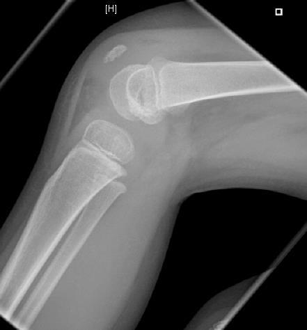
13 XRAY
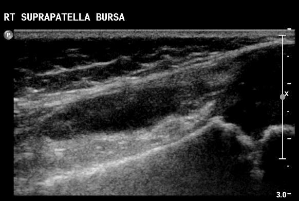
14 ULTRASOUND
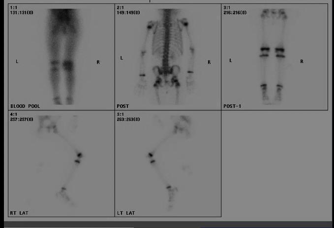
15 BONE SCAN
16
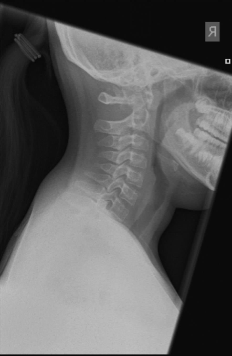
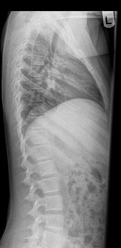
17 DISCITIS 2 peaks: 6 months 4 years Lumbar spine L3/4 and L2/ years Thoracic spine Staph aureus Atypical infections Salmonella. TB.
18 MAKING THE DIAGNOSIS Xray Nuclear Medicine MRI Intervention
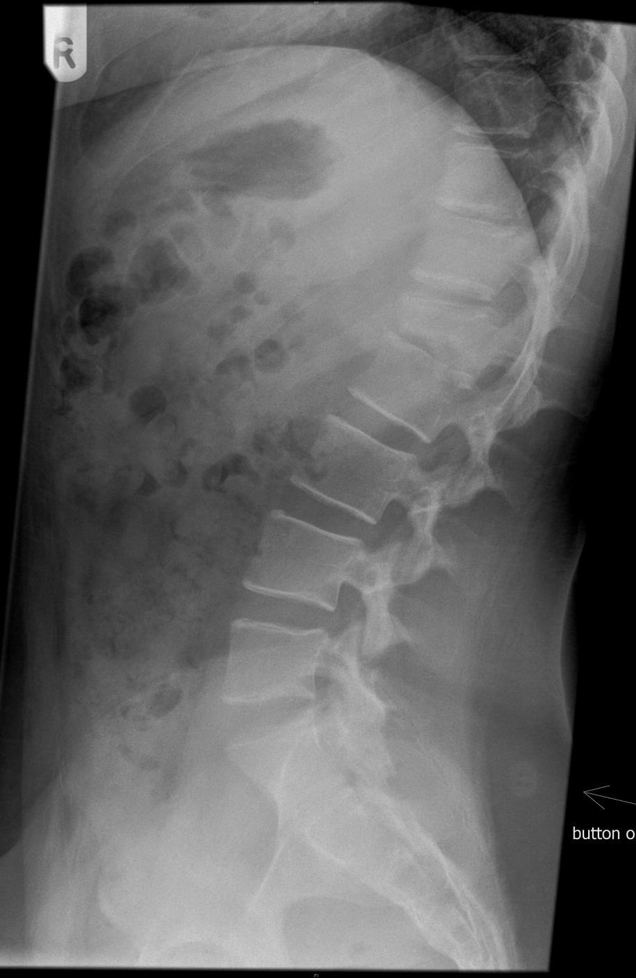
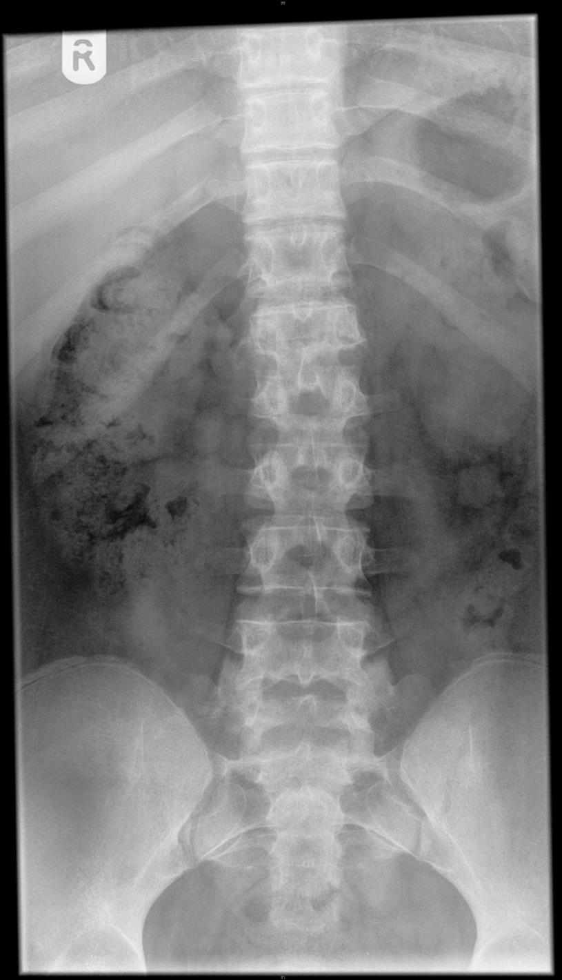
19 XRAY
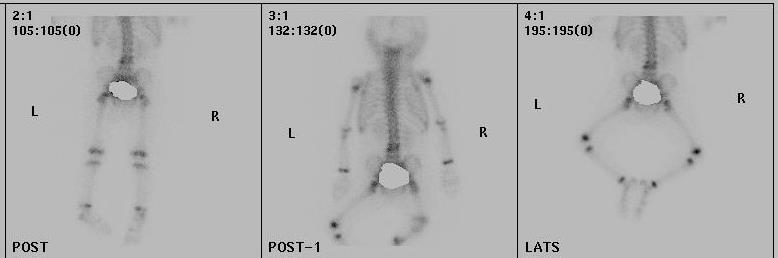
20 BONE SCAN
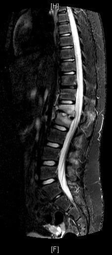
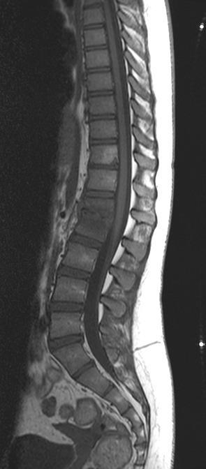
21 MRI
22 PYOMYOSITIS 5 9 years Male > Female Proximal Lower Limb and Pelvis 90% Staph aureus Previous trauma reported in 25 35% patients Iliopsoas infection associated with GI/GU/Spinal infection.
23 MAKING THE DIAGNOSIS Xray Ultrasound MRI CT Intervention
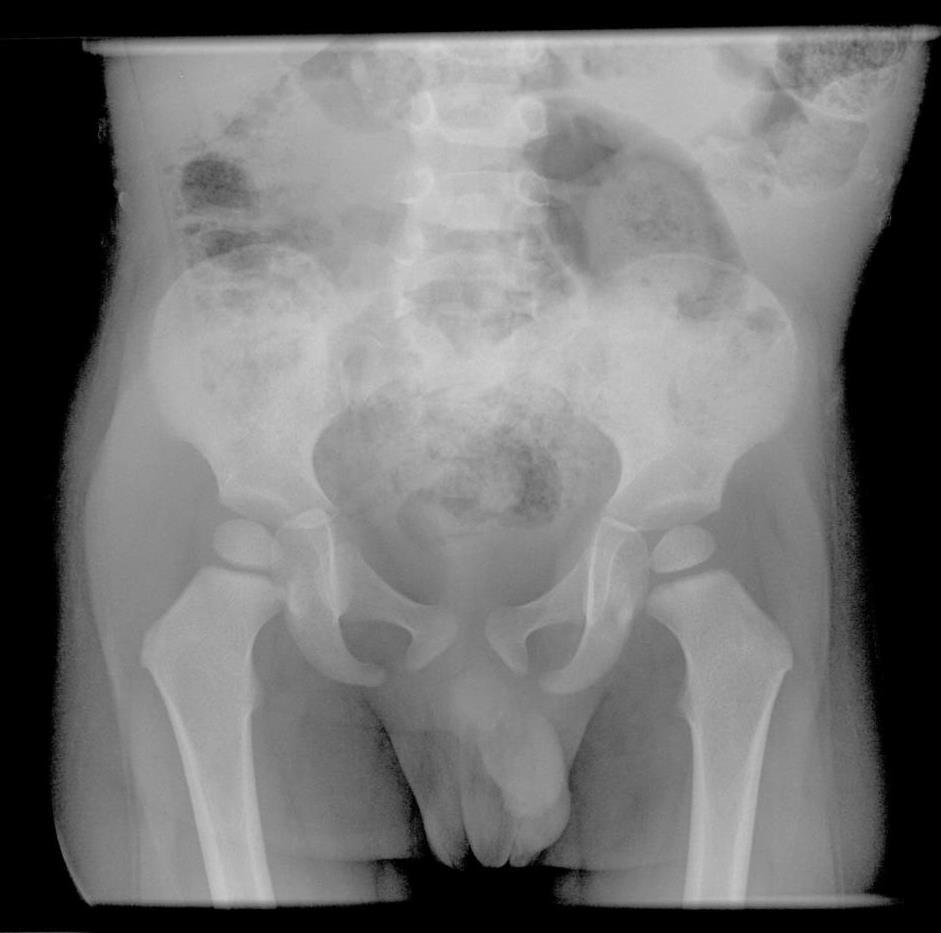
24 XRAY
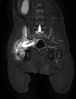
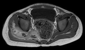
25 MRI
26 C.R.M.O. Multifocal non-pyogenic inflammatory bone lesions Most common between 9 and 14 years of age. Female > Male Unknown pathogenesis Genetic susceptibility focus identified at 18q Associated with skin disorders and inflammatory bowel disease Most common in lower extremities The tibia is the most common bone The metaphysis is most common site Chronic Recurrent Multifocal Osteomyelitis
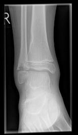
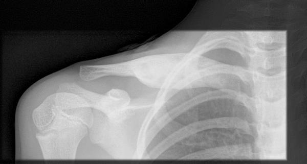
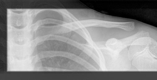
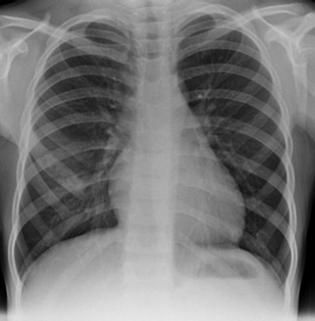
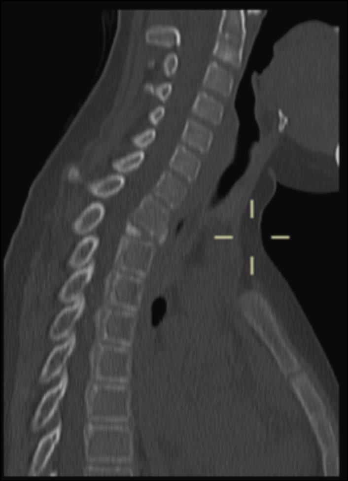
27 S.A.P.H.O. Most common site is upper anterior chest wall 70-90% involve sternocostoclavicular region No known pathogenesis Usually seen in young adults, but can be seen in children Male = Female Synovitis Acne Palmoplantar pustulosis Hyperostosis Osteitis
28 MAKING THE DIAGNOSIS Xray Nuclear Medicine MRI Intervention
29
30

31 XRAY AND BONE SCAN
32
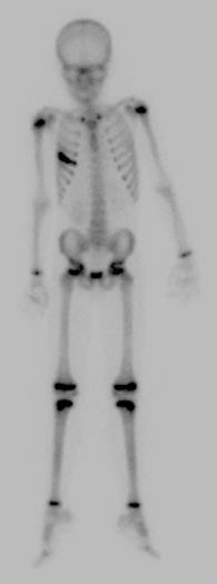
33 BONE SCAN
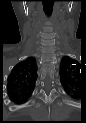
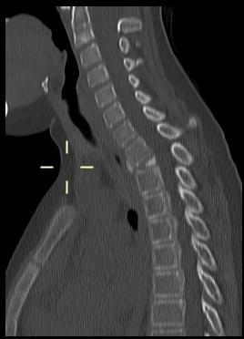
34 CT
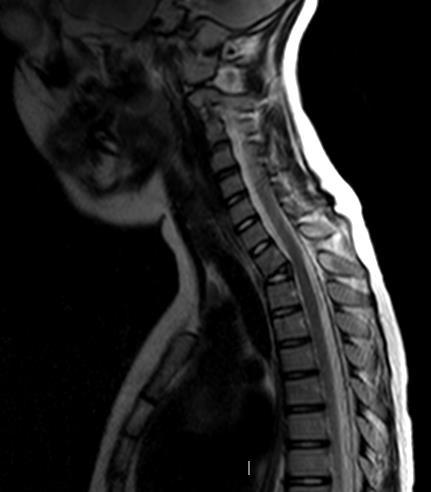
35 AND MRI
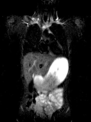
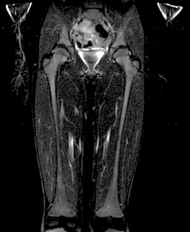
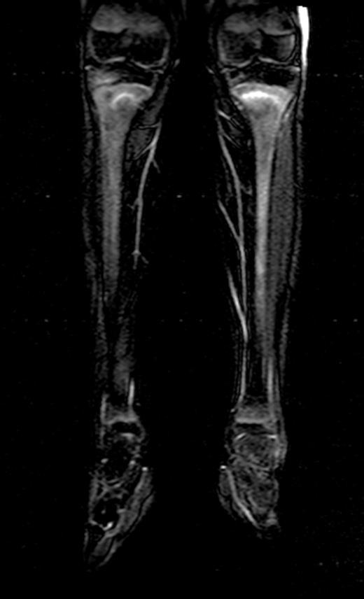
36 MRI Increasing use Identify area(s) of pathology Whole body STIR
37
38 CRMO. OR NOT CRMO? Isolated lesion Absence of other clinical features or comorbidity Rare locations Spine Mandible If in doubt. Biopsy
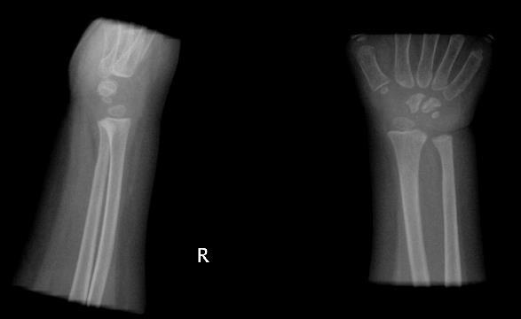
39 PROBLEMS AND PITFALLS The growing skeleton. What you can t see. The differential diagnoses: Septic Arthritis.v. Inflammatory Arthritis Osteomyelitis.v. Osteolysis C.R.M.O.v. S.A.P.H.O.
40 UNOSSIFIED PATELLA

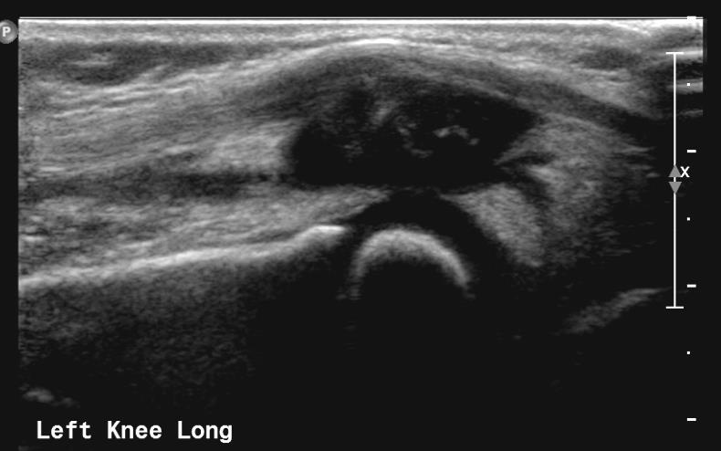
41 SOFT TISSUE COLLECTION
42
43 A SALUTARY TALE
44
45
46
47
48
49
50
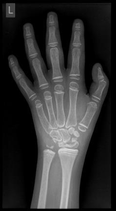
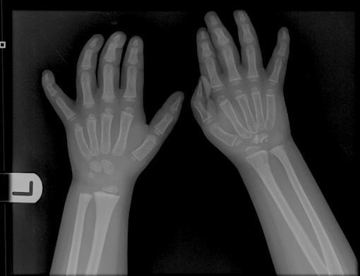
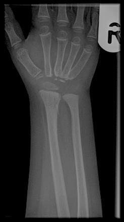
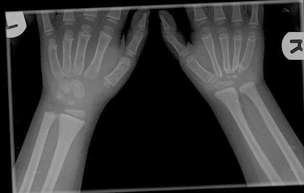
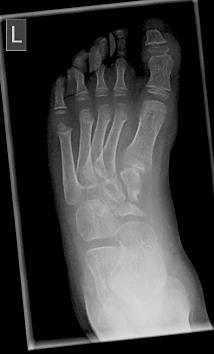
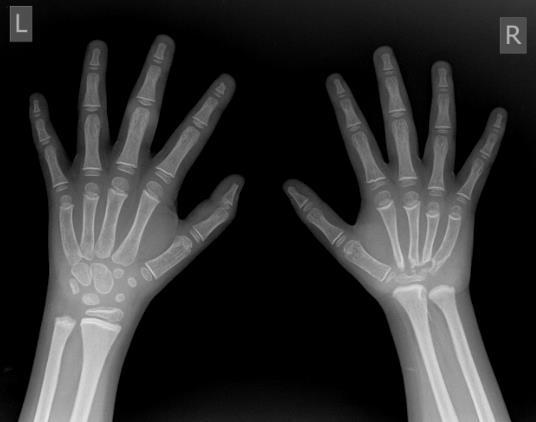
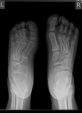
51 Carpotarsal osteolysis
52 SUMMARY Consider musculoskeletal infection in any child with PUO Plain film and ultrasound as first line investigations Increasing use of MRI Multidisciplinary Team Approach Tertiary centre support Differential Diagnoses

53 THANK YOU Any questions?
Osteomyelitis in infancy and childhood: A clinical and diagnostic overview M. Mearadji
 Osteomyelitis in infancy and childhood: A clinical and diagnostic overview M. Mearadji International Foundation for Pediatric Imaging Aid Introduction Osteomyelitis is a relative common disease in infancy
Chronic non-bacterial Osteomyelitis/Osteitis (or CRMO)
") www.printo.it/pediatric-rheumatology/gb/intro Chronic non-bacterial Osteomyelitis/Osteitis (or CRMO) Version of 2016 1. WHAT IS CRMO 1.1 What is it? Chronic Recurrent Multifocal Osteomyelitis (CRMO) is
www.printo.it/pediatric-rheumatology/gb/intro Chronic non-bacterial Osteomyelitis/Osteitis (or CRMO) Version of 2016 1. WHAT IS CRMO 1.1 What is it? Chronic Recurrent Multifocal Osteomyelitis (CRMO) is
Acute Osteomyelitis: similar to septic arthritis but up to 40% may be afebrile swelling overlying the bone & tenderness
 Osteomyelitis / Bone and Joint Infections Bone infections in children are usually from haematogenous bacterial seeding to a single joint, usually the lower limbs, but may be multifocal. Approximately 10%
Spondylarthropathies. Outline. Introduction. Spondylarthropathy other than AS. Mimickers of spondylarthropathy. Conclusions.
 Spondylarthropathies Filip M. Vanhoenacker Johan Van Goethem General Hospital St-Maarten Duffel-Mechelen Universities of Antwerp and Ghent Outline Introduction Spondylarthropathy other than AS Mimickers
Osteomieliti STEOMIE
 OsteomielitiSTEOMIE Osteomyelitis is the inflammation of bone caused by pyogenic organisms. Major sources of infection: - haematogenous spread - tracking from adjacent foci of infection - direct inoculation
Complex Fractures and Hip Dislocations
 IMAGING OF HIP PAIN Patients may present with acute (< 2 weeks) or chronic hip pain. Acute pain may be related or not related to an acute traumatic event such as fall or trauma from a motor vehicle accident.
OSTEOMYELITIS. If it occurs in adults, then the axial skeleton is the usual site.
 OSTEOMYELITIS Introduction Osteomyelitis is an acute or chronic inflammatory process of the bone and its structures secondary to infection with pyogenic organisms. Pathophysiology Osteomyelitis may be
ISPUB.COM. Spectrum Of MRI Findings In Musculoskeletal Tuberculosis: Pictoral Essay. P Chudgar INTRODUCTION SPINE
 ISPUB.COM The Internet Journal of Radiology Volume 8 Number 2 Spectrum Of MRI Findings In Musculoskeletal Tuberculosis: Pictoral Essay P Chudgar Citation P Chudgar.. The Internet Journal of Radiology.
Clinical cases and pitfalls using FDG-PET. François Jamar Université Catholique de Louvain, Brussels, Belgium
 Clinical cases and pitfalls using FDG-PET François Jamar Université Catholique de Louvain, Brussels, Belgium Case history (1) sepsis 60 yr old female Diabetes (1)- hypertension, alcohol Renal failure,
Case Report Sequential MR Images and Radiographs of Epiphyseal Osteomyelitis in the Distal Femur of an Infant
 Case Reports in Radiology Volume 2013, Article ID 672815, 4 pages http://dx.doi.org/10.1155/2013/672815 Case Report Sequential MR Images and Radiographs of Epiphyseal Osteomyelitis in the Distal Femur
Screening for and Assessment of Osteonecrosis in Oncology Patients. Sue C. Kaste, DO SPR Postgraduate Course 2015
 Screening for and Assessment of Osteonecrosis in Oncology Patients Sue C. Kaste, DO SPR Postgraduate Course 2015 The author declares no potential conflicts of interest or financial disclosures Osteonecrosis
Assesment by MRI in the diagnosing of osteomyelitis in children
 Assesment by MRI in the diagnosing of osteomyelitis in children Poster No.: C-1295 Congress: ECR 2011 Type: Educational Exhibit Authors: M. Teixidor Viñas, J. L. Ribó, J. muxart, J. Blanch, L. Riaza ;
Imaging Of The Pelvis
 Imaging Of The Pelvis 1 / 6 2 / 6 3 / 6 Imaging Of The Pelvis MRI of the pelvis may be more focused on the organs, soft tissues, and vessels, rather than on the bones themselves. In many instances, MRI
CLINICAL PRESENTATION AND RADIOLOGY QUIZ QUESTION
 Donald L. Renfrew, MD Radiology Associates of the Fox Valley, 333 N. Commercial Street, Suite 100, Neenah, WI 54956 11/24/2012 Radiology Quiz of the Week # 100 Page 1 CLINICAL PRESENTATION AND RADIOLOGY
Types of osteoarthritis
 ARTHRITIS Osteoarthritis is a degenerative joint disease is the most common joint disorder. It is a frequent part of aging and is an important cause of physical disability in persons older than 65 years
Spinal infection. Outline ANATOMY 6/2/2017. Anatomy Pathogen
 Outline Spinal infection Pramot Tanutit, M.D. Department of Radiology, Songklanagarind Hospital Faculty of Medicine, Prince of Songkla University Anatomy Pathogen Pyogenic spondylodiscitis Tuberculous
Case Report Nonbacterial Osteitis of the Clavicle: Longitudinal Imaging Series from Initial Diagnosis to Clinical Improvement
 Case Reports in Rheumatology Volume 2015, Article ID 182731, 4 pages http://dx.doi.org/10.1155/2015/182731 Case Report Nonbacterial Osteitis of the Clavicle: Longitudinal Imaging Series from Initial Diagnosis
Special Imaging MUSCULOSKELETAL INFECTION. Special Imaging. Special Imaging. 18yr old male pt What is it? Additional Imaging
 MUSCULOSKELETAL INFECTION Additional Imaging May assist in diagnosis and, possibly, treatment Help create the picture May help differentiate from neoplasia 18yr old male pt What is it? Lymphoma Ewings
Pediatric TB Intensive Houston, Texas October 14, 2013
 Pediatric TB Intensive Houston, Texas October 14, 2013 Radiologic Presentation of Childhood TB Susan D. John, MD, FACR October 14, 2013 Disclosures I have no disclosures or conflicts of interest to report
Types of bone/joint infections. Bone and Joint Infections. Septic Arthritis. Pathogenesis. Pathogenesis. Bacterial arthritis: predisposing factors
 Bone and Joint Infections Types of bone/joint infections Arthritis (infective/septic) Osteomyelitis Prosthetic bone and joint infections Septic Arthritis Common destructive athroplasty Mono-articular Poly-articular
Pediatric TB Intensive Houston, Texas
 Pediatric TB Intensive Houston, Texas November 13, 2009 Radiographic Manifestations of Pediatric TB Susan D. John, MD, FACR November 13, 2009 Radiologic Presentation of Childhood TB Susan D. John, MD,
A Patient s Guide to Limping in Children
 A Patient s Guide to Limping in Children 651 Old Country Road Plainview, NY 11803 Phone: 5166818822 Fax: 5166813332 p.lettieri@aol.com DISCLAIMER: The information in this booklet is compiled from a variety
ADI Procedure Codes. August 2016 Revised April 2017 Page 1 of 7 ADI Procedure Codes
 Code Description 70450 CT Head without contrast 70460 CT Head with contrast 70470 CT Head with & without contrast 70480 CT Orbit, et al without contrast 70481 CT Orbit, et al with contrast 70482 CT Orbit,
PSOAS ABSCESS. Dr Noman Ullah Wazir
 PSOAS ABSCESS Dr Noman Ullah Wazir Psoas Major muscle The psoas major is a long fusiform muscle located on the side of the lumbar region of the vertebral column and brim of the lesser pelvis. Psoas Major
Disseminated Primary Non-Hodgkin s Lymphoma of Bone : A Case Re p o r t 1
 Disseminated Primary Non-Hodgkin s Lymphoma of Bone : A Case Re p o r t 1 Hee-Jin Park, M.D., Sung-Moon Lee, M.D., Hee-Jung Lee, M.D., Jung-Sik Kim, M.D., Hong Kim, M.D. Primary lymphoma of bone is uncommon
Topics. Musculoskeletal Infection Extremities. Detection of Infection. Role of Imaging in Extremity Infection. Detection of Infection
 Topics Musculoskeletal Infection Extremities Nuttaya Pattamapaspong M.D. Department of Radiology, Faculty of Medicine, Chiang Mai University, Chiang Mai, Thailand Role of imaging in extremity infection
Extra-spinal musculoskeletal manifestations of mycobacterium tuberculosis (TB) infection: A review of radiological findings
 infection: A review of radiological findings") Extra-spinal musculoskeletal manifestations of mycobacterium tuberculosis (TB) infection: A review of radiological findings Poster No.: P-0118 Congress: ESSR 2013 Type: Scientific Exhibit Authors: I. Anwar,
Extra-spinal musculoskeletal manifestations of mycobacterium tuberculosis (TB) infection: A review of radiological findings Poster No.: P-0118 Congress: ESSR 2013 Type: Scientific Exhibit Authors: I. Anwar,
Spondyloarthritis: A Gouty Display
 Spondyloarthritis: A Gouty Display Preetam Gongidi 1*, Shawn Gough-Fibkins 2 1. Nova Southeastern University College of Osteopathic Medicine, Fort Lauderdale, FL, USA 2. Broward General Medical Center,
Radiology. General radiology department. X-ray
 The radiology directorate provides a diagnostic, interventional and therapeutic service for its local population, and a tertiary service for the region. It also provides support to some national work such
A 4 year old with hip pain: Legg-Calvé-Perthes Disease
 A 4 year old with hip pain: Legg-Calvé-Perthes Disease Cyndie Seraphin Harvard Medical School Year III Our Patient A 4 year-old boy is complaining of severe L hip pain. The differential diagnosis of acute
Assessment of limping child (beware the child who does not weight bear at all):
:") Department of Paediatrics Clinical Guideline Acutely Limping Child and Septic Arthritis Assessment of limping child (beware the child who does not weight bear at all): History Careful history of any significant
Department of Paediatrics Clinical Guideline Acutely Limping Child and Septic Arthritis Assessment of limping child (beware the child who does not weight bear at all): History Careful history of any significant
PITFALLS IN THE DIAGNOSIS OF SKELETAL TUBERCULOSIS IN CHILDREN
 PITFALLS IN THE DIAGNOSIS OF SKELETAL TUBERCULOSIS IN CHILDREN DR.JANANI SANKAR SENIOR CONSULTANT - PEDIATRICS DEPARTMENT OF PEDIATRICS & PEDIATRIC ORTHOPEDICS KANCHI KAMAKOTI CHILDS TRUST HOSPITAL CHENNAI,
GALEN ADVANCED Course
 GALEN ADVANCED Course Education in partnership Course information The course is aimed at senior residents, board-certified radiologists and fellows and is designed to advance the knowledge of an array
BONES & JOINTS INFECTION BONE TUMOURS
 BONES & JOINTS INFECTION BONE TUMOURS IMPORTANT SERIOUS CONSEQUENCE PLEASE DON T MISS!! EARLY DIAGNOSIS & PROPER TREATMENT HOW?? AWARE of THEIR EXISTENCE (Knowledge) PREPARE for THEIR OCCURRENCE A HIGH
Question 1 History. Likely Diagnosis Differential. Further Investigation or Management. Requires Paediatric Surgical referral for laparotomy
 Question 1 Male newborn spilling green tinged vomit day 1 of life Imaging Abdominal X-Rays performed on 03/05/2012 Upper and lower gastrointestinal contrast studies performed on 03/05/2012 Abdominal X-Rays
Pigmented Villonodular Synovitis PVNS
 February 2002 Pigmented Villonodular Synovitis PVNS Amy Gillis, Harvard Medical School Year III 47 year old female Our Patient Right hip pain since age 20 No history of trauma Diagnosed with DJD of R hip
CHAPTER 13 SKELETAL SYSTEM
 CHAPTER 13 SKELETAL SYSTEM Structure and Function Functions of the skeletal system Provides shape and support Protects internal organs Stores minerals and fat Produces blood cells and platelets Assists
Imaging and intervention of sacroiliac joint. Dr Ryan Lee Ka Lok Associate Consultant Prince of Wales Hospital
 Imaging and intervention of sacroiliac joint Dr Ryan Lee Ka Lok Associate Consultant Prince of Wales Hospital Introduction 15%-25% of low back pain is related to sacroiliac joint (SIJ) pain SIJ pain is
FOLLICULAR / OVULATION STUDY USG HIP JOINT (LEFT) USG HIP JOINT (RIGHT) USG KNEE JOINT (LEFT) USG KNEE JOINT (RIGHT) USG KUB USG MUSKULOSKELETAL USG
 USG HIP JOINT (RIGHT) USG KNEE JOINT (LEFT) USG KNEE JOINT (RIGHT) USG KUB USG MUSKULOSKELETAL USG") RADIOLOGY TESTS SONOGRAPHY,COLOR DOPPLER 3D/4D ANAMOLY SCAN 3D/4D ANAMOLY SCAN TWINS 3D/4D USG PELVIS ABDOMEN & PELVIS USG ABDOMEN UPPER USG ANKLE JOINT (LEFT) USG ANKLE JOINT (RIGHT) USG B SCAN BREAST
RADIOLOGY TESTS SONOGRAPHY,COLOR DOPPLER 3D/4D ANAMOLY SCAN 3D/4D ANAMOLY SCAN TWINS 3D/4D USG PELVIS ABDOMEN & PELVIS USG ABDOMEN UPPER USG ANKLE JOINT (LEFT) USG ANKLE JOINT (RIGHT) USG B SCAN BREAST
ESPID New Bone and Joint Infection Guidelines
 ESPID New Bone and Joint Infection Guidelines Theoklis Zaoutis, MD, MSCE Professor of Pediatrics and Epidemiology Perelman School of Medicine at the University of Pennsylvania Chief, Division of Infectious
Digital tomosynthesis (DT) has been well described as a
 has been well described as a") Case Report The Usefulness of Digital Tomosynthesis (DT) in Assisting in Cases of Doubtful Routine Radiography and/or Computed Tomography (CT) Image. Abstract Digital tomosynthesis is useful in assisting
Case Report The Usefulness of Digital Tomosynthesis (DT) in Assisting in Cases of Doubtful Routine Radiography and/or Computed Tomography (CT) Image. Abstract Digital tomosynthesis is useful in assisting
Bone PET/MRI : Diagnostic yield in bone metastases and malignant primitive bone tumors
 Bone PET/MRI : Diagnostic yield in bone metastases and malignant primitive bone tumors Lars Stegger, Benjamin Noto Department of Nuclear Medicine University Hospital Münster, Germany Content From PET to
COMPETENCY REQUIREMENTS for the CERTIFICATION EXAMINATION
 COMPETENCY REQUIREMENTS for the 10/2013 CERTIFICATION BOARD FOR RADIOLOGY PRACTITIONER ASSISTANTS CERTIFICATION EXAMINATION Note: The competency requirements contained in this document will be in effect
Course specification
 Al-Azhar University Faculty of Medicine for Men Course specification For Master of Radiodiagnosis ( 2014 2015 ) University : Al-Azhar Faculty : Medicine for men Course specification - Programmers on which
Image Interpretation and Evaluation
 Semester 2 / Commences January 20 Credits Each Course is composed of Modules & Activities. Modules: Image Interpretation Thoracic radiography Abdominal pelvic imaging Musculoskeletal Imaging Neuroimaging
Sacroiliac joint infection
 Case Report Brunei Int Med J. 2015; 11 (2): 110-114 Sacroiliac joint infection Jon CHUA 1, Kamal JAMIL 1, Kamalnizat IBRAHIM 1, Suraya AZIZ 2 1 Department of Orthopaedic and Traumatology, Faculty of Medicine,
Diagnostic Imaging Exams
 Guide for Chiropractors Diagnostic Imaging Exams CREATED FOR OUR CHIROPRACTIC PARTNERS This document has been prepared by the specialized, board-certified radiologists who interpret patient exams for Center
INFECTION & INFLAMMATION IMAGING
 INFECTION & INFLAMMATION IMAGING Radiopharmaceutical Drug Interactions & Other Interesting Case Studies MICHELLE RUNDIO, CNMT NCT MBA PCI NUCLEAR IN-111 WHITE BLOOD CELL IMAGING Interactions, Imaging Parameters
Spine MRI and Spine CT Test Request Tip Sheet
 Spine MRI and Spine CT With/Without Contrast CT, MRI Studies should NOT be ordered simultaneously as dual studies (i.e., with and without contrast). Radiation exposure is doubled and both views are rarely
Osteo-articular TB in Children
 Osteo-articular TB in Children H Simon Schaaf Desmond Tutu TB Centre, Department of Paediatrics and Child Health, Stellenbosch University, Cape Town, South Africa Questions Spinal TB is the most common
Skeletal changes in endocrine disorders
 Skeletal changes in endocrine disorders Poster No.: P-0100 Congress: ESSR 2015 Type: Authors: Educational Poster A. C. O'Brien 1, H. L. khosa 2, A. levai 2, N. Ramesh 2 ; 1 Dublin/IE, 2 Portlaoise/IE Keywords:
RADIOLOGY REQUEST MANUAL. (615)
") RADIOLOGY REQUEST MANUAL www.vanderbiltchildrens.com RADIOLOGY REQUEST MANUAL EXAM PROTOCOL QUESTIONS? Please call: DIAGNOSTIC RADIOLOGY (X-RAY) Pager (615) 835-1714 CT (615) 936-4920 MRI (615) 936-4933
RADIOLOGY REQUEST MANUAL www.vanderbiltchildrens.com RADIOLOGY REQUEST MANUAL EXAM PROTOCOL QUESTIONS? Please call: DIAGNOSTIC RADIOLOGY (X-RAY) Pager (615) 835-1714 CT (615) 936-4920 MRI (615) 936-4933
Primary care referral criteria for musculoskeletal MRI scans
 Appendix 1 Primary care referral criteria for musculoskeletal MRI scans Accepted Criteria for Direct Access MRI Body Part Symptoms Imaging indicated Lumbar Spine Low Back Pain with adverse symptoms or
RADIOLOGY - X-RAY - COMPUTERIZED AXIAL TOMMOGRAPHY - MAGNETIC RESONENCE IMAGING For the Time Period : 10/01/16 and 09/30/2017
 RADIOLOGY - X-RAY - COMPUTERIZED AXIAL TOMMOGRAPHY - MAGNETIC RESONENCE IMAGING For the Time Period : 10/01/16 and 09/30/2017 IF YOU ARE COVERED BY HEALTH INSURANCE, YOU ARE STRONGLY ENCOURAGED TO CONSULT
Accuracy of Point-of-Care Ultrasonography for Pediatric Ankle Sprain Injuries
 Accuracy of Point-of-Care Ultrasonography for Pediatric Ankle Sprain Injuries S Jones 1, K Colaco 2, J Fischer 2, J Stimec 2, C Kwan 2, K Boutis 2 1 Alder Hey Children s NHS Foundation Trust, Prescot Rd,
Recommendations for cross-sectional imaging in cancer management, Second edition
 www.rcr.ac.uk Recommendations for cross-sectional imaging in cancer management, Second edition Musculoskeletal tumours Faculty of Clinical Radiology www.rcr.ac.uk Contents Primary bone tumours 3 Clinical
MRI Non-Joint Extremity Questionnaire
 MRI n-joint Extremity Questionnaire INSTRUCTIONS FOR COMPLETING QUESTIONNAIRE: Answer all of the initial questions (Pages 1 and 2) Select the reason for imaging by answering question #6. Based on your
RADIOLOGY - X-RAY - COMPUTERIZED AXIAL TOMMOGRAPHY - MAGNETIC RESONENCE IMAGIN For the Time Period : 10/01/16 and 09/30/2017
 RADIOLOGY - X-RAY - COMPUTERIZED AXIAL TOMMOGRAPHY - MAGNETIC RESONENCE IMAGIN For the Time Period : 10/01/16 and 09/30/2017 IF YOU ARE COVERED BY HEALTH INSURANCE, YOU ARE STRONGLY ENCOURAGED TO CONSULT
Mr Simon Jennings BSc, MB BS, FRCS, Dip Sports Med FRCS (Trauma & Orthopaedics)
") Mr Simon Jennings BSc, MB BS, FRCS, Dip Sports Med FRCS (Trauma & Orthopaedics) Consultant Orthopaedic Surgeon Northwick Park Hospital 107 Harley Street RSM 16 th September 2010 Orthopaedic Surgeon Knee
Mr Simon Jennings BSc, MB BS, FRCS, Dip Sports Med FRCS (Trauma & Orthopaedics) Consultant Orthopaedic Surgeon Northwick Park Hospital 107 Harley Street RSM 16 th September 2010 Orthopaedic Surgeon Knee
Chronic non bacterial osteitis- a multicentre study
 Bhat et al. Pediatric Rheumatology (2018) 16:74 https://doi.org/10.1186/s12969-018-0290-5 RESEARCH ARTICLE Chronic non bacterial osteitis- a multicentre study Open Access Chandrika S. Bhat 1, Catriona
Spine MRI and Spine CT Test Request Tip Sheet
 Spine MRI and Spine CT MRI is almost always preferred over CT scan; if ordering CT, CLEARLY document why MRI is not appropriate. In cases of back pain without red flags, six weeks of multimodality supervised
Original Date: December 2015 Page 1 of 8 FOR CMS (MEDICARE) MEMBERS ONLY
 MEMBERS ONLY") National Imaging Associates, Inc. Clinical guidelines TOTAL JOINT ARTHROPLASTY -Total Hip Arthroplasty -Total Knee Arthroplasty -Replacement/Revision Hip or Knee Arthroplasty CPT4 Codes: Please refer to
National Imaging Associates, Inc. Clinical guidelines TOTAL JOINT ARTHROPLASTY -Total Hip Arthroplasty -Total Knee Arthroplasty -Replacement/Revision Hip or Knee Arthroplasty CPT4 Codes: Please refer to
Ligamentous Integrity in Spinal Cord Injury without Radiographic Abnormality. Dr Anria Horn Dr Stewart Dix-Peek
 Ligamentous Integrity in Spinal Cord Injury without Radiographic Abnormality Dr Anria Horn Dr Stewart Dix-Peek Introduction Spinal Cord Injury Without Radiographic Abnormality SCIWORA Pang, Wilberger 1982
Icd 10 code for ct pelvis with contrast
 Icd 10 code for ct pelvis with contrast November 16, 2009. How to Code for CT Angiography. By Anthony McCallum, CPC, CCS, CIRCC, CPC-I Radiology Today Vol. 10 No. 18 P. 12. CT. procedure code and description
Overview. Imaging Indications. Paediatric Radiation Safety 2015/03/12. Paediatric radiation safety General guidelines Protocols
 Overview Paediatric radiation safety General guidelines Protocols Paediatric Radiation Safety Paediatric patients are unique Children are more susceptible to radiation induced cancer than adults Younger
RADPrimer Curriculum Breast Topics Covered Basic Intermediate 225
 Breast Anatomy & Normal Variants 11 Breast Imaging Modalities 13 BI RADS Lexicon 3 Mammography: Masses 9 Mammography: Calcifications 17 Mammography: Additional Findings 8 Ultrasound Features 10 Ultrasound
MSK Interesting Case (17/8/2017) Dr Yap Sheau Huey
 Dr Yap Sheau Huey") MSK Interesting Case (17/8/2017) Dr Yap Sheau Huey 3 cases of clavicular swelling with different diagnoses. Case 1 WKW, 48 y.o lady Left mid clavicular and supraclavicular fossa pain 2 months. Rest and
MSK Interesting Case (17/8/2017) Dr Yap Sheau Huey 3 cases of clavicular swelling with different diagnoses. Case 1 WKW, 48 y.o lady Left mid clavicular and supraclavicular fossa pain 2 months. Rest and
Pyogenic spondylitis as a complication of ear piercing : Differentiating between spondylitis and discitis
 Acta Orthop. Belg., 2007, 73, 128-132 CASE REPORT Pyogenic spondylitis as a complication of ear piercing : Differentiating between spondylitis and discitis Miguel SEWNATH, Tina FABER, Rene CASTELEIN From
MUSCULOSKELETAL RADIOLOGY
 MUSCULOSKELETAL RADOLOGY SECTON www.cambridge.org Achilles tendonopathy/rupture Characteristics Describes pathology of the combined tendon of the gastro-soleus complex, which inserts onto the calcaneum.
A Patient s Guide to Diffuse Idiopathic Skeletal Hyperostosis (DISH)
") A Patient s Guide to Diffuse Idiopathic Skeletal Hyperostosis (DISH) 6565 Fannin Street Houston, TX 77030 Phone: 713-790-3333 DISCLAIMER: The information in this booklet is compiled from a variety of sources.
A Patient s Guide to Diffuse Idiopathic Skeletal Hyperostosis (DISH) 6565 Fannin Street Houston, TX 77030 Phone: 713-790-3333 DISCLAIMER: The information in this booklet is compiled from a variety of sources.
Paediatric rheumatology. Ten-year review of Danish children with chronic non-bacterial osteitis
 Paediatric rheumatology Ten-year review of Danish children with chronic non-bacterial osteitis A. Ziobrowska-Bech 1, B. Fiirgaard 2, C. Heuck 1, O. Ramsgaard Hansen 1, T. Herlin 1 1 Paediatric Rheumatology
The Child With a Limp
 KID WITH A LIMP Common in ED, common in Exams Differential diagnosis is very wide Most causes benign, but mustn't miss Septic arthritis Osteomyelitis Fractures / NAI SUFE (older, heavier children) The
Message of the Month for GPs June 2013
 Message of the Month for GPs June 2013 Dr Winn : Consultant Musculoskeletal Radiologist, Manchester Royal Infirmary Imaging of the musculoskeletal system Musculoskeletal pain is a common problem in the
Peggers Super Summaries: Paediatric Hip
 EMBRYOLOGY Development o Mesenchymal stem cells cartilage blood supply bone Dates o 6/40 Limb development o 8-11/40 hip development (acetabulum and hip formed from one bone splitting by apoptosis) o 16/40
Imaging of sternocostoclavicular joint in spondyloarthropaties and other rheumatic conditions
 Imaging of sternocostoclavicular joint in spondyloarthropaties and other rheumatic conditions G. Guglielmi 1,2, A. Cascavilla 1, G. Scalzo 1, F. Salaffi 3, W. Grassi 3 1 Department of Radiology, University
CLINICAL PRESENTATION AND RADIOLOGY QUIZ QUESTION
 Donald L. Renfrew, MD Radiology Associates of the Fox Valley, 333 N. Commercial Street, Suite 100, Neenah, WI 54956 11/17/2012 Radiology Quiz of the Week # 99 Page 1 CLINICAL PRESENTATION AND RADIOLOGY
INDICATIONS FOR IMAGING IN DISORDERS OF THE AXIAL SKELETON
 INDICATIONS FOR IMAGING IN DISORDERS OF THE AXIAL SKELETON Geert Vanderschueren, MD, PhD Department of Musculoskeletal Radiology UZ Leuven, Leuven, Belgium 1111 DIFFERENT TOPICS 1. MRI: PATIENT S CHECKLIST
Sacroiliac Joint Imaging
 Sacroiliac Joint Imaging Jacob Jaremko, MD, PhD Edmonton, Canada SPR, May 2017 Longview, Alberta Overview SI joint anatomy Sacroiliitis pathophysiology Sacroiliitis imaging Disease features Imaging protocols
Publication for the Philips MRI Community
 FieldStrength Publication for the Philips MRI Community Issue 38 Summer 2009 Pediatric MSK imaging benefits from tailored scan protocols Vanderbilt University Children s Hospital builds dedicated scans
PREAMBLE GENERAL DIAGNOSTIC RADIOLOGY
 PREAMBLE The General Diagnostic Radiology category is intended to cover the body of knowledge a practicing board certified Diagnostic Radiologist should know. Since the range of content relevant to the
Spine MRI and Spine CT Test Request Tip Sheet
 Spine MRI and Spine CT With/Without Contrast CT, MRI The study considered best for a specific clinical scenario should be ordered. The second study should be done ONLY if the first study does not provide
Effective Utilization of Imaging. John V. Roberts, M.D. Premier Radiology Abdominal Imaging
 Effective Utilization of Imaging John V. Roberts, M.D. Premier Radiology Abdominal Imaging Safety Contrast and Radiation What to order Abdomen/Pelvis Brain/Spine Chest Musculoskeletal Ob/Gyn Head and Neck
screening; including image post processing CT, heart; without contrast material; with Requires authorization
 0042T Cerebral perfusion analysis using CT; with ; including of parametric maps with determination of cerebral blood flow, cerebral blood volume, and mean transit time 74263 Computed tomographic (CT) colonography,
Friday Teaching. Bones
 Friday Teaching Bones Regarding slipped femoral capital epiphysis It represents Salter Harris type V injury 20% are bilateral There is slight widening of the joint space Slip is typically posteromedial
To review the mechanisms, characteristics and diagnostic roles of fatsuppressed water bright magnetic resonance images such as short tau
 Differential diagnosis of non-neoplastic vertebral and paravertebral disorders with increased signal intensity on short tau inversion recovery (STIR) or fat-suppressed T2weighted images Poster No.: C-1375
P-1 (Former P-1) Are pediatric patients on oral or intravenous steroids at an increased risk of developing septic arthritis?
 Are pediatric patients on oral or intravenous steroids at an increased risk of developing septic arthritis?") Pediatrics Prevention P-1 (Former P-1) Are pediatric patients on oral or intravenous steroids at an increased risk of developing septic arthritis? RESEARCHED BY: Muhammad Amin Chinoy MD, Pakistan Literature:
Pediatrics Prevention P-1 (Former P-1) Are pediatric patients on oral or intravenous steroids at an increased risk of developing septic arthritis? RESEARCHED BY: Muhammad Amin Chinoy MD, Pakistan Literature:
Prof Oluwadiya KS FMCS (Orthop) Consultant Orthopaedic Surgeon / Associate Professor Division of Orthopaedics and Traumatology Department of Surgery
 Consultant Orthopaedic Surgeon / Associate Professor Division of Orthopaedics and Traumatology Department of Surgery") Prof Oluwadiya KS FMCS (Orthop) Consultant Orthopaedic Surgeon / Associate Professor Division of Orthopaedics and Traumatology Department of Surgery College of Health Sciences Ladoke Akintola University
Prof Oluwadiya KS FMCS (Orthop) Consultant Orthopaedic Surgeon / Associate Professor Division of Orthopaedics and Traumatology Department of Surgery College of Health Sciences Ladoke Akintola University
Louisa Fleure. Advanced Prostate Cancer Clinical Nurse Specialist. Guys and St Thomas NHS Trust
 Louisa Fleure Advanced Prostate Cancer Clinical Nurse Specialist Guys and St Thomas NHS Trust The classification of advanced prostate cancer The incidence of patients presenting with, or developing advanced
Spine MRI and Spine CT Test Request Tip Sheet
 Spine MRI and Spine CT With/Without Contrast CT, MRI The study considered best for a specific clinical scenario should be ordered. The second study should be done ONLY if the first study does not provide
A free online interactive information resource for clinicians.
 A free online interactive information resource for clinicians www.pmmonline.org The limping child Helen Foster Professor of Paediatric Rheumatology Newcastle University Honorary Consultant Great North
Hypertrophic Osteoarthropathy
 September 2005 Hypertrophic Osteoarthropathy Roxanne Landesman, Harvard Medical School Year III Hypothetical Patient A patient presents with persistent right ankle pain, and no history of trauma. As the
MARK D. MURPHEY MD, FACR. Physician-in-Chief, AIRP. Chief, Musculoskeletal Imaging
 ALPHABET SOUP AND CYSTIC LESIONS OF THE BONE MARK D. MURPHEY MD, FACR Physician-in-Chief, AIRP Chief, Musculoskeletal Imaging ALPHABET SOUP AND CYSTIC LESIONS OF THE BONE Giant cell tumor (GCT) Unicameral
Musculoskeletal Imaging What to order? Brian Cole, MD
 Musculoskeletal Imaging What to order? Brian Cole, MD my background: 1994 University of Illinois 1998 MD University of Illinois College of Medicine 1999-2003 Diagnostic Radiology Mayo Clinic 2004 Fellowship
Andrea Marmor, MD Associate Clinical Professor, Pediatrics UCSF San Francisco General Hospital
 Andrea Marmor, MD Associate Clinical Professor, Pediatrics UCSF San Francisco General Hospital Carambola is a 16 mo old girl brought to the ED for crying nonstop She has been not herself for about a week,
Hip and Thigh Cases: Surprises
 Hip and Thigh Cases: Surprises Mary Lloyd Ireland, M.D. Associate Professor University of Kentucky Dept. of Orthopaedic Surgery and Sports Medicine Lexington, Kentucky www.marylloydireland.com Learning
6/23/2017. What do you see? skull fracture
 What do you see? skull fracture 1 Head CT On soft tissue windows, posterior soft tissues swelling and hemorrhage, no definite evidence of fracture Head CT On bone windows, fracture now seen subjacent to
Bone and Joint Infections in Diabetics: Diagnosis and Management of Diabetic Foot and Other Common Lower Extremity Infections
 Bone and Joint Infections in Diabetics: Diagnosis and Management of Diabetic Foot and Other Common Lower Extremity Infections Objectives How do you to diagnose, classify and manage DFI? How do you diagnose
Contributors. Thanks to Peter Miller, MD; LCDR Kevin Preston, MD; and Keith Newbrough, MD for their generous contribution of images:
 Contributors Thanks to Peter Miller, MD; LCDR Kevin Preston, MD; and Keith Newbrough, MD for their generous contribution of images: Peter Miller, MD, Indiana University School of Medicine Chapter 1: Figure
Imaging and Management of the Charcot Spine Following Spinal Injury
 Imaging and Management of the Charcot Spine Following Spinal Injury Poster No.: P-0023 Congress: ESSR 2012 Type: Scientific Exhibit Authors: A. Isaac, P. A. Tyler; Stanmore/UK Keywords: Musculoskeletal
Chronic recurrent multifocal osteomyelitis (CRMO) advancing the diagnosis
 advancing the diagnosis") Roderick et al. Pediatric Rheumatology (2016) 14:47 DOI 10.1186/s12969-016-0109-1 SHORT REPORT Chronic recurrent multifocal osteomyelitis (CRMO) advancing the diagnosis M. R. Roderick 1,2*, R. Shah 2,
Roderick et al. Pediatric Rheumatology (2016) 14:47 DOI 10.1186/s12969-016-0109-1 SHORT REPORT Chronic recurrent multifocal osteomyelitis (CRMO) advancing the diagnosis M. R. Roderick 1,2*, R. Shah 2,