MSK Interesting Case (17/8/2017) Dr Yap Sheau Huey
|
|
|
- Jessie Stafford
- 5 years ago
- Views:
Transcription
1 MSK Interesting Case (17/8/2017) Dr Yap Sheau Huey
2 3 cases of clavicular swelling with different diagnoses.
3 Case 1 WKW, 48 y.o lady Left mid clavicular and supraclavicular fossa pain 2 months. Rest and nocturnal pain, no trauma.

4 Plain XR
, Oncontrol Drill (11G) failed to get bone")
5 CT Biopsy ( 11/4/2017) Very hard sclerotic bone. Osteobell (9G), Oncontrol Drill (11G) failed to get bone tissue.
6 Coronal view Finally, obtained the soft tissue lesion with 16G Temno.


7 CT 11/7/2017 Lesion unchanged in 3 months.
8 HPE Features are most suggestive of epitheloid vascular tumour including epitheloid hemangioma or hemangioendothelioma.
9 Discussion Vascular tumour of the bone From benign intermediate malignant. Epithelioid hemangioma Histiocytoid hemangioma, angiolymphoid hyperplasia w eosinophilia. New entity in 2013 WHO classification Composed of small vessels lined by epitheloid endothelial cells. Long bone > flat bone > vertebra. Locally aggressive, recurrence ~ 10%.
10 Radiology: Ranges from well defined osteolytic lesion, sclerotic margin eccentrically located Mixed lytic sclerotic, cortical expansion and thinning. Treatment: Primary curettage, marginal en bloc resection.
11 Case 2 LTW, 9 y.o boy Presented for painful left clavicular swelling for 2 months. No constitutional symptoms.

12 Plain XR


13 CT


14 Expansile lytic lesion with periosteal reaction and cortical disruption.

15 Bone Scan
16 Diagnosis Histopathology: Consistent with Langerhans cell histiocytosis.
17 Langerhans Cell Histiocytosis A group of diseases due to abnormal proliferation of Langerhans cells in > organs. Causes:unknown. Divided into: unifocal(localized), multifocal unisystem, multifocal multisystem. Most common in children. Osseous manifestation: most common ( >flat bone)
18 Long bone: diaphysis/ metaphysis. Intramedullary lesion Early lesion: lytic, expansile, aggressive Mature lesion: sharply defined sclerotic margin Chronic: may resolve/appear sclerotic.
19 Case 3 KKM, 52 y.o man Left clavicular painless swelling for 2 years. Swelling is fluctuated in size.


20 CT Severe hypertrophy, hyperostosis, sclerosis of both clavicle ( >at left)


21 Ankylosis of bilateral sternocostal joint, calcifications of costocalvicular ligament

22 Sclerosis of anterior edge of T4-T6 vertebrae. Ankylosis of manubriosternal joint


23 PDFS 1: SCJ MRI


24 Post Contrast
25 SAPHO Acronym of Synovitis, Acne, Pustulosis, Hyperostosis, Osteitis Inflammatory condition whose denominator is Aseptic osteoarticular lesions Skin lesions Both manifestations need not co-exist for diagnosis
26 Osteoarticular lesions: Synovitis, hyperostosis, osteitis, arthropathy, enthesopathy Osteodestructive (early stage); osteoproliferative ( later stage) Adult: Anterior chest wall ( sterno-costo-clavicular junction) > axial skeletal ( spine, SI Joint) Can have surrounding soft tissue inflammation. NO abscess, fistula, large paravertebral masses.
27 Case 4 18 y.o man. Recurrent hip pain at anterior hip and greater trochanter.


28 Plain XR

29 MRI Coronal View
30 Supraacetabular fossa (Pseudocartilage defect) Normal variant, in about 10% of MRI hip. Type 1: with contrast Type 2: with cartilage Location: 12 o'clock of acetabular roof Distinguished with osteochondral/chondral defect by: Location Normal underlying marrow signal No cartilage defect in arthroscopy
31 Case 5 YLY 65 y.o lady Initially has sudden onset right wrist swelling and mild pain 6 months ago. Swelling later 'moved' to the distal forearm. Stable in size. No h/o trauma.


32 Clinical

33 Xray


34 US

35 Left Right

36


37


38
39 Diagnosis Partial tear of flexor carpi radialis secondary to triscaphe osteoarthtitis.

40 Discussion Flexor carpi radialis (FCR) muscle: from humerus epicondyle and inserts to base of 2 nd and 3 rd metacarpal. Adapted from Flexor carpi radialis tendon ultrasound pictorial essay, Dien et al

41 Adapted from Flexor carpi radialis tendon ultrasound pictorial essay, Dien et al
42 FCR tendon lies in close contact with STT joints; the tendon sheath smtm communicates. FCR tendon rupture/tear RA/chronic inflammatory disorder Non RA: most common cause: OA Bony spur penetrates tendon sheath, tendinosis, then tear. Other causes: trauma Treatment: usually conservative dt minor functional deficit.
43 Case 6 History of Ultrasound diathermy for physiotherapy.


44 MRI: Left Knee


45


46 MRI: Right Knee


47
48 Bone lesion as a complication of US diathermy Ultrasonic diathermy: Use in physiotherapy Generate heating effect deep in the soft tissue At high intensities, instantaneous necrosis can occur.
49 Imaging of bone lesions by US diathermy: Similar with osteonecrosis Location: superficial location, at the site of bone facing the body surface, not involved deeper marrow F/U MRI: resolutions of lesion
50 Case 7 CHT 24 y.o. Football/rugby player Has injury over the Rt greater toe on 19/2/2017 during football game. Unable to flex IPJ. Had extracorporeal shock wave therapy. However, no improvement, slightly worsening.


51 Plain XR (25 April 2017)

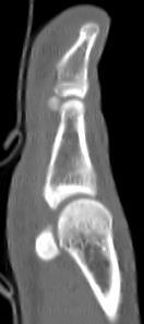
52 CT


53 PD MRI May 2017


54 PDFS

55 Abnormal linear signal in metatarsal head. Serpiginous line in the base of distal phalanx and proximal phalanx.


56 MRI July 2017


57


58
59 Discussion Extracorporeal shockwave therapy (ESWT) Treatment of sports related disorder Plantar fascitis, lateral epicondylitis, calcified/non calcified SST tendinosis, patellar tendinopathy Also in treatment of non-union long bone fracture, femoral head AVN. Shockwave induced tissue repair & regeneration, neovascularization Usually no severe complication ( local soft tissue swelling, cutaneous erosion, petechial or local subcutaneous hematoma).
60 One reported complication from ESWT for calcified SST tendinosis, ie AVN humeral head. by Durst et al. Possible pathomechanism: Shockwaves cause damage to blood vessels, can result in arterial occlusion, capillary extravasation, or vessel wall ruptured. Leads to AVN.
61 Case 8 NCY 29 y.o lady, h/o RTA in Sustained laceration wound with foreign body of left shin, surgery done. C/O left pretibial mass, which is painful after prolonged standing.


62

63 Normal
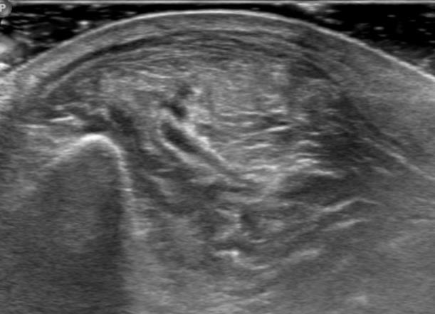
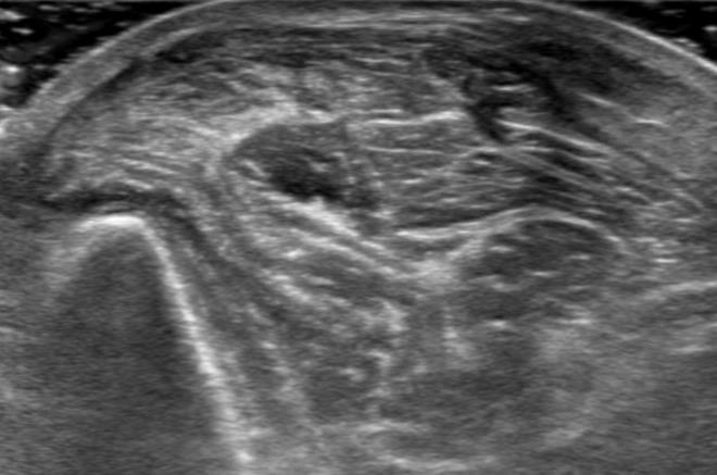
64 Ultrasound
65 Tibialis anterior muscle herniation from fascial defect secondary to trauma.
66 Discussion Muscle hernia: protrusion of muscle belly through an acquired/congenital fascial defect. Potentiate by increases in intracompartment pressure ( muscle hypertrophy). Most commonly in leg, and mostly affected tibialis anterior muscle (its fascia is most vulnerable to trauma). Clinical: Anterior tibial mass, vary in size; enlarged during leg dorsiflexion, and smaller at rest.
67 Dynamic ultrasound: Muscle bulge through fascia defect on muscle contraction, retraction on relaxation.
68 THANK YOU!!
MSK Interesting Cases. Dr Yap Sheau Huey
 MSK Interesting Cases Dr Yap Sheau Huey Case 1: History 41 y.o man, surf skier C/o pain over anterior left 5 th to 8 th ribs. Worse after sport activity. Chest Radiograph US Periostitis and early callus
Imaging of Ankle and Foot pain
 Imaging of Ankle and Foot pain Pramot Tanutit, M.D. Department of Radiology Faculty of Medicine, Prince of Songkla University 1 Outlines Plain film: anatomy Common causes of ankle and foot pain Exclude:
The Radiology Assistant : Bone tumor - well-defined osteolytic tumors and tumor-like lesions
 Bone tumor - well-defined osteolytic tumors and tumor-like lesions Henk Jan van der Woude and Robin Smithuis Radiology department of the Onze Lieve Vrouwe Gasthuis, Amsterdam and the Rijnland hospital,
MARK D. MURPHEY MD, FACR. Physician-in-Chief, AIRP. Chief, Musculoskeletal Imaging
 ALPHABET SOUP AND CYSTIC LESIONS OF THE BONE MARK D. MURPHEY MD, FACR Physician-in-Chief, AIRP Chief, Musculoskeletal Imaging ALPHABET SOUP AND CYSTIC LESIONS OF THE BONE Giant cell tumor (GCT) Unicameral
ELENI ANDIPA General Hospital of Athens G. Gennimatas
 ELENI ANDIPA General Hospital of Athens G. Gennimatas Technological advances over the last years have caused a dramatic improvement in ultrasound quality and resolution An established imaging modality
17/10/2017. Foot and Ankle
 17/10/2017 Alicia M. Yochum RN, DC, DACBR, RMSK Foot and Ankle Plantar Fasciitis Hallux Valgus Deformity Achilles Tendinosis Posterior Tibialis Tendon tendinopathy Stress Fracture Ligamentous tearing Turf
MRI XR, CT, NM. Principal Modality (2): Case Report # 2. Date accepted: 15 March 2013
: Case Report # 2. Date accepted: 15 March 2013") Radiological Category: Musculoskeletal Principal Modality (1): Principal Modality (2): MRI XR, CT, NM Case Report # 2 Submitted by: Hannah Safia Elamir, D.O. Faculty reviewer: Naga R. Chinapuvvula, M.D.
Radiological Category: Musculoskeletal Principal Modality (1): Principal Modality (2): MRI XR, CT, NM Case Report # 2 Submitted by: Hannah Safia Elamir, D.O. Faculty reviewer: Naga R. Chinapuvvula, M.D.
Urgent Cases and Foreign Bodies
 Urgent Cases and Foreign Bodies Catherine J. Brandon, MD, MS University of Michigan Ann Arbor, MI, USA Introduction: Patients added on to the schedule from the emergency department or as urgent add-on
MUSCULOSKELETAL INFECTIONS IN CHILDREN. Dr Caren Landes Alder Hey Children s NHS Foundation Trust Liverpool
 MUSCULOSKELETAL INFECTIONS IN CHILDREN Dr Caren Landes Alder Hey Children s NHS Foundation Trust Liverpool MUSCULOSKELETAL INFECTIONS Common and uncommon infections Common and uncommon presentations Imaging
MY PATIENT HAS KNEE PAIN. David Levi, MD Chief, Division of Musculoskeletal l limaging Atlantic Medical Imaging
 MY PATIENT HAS KNEE PAIN David Levi, MD Chief, Division of Musculoskeletal l limaging Atlantic Medical Imaging Causes of knee pain Non traumatic Trauma Osteoarthritis Patellofemoral pain Menisci or ligaments
12 Interesting MSK Cases
 12 Interesting MSK Cases James F Griffith Department of Imaging and Interventional Radiology Prince of Wales Hospital Case 1: 12-year-old boy Slipped and fell. Anterior knee pain and swelling Knee pain
Overuse Injuries & special skeletal injuries Dr M.Taghavi Director of sport medicine center of olympic academy
 Overuse Injuries & special skeletal injuries Dr M.Taghavi Director of sport medicine center of olympic academy Prevalence of Overuse Injuries 30 to 50% of all sport injuries are from overuse In some sports
Management of Brachial Plexus & Peripheral Nerves Blast Injuries. First Global Conflict Medicine Congress
 Management of Brachial Plexus & Peripheral Nerves Blast Injuries Joseph BAKHACH First Global Conflict Medicine Congress Hand & Microsurgery Department American University of Beirut Medical Centre Brachial
MUSCULOSKELETAL RADIOLOGY
 MUSCULOSKELETAL RADOLOGY SECTON www.cambridge.org Achilles tendonopathy/rupture Characteristics Describes pathology of the combined tendon of the gastro-soleus complex, which inserts onto the calcaneum.
Extracorporeal Shockwave Therapy. Outcomes in Shoulder Tendinopathy and Plantar Fasciitis. American University of Beirut Medical Center
 Extracorporeal Shockwave Therapy. Outcomes in Shoulder Tendinopathy and Plantar Fasciitis Nagham HADDAD, PT Nagham HADDAD, PT American University of Beirut Medical Center Introduction: Tendinosis is the
Ultrasound Evaluation of Masses
 Ultrasound Evaluation of Masses Jon A. Jacobson, M.D. Professor of Radiology Director, Division of Musculoskeletal Radiology University of Michigan Disclosures: Consultant: Bioclinica Advisory Panel: GE,
MR IMAGING OF THE WRIST
 MR IMAGING OF THE WRIST Wrist Instability Dissociative Pattern apparent on routine radiographs Non-dissociative Stress / positional radiographs Dynamic fluoroscopy during stress Arthrography MRI / MR arthrography
Surgical Care at the District Hospital. EMERGENCY & ESSENTIAL SURGICAL CARE
 Surgical Care at the District Hospital 1 18 Orthopedic Trauma Key Points 2 18.1 Upper Extremity Injuries Clavicle Fractures Diagnose fractures from the history and by physical examination Treat with a
Fluid-fluid levels in bone tumors: A pictorial review
 Fluid-fluid levels in bone tumors: A pictorial review Poster No.: C-578 Congress: ECR 2009 Type: Educational Exhibit Topic: Musculoskeletal Authors: L. Figueroa Nasra, C. Martín Hervás, M. Tapia-Viñé,
Sonography of Pediatric Superficial Lumps and Bumps: Illustrative Examples from Head to Toe
 Sonography of Pediatric Superficial Lumps and umps: Illustrative Examples from Head to Toe nmol Gupta ansal, MD Henrietta Kotlus Rosenberg, MD, FCR, FP Mount Sinai Hospital Icahn School of Medicine at
Review relevant anatomy of the foot and ankle. Learn the approach to examining the foot and ankle
 Objectives Review relevant anatomy of the foot and ankle Learn the approach to examining the foot and ankle Learn the basics of diagnosis and treatment of ankle sprains Overview of other common causes
E-Shock Wave in Physical Therapy. Mohammed TA Omar PhD PT Rehabilitation Health Science
 E-Shock Wave in Physical Therapy Mohammed TA Omar PhD PT Rehabilitation Health Science Objectives Following completion of this lecture the student will be able to: Describe the mechanical characteristics
Message of the Month for GPs June 2013
 Message of the Month for GPs June 2013 Dr Winn : Consultant Musculoskeletal Radiologist, Manchester Royal Infirmary Imaging of the musculoskeletal system Musculoskeletal pain is a common problem in the
Copyright 2004 Lippincott Williams & Wilkins. 2. Bone Structure. Copyright 2004 Lippincott Williams & Wilkins
 Chapter 7 The Skeleton: Bones and Joints The Skeleton Skeletal system is made up of bones and joints and supporting connective tissue. 1. Bone Functions 1. To store calcium salts 2. To protect delicate
Pragmatic ultrasound in the diagnosis of soft tissue rheumatic pain. Plamen Todorov
 Pragmatic ultrasound in the diagnosis of soft tissue rheumatic pain Plamen Todorov INTRODUCTION Soft tissue rheumatism: nonsystemic, focal pathological syndromes involving the periarticular structures.
emoryhealthcare.org/ortho
 COMMON SOCCER INJURIES Oluseun A. Olufade, MD Assistant Professor, Department of Orthopedics and PM&R 1/7/18 GOALS Discuss top soccer injuries and treatment strategies Simplify hip and groin injuries in
Topics. Musculoskeletal Infection Extremities. Detection of Infection. Role of Imaging in Extremity Infection. Detection of Infection
 Topics Musculoskeletal Infection Extremities Nuttaya Pattamapaspong M.D. Department of Radiology, Faculty of Medicine, Chiang Mai University, Chiang Mai, Thailand Role of imaging in extremity infection
Doc, I've done my groin. Groin Pain. Peter Brukner. Doc, I've done my groin 1. acute chronic
 Doc, I've done my groin Peter Brukner Associate Professor in Sports Medicine Centre for Sports Medicine Research and Education School of Physiotherapy 9/22/2006 The University of Melbourne Groin Pain acute
Clinical Application of the EMS Swiss DolorClast
 Chapter 12.fm Page 119 Tuesday, November 21, 2006 6:38 PM 12 Clinical Application of the EMS Swiss DolorClast L. Gerdesmeyer, M. Henne, P. Diehl, H. Gollwitzer, M. Göbel In general, the following recommendations
The Radiology Assistant : Bone tumor - ill defined osteolytic tumors and tumor-like lesions
 Bone tumor - ill defined osteolytic tumors and tumor-like lesions Henk Jan van der Woude and Robin Smithuis Radiology department of the Onze Lieve Vrouwe Gasthuis, Amsterdam and the Rijnland hospital,
Imaging and intervention of sacroiliac joint. Dr Ryan Lee Ka Lok Associate Consultant Prince of Wales Hospital
 Imaging and intervention of sacroiliac joint Dr Ryan Lee Ka Lok Associate Consultant Prince of Wales Hospital Introduction 15%-25% of low back pain is related to sacroiliac joint (SIJ) pain SIJ pain is
Osteonecrosis of the knee Treatment with ESWT. Dr Shrenik Shah Shrey hospital Ahmedabad
 Osteonecrosis of the knee Treatment with ESWT Dr Shrenik Shah Shrey hospital Ahmedabad Osteonecrosis(ON) of the knee SPONK- Spontaneous ON of the knee Secondary ON of the knee Postarthrosopic ON of the
STAGING, BIOPSY AND NATURAL HISTORY OF TUMORS SCOTT D WEINER MD
 STAGING, BIOPSY AND NATURAL HISTORY OF TUMORS SCOTT D WEINER MD WHAT DO YOU DO WHEN THIS SHOWS UP IN YOUR OFFICE? besides panicking KEY PRINCIPLE!!! Reactive zone is the edema, neovascularity and inflammation
Shoulder Case Studies
 Shoulder Case Studies Eden Raleigh Orthopaedic Surgeon Shoulder & Knee Surgery Ph: 9421 1900 0402697115 dredenraleigh@gmail.com My Background Specialising in Shoulder and Knee Surgery Main focus on Arthroscopic/Sports
Anterior Tibialis Tendon Rupture: The Other Cause of Foot Drop. Alicia Rozario, DPM PGY-3 DVA Puget Sound Healthcare System
 Anterior Tibialis Tendon Rupture: The Other Cause of Foot Drop Alicia Rozario, DPM PGY-3 DVA Puget Sound Healthcare System Disclosures Nothing to Disclose. ANATOMY https://osteopathysingapore.files.wordpress.com/2015/05/tibialis-anterior-muscle1.png
In which arm muscle are intramuscular injections most often given? (not in text)
") AP1 Lab 9 - Muscles of the Arms and Legs Locate the following muscles on the models and on yourself. Recall anatomical position. Directional terms such as anterior, posterior, lateral, etc. all assume
AP1 Lab 9 - Muscles of the Arms and Legs Locate the following muscles on the models and on yourself. Recall anatomical position. Directional terms such as anterior, posterior, lateral, etc. all assume
Knee Contusions and Stress Injuries. Laura W. Bancroft, M.D.
 Knee Contusions and Stress Injuries Laura W. Bancroft, M.D. Objectives Review 5 types of contusion patterns Pivot shift Dashboard Hyperextension Clip Lateral patellar dislocation Demonstrate various stress
Osteomyelitis in infancy and childhood: A clinical and diagnostic overview M. Mearadji
 Osteomyelitis in infancy and childhood: A clinical and diagnostic overview M. Mearadji International Foundation for Pediatric Imaging Aid Introduction Osteomyelitis is a relative common disease in infancy
ISPUB.COM. Spectrum Of MRI Findings In Musculoskeletal Tuberculosis: Pictoral Essay. P Chudgar INTRODUCTION SPINE
 ISPUB.COM The Internet Journal of Radiology Volume 8 Number 2 Spectrum Of MRI Findings In Musculoskeletal Tuberculosis: Pictoral Essay P Chudgar Citation P Chudgar.. The Internet Journal of Radiology.
Update - Imaging of the Spondyloarthropathies. Spondyloarthropathies. Spondyloarthropathies
 Update - Imaging of the Spondyloarthropathies Donald J. Flemming, M.D. Dept of Radiology Penn State Hershey Medical Center Spondyloarthropathies Family of inflammatory arthritides of synovium and entheses
Plantar fasciitis occurs when the strong band of tissue that supports the arch of your foot becomes irritated and inflamed.
 Plantar Fasciitis and Bone Spurs Plantar fasciitis (fashee-eye-tiss) is the most common cause of pain on the bottom of the heel. Approximately 2 million patients are treated for this condition every year.
Rad Lab 6 Unknowns: Musculoskeletal
 Rad Lab 6 Unknowns: Musculoskeletal Peter Clarke MD Associate Clerkship Director for Radiology Harvard Medical School Brigham and Women s Hospital Dana Farber Cancer Institute Here are two men, one 70,
PITFALLS IN THE DIAGNOSIS OF SKELETAL TUBERCULOSIS IN CHILDREN
 PITFALLS IN THE DIAGNOSIS OF SKELETAL TUBERCULOSIS IN CHILDREN DR.JANANI SANKAR SENIOR CONSULTANT - PEDIATRICS DEPARTMENT OF PEDIATRICS & PEDIATRIC ORTHOPEDICS KANCHI KAMAKOTI CHILDS TRUST HOSPITAL CHENNAI,
Seronegative Spondyloarthropathies: A Radiological Persepctive
 Seronegative Spondyloarthropathies: A Radiological Persepctive Poster No.: C-1816 Congress: ECR 2016 Type: Educational Exhibit Authors: K. Shindi, H. Nejadhamzeeigilani, P. Nagtode, C. Nel ; 1 1 2 2 3
Imaging in Groin Pain What the Team Physician Needs to Know
 Imaging in Groin Pain What the Team Physician Needs to Know Üstün Aydıngöz, MD Professor of Radiology Hacettepe University School of Medicine Ankara, Turkey ustunaydingoz@yahoo.com No conflicts of interest
New 2010 CPT Codes (italic font represents a new or revised code/description)
") New 2010 CPT Codes (italic font represents a new or revised code/description) 14301 Adjacent tissue transfer or rearrangement, any area; defect 30.1 sq cm to 60.0 sq cm 14302 each additional 30.0 sq cm,
New 2010 CPT Codes (italic font represents a new or revised code/description) 14301 Adjacent tissue transfer or rearrangement, any area; defect 30.1 sq cm to 60.0 sq cm 14302 each additional 30.0 sq cm,
Ankle Tendons in Athletes. Laura W. Bancroft, M.D.
 Ankle Tendons in Athletes Laura W. Bancroft, M.D. Outline Protocols Normal Anatomy Tendinopathy, partial and complete tears Posterior tibial, Flexor Hallucis Longus, Achilles, Peroneal and Anterior Tibial
Case 1 7 yo male Right elbow injury 3 months ago Medial elbow pain and tenderness over medial epicondyle Long arm cast given but off himself 1 month a
 Case presentations Case 1 7 yo male Right elbow injury 3 months ago Medial elbow pain and tenderness over medial epicondyle Long arm cast given but off himself 1 month after Progressive limited elbow flexion
ORTHOPEDICS BONE Recalcitrant nonunions In total hip replacement total knee surgery increased callus volume
 ORTHOPEDICS Orthopedics has to do with a variety of tissue: bone, cartilage, tendon, ligament, muscle. In this regard orthopedic and sports medicine share the same tissue targets. Orthopedics is mostly
Imaging the musculoskeletal system. An Introduction
 Imaging the musculoskeletal system An Introduction Objectives Discuss: commonly used imaging modalities in the musculoskeletal system normal imaging anatomy in the extremities fracture description Imaging
Common%Work%Related%Foot% and%ankle%problems
 Common%Work%Related%Foot% and%ankle%problems Dr. George H. Theodore Massachusetts General Hospital Harvard Medical School Foot and Ankle Consultant Boston Red Sox New England Patriots Boston Bruins Work%Related%Foot%and%Ankle%
Extracorporeal Shock Wave Therapy (ESWT) Jeff Blea, DVM Von Bluecher, Blea, Hunkin, Inc. Equine Medicine and Surgery Sierra Madre, CA
 Jeff Blea, DVM Von Bluecher, Blea, Hunkin, Inc. Equine Medicine and Surgery Sierra Madre, CA") Extracorporeal Shock Wave Therapy (ESWT) Jeff Blea, DVM Von Bluecher, Blea, Hunkin, Inc. Equine Medicine and Surgery Sierra Madre, CA History Shock waves used in human medicine as early as 1980s: Break
Extracorporeal Shock Wave Therapy (ESWT) Jeff Blea, DVM Von Bluecher, Blea, Hunkin, Inc. Equine Medicine and Surgery Sierra Madre, CA History Shock waves used in human medicine as early as 1980s: Break
Gout. Crystal deposition disease: Imaging perspectives. Crystal associated arthropathies. Clinical Stages of Gout 07/06/60
 Crystal associated arthropathies Crystal deposition disease: Imaging perspectives Warapat Virayavanich, MD Ramathibodi hospital, Mahidol University Commonly seen arthropathy MSU (gout) CPPD HADD Uncommon
Contents of the Posterior Fascial Compartment of the Thigh
 Contents of the Posterior Fascial Compartment of the Thigh 1-Muscles: B i c e p s f e m o r i s S e m i t e n d i n o s u s S e m i m e m b r a n o s u s a small part of the adductor magnus (h a m s t
Chapter 5 The Skeletal System
 Chapter 5 The Skeletal System The Skeletal System Parts of the skeletal system Bones (skeleton) Joints Cartilages Ligaments (bone to bone)(tendon=bone to muscle) Divided into two divisions Axial skeleton:
2017 OMA Sport and Exercise Medicine RADIOLOGY QUIZ. Dr. Ali Rendely Resident Representative PGY2
 2017 OMA Sport and Exercise Medicine RADIOLOGY QUIZ Dr. Ali Rendely Resident Representative PGY2 Acknowledgement Dr. R. Bleakney Assistant Professor Department of Medical Imaging, University of Toronto
 www.fisiokinesiterapia.biz Shoulder Problems Fractures Instability Impingement Miscellaneous Anatomy Bones Joints / Ligaments Muscles Neurovascular Anatomy Anatomy Supraspinatus Anterior Posterior Anatomy
Anatomy and Physiology II. Review Shoulder Girdle New Material Upper Extremities - Bones
 Anatomy and Physiology II Review Shoulder Girdle New Material Upper Extremities - Bones Anatomy and Physiology II Shoulder Girdle Review Questions From Last Lecture Can you identify the following muscles?
Take Pride in Performance
 2017 Take Pride in Performance Knee: Meniscal Tear FSE PD - Sagittal FSE PD - Coronal FSTIR - Coronal Knee: ACL Tibial Avulsion 3D SHARC ISO - Sagittal FSE PD - Sagittal FSTIR - Coronal Knee: Subchondral
Anatomy and Physiology 2016
 Anatomy and Physiology 2016 O = Temporal line I = coronoid process (Mandible) A = elevates mandible (chewing) O = galea aponeurotica (layer of dense fibrous tissue which covers the upper part of the cranium)
Leg. Dr. Heba Kalbouneh Associate Professor of Anatomy and Histology
 Leg Dr. Heba Kalbouneh Associate Professor of Anatomy and Histology Skin of the Leg Cutaneous Nerves Medially: The saphenous nerve, a branch of the femoral nerve supplies the skin on the medial surface
Plantar fasciopathy (PFs)
") Plantar fasciopathy (PFs) 2016. 04. 30. Jung-Soo Lee, M.D., Ph.D. Department of Rehabilitation Medicine, Uijeongbu St. Mary's Hospital, College of Medicine, The Catholic University of Korea Anatomy of
Plantar fasciopathy (PFs) 2016. 04. 30. Jung-Soo Lee, M.D., Ph.D. Department of Rehabilitation Medicine, Uijeongbu St. Mary's Hospital, College of Medicine, The Catholic University of Korea Anatomy of
KNEE DISLOCATION. The most common injury will be an anterior dislocation, and this usually results from a hyperextension mechanism.
 KNEE DISLOCATION Introduction Dislocation of the knee is a severe injury associated with major soft tissue injury and a high incidence of damage to the popliteal artery. There is displacement of the tibia
Ultrasound Evaluation of Posteromedial Ankle Pathology. Andrew C Cordle, M.D., Ph.D. 9/21/2018
 Ultrasound Evaluation of Posteromedial Ankle Pathology Andrew C Cordle, M.D., Ph.D. 9/21/2018 Overview: Pathology of the Posteromedial Ankle Flexor Tendon Pathology Accessory Navicular Bone Pathology Tarsal
Why? Ultrasound of the Foot. Ultrasound of the Foot. General Rules. Plantar Fascia. Plantar Fasciitis 18/09/2018
 Ultrasound of the Foot Why? Ultrasound of the Foot Plantar fasciitis Plantar fascia fibromatosis Morton s neuroma Intermetatarsal bursitis Adventitial bursitis Plantar plate tears MTP joint synovitis Ganglia
Anatomy. Anatomy deals with the structure of the human body, and includes a precise language on body positions and relationships between body parts.
 Anatomy deals with the structure of the human body, and includes a precise language on body positions and relationships between body parts. Proper instruction on safe and efficient exercise technique requires
MRI of the Shoulder What to look for and how to find it? Dr. Eric Handley Musculoskeletal Radiologist Cherry Creek Imaging
 MRI of the Shoulder What to look for and how to find it? Dr. Eric Handley Musculoskeletal Radiologist Cherry Creek Imaging MRI of the Shoulder Benefits of Ultrasound: * Dynamic * Interactive real time
Aetiology: Pressure of Distal intermetatarsal ligament against common digital nerve. Lumbar radiculopathy Instability MTPJ joint or inflammatory MPJ
 MORTON S NEUROMA 80% III web space (next common is II). Never occurs in III or IV Common in females in fifties Aetiology: Pressure of Distal intermetatarsal ligament against common digital nerve Rule out
Basic Radiographic Principles Part II
 Basic Radiographic Principles Part II Kristopher Avant, D.O. October 19 th, 2016 I have no disclosures relevant to the material presented in this discussion. Good Stuff!!! 1 Really? Really! Musculoskeletal
Ultrasound of Mid and Hindfoot Pathology
 Ultrasound of Mid and Hindfoot Pathology Levon N. Nazarian, M.D. Professor of Radiology Thomas Jefferson University Hospital Disclosures None relevant to this presentation Educational Objective Following
EXTRACORPOREAL SHOCK WAVE THERAPY (ESWT) FOR CHRONIC TENDINITIS AND RELATED DISORDERS
 FOR CHRONIC TENDINITIS AND RELATED DISORDERS") BACKGROUND EXTRACORPOREAL SHOCK WAVE THERAPY (ESWT) FOR CHRONIC TENDINITIS AND RELATED DISORDERS REVIEW OF THE MEDICAL LITERATURE FOR EFFICACY, SAFETY, AND COST-EFFECTIVENESS Low energy ESWT is a byproduct
BACKGROUND EXTRACORPOREAL SHOCK WAVE THERAPY (ESWT) FOR CHRONIC TENDINITIS AND RELATED DISORDERS REVIEW OF THE MEDICAL LITERATURE FOR EFFICACY, SAFETY, AND COST-EFFECTIVENESS Low energy ESWT is a byproduct
A 76 year old male presented with sudden increase of dyspnoea on 15 November 2014, following a biopsy. A previous CXR was reviewed.
 Question 1 A 76 year old male presented with sudden increase of dyspnoea on 15 November 2014, following a biopsy. A previous CXR was reviewed. Imaging A CXR was performed on 28 May 2014. A CT of the chest
Extra-spinal musculoskeletal manifestations of mycobacterium tuberculosis (TB) infection: A review of radiological findings
 infection: A review of radiological findings") Extra-spinal musculoskeletal manifestations of mycobacterium tuberculosis (TB) infection: A review of radiological findings Poster No.: P-0118 Congress: ESSR 2013 Type: Scientific Exhibit Authors: I. Anwar,
Extra-spinal musculoskeletal manifestations of mycobacterium tuberculosis (TB) infection: A review of radiological findings Poster No.: P-0118 Congress: ESSR 2013 Type: Scientific Exhibit Authors: I. Anwar,
Lower Limb. Hamstring Strains. Risk Factors. Dr. Peter Friis 27/04/15. 16% missed games AFL 6-15% injury in rugby 30% recurrent
 Lower Limb Dr. Peter Friis MB BS FACSP Sports Physician Hamstring Strains 16% missed games AFL 6-15% injury in rugby 30% recurrent Risk Factors Modifiable Warm up Fatigue Strength Flexibility L/Spine Pelvic
Brain Atrophy. Brain Atrophy
 Aging Central Nervous System Processes Age related brain atrophy Non-age related brain atrophy Cerebrovascular disease Cerebral infarction Hypertensive hemorrhage Carotid artery stenosis and occlusion
MUSCULOSKELETAL DISORDERS: THE BIGGEST JOB SAFETY PROBLEM. What Are Musculoskeletal Disorders
 MUSCULOSKELETAL DISORDERS: THE BIGGEST JOB SAFETY PROBLEM What Are Musculoskeletal Disorders Every year more than 1.8 million workers in the United States suffer painful back and repetitive strain injuries,
The Elbow and the cubital fossa. Prof Oluwadiya Kehinde
 The Elbow and the cubital fossa Prof Oluwadiya Kehinde www.oluwadiya.com Elbow and Forearm Anatomy The elbow joint is formed by the humerus, radius, and the ulna Bony anatomy of the elbow Distal Humerus
Mr Simon Jennings BSc, MB BS, FRCS, Dip Sports Med FRCS (Trauma & Orthopaedics)
") Mr Simon Jennings BSc, MB BS, FRCS, Dip Sports Med FRCS (Trauma & Orthopaedics) Consultant Orthopaedic Surgeon Northwick Park Hospital 107 Harley Street RSM 16 th September 2010 Orthopaedic Surgeon Knee
Mr Simon Jennings BSc, MB BS, FRCS, Dip Sports Med FRCS (Trauma & Orthopaedics) Consultant Orthopaedic Surgeon Northwick Park Hospital 107 Harley Street RSM 16 th September 2010 Orthopaedic Surgeon Knee
divided by the bones ( redius and ulna ) and interosseous membrane into :
 and interosseous membrane into :") fossa Cubital Has: * floor. * roof : - Skin - superficial fasica - deep fascia ( include bicipital aponeurosis ) Structures within the roof : -cephalic and basilic veins -and between them median cubital
fossa Cubital Has: * floor. * roof : - Skin - superficial fasica - deep fascia ( include bicipital aponeurosis ) Structures within the roof : -cephalic and basilic veins -and between them median cubital
Alberta Health Care Insurance Plan. Schedule Of Anaesthetic Rates Applicable To Podiatry. Procedure List. As Of. 01 April Government of Alberta
 Alberta Health Care Insurance Plan Procedure List As Of 01 April 2017 Alberta Health Care Insurance Plan Page i Generated 2017/03/14 TABLE OF CONTENTS As of 2017/04/01 II. OPERATIONS ON THE NERVOUS SYSTEM.......................
Dr abedi yekta. Assistant Professor of Sports and Exercise Medicine Faculty of Medicine shahid beheshti University of Medical Sciences
 Dr abedi yekta Assistant Professor of Sports and Exercise Medicine Faculty of Medicine shahid beheshti University of Medical Sciences Pain in midportion of Achilles tendon. Morning stiffness Tendinitis
Mr. Duy Thai Orthopaedic Surgeon, Melbourne VIC
 Mr. Duy Thai Orthopaedic Surgeon, Melbourne VIC International Convention of the Vietnamese Physicians, Dentists and Pharmacists of the Free World Melbourne 8 10 August 2014 Conflict of Interest None Subacromial
AMG Virtual CME Series Plantar Fasciitis Brian T. Dix, DPM, FACFAS Board Certified in Foot and Reconstructive Hindfoot & Ankle Surgery
 AMG Virtual CME Series Plantar Fasciitis 11-9-17 Brian T. Dix, DPM, FACFAS Board Certified in Foot and Reconstructive Hindfoot & Ankle Surgery Anatomy 3 bands of dense connective tissue, which originate
CLINICAL PRESENTATION AND RADIOLOGY QUIZ QUESTION
 Donald L. Renfrew, MD Radiology Associates of the Fox Valley, 333 N. Commercial Street, Suite 100, Neenah, WI 54956 12/01/2012 Radiology Quiz of the Week # 101 Page 1 CLINICAL PRESENTATION AND RADIOLOGY
Pigmented Villonodular Synovitis PVNS
 February 2002 Pigmented Villonodular Synovitis PVNS Amy Gillis, Harvard Medical School Year III 47 year old female Our Patient Right hip pain since age 20 No history of trauma Diagnosed with DJD of R hip
Point of Care Ultrasound on the Field of Play K AT I E N ANOS, MD
 Point of Care Ultrasound on the Field of Play K AT I E N ANOS, MD H I GH P ERFORMANCE S PORTS MEDICINE P HYSI ATRIST, P R ACTICING S PORTS MEDI CINE No disclosures No disclosures Who am I? Objectives Over
4/28/2010. Fractures. Normal Bone and Normal Ossification Bone Terms. Epiphysis Epiphyseal Plate (physis) Metaphysis
 Metaphysis") Fractures Normal Bone and Normal Ossification Bone Terms Epiphysis Epiphyseal Plate (physis) Metaphysis Diaphysis 1 Fracture Classifications A. Longitudinal B. Transverse C. Oblique D. Spiral E. Incomplete
Fractures Normal Bone and Normal Ossification Bone Terms Epiphysis Epiphyseal Plate (physis) Metaphysis Diaphysis 1 Fracture Classifications A. Longitudinal B. Transverse C. Oblique D. Spiral E. Incomplete
Table of Compensation for Injury 1/2015
 Table of Compensation for 1/2015 Valid from 01.04.2015 1. CRANIAL INJURIES 1.1 Cranial bone fractures: a) fracture of cranial dome 10 b) fracture of cranial base 15 c) fracture of cranial dome and base
Practical Reporting of Musculoskeletal Imaging Studies: MRI Elbow
 Practical Reporting of Musculoskeletal Imaging Studies: MRI Elbow James F Griffith History Where is pain located? For how long? Trauma if so, what and when Radiographers can get this info Grade. Don t
Case Report Nonbacterial Osteitis of the Clavicle: Longitudinal Imaging Series from Initial Diagnosis to Clinical Improvement
 Case Reports in Rheumatology Volume 2015, Article ID 182731, 4 pages http://dx.doi.org/10.1155/2015/182731 Case Report Nonbacterial Osteitis of the Clavicle: Longitudinal Imaging Series from Initial Diagnosis
31yo M with chronic basilar thumb and wrist pain that started after cross-country bicycle ride 5 yrs ago.
 31yo M with chronic basilar thumb and wrist pain that started after cross-country bicycle ride 5 yrs ago. EPL EPB APL Full-thickness tear involving the dorsal deltoid ligament of the first carpometacarpal
SHOULDER Highly mobile, so less stable. Abnormalities cloaked within extensive musculature, dx can be difficult Bony abnormalities less common than li
 SPORTS MEDICINE CASES A quick tour of some local joints Featuring gco common o and unusual problems SHOULDER Highly mobile, so less stable. Abnormalities cloaked within extensive musculature, dx can be
Current Thinking of the Osteochondroses. Diego Jaramillo, M.D., M.P.H. Department of Radiology Stanford Children s Hospital
 Current Thinking of the Osteochondroses Diego Jaramillo, M.D., M.P.H. Department of Radiology Stanford Children s Hospital What is an osteochondrosis? Abnormal endochondral ossification and epiphyseal
Salisbury Foundation Trust Radiology Department Referral Guidelines for Primary Care: Musculoskeletal Imaging
 Salisbury Foundation Trust Radiology Department Referral Guidelines for Primary Care: Musculoskeletal Imaging These guidelines have been issued in conjunction with the Royal College of Radiology referral
Scrotum-like protrusion of lipoma arising from the proximal thigh
 Upsala J Med sci 109: 261 265, 2004 Scrotum-like protrusion of lipoma arising from the proximal thigh Report of two cases Koshi Hattori, 1 Masahito Hatori, 1 Mika Watanabe, 2 Toshihisa Osanai, 3 Shoichi
MRI of Pediatric Ankle and Foot. Mahesh Thapa, MD Associate Professor Seattle Children s University of Washington School of Medicine
 MRI of Pediatric Ankle and Foot Mahesh Thapa, MD Associate Professor Seattle Children s University of Washington School of Medicine Disclosures Under contract with Lippincott Williams and Wilkins (LWW)
Impingement Syndromes of the Ankle. Noaman W Siddiqi MD 5/4/2006
 Impingement Syndromes of the Ankle Noaman W Siddiqi MD 5/4/2006 Ankle Impingement Overview Clinical DX Increasingly recognized cause of chronic ankle pain Etiology can be soft tissue or osseous Professional
Superficial Lumps and Bumps: Ultrasound Assessment
 Posterior knee Superficial Lumps and Bumps: Ultrasound Assessment Walter Mak, MD Department of Medical Imaging St. Michael s Hospital SM SM MGas MGas MGas MGas Synovial lined Synovial cyst: extrusion of
Index. Note: Page numbers of article titles are in boldface type.
 Note: Page numbers of article titles are in boldface type. A ACJ. See Acromioclavicular joint (ACJ) Acromioclavicular joint (ACJ) procedures of, 557 559 Ankle and foot procedures of, 649 671 (See also
The Lower Limb VI: The Leg. Anatomy RHS 241 Lecture 6 Dr. Einas Al-Eisa
 The Lower Limb VI: The Leg Anatomy RHS 241 Lecture 6 Dr. Einas Al-Eisa Muscles of the leg Posterior compartment (superficial & deep): primary plantar flexors of the foot flexors of the toes Anterior compartment: