Chest imaging II. Interstitial lung diseases
|
|
|
- Kristin Jessica Morton
- 6 years ago
- Views:
Transcription


1 Chest imaging II. Interstitial lung diseases Dávid L. Tárnoki MD, PhD Ádám D. TárnokiMD, PhD Department of Radiology Semmelweis University

2 Topics 1. Interstitial lung diseases 2. Occupational lung diseases 3. Pleura 2
3 Interstitial lung diseases 3


 bronchiolus")

4 Anatomy Interstitial lung diseases Secondary lobule: 1-2,5 cm ø Centrilobular artery: 0,5-0,7 mm ø Centrilobular bronchiole (wall: 0,15 mm) bronchiolus terminalis terminal ducts acini alveoli Radiologyassistant; Webb WR, Radiology 2006;239 4


5 Interstitial lung diseases Anatomy Interstitium: Pulmonary arteries and veins Lymph vessels, lymph nodes Nerves Fibroblasts Collagen fibers Elastic fibers Lynch PJ, Creative Commons, 5
 Granuloma")
6 6 Interstitial lung diseases Pathogenesis - Overview Noxen (aerogenous, circulatory) Damage of the alveolar /from circulation/ endothelial membrane Remodeling Inflammation cells from circulation / interstitium (activation and recruitment) Granuloma Fibrosis


 GGO")
7 7 Interstitial lung diseases Pathogenesis - Overview Alveolar block (real diffusion disorder) Reduction of the alveolar space Reduction of the capillary diffusion area Reduction of the Diffusion constants (in anemia) GGO Reticulation Honeycombing

8 Definition Interstitial lung diseases Caused by chronic inflammation of the pulmonary interstitium with a scarred reconstruction of connective tissue proliferation due to different causes Over 150 triggers (allergens, Medicines, etc.) Half of the cases remain unclear Mostly affected alveoli and interstitium Airways and pulmonary vessels are often secondary affected 8

 nails Basal crackling")

9 Interstitial lung diseases Clinics Unproductive cough Stress dyspnoea Drumstick fingers Clubbed (Watch glass) nails Basal crackling Weakness, weight loss Full history required: - Medicines - Job - Hobbies - "pleasure poisons" 9
 Imaging (Rö and HRCT) Bronchoscopy with transbronchial biopsy (TBB) and bronchoalveolar lavage (BAL) Open lung")
10 Diagnostics Interstitial lung diseases Ideally by multidisciplinary ILD board: pneumologists, rheumatologists, radiologists and pathologists Laboratory: - Kidney functions, electrolytes, CRP - Differential blood counts - Antibodies Lung function (Restrictive ventilation fault) Imaging (Rö and HRCT) Bronchoscopy with transbronchial biopsy (TBB) and bronchoalveolar lavage (BAL) Open lung biopsy (VATS) 10
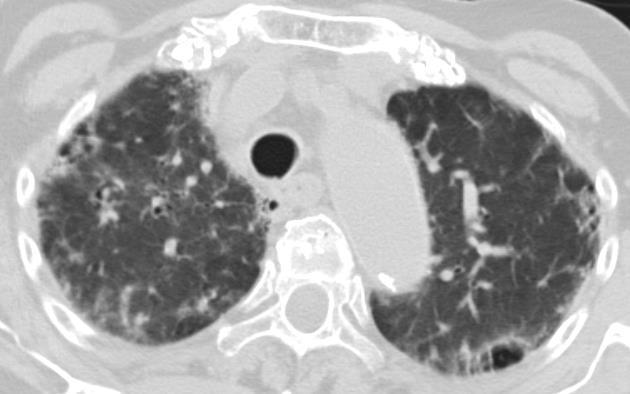
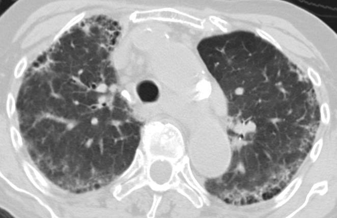
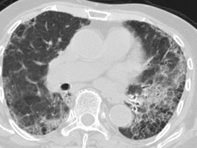
11 Interstitial lung diseases Clinics What kind of information the clinician needs from us? Diagnosis UIP (Usual interstitial pneumonia) biopsy is not necessary Other DD? biopsy is necessary Prognosis acute / cronic Comparison with previuos HRCT: progression / regression? Therapeutic options - Steroids - Lung transplantation Prognosis - Median survival after diagnosis years 11
12 12 Interstitial lung diseases Typical patterns of an ILD Ground glass opacity, consolidation Ground glass opacity (GGO) Ground glass opacities (Milk glass opacity - increased parenchyma decreasement) partially filled and / or collapsed alveoli e.g. active inflammation Consolidation - Consolidation completely filled and / or collapsed alveoli (Accumulation of exudate, transudate, or other tissue in the alveoli)


13 Interstitial lung diseases Ground glass opacity, consolidation Consolidation GGO 13
14 Interstitial lung diseases Idiopathic pulmonary fibrosis (heterogeneous entity) AIP (acute interstitial pneumonitis) UIP (usual interstitial pneumonitis) 70% DIP (desquamative interstitial pneumonia) RBILD (respiratory bronchiolitis ILD) NSIP (non specific interstial pneumonia) BOOP=COP (bronchiolitis obliterans organizing pneumonia = cryptogenic organizing pneumonia) Why do we need to subtype the idiopathic interstitial Pneumonias? Different prognosis which results different therapeutic approaches 14


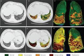
15 Interstitial lung diseases X-Ray - Fine-stripping-reticular pattern that is superimposed by a diffuse transparency reduction - Increased reticulation with basal dominance NORMAL FIBROSIS 15


16 Interstitial lung diseases HRCT Plays a central role in detection, diagnosis and differential diagnosis Sufficiency of assessability with a layer thickness of 1.25 mm or less Acut Alveolitis: GGO the location of optimal bronchoscopic tissue collection Chronic: Nodules, lines and bands, pleural thickening Final stadium: Honeycombing Beehive pattern" 16


17 Interstitial lung diseases Basic pattern in HRCT linear reticular pattern nodular pattern Structures with density reduction (Lesions with "less air") Structures with density increase (Lesions with more air") 17


18 Interstitial lung diseases ILD classification according to ATS / ERS 18
 Acute alveolitis: milk opacity (density increase with still recognizable bronchovascular structures) \"idiopathic form of ARDS\" / ARDS of")
19 19 Interstitial lung diseases 1. AIP (acute interstitial pneumonia) Acute alveolitis: milk opacity (density increase with still recognizable bronchovascular structures) "idiopathic form of ARDS" / ARDS of unclear cause Acute and short prognosis HRCT image: Acute: - homogeneous GGO (alveolitis) - diffuse consolidation Chronic: honeycombing, fibrosis


20 Interstitial lung diseases 2. NSIP (Non-specific interstitial pneumonia) Chronic form Manifestation: 4. Life decade Not tobacco-associated HRCT Image: - GGO - Peripheral, basal, subpleural, symmetrical fine reticular condensation, traction bronchiolectasis - no honeycomb pattern (no honeycombing) Usually occurs as a pulmonary involvement in the context of collagenosis Importantly, the NSIP. can be treated with systemic glucocorticosteroids prognosis more favorable than with UIP 20
 Simultaneity of inflammation, proliferation and fibrosis HRCT is very specific: - GGO (signs of active inflammation and fibroblast proliferation) - Honeycomb")
21 Interstitial lung diseases 3. UIP (Usual Interstitial Pneumonia) Simultaneity of inflammation, proliferation and fibrosis HRCT is very specific: - GGO (signs of active inflammation and fibroblast proliferation) - Honeycomb patterns (honeycombing), bronchiectasis as signs of fibrosis (not present in NSIP!) - subpleural and basally stressed localization - Reticular pattern - Lack of changes that are not associated with UIP are compatible. Probability a correct diagnosis of a UIP at nearly 100% with HRCT Prognosis: poor 21 manju-imagingxpert.blogspot.com
22 Interstitial lung diseases Severe pulmonary parenchymal changes Normal or pathologic image? Fibrosis ( 22


23 Interstitial lung diseases Honeycombing: subpleural localized, multi-rowed, small cystic changes 23


 Lower")
24 24 Interstitial lung diseases Regardless of the cause End stage: honeycomb lung Upper lobes: signs of activity (GGO, alveolitis) Lower lobes: honeycombing


25 Interstitial lung diseases Lung fibrosis: honeycombing pattern Patients with a typical UIP pattern in HRCT: does not need lung biopsy 25


 43 year")
26 Interstitial lung diseases DD: Fibrosis after radiation (due to breast cancer) 43 year old woman 26


27 HRCT Interstitial lung diseases 3D reconstructions Lung volumen Pattern softwares 27
 Diaphragm movement disorders - ultrasound")

28 Interstitial lung diseases UIP (Usual Interstitial Pneumonia) Diaphragm movement disorders - ultrasound M-mode Complication: shrinking lung 28
 - Systemic Lupus Erythematosus (SLE) - Sjögren syndrome - Systemic sclerosis / scleroderma - Dermatopolymyositis -")
29 Interstitial lung diseases 4. Collagenoses and vasculitides Pulmonary manifestations: - Rheumatoid arthritis (RA) - Systemic Lupus Erythematosus (SLE) - Sjögren syndrome - Systemic sclerosis / scleroderma - Dermatopolymyositis - Wegener granulomatosis X-ray: less sensitive In pronounced cases: Basal-dominant reticulonodular pattern HRCT: Patological findings in 30% pleural thickening Late stage: fibrosis What will the clinician ask from the radiologist? Disease characterization and expansion. UIP or NSIP patterns? 29
30 Occupational lung diseases 30
31 Interstitial lung diseases ILD classification according to ATS / ERS 31

32 Occupational lung diseases New diseases due to occupational regulations Chronic inhalation of inorganic dusts (e.g., silicate) Long-time exposure (e.g., mineral workers) Alveolar phagocytosis of inhaled particles and interstitial deposition Interstitial reticulo-granuloma formation, sometimes massive fibrosis Therapy: Exposure stop I. DISEASES OF IMMUNOLOGICAL / UNCLEAR AETIOLOGY II. PNEUMOCONIOSIS (Inhaled Particles) X-Ray: Nodular herd often with calcifications Hilary / mediastinal lymph nodes with calcifications ("laryngeal calcification") HRCT: X-ray patterns + Micronodular lesions pulmonary fibrosis 32
 Idiopathic form: Cryptogen-organizing pneumonia (COP) Non-infectious inflammation that may occur in collagenous diseases, chronic eosinophilic pneumonia, exogenous allergic")
33 Occupational lung diseases I. DISEASES OF IMMUNOLOGICAL / UNCLEAR AETIOLOGY 1. Organising pneumonia (OP) Idiopathic form: Cryptogen-organizing pneumonia (COP) Non-infectious inflammation that may occur in collagenous diseases, chronic eosinophilic pneumonia, exogenous allergic alveolitis, infections or even as a medication reaction Symptoms: chronic subfebrile temperatures, dyspnoea and cough HRCT: Band-shaped, subpleural and peribronchial stressed focal consolidations with basal predominance - migrating consolidations Atoll sign: by a ring-like consolidation with central milk glass Prosch H. Journal für Pneumologie 2015; 3 (1),

 Immune reaction of the alveoli and bronchioles")
, chemotherapeutic agents Anamnesis!")
 reticular pattern")
34 Occupational lung diseases I. DISEASES OF IMMUNOLOGICAL / UNCLEAR AETIOLOGY 2. Exogen Allergic Alveolitis, EAA ( Farmer lung, Hypersensitivity pneumonitis, HP) Immune reaction of the alveoli and bronchioles Inhalation of organic dusts (actinomycetes, aspergilli, excrements, flour), chemotherapeutic agents Anamnesis! X-ray: normal in the acute / subacute stage HRCT: Acute / subacute stage: Milk glass infiltrates (centrilobular) reticular pattern bronchial wall thickening Chronic stage: Fibrosis (X-Ray) Prosch H. Journal für Pneumologie 2015; 3 (1),


35 3. Silikosis Occupational lung diseases II. PNEUMOCONIOSIS (Inhaled Particles) Inhalation of mineral dusts (e.g., SiO2, coal) CT: - Nodules - Hiliary and mediastinal lymphadenopathy Eggshell calcifications - Complicated pneumoconiosis: fibrosis
 Inhalation of")


36 4. Asbestosis Occupational lung diseases II. PNEUMOCONIOSIS (Inhaled Particles) Inhalation of asbestos fibers Manifestation years after exposure CT: - interlobular septal thickening - Honeycomb pattern (honeycombing) - Pleural plaques Malignisation: pleural mesothelioma, bronchial carcinoma 36


37 37 Asbestosis Occupational lung diseases Community Houses
38 Pleura 38


39 Ultrasound Pleura Artefacts: B Lines In ILD patients pleural effusion is not typical. Pleura irregularities! Pleura lines 39


40 40 Pleura Mesothelioma % of all asbestosis cases develop with a latency of years - High-risk groups: construction workers, carpenters, electricians, vehicle workshops - Clinical: chest pain, dyspnoea, weight loss, sometimes fever - Infiltrations of the thorax wall and the ribs come before survival is an average of 18 to 28 months - X-Ray, CT: elongated or glandular pleural thickening, thickness over 10 mm pleural calcifications X-Ray CT US
41 Summary For the characterization of interstitial changes, the Computed tomography (HRCT) is the method of choice None of the samples presented is specific for a disease (UIP only) The suspicious diagnosis results from the morphology, the distribution of findings, the dynamics and the clinic discussion with the clinicans (!) 41
: GGO Chronic: Honeycombing 4.")
42 Summary 1. What kind of information needed for the clinicians? - Diagnosis of the ILDs, biopsy, alveolitis or fibrosis patterns 2. X-Ray: Fine-striated reticular pattern, basal dominance 3. HRCT-Signs: Acute inflammation (alveolitis): GGO Chronic: Honeycombing 4. Difference between NSIP and UIP: NSIP: no honeycombing UIP is very specific: honeycombing 42

43 Summary 5. Consequences of asbestosis: pleural mesothelioma, bronchial carcinoma 6. X-ray and CT signs of sarcoidosis: Bilateral hilar and mediastinal lymphnodes, intrapulmonary changes. 43
 Thank you!")

44 44 You only see what you know (Johann Wolfgang von Goethe) Thank you! Thank you for the images: Dr. Monostori und Dr. Judit Pápay
Financial disclosure COMMON DIAGNOSES IN HRCT. High Res Chest HRCT. HRCT Pre test. I have no financial relationships to disclose. Anatomy Nomenclature
 Financial disclosure I have no financial relationships to disclose. Douglas Johnson D.O. Cardiothoracic Imaging Gaston Radiology COMMON DIAGNOSES IN HRCT High Res Chest Anatomy Nomenclature HRCT Sampling
Differential diagnosis
 Differential diagnosis Idiopathic pulmonary fibrosis (IPF) is part of a large family of idiopathic interstitial pneumonias (IIP), one of four subgroups of interstitial lung disease (ILD). Differential
Outline Definition of Terms: Lexicon. Traction Bronchiectasis
 HRCT OF IDIOPATHIC INTERSTITIAL PNEUMONIAS Disclosures Genentech, Inc. Speakers Bureau Tadashi Allen, MD University of Minnesota Assistant Professor Diagnostic Radiology 10/29/2016 Outline Definition of
HRCT in Diffuse Interstitial Lung Disease Steps in High Resolution CT Diagnosis. Where are the lymphatics? Anatomic distribution
 Steps in High Resolution CT Diagnosis Pattern of abnormality Distribution of disease Associated findings Clinical history Tomás Franquet MD What is the diagnosis? Hospital de Sant Pau. Barcelona Secondary
Manish Powari Regional Training Day 10/12/2014
 Manish Powari Regional Training Day 10/12/2014 Large number of different types of Interstitial Lung Disease (ILD). Most are very rare Most patients present with one of a smaller number of commoner diseases
Liebow and Carrington's original classification of IIP
 Liebow and Carrington's original classification of IIP-- 1969 Eric J. Stern MD University of Washington UIP Usual interstitial pneumonia DIP Desquamative interstitial pneumonia BIP Bronchiolitis obliterans
11/10/2014. Multi-disciplinary Approach to Diffuse Lung Disease: The Imager s Perspective. Radiology
 Multi-disciplinary Approach to Diffuse Lung Disease: The Imager s Perspective Radiology Pathology Clinical 1 Role of HRCT Diagnosis Fibrosis vs. inflammation Next step in management Response to treatment
INTERSTITIAL LUNG DISEASE Dr. Zulqarnain Ashraf
 Indep Rev Jul-Dec 2018;20(7-12) Dr. Zulqarnain Ashraf IR-653 Abstract: ILD is a group of diseases affect interstitium of the lung. Repeated insult to the lung cause the interstitium to be damaged. Similarly
INTERSTITIAL LUNG DISEASE. Radhika Reddy MD Pulmonary/Critical Care Long Beach VA Medical Center January 5, 2018
 INTERSTITIAL LUNG DISEASE Radhika Reddy MD Pulmonary/Critical Care Long Beach VA Medical Center January 5, 2018 Interstitial Lung Disease Interstitial Lung Disease Prevalence by Diagnosis: Idiopathic Interstitial
Case 1 : Question. 1.1 What is the intralobular distribution? 1. Centrilobular 2. Perilymphatic 3. Random
 Interesting case Case 1 Case 1 : Question 1.1 What is the intralobular distribution? 1. Centrilobular 2. Perilymphatic 3. Random Case 1: Answer 1.1 What is the intralobular distribution? 1. Centrilobular
Usual Interstitial pneumonia and Nonspecific Interstitial Pneumonia. Nitra and the Gangs.
 Usual Interstitial pneumonia and Nonspecific Interstitial Pneumonia Nitra and the Gangs. บทน ำและบทท ๓, ๑๐, ๑๒, ๑๓, ๑๔, ๑๕, ๑๗ Usual Interstitial Pneumonia (UIP) Most common & basic pathologic pattern
A Review of Interstitial Lung Diseases. Paul J. Wolters, MD Associate Professor Department of Medicine University of California San Francisco
 A Review of Interstitial Lung Diseases Paul J. Wolters, MD Associate Professor Department of Medicine University of California San Francisco Outline Overview of diagnosis in ILD Why it is important Definition/Classification
A Review of Interstitial Lung Diseases
 Outline A Review of Interstitial Lung Diseases Paul J. Wolters, MD Associate Professor Department of Medicine University of California San Francisco Overview of diagnosis in ILD Why it is important Definition/Classification
5/9/2015. Multi-disciplinary Approach to Diffuse Lung Disease: The Imager s Perspective. No, I am not a pulmonologist! Radiology
 Multi-disciplinary Approach to Diffuse Lung Disease: The Imager s Perspective No, I am not a pulmonologist! Radiology Pathology Clinical 1 Everyone needs a CT Confidence in diagnosis Definitive HRCT +
Restrictive lung diseases
 Restrictive lung diseases Restrictive lung diseases are diseases that affect the interstitium of the lung. Interstitium of the lung is the very thin walls surrounding the alveoli, it s formed of epithelium
Epidemiology and classification of smoking related interstitial lung diseases
 Epidemiology and classification of smoking related interstitial lung diseases Šterclová M. Department of Respiratory Diseases, Thomayer Hospital, Prague, Czech Republic Supported by an IGA Grant No G 1207
Acute and Chronic Lung Disease
 KATHOLIEKE UNIVERSITEIT LEUVEN Faculty of Medicine Acute and Chronic Lung Disease W De Wever, JA Verschakelen Department of Radiology, University Hospitals Leuven, Belgium Clinical utility of HRCT To detect
Bronkhorst colloquium Interstitiële longziekten. Katrien Grünberg, klinisch patholoog
 Bronkhorst colloquium 2013-2014 Interstitiële longziekten De pathologie achter de CT Katrien Grünberg, klinisch patholoog K.grunberg@vumc.nl Preparing: introduction and 3 cases The introduction on microscopic
Non-neoplastic Lung Disease II
 Pathobasic Non-neoplastic Lung Disease II Spasenija Savic Prince Pathology Program Systematic approach to surgical lung biopsies with ILD Examples (chronic ILD): Idiopathic interstitial pneumonias: UIP,
The Egyptian Journal of Hospital Medicine (July 2017) Vol.68 (2), Page
 Vol.68 (2), Page") The Egyptian Journal of Hospital Medicine (July 2017) Vol.68 (2), Page 1135-1140 Role of High Resolution Computed Tomography in Diagnosis of Interstitial Lung Diseases in Patients with Collagen Diseases
The Egyptian Journal of Hospital Medicine (July 2017) Vol.68 (2), Page 1135-1140 Role of High Resolution Computed Tomography in Diagnosis of Interstitial Lung Diseases in Patients with Collagen Diseases
Systemic lupus erythematosus (SLE): Pleuropulmonary Manifestations
: Pleuropulmonary Manifestations") 08/30/10 09/26/10 Systemic lupus erythematosus (SLE): Pleuropulmonary Manifestations Camila Downey S. Universidad de Chile, School of Medicine, Year VII Harvard University, School of Medicine Sept 17,
08/30/10 09/26/10 Systemic lupus erythematosus (SLE): Pleuropulmonary Manifestations Camila Downey S. Universidad de Chile, School of Medicine, Year VII Harvard University, School of Medicine Sept 17,
Daria Manos RSNA 2016 RC 401. https://medicine.dal.ca/departments/depar tment-sites/radiology/contact/faculty/dariamanos.html
 Daria Manos RSNA 2016 RC 401 https://medicine.dal.ca/departments/depar tment-sites/radiology/contact/faculty/dariamanos.html STEP1: Is this fibrotic lung disease? STEP 2: Is this a UIP pattern? If yes:
Case Presentations in ILD. Harold R. Collard, MD Department of Medicine University of California San Francisco
 Case Presentations in ILD Harold R. Collard, MD Department of Medicine University of California San Francisco Outline Overview of diagnosis in ILD Definition/Classification High-resolution CT scan Multidisciplinary
Replacement of air with fluid, inflammatory. cells or cellular debris. Parenchymal, Interstitial (Restrictive) and Vascular Diseases.
 and Vascular Diseases.") Parenchymal, Interstitial (Restrictive) and Vascular Diseases Alain C. Borczuk, M.D. Dept of Pathology Replacement of air with fluid, inflammatory cells Pulmonary Edema Pneumonia Hemorrhage Diffuse alveolar
Parenchymal, Interstitial (Restrictive) and Vascular Diseases Alain C. Borczuk, M.D. Dept of Pathology Replacement of air with fluid, inflammatory cells Pulmonary Edema Pneumonia Hemorrhage Diffuse alveolar
Radiologists toolbox to differentiate alveolar versus interstitial lung diseases
 Radiologists toolbox to differentiate alveolar versus interstitial lung diseases Dr Sumer Shikhare, Dr Trishna Shimpi, Dr Ashish Chawla Khoo Teck Puat Hospital Singapore. Relevant financial disclosures
Case 1: Question. 1.1 What is the main pattern of this HRCT? 1. Intralobular line 2. Groundglass opacity 3. Perilymphatic nodule
 HRCT WORK SHOP Case 1 Case 1: Question 1.1 What is the main pattern of this HRCT? 1. Intralobular line 2. Groundglass opacity 3. Perilymphatic nodule Case 1: Question 1.2 What is the diagnosis? 1. Hypersensitivity
HYPERSENSITIVITY PNEUMONITIS
 HYPERSENSITIVITY PNEUMONITIS A preventable fibrosis MOSAVIR ANSARIE MB., FCCP INTERSTITIAL LUNG DISEASES A heterogeneous group of non infectious, non malignant diffuse parenchymal disorders of the lower
Thoracic lung involvement in rheumatoid arthritis: Findings on HRCT
 Thoracic lung involvement in rheumatoid arthritis: Findings on HRCT Poster No.: C-2488 Congress: ECR 2015 Type: Educational Exhibit Authors: R. E. Correa Soto, M. J. Martín Sánchez, J. M. Fernandez 1 1
IPF: Epidemiologia e stato dell arte
 IPF: Epidemiologia e stato dell arte Clinical Classification Diffuse parenchimal lung diseases Exposure-related: - occupational - environmental - medication Desquamative interstitial pneumonia Idiopathic
4/17/2010 C ini n ca c l a Ev E a v l a ua u t a ion o n of o ILD U dat a e t e i n I LDs
 Update in ILDs Diagnosis 101: Clinical Evaluation April 17, 2010 Jay H. Ryu, MD Mayo Clinic, Rochester MN Clinical Evaluation of ILD Outline General aspects of ILDs Classification of ILDs Clinical evaluation
Progress in Idiopathic Pulmonary Fibrosis
 Progress in Idiopathic Pulmonary Fibrosis David A. Lynch, MB Disclosures Progress in Idiopathic Pulmonary Fibrosis David A Lynch, MB Consultant: t Research support: Perceptive Imaging Boehringer Ingelheim
DISEASES OF THE RESPIRATORY SYSTEM LECTURE 5 DR HEYAM AWAD FRCPATH
 DISEASES OF THE RESPIRATORY SYSTEM LECTURE 5 DR HEYAM AWAD FRCPATH RESTRICTIVE, INTERSTITIAL LUNG DISESAES. FIROSING DISESES. GRANULOMATOUS DISEASES. EOSINOPHILIC. SMOKING RELATED. FIBROSING DISEASES
Idiopathic interstitial pneumonias (IIPs) are a group of
 are a group of") SYMPOSIA C. Isabela S. Silva, MD, PhD and Nestor L. Müller, MD, PhD Abstract: The idiopathic interstitial pneumonias (IIPs) are a group of diffuse parenchymal lung diseases of unknown etiology characterized
SYMPOSIA C. Isabela S. Silva, MD, PhD and Nestor L. Müller, MD, PhD Abstract: The idiopathic interstitial pneumonias (IIPs) are a group of diffuse parenchymal lung diseases of unknown etiology characterized
Histopathologic Approach to Interstitial Lung Disease
 Histopathologic Approach to Interstitial Lung Disease Kirk D. Jones, MD UCSF Dept of Pathology kirk.jones@ucsf.edu Disclosures I have nothing to disclose 1 Why? Much of interstitial lung disease biopsies
The crazy-paving pattern: A radiological-pathological correlated and illustrated overview
 The crazy-paving pattern: A radiological-pathological correlated and illustrated overview Poster No.: C-0827 Congress: ECR 2010 Type: Educational Exhibit Topic: Chest Authors: W. F. M. De Wever, J. Coolen,
Dr.kassim.m.sultan F.R.C.P
 Dr.kassim.m.sultan F.R.C.P inflammatory disorder of the lung, involving alveolar walls and terminal airways, that is induced, in a susceptible host, by repeated inhalation of a variety of organic agents.
Chronic Interstitial (Restrictive) Lung Disease
 Lung Disease") Chronic Interstitial (Restrictive) Lung Disease Fibrosing Usual interstitial pneumonia (idiopathic pulmonary fibrosis) IPF/UIP Nonspecific interstitial pneumonia(nsip) Cryptogenic organizing pneumonia(cop)
Chronic Interstitial (Restrictive) Lung Disease Fibrosing Usual interstitial pneumonia (idiopathic pulmonary fibrosis) IPF/UIP Nonspecific interstitial pneumonia(nsip) Cryptogenic organizing pneumonia(cop)
Medical Policy An independent licensee of the Blue Cross Blue Shield Association
 Idiopathic Pulmonary Fibrosis Page 1 of 10 Medical Policy An independent licensee of the Blue Cross Blue Shield Association Title: Idiopathic Pulmonary Fibrosis (Esbriet /pirfenidone, Ofev /nintedanib)
Criteria for confident HRCT diagnosis of usual interstitial pneumonia (UIP)
") Criteria for confident HRCT diagnosis of usual interstitial pneumonia (UIP) Assem El Essawy (1) & Amr A. Nassef (٢) Abstract Identification of interstitial pneumonia (IP) was mainly based on histological
Criteria for confident HRCT diagnosis of usual interstitial pneumonia (UIP) Assem El Essawy (1) & Amr A. Nassef (٢) Abstract Identification of interstitial pneumonia (IP) was mainly based on histological
Lines and crackles. Making sense of ILD
 Lines and crackles Making sense of ILD Case JM 65 year old male Gradual shortness of breath, going on over a year Some dry cough Ex-smoker, quit 10 years ago Crackles in the bases CXR presented Sent to
Parenchymal, Interstitial i (Restrictive) i and Vascular Diseases
 i and Vascular Diseases") Pulmonary Diseases: Structure-Function Correlation II Parenchymal, Interstitial i (Restrictive) i and Vascular Diseases Alain C. Borczuk, M.D. Dept of Pathology Pulmonary Diseases: Structure-Function Correlation
Pulmonary Diseases: Structure-Function Correlation II Parenchymal, Interstitial i (Restrictive) i and Vascular Diseases Alain C. Borczuk, M.D. Dept of Pathology Pulmonary Diseases: Structure-Function Correlation
CLEARING THE AIR ON DIFFUSE PARENCHYMAL (INTERSTITIAL) LUNG DISEASE (ILD)
 LUNG DISEASE (ILD)") CLEARING THE AIR ON DIFFUSE PARENCHYMAL (INTERSTITIAL) LUNG DISEASE (ILD) David Northrop MBA, RRT Assistant Director of Respiratory Therapy Services The University of Kansas Health System Clinical Assistant
CLEARING THE AIR ON DIFFUSE PARENCHYMAL (INTERSTITIAL) LUNG DISEASE (ILD) David Northrop MBA, RRT Assistant Director of Respiratory Therapy Services The University of Kansas Health System Clinical Assistant
International consensus statement on idiopathic pulmonary fibrosis
 Eur Respir J 2001; 17: 163 167 Printed in UK all rights reserved Copyright #ERS Journals Ltd 2001 European Respiratory Journal ISSN 0903-1936 PERSPECTIVE International consensus statement on idiopathic
Radiologic Approach to Smoking Related Interstitial Lung Disease
 Radiologic Approach to Smoking Related Interstitial Lung Disease Poster No.: C-1854 Congress: ECR 2013 Type: Educational Exhibit Authors: K.-N. Lee, J.-Y. Han, E.-J. Kang, J. Kang; Busan/KR Keywords: Toxicity,
Cryptogenic Organizing Pneumonia Diagnosis Approach Based on a Clinical-Radiologic-Pathologic Consensus
 Cryptogenic Organizing Pneumonia Diagnosis Approach Based on a Clinical-Radiologic-Pathologic Consensus Poster No.: C-1622 Congress: ECR 2012 Type: Scientific Exhibit Authors: C. Cordero Lares, E. Zorita
IPF - Inquadramento clinico
 IPF - Inquadramento clinico Sergio Harari Unità Operativa di Pneumologia UTIR Servizio di Fisiopat. Resp. e Emodinamica Polmonare Ospedale S. Giuseppe, Milano Clinical Classification Diffuse parenchimal
Imaging Small Airways Diseases: Not Just Air trapping. Eric J. Stern MD University of Washington
 Imaging Small Airways Diseases: Not Just Air trapping Eric J. Stern MD University of Washington What we are discussing SAD classification SAD imaging with MDCT emphasis What is a small airway? Airway with
September 2014 Imaging Case of the Month. Michael B. Gotway, MD. Department of Radiology Mayo Clinic Arizona Scottsdale, AZ
 September 2014 Imaging Case of the Month Michael B. Gotway, MD Department of Radiology Mayo Clinic Arizona Scottsdale, AZ Clinical History: A 57-year-old non-smoking woman presented to her physician as
Mimics in chest disease: interstitial opacities
 Insights Imaging (2013) 4:9 27 DOI 10.1007/s13244-012-0207-7 PICTORIAL REVIEW Mimics in chest disease: interstitial opacities Anastasia Oikonomou & Panos Prassopoulos Received: 19 June 2012 / Revised:
NONE OVERVIEW FINANCIAL DISCLOSURES UPDATE ON IDIOPATHIC PULMONARY FIBROSIS/IPF (UIP) FOR PATHOLOGISTS. IPF = Idiopathic UIP Radiologic UIP Path UIP
 FOR PATHOLOGISTS. IPF = Idiopathic UIP Radiologic UIP Path UIP") UPDATE ON IDIOPATHIC PULMONARY FIBROSIS/IPF () FOR PATHOLOGISTS Thomas V. Colby, M.D. Professor of Pathology (Emeritus) Mayo Clinic Arizona FINANCIAL DISCLOSURES NONE OVERVIEW IPF Radiologic Dx Pathologic
UPDATE ON IDIOPATHIC PULMONARY FIBROSIS/IPF () FOR PATHOLOGISTS Thomas V. Colby, M.D. Professor of Pathology (Emeritus) Mayo Clinic Arizona FINANCIAL DISCLOSURES NONE OVERVIEW IPF Radiologic Dx Pathologic
I have no relevant conflicts of interest to disclose
 I have no relevant conflicts of interest to disclose Diffuse parenchymal lung disease (DPLD) and its associations Secondary lobular anatomy DPLD History, clinical findings, temporal evolution, and exposures
Pathologic Assessment of Interstitial Lung Disease
 Pathologic Assessment of Interstitial Lung Disease Dry and itchy? It could be eczema or fungal infection. We don t need to worry, the drugs aren t that dangerous. Kirk D. Jones, MD UCSF Dept. of Pathology
Challenges in the Diagnosis of Interstitial Lung Disease
 Challenges in the Diagnosis of Interstitial Lung Disease Kirk D. Jones, MD UCSF Dept. of Pathology kirk.jones@ucsf.edu Overview New Classification of IIP Prior classification Modifications for new classification
Bronchoalveolar Lavage and Histopathologic Diagnosis Based on Biopsy
 Idiopathic Pulmonary Fibrosis Bronchoalveolar Lavage and Histopathologic Diagnosis Based on Biopsy JMAJ 46(11): 469 474, 2003 Yukihiko SUGIYAMA Professor, Division of Pulmonary Medicine, Department of
Radiological Imaging of Drug-Induced Pulmonary Lesions
 Review Article imedpub Journals www.imedpub.com Journal of Clinical Radiology and Case Reports Radiological Imaging of Drug-Induced Pulmonary Lesions D souza M *, Rajiah P, Khan A and Irion K Department
I don t need you. Disclosure Statement. Pathology Approach to ILD 11/5/2016. Kirk D. Jones, MD UCSF Dept of Pathology
 Pathology Approach to ILD Disclosure Statement Relevant financial relationships with a commercial interest: Boeringer Ingleheim, speaker Kirk D. Jones, MD UCSF Dept of Pathology kirk.jones@ucsf.edu I don
Disclosures. Fibrotic lung diseases: Basic Principles, Common Problems, and Reporting. Relevant financial relationships: None. Off-label usage: None
 Fibrotic lung diseases: Basic Principles, Common Problems, and Reporting Brandon T. Larsen, MD, PhD Senior Associate Consultant Department of Laboratory Medicine and Pathology Mayo Clinic Arizona Arizona
Diffuse Interstitial Lung Diseases: Is There Really Anything New?
 : Is There Really Anything New? Sujal R. Desai, MBBS, MD ESTI SPEAKER SUNDAY Society of Thoracic Radiology San Antonio, Texas March 2014 Diffuse Interstitial Lung Disease The State of Play DILDs Is There
CTD-related Lung Disease
 13 th Cambridge Chest Meeting King s College, Cambridge April 2015 Imaging of CTD-related Lung Disease Dr Sujal R Desai King s College Hospital, London Disclosure Statement No Disclosures / Conflicts of
Interstitial Lung Diseases(ILD) By : Dr. Shaher M. Samrah Done by : Ibrahim M. sun
 By : Dr. Shaher M. Samrah Done by : Ibrahim M. sun") Interstitial Lung Diseases(ILD) By : Dr. Shaher M. Samrah Done by : Ibrahim M. sun. 26.11.11 Introduction Interstitial Lung Diseases (ILD) are group of diseases that affect the interstitium of the lungs,
Interstitial Lung Diseases(ILD) By : Dr. Shaher M. Samrah Done by : Ibrahim M. sun. 26.11.11 Introduction Interstitial Lung Diseases (ILD) are group of diseases that affect the interstitium of the lungs,
T he diagnostic evaluation of a patient with
 546 REVIEW SERIES Challenges in pulmonary fibrosis? 1: Use of high resolution CT scanning of the lung for the evaluation of patients with idiopathic interstitial pneumonias Michael B Gotway, Michelle M
Spectrum of Findings on HRCT in Evaluation of Interstitial Lung Diseases: A Single Centre Prospective Observational Study
 IOSR Journal of Dental and Medical Sciences (IOSR-JDMS) e-issn: 2279-0853, p-issn: 2279-0861.Volume 17, Issue 11 Ver. 7 (November. 2018), PP 53-63 www.iosrjournals.org Spectrum of Findings on HRCT in Evaluation
Challenges in the Diagnosis of Interstitial Lung Disease
 Challenges in the Diagnosis of Interstitial Lung Disease Kirk D. Jones, MD UCSF Dept. of Pathology kirk.jones@ucsf.edu Overview New Classification of IIP Prior classification Modifications for new classification
Imaging of the Lung. István Battyány
 Imaging of the Lung István Battyány Anatomy of airways: (asszimetric dichotomy) trachea Main bronchus Lobar bronchus }lung, lobe 1. segmental bronchus subsegmental bronchus bronchus lobularis bronchiolus
C h a p t e r 1 3 Interstitial Lung Disease
 C h a p t e r 1 3 Interstitial Lung Disease Shirish P Shah 1, Somesh Chaudhary 2, P Shetty 2 1 Consultant Chest Physician, Nanavati Hospital, Mumbai, Former Head of Department of Chest Medicine, BYL Nair
How to identify interstitial pneumonias.
 How to identify interstitial pneumonias. Poster No.: C-0804 Congress: ECR 2014 Type: Educational Exhibit Authors: S. claret loaiza, M. C. Cañete Moslero, R. Carreño Gonzalez, C. de la Torre; Malaga/ES
The radiological differential diagnosis of the UIP pattern
 5th International Conference on Idiopathic Pulmonary Fibrosis, Modena, 2015, June 12th The radiological differential diagnosis of the UIP pattern Simon Walsh King s College Hospital Foundation Trust London,
10/17/2016. Nuts and Bolts of Thoracic Radiology. Objectives. Techniques
 Nuts and Bolts of Thoracic Radiology October 20, 2016 Carleen Risaliti Objectives Understand the basics of chest radiograph Develop a system for interpreting chest radiographs Correctly identify thoracic
UIP Possibile e Probabile
 UIP Possibile e Probabile Sergio Harari U.O. di Pneumologia e UTIR Servizio di Emodinamica e Fisiopatologia Respiratoria Ospedale San Giuseppe - Milano Current definition of IPF IPF is a distinct type
Definition, classification and epidemiology
 Interstitial Lung Diseases Definition, classification and epidemiology Haluk Türktaş Professor of Pulmonary Medicine Gazi University Ankara Interstitial Lung Diseases Definition of ILD A diverse group
Diagnostic challenges in IPF
 Medicine, Nursing and Health Sciences Diagnostic challenges in IPF Dr Ian Glaspole Central and Eastern Clinical School, Alfred Hospital and Monash University March 2015 Disclosures Consultancy fees from
Radiologic findings of drug-induced lung disease
 Radiologic findings of drug-induced lung disease Poster No.: P-0115 Congress: ESTI 2015 Type: Educational Poster Authors: A. I. C. Santos, A. F. Roque, R. Mamede, L. Oliveira, T. Saldanha; Lisbon/PT Keywords:
Connective Tissue Disorder- Associated Interstitial Lung Disease (CTD-ILD) and Updates
 and Updates") Connective Tissue Disorder- Associated Interstitial Lung Disease (CTD-ILD) and Updates Maria Elena Vega, M.D Assistant Professor of Medicine Lewis Katz School of Medicine at Temple University Nothing to
Connective Tissue Disorder- Associated Interstitial Lung Disease (CTD-ILD) and Updates Maria Elena Vega, M.D Assistant Professor of Medicine Lewis Katz School of Medicine at Temple University Nothing to
Smoking-related Interstitial Lung Diseases: High-Resolution CT Findings
 Smoking-related Interstitial Lung Diseases: High-Resolution CT Findings Poster No.: C-2358 Congress: ECR 2013 Type: Educational Exhibit Authors: V. Cuartero Revilla, M. Nogueras Carrasco, P. Olmedilla
Overview of Idiopathic Pulmonary Fibrosis: Diagnosis and Therapy
 Overview of Idiopathic Pulmonary Fibrosis: Diagnosis and Therapy Jeff Swigris, DO, MS Director, ILD Program National Jewish Health Disclosures Speaker - Boehringer Ingelheim and Genentech Objectives Describe
Asbestosis: diagnosis and attribution criteria. Eduardo Algranti Serviço de Medicina FUNDACENTRO
 Asbestosis: diagnosis and attribution criteria Eduardo Algranti Serviço de Medicina FUNDACENTRO Asbestosis A declining disease in developed countries Most cases of asbestosis are due to occupational exposure
LUNG DISEASES DUE TO ORGANIC&INORGANIC DUSTS. Dr.kassim.m.sultan F.R.C.P
 LUNG DISEASES DUE TO ORGANIC&INORGANIC DUSTS Dr.kassim.m.sultan F.R.C.P efinition of hypersensitivity pneumonitis(extrinsic allergic alveolitis): Inflammatory disorder of the lung, involving alveolar walls
The Imaging Analysis of Pulmonary Sarcodiosis
 www.cancercellresearch.org ISSN: 2161-2609 Article The Imaging Analysis of Pulmonary Sarcodiosis Xin He, Chuanyu Zhang* Department of Radiology, Affiliated Hospital of Qingdao University, Qingdao, China
Diagnosing ILD. What is important in 2016? Chris Grainge
 Diagnosing ILD What is important in 2016? Chris Grainge Senior Staff Specialist Respiratory Medicine John Hunter Hospital Conjoint A/Prof University of Newcastle Conflict of interest I have acted as a
Spectrum of Cystic Lung Disease and its Mimics. Kathleen Jacobs MD and Elizabeth Weihe MD UC San Diego Medical Center, Department of Radiology
 Spectrum of Cystic Lung Disease and its Mimics Kathleen Jacobs MD and Elizabeth Weihe MD UC San Diego Medical Center, Department of Radiology No Financial Disclosures Learning Objectives 1. Review the
ARTICLE IN PRESS. Ahuva Grubstein a, Daniele Bendayan b, Ithak Schactman c, Maya Cohen a, David Shitrit b, Mordechai R. Kramer b,
 Respiratory Medicine (2005) 99, 948 954 Concomitant upper-lobe bullous emphysema, lower-lobe interstitial fibrosis and pulmonary hypertension in heavy smokers: report of eight cases and review of the literature
DISEASES OF THE RESPIRATORY SYSTEM 2017 DR HEYAM AWAD LECTURE 5: restrictive lung diseases, part 1: fibrosing lung diseases
 DISEASES OF THE RESPIRATORY SYSTEM 2017 DR HEYAM AWAD LECTURE 5: restrictive lung diseases, part 1: fibrosing lung diseases Reference: Robbins, 9 th : 472-478, 10 th : 506-512 INTRODUCTION: RESTRICTIVE
Interstitial Lung Disease
 Interstitial Lung Disease Interstitial lung disease (ILD) is a broad category of lung diseases that includes more than 130 disorders which are characterized by scarring (i.e. fibrosis ) and/or inflammation
Diagnostic Imaging of Diffuse Infiltrative Disease of the Lung
 Thematic Review Series Respiration 2004;71:4 19 DOI: 10.1159/000075642 Diagnostic Imaging of Diffuse Infiltrative Disease of the Lung Maurizio Zompatori a Claudio Bnà a Venerino Poletti c Enrica Spaggiari
An Image Repository for Chest CT
 An Image Repository for Chest CT Francesco Frajoli for the Chest CT in Antibody Deficiency Group An Image Repository for Chest CT he Chest CT in Antibody Deficiency Group is an international and interdisciplinary
RADIOLOGICALL ANALYSIS OF INTERSTITIAL LUNG DISEASES
 Original Research Article RADIOLOGICALL ANALYSIS OF INTERSTITIAL LUNG DISEASES Meraj Rentia 1*, Himanshu Singla 1, Divya Malpani 2, Tushar Vaishnav 3, Pradeep Jhala 3 1 1 st year Resident, 2 3 rd year
Chest XRay interpretation INTERPRETATIONS Identifications: Name & Date Technical evaluation Basic Interpretations
 Chest XRay interpretation INTERPRETATIONS Identifications: Name & Date Technical evaluation Basic Interpretations TECHNICAL EVALUATION 1. Projection: AP/PA view To differentiate between AP & PA films,
Imaging findings in Hypersensitivity Pneumonitis - a pictorical review.
 Imaging findings in Hypersensitivity Pneumonitis - a pictorical review. Poster No.: C-1655 Congress: ECR 2014 Type: Educational Exhibit Authors: B. M. Araujo, A. F. S. Simões, M. S. C. Rodrigues, J. Pereira;
Nonspecific interstitial pneumonia and usual interstitial pneumonia: comparison of the clinicopathologic features and prognosis
 Original Article Nonspecific interstitial pneumonia and usual interstitial pneumonia: comparison of the clinicopathologic features and prognosis Xia Li 1, Chang Chen 2, Jinfu Xu 1, Jinming Liu 1, Xianghua
Case 4 History. 58 yo man presented with prox IP joint swelling 2 months later pain and swelling in multiple joints Chest radiograph: bi-basilar
 Case 4 History 58 yo man presented with prox IP joint swelling 2 months later pain and swelling in multiple joints Chest radiograph: bi-basilar basilar infiltrates suggestive of pulmonary fibrosis Open
Diagnosing Idiopathic Pulmonary Fibrosis on Evidence-Based Guidelines
 Diagnosing Idiopathic Pulmonary Fibrosis on Evidence-Based Guidelines Rebecca Keith, MD Assistant Professor, Division of Pulmonary and Critical Care Medicine National Jewish Health, Denver, CO Objectives
Atopic Pulmonary Disease: Findings on Thoracic Imaging
 July 2003 Atopic Pulmonary Disease: Findings on Thoracic Imaging Rebecca G. Breslow Harvard Medical School Year IV Churg-Strauss Syndrome Hypersensitivity Pneumonitis Asthma Atopic Pulmonary Disease Allergic
Katerina M. Antoniou, MD, PhD As. Professor in Thoracic Medicine ERS ILD Group Secretary Medical School, University of Crete Prague, June 2014
 Hypersensitivity pneumonitis: Causes, clinical course, diagnosis and differential diagnosis, treatment Katerina M. Antoniou, MD, PhD As. Professor in Thoracic Medicine ERS ILD Group Secretary Medical School,
7:30 7:45 AM Case of the Day Melissa L. Rosado de Christenson, MD. Scanlon Symposium: Assessment of Diffuse Lung Disease Part I
 April 4, 2001 General Session Grand GHIJ Wednesday 7:30 8:00 AM Coffee and Pastries Grand Assembly Cases of the Day Moderator: Melissa L. Rosado de Christenson, MD 7:30 7:45 AM Case of the Day Melissa
Hypersensitivity Pneumonitis: Spectrum of High-Resolution CT and Pathologic Findings
 CT of Hypersensitivity Pneumonitis Chest Imaging Pictorial Essay C. Isabela S. Silva 1 ndrew Churg 2 Nestor L. Müller 1 Silva CIS, Churg, Müller NL Keywords: high-resolution CT, hypersensitivity pneumonitis,
SCLERODERMA LUNG DISEASE: WHAT THE PATIENT SHOULD KNOW
 SCLERODERMA LUNG DISEASE: WHAT THE PATIENT SHOULD KNOW Lung disease can be a serious complication of scleroderma. The two most common types of lung disease in patients with scleroderma are interstitial
Unpaid scientific collaborator & advisor with Veracyte, Inc.
 Diagnosis and Classification of Idiopathic Interstitial Pneumonias: Role of Histopathology in the Golden Age of Consensus Jeffrey L. Myers, M.D. A. James French Professor of Diagnostic Pathology Vice Chair
Pulmonary Manifestations Of Skeletal Disorders
 Pulmonary Manifestations Of Skeletal Disorders U. A. Saeed, MBBS FCPS, J. Nair, MBBS MD, R. Khosla, MD FRCR, K. Sayegh, MD FRCPC, J. Kosiuk, MD FRCPC, J. Taylor, MD FRCPC; Department of Radiology, McGill