Diastology State of The Art Assessment
|
|
|
- Scott James
- 6 years ago
- Views:
Transcription


1 Diastology State of The Art Assessment Dr. Mohammad AlGhamdi Assistant professor, KSAU-HS Consultant Cardiologist King AbdulAziz Cardiac Center Ministry of National Guard Health Affairs
2 Diagnostic Clinical implications of diastolic assessment o identify the underlying disease process o Differentiate pathophysiologic etiologies Therapeutic o HFrEF vs HFpEF o Restriction vs constriction Prognostic o Prediction of mortality o Prediction of response to treatment
3 Cardiac function Physiology Filling Pumping Pathology Impaired diastolic function Impaired systolic function Clinical HFpEF HFrEF Echo Diastolic function assessment Systolic function assessment
4 Normal diastolic filling Early diastolic filling 75 80% of LV filling Passive flow (needs LV relaxation) Late diastolic filling 20 25% of LV filling Due to atrial systole (needs compliant LV) Early filling Diastasis Late filling Diastasis State of equilibrium No flow or minimal flow

5 Determinants of LV filling

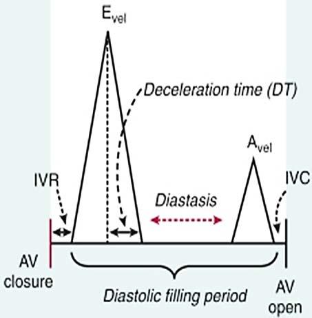
6 Normal mitral inflow


7 LV filling is load dependent Before dialysis After dialysis


8 Pathophysiology of diastolic dysfunction Caused by structural abnormalities that lead to adverse hemodynamic consequences Common predisposing conditions include: o Hypertension o Renal failure o Coronary artery disease o Dilated CM o Hypertrophic CM o Restrictive CM Myocardial ischemia LV hypertrophy LV fibrosis or infiltration Impaired LV relaxation Increased LV stiffness Inability of the heart to fill optimally under normal loading conditions

9 Spectrum of diastolic dysfunction Mitral flow Tissue Doppler

10 Essentials Technical o o o o o o o Competent sonographer Adequate study time Comprehensive assessment Accurate measurements Up-to-date technology Good quality images Cooperative patient Interpretation o o o o Well trained cardiologist Comprehensive Integrated Conclusive

11 Latest guidelines (J Am Soc Echocardiogr 2016;29: )
12 Assessment tools 2D and M mode Flow Doppler Tissue Doppler

13 Common substrates Dilated CM Restrictive CM IHD Scar tissue Hypertrophic CM Aortic stenosis
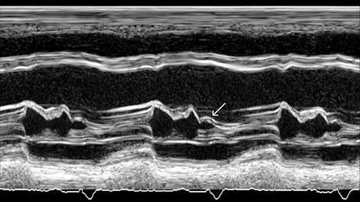
14 Hemodynamic effects B bump Dilated LA High PAP
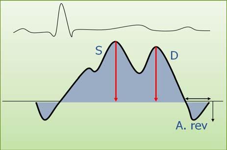
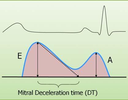
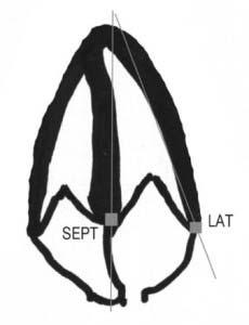
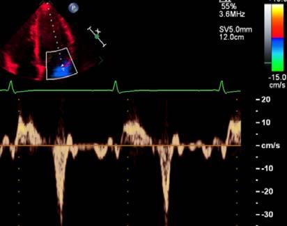
15 Diastolic parameters Flow Doppler Mitral inflow: E velocity A velocity & duration E/A ratio Valsalva maneuver DT L velocity Pulmonary vein flow: S wave D wave S/D ratio A reversal (V & dur) Shared: E/e ratio Ar-A duration Tissue Doppler Mitral annulus: Lat. Velocity (e ) Sept. Velocity (e ) Others LA volume index TR velocity Secondary: IVRT C. M-mode Vp T E-e E/Vp ratio
16 Doppler assessment
17 Normal flow patterns

18 Spectrum of mitral inflow patterns

19 Pulmonary vein flow
20 We need to answer the following questions Is there diastolic dysfunction or not? o Yes o No o Indeterminate How severe is the diastolic dysfunction? o Mild o Moderate o Severe Is the left atrial pressure normal or high? o Low (normal) o High

21 Cut off values for diastolic dysfunction
22 Major criteria for presence of diastolic dysfunction
23 Grading of LAP and diastolic dysfunction


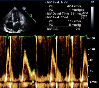
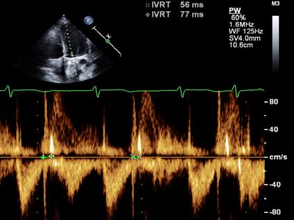
24 A young healthy male Para. Value Normal E vel A vel DT ms E/A e vel Sep 12 S 7 L 10 E/e 7 < 10 LAVI 29 < 34 TR vel 1.5 < 2.8 E/A NA < 0.50 S/D <1 > 1 Ar A NA < 0 ms Vel. (m/s), e vel (cm/s), LAVI (ml/m 2 ), NA (not available)


25 65 y old male with IHD Para. Value Normal E vel A vel DT ms E/A e vel NA S 7 L 10 E/e NA < 10 LAVI NA < 34 TR vel NA < 2.8 E/A NA < 0.50 S/D <1 > 1 Ar A 90 < 0 ms Vel. (m/s), e vel (cm/s), LAVI (ml/m 2 ), NA (not available)


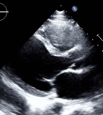
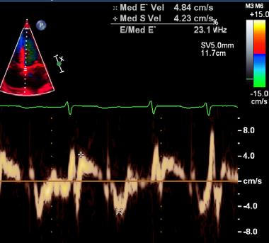
26 93 y old male with severe AS Para. Value Normal E vel A vel DT ms E/A e vel Sept 4 S 7 L 10 E/e 14 < 10 LAVI 31 < 34 TR vel NA < 2.8 E/A NA < 0.50 S/D NA > 1 Ar A NA < 0 ms Vel. (m/s), e vel (cm/s), LAVI (ml/m 2 ), NA (not available)


27 58 y old male with HTN Para. Value Normal E vel A vel DT ms E/A e vel Sep 6 S 7 L 10 E/e 9 < 10 LAVI 34 < 34 TR vel 2 < 2.8 E/A NA < 0.50 S/D NA > 1 Ar A NA < 0 ms Vel. (m/s), e vel (cm/s), LAVI (ml/m 2 ), NA (not available)


28 85 y old female with HTN Para. Value Normal E vel A vel DT ms E/A e vel S 7 4 & 7 L 10 E/e 13 < 10 LAVI 38 < 34 TR vel 2.5 < 2.8 E/A NA < 0.50 S/D >1 > 1 Ar A NA < 0 ms Vel. (m/s), e vel (cm/s), LAVI (ml/m 2 ), NA (not available)


, e vel (cm/s), LAVI")
29 66 y old female with HTN Para. Value Normal E vel A vel DT ms E/A e vel S 7 7 & 4 L 10 E/e 13 < 10 LAVI NA < 34 TR vel 2.7 < 2.8 E/A NA < 0.50 S/D >1 > 1 Ar A NA < 0 ms Vel. (m/s), e vel (cm/s), LAVI (ml/m 2 ), NA (not available)


, LAVI (ml/m")
30 75 y old female with HTN Para. Value Normal E vel A vel DT ms E/A e vel Sep 6 S 7 L 10 E/e 14 < 10 LAVI < 34 TR vel NA < 2.8 E/A NA < 0.50 S/D NA > 1 Ar A NA < 0 ms Vel. (m/s), e vel (cm/s), LAVI (ml/m 2 ), NA (not available)


, LAVI")
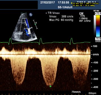
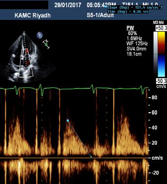
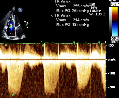
31 47 y old female with ESRD Para. Value Normal E vel A vel DT ms E/A e vel Sept 8 S 7 L 10 E/e 12.5 < 10 LAVI < 34 TR vel 3.6 < 2.8 E/A 57% < 0.50 S/D <1 > 1 Ar A NA < 0 ms Vel. (m/s), e vel (cm/s), LAVI (ml/m 2 ), NA (not available)


, e vel")
32 68 y old male with DCM Para. Value Normal E vel 92/ A vel 34/ DT ms E/A 2.7/ e vel Lat 4 S 7 L 10 E/e 23 < 10 LAVI < 34 TR vel NA < 2.8 E/A 63% < 0.50 S/D <1 > 1 Ar A NA < 0 ms Vel. (m/s), e vel (cm/s), LAVI (ml/m 2 ), NA (not available)


33 56 y old female with ACS/NSTEMI Para. Value Normal E vel A vel DT ms E/A e vel Sept 3 S 7 L 10 E/e 32 < 10 LAVI 44 < 34 TR vel NA < 2.8 E/A NA < 0.50 S/D <1 > 1 Ar A NA < 0 ms Vel. (m/s), e vel (cm/s), LAVI (ml/m 2 ), NA (not available)


34 66 y old male with DCM Para. Value Normal E vel A vel DT ms E/A e vel S 7 4 & 5 L 10 E/e 31 < 10 LAVI NA < 34 TR vel NA < 2.8 E/A NA < 0.50 S/D <1 > 1 Ar A NA < 0 ms Vel. (m/s), e vel (cm/s), LAVI (ml/m 2 ), NA (not available)


, e vel")
35 50 y old male with DCM Para. Value Normal E vel A vel DT ms E/A e vel sept 4 S 7 L 10 E/e 33 < 10 LAVI 47 < 34 TR vel NA < 2.8 E/A NA < 0.50 S/D <1 > 1 Ar A <0 < 0 ms Vel. (m/s), e vel (cm/s), LAVI (ml/m 2 ), NA (not available)


, e vel")
36 60 y old female with high BP Para. Value Normal E vel A vel DT ms E/A e vel Sept 5 S 7 L 10 E/e 23 < 10 LAVI 31 < 34 TR vel NA < 2.8 E/A NA < 0.50 S/D <1 > 1 Ar A NA < 0 ms Vel. (m/s), e vel (cm/s), LAVI (ml/m 2 ), NA (not available)


, e vel")
37 37 y old male with BP of 209/120 mmhg Para. Value Normal E vel A vel DT ms E/A e vel Lat 8 S 7 L 10 E/e 16 < 10 LAVI 45 < 34 TR vel NA < 2.8 E/A NA < 0.50 S/D <1 > 1 Ar A NA < 0 ms Vel. (m/s), e vel (cm/s), LAVI (ml/m 2 ), NA (not available)


. DT of pulmonary")
. E/Vp ratio (1.")
38 Peak acceleration rate of mitral E velocity (1,900 cm/s2). Atrial fibrillation (Normal values) IVRT (65 ms). DT of pulmonary venous diastolic velocity (< 220 ms). E/Vp ratio (1.4). septal E/e= ratio (>11)
39 Atrial fibrillation
40 Conclusions Diastolic assessment is an essential component of echocardiographic study Mitral inflow Doppler, Tissue Doppler, LA volume and TR velocity are the most important components of diastolic assessment New criteria-based algorithms makes interpretation easier, consistent and more clinically oriented
41 Thank you
Μαρία Μπόνου Διευθύντρια ΕΣΥ, ΓΝΑ Λαϊκό
 Μαρία Μπόνου Διευθύντρια ΕΣΥ, ΓΝΑ Λαϊκό Diastolic HF DD: Diastolic Dysfunction DHF: Diastolic HF HFpEF: HF with preserved EF DD Pathophysiologic condition: impaired relaxation, LV compliance, LV filling
Diastolic Function Assessment New Guideline Update Practical Approach
 Mayo Clinic Department of Cardiovascular Diseases Mayo Clinic Echocardiography Review Course for Boards and Recertification Diastolic Function Assessment New Guideline Update Practical Approach Jae K.
An Integrated Approach to Study LV Diastolic Function
 An Integrated Approach to Study LV Diastolic Function Assoc. Prof. Adriana Ilieşiu, FESC University of Medicine Carol Davila Bucharest, Romania LV Diastolic Dysfunction impaired relaxation (early diastole)
Diastolic Heart Function: Applying the New Guidelines Case Studies
 Diastolic Heart Function: Applying the New Guidelines Case Studies Mitral Regurgitation The New ASE William Guidelines: A. Zoghbi Role MD, of FASE, 2D/3D MACCand CMR Professor and Chairman, Department
Diastolic Function Assessment Practical Ways to Incorporate into Every Echo
 Diastolic Function Assessment Practical Ways to Incorporate into Every Echo Jae K. Oh, MD Echo Hawaii 2018 2018 MFMER 3712003-1 Learning Objectives My presentation will help you to Appreciate the importance
Evalua&on)of)Le-)Ventricular)Diastolic) Dysfunc&on)by)Echocardiography:) Role)of)Ejec&on)Frac&on)
of)Le-)Ventricular)Diastolic) Dysfunc&on)by)Echocardiography:) Role)of)Ejec&on)Frac&on)") Evalua&on)of)Le-)Ventricular)Diastolic) Dysfunc&on)by)Echocardiography:) Role)of)Ejec&on)Frac&on) N.Koutsogiannis) Department)of)Cardiology) University)Hospital)of)Patras)! I have no conflicts of interest
Evalua&on)of)Le-)Ventricular)Diastolic) Dysfunc&on)by)Echocardiography:) Role)of)Ejec&on)Frac&on) N.Koutsogiannis) Department)of)Cardiology) University)Hospital)of)Patras)! I have no conflicts of interest
How to Assess Diastolic Dysfunction?
 How to Assess Diastolic Dysfunction? Fausto J Pinto, MD, PhD, FESC, FACC, FASE Lisbon University Dyastolic Dysfunction Impaired relaxation Elevated filling pressures Ischemic heart disease Cardiomyopathies
Basic Approach to the Echocardiographic Evaluation of Ventricular Diastolic Function
 Basic Approach to the Echocardiographic Evaluation of Ventricular Diastolic Function J A F E R A L I, M D U N I V E R S I T Y H O S P I T A L S C A S E M E D I C A L C E N T E R S T A F F C A R D I O T
Choose the grading of diastolic function in 82 yo woman
 Question #1 Choose the grading of diastolic function in 82 yo woman E= 80 cm/s A= 70 cm/s LAVI < 34 ml/m 2 1= Grade 1 2= Grade 2 3= Grade 3 4= Normal 5= Indeterminate 2018 MFMER 3712003-1 Choose the grading
LV FUNCTION ASSESSMENT: WHAT IS BEYOND EJECTION FRACTION
 LV FUNCTION ASSESSMENT: WHAT IS BEYOND EJECTION FRACTION Jamilah S AlRahimi Assistant Professor, KSU-HS Consultant Noninvasive Cardiology KFCC, MNGHA-WR Introduction LV function assessment in Heart Failure:
Diastolic Function: What the Sonographer Needs to Know. Echocardiographic Assessment of Diastolic Function: Basic Concepts 2/8/2012
 Diastolic Function: What the Sonographer Needs to Know Pat Bailey, RDCS, FASE Technical Director Beaumont Health System Echocardiographic Assessment of Diastolic Function: Basic Concepts Practical Hints
GENERAL PRINCIPLES FOR ECHO ASSESSMENT OF DIASTOLIC FUNCTION (For full recommendation refer to the Left Ventricular Diastolic Function Guideline)
") 1 THE AMERICAN SOCIETY OF ECHOCARDIOGRAPHY RECOMMENDATIONS FOR THE EVALUATION OF LEFT VENTRICULAR DIASTOLIC FUNCTION BY ECHOCARDIOGRAPHY: A QUICK REFERENCE GUIDE FROM THE ASE WORKFLOW AND LAB MANAGEMENT
1 THE AMERICAN SOCIETY OF ECHOCARDIOGRAPHY RECOMMENDATIONS FOR THE EVALUATION OF LEFT VENTRICULAR DIASTOLIC FUNCTION BY ECHOCARDIOGRAPHY: A QUICK REFERENCE GUIDE FROM THE ASE WORKFLOW AND LAB MANAGEMENT
The Patient with Atrial Fibrilation
 Assessment of Diastolic Function The Patient with Atrial Fibrilation Assoc. Prof. Adriana Ilieşiu, FESC University of Medicine Carol Davila Bucharest, Romania Associated Conditions with Atrial Fibrillation
HFpEF. April 26, 2018
 HFpEF April 26, 2018 (J Am Coll Cardiol 2017;70:2476 86) HFpEF 50% or more (40-71%) of patients with CHF have preserved LV systolic function. HFpEF is an increasingly frequent hospital discharge. Outcomes
Left ventricular diastolic function and filling pressure in patients with dilated cardiomyopathy
 Left ventricular diastolic function and filling pressure in patients with dilated cardiomyopathy Bogdan A. Popescu University of Medicine and Pharmacy Bucharest, Romania My conflicts of interest: I have
Heart Failure in Women: Dr Goh Ping Ping Cardiologist Asian Heart & Vascular Centre
 Heart Failure in Women: More than EF? Dr Goh Ping Ping Cardiologist Asian Heart & Vascular Centre Overview Review pathophysiology as it relates to diagnosis and management Rational approach to workup:
OPTIMIZING ECHO ACQUISTION FOR STRAIN AND DIASTOLOGY
 OPTIMIZING ECHO ACQUISTION FOR STRAIN AND DIASTOLOGY October 8, 2017 Deborah Agler, ACS, RDCS, FASE Coordinator of Education and Training Cleveland Clinic General Principles Diastology Clinical Data Heart
Objectives. Diastology: What the Radiologist Needs to Know. LV Diastolic Function: Introduction. LV Diastolic Function: Introduction
 Objectives Diastology: What the Radiologist Needs to Know. Jacobo Kirsch, MD Cardiopulmonary Imaging, Section Head Division of Radiology Cleveland Clinic Florida Weston, FL To review the physiology and
Imaging in Heart Failure: A Multimodality Approach. Thomas Ryan, MD
 Imaging in Heart Failure: A Multimodality Approach Thomas Ryan, MD Heart Failure HFrEF HFpEF EF50% Lifetime risk 20% Prevalence 6M Americans Societal costs - $30B 50% 5-year survival 1 Systolic
Review of Cardiac Imaging Modalities in the Renal Patient. George Youssef
 Review of Cardiac Imaging Modalities in the Renal Patient George Youssef ECHO Left ventricular hypertrophy (LVH) assessment Diastolic dysfunction Stress ECHO Cardiac CT angiography Echocardiography - positives
LV geometric and functional changes in VHD: How to assess? Mi-Seung Shin M.D., Ph.D. Gachon University Gil Hospital
 LV geometric and functional changes in VHD: How to assess? Mi-Seung Shin M.D., Ph.D. Gachon University Gil Hospital LV inflow across MV LV LV outflow across AV LV LV geometric changes Pressure overload
Value of echocardiography in chronic dyspnea
 Value of echocardiography in chronic dyspnea Jahrestagung Schweizerische Gesellschaft für /Schweizerische Gesellschaft für Pneumologie B. Kaufmann 16.06.2016 Chronic dyspnea Shortness of breath lasting
Dr.Fayez EL Shaer Consultant cardiologist Assistant professor of cardiology KKUH
 Pulmonary Hypertension in patients with Heart Failure with Preserved Ejection Fraction Dr.Fayez EL Shaer Consultant cardiologist Assistant professor of cardiology KKUH Recent evaluation of available data
LA Function analysis Marcia Barbosa Vice Presidente - Brazilian Soc of Cardiology President-elect - Interamerican Soc of Cardiology
 LA Function analysis Marcia Barbosa Vice Presidente - Brazilian Soc of Cardiology President-elect - Interamerican Soc of Cardiology Belo Horizonte Brazil DECLARATION OF CONFLICT OF INTEREST Nothing to
Diastolic Function Overview
 Diastolic Function Overview Richard Palma BS, RDCS, RCS, APS, FASE Director and Clinical Coordinator The Hoffman Heart and Vascular Institute School of Cardiac Ultrasound None Disclosures Learning Objectives
Pericardial Diseases. Smonporn Boonyaratavej, MD. Division of Cardiology, Department of Medicine Chulalongkorn University
 Pericardial Diseases Smonporn Boonyaratavej, MD Division of Cardiology, Department of Medicine Chulalongkorn University Cardiac Center, King Chulalongkorn Memorial Hospital 21 AUGUST 2016 Pericardial
E/Ea is NOT an essential estimator of LV filling pressures
 Euroecho Kopenhagen Echo in Resynchronization in 2010 E/Ea is NOT an essential estimator of LV filling pressures Wilfried Mullens, MD, PhD December 10, 2010 Ziekenhuis Oost Limburg Genk University Hasselt
P = 4V 2. IVC Dimensions 10/20/2014. Comprehensive Hemodynamic Evaluation by Doppler Echocardiography. The Simplified Bernoulli Equation
 Comprehensive Hemodynamic Evaluation by Doppler Echocardiography Itzhak Kronzon, MD North Shore LIJ/ Lenox Hill Hospital New York, NY Disclosure: Philips Healthcare St. Jude Medical The Simplified Bernoulli
Advanced imaging of the left atrium - strain, CT, 3D, MRI -
 Advanced imaging of the left atrium - strain, CT, 3D, MRI - Monica Rosca, MD Carol Davila University of Medicine and Pharmacy, Bucharest, Romania Declaration of interest: I have nothing to declare Case
DOPPLER HEMODYNAMICS (1) QUANTIFICATION OF PRESSURE GRADIENTS and INTRACARDIAC PRESSURES
 QUANTIFICATION OF PRESSURE GRADIENTS and INTRACARDIAC PRESSURES") THORAXCENTRE DOPPLER HEMODYNAMICS (1) QUANTIFICATION OF PRESSURE GRADIENTS and INTRACARDIAC PRESSURES J. Roelandt DOPPLER HEMODYNAMICS Intracardiac pressures and pressure gradients Volumetric measurement
THORAXCENTRE DOPPLER HEMODYNAMICS (1) QUANTIFICATION OF PRESSURE GRADIENTS and INTRACARDIAC PRESSURES J. Roelandt DOPPLER HEMODYNAMICS Intracardiac pressures and pressure gradients Volumetric measurement
Ref 1. Ref 2. Ref 3. Ref 4. See graph
 Ref 1 Ref 2 Ref 3 1. Ages 6-23 y/o 2. Significant LVM differences by gender 3. For males 95 th percentiles: a. LVM/BSA = 103 b. LVM/height = 100 4. For females 95 th percentiles: a. LVM/BSA = 84 b. LVM/height
Adult Echocardiography Examination Content Outline
 Adult Echocardiography Examination Content Outline (Outline Summary) # Domain Subdomain Percentage 1 2 3 4 5 Anatomy and Physiology Pathology Clinical Care and Safety Measurement Techniques, Maneuvers,
Diastolic Heart Failure
 Chronic Heart Failure Prevalence overall = 2-3 % Diastolic Heart Failure Patrick Wouters University Hospital Ghent Belgium (Heart Failure + Asymptomatic Ventricular Dysfunction) Prevalence > 70 y = 10-20
HFPEF Echo with Strain vs. MRI T1 Mapping
 HFPEF Echo with Strain vs. MRI T1 Mapping Erik Schelbert, MD MS Director, Cardiovascular Magnetic Resonance Assistant Professor of Medicine Heart & Vascular Institute University of Pittsburgh Disclosures
Atrial dyssynchrony syndrome: An overlooked cause of heart failure with normal ejection fraction
 Atrial dyssynchrony syndrome: An overlooked cause of heart failure with normal ejection fraction JC Eicher, G Laurent, O Barthez, A Mathé, G Bertaux, JE Wolf Heart Failure Treatment Unit, Rhythmology and
Highlights from EuroEcho 2009 Echo in cardiomyopathies
 Highlights from EuroEcho 2009 Echo in cardiomyopathies Bogdan A. Popescu University of Medicine and Pharmacy, Bucharest, Romania ESC Congress 2010 Hypertrophic cardiomyopathy To determine the differences
Rotation: Echocardiography: Transthoracic Echocardiography (TTE)
") Rotation: Echocardiography: Transthoracic Echocardiography (TTE) Rotation Format and Responsibilities: Fellows rotate in the echocardiography laboratory in each clinical year. Rotations during the first
Rotation: Echocardiography: Transthoracic Echocardiography (TTE) Rotation Format and Responsibilities: Fellows rotate in the echocardiography laboratory in each clinical year. Rotations during the first
Echo Doppler Assessment of Right and Left Ventricular Hemodynamics.
 Echo Doppler Assessment of Right and Left Ventricular Hemodynamics. Itzhak Kronzon, MD, FASE, FACC, FESC, FAHA, FACP, FCCP Northwell, Lenox Hill Hospital, New York Professor of Cardiology Hofstra University
Diastolic Functions: Evaluation & Clinical Applications
 Special Articles Diastolic Functions: Evaluation & Clinical Applications Senior Consultant Cardiologist, Metro Heart Institute, Delhi Immediate Past President, Cardiological Society of India (Cardiovasc.
Echocardiographic and Doppler Assessment of Cardiac Functions in Patients of Non-Insulin Dependent Diabetes Mellitus
 ORIGINAL ARTICLE JIACM 2002; 3(2): 164-8 Echocardiographic and Doppler Assessment of Cardiac Functions in Patients of Non-Insulin Dependent Diabetes Mellitus Rajesh Rajput*, Jagdish**, SB Siwach***, A
HFNEF. Heart Failure is
 HFNEF Bijoy K. Khandheria, MD. FASE, FACP, FACC FESC Professor of Medicine University of Wisconsin Director. Echocardiography Services Aurora Health Care No conflicts or off label use CP1173868-1 Heart
The importance of left atrium in LV diastolic function
 II Baltic Heart Failure Meeting and Congress of Latvian Society of Cardiology The importance of left atrium in LV diastolic function Dr. Artem Kalinin Eastern Clinical University Hospital Riga 30.09.2010.
The Causes of Heart Failure
 The Causes of Heart Failure Andy Birchall HFSN Right heart failure LVSD - HFREF Valve regurgitation or stenosis Dropsy CCF congestive cardiac failure Cor pulmonale Pulmonary hypertension HFPEF LVF Definitions
Diastole is Not a Single Entity Four Components of Diastolic Dysfunction
 Physiology of Diastolic Function Made Easy James D. Thomas, MD, FACC, FASE Director, Center for Heart Valve Disease Bluhm Cardiovascular Institute Professor of Medicine, Feinberg School of Medicine, Northwestern
Jong-Won Ha*, Jeong-Ah Ahn, Jae-Yun Moon, Hye-Sun Suh, Seok-Min Kang, Se-Joong Rim, Yangsoo Jang, Namsik Chung, Won-Heum Shim, Seung-Yun Cho
 Eur J Echocardiography (2006) 7, 16e21 CLINICAL/ORIGINAL PAPERS Triphasic mitral inflow velocity with mid-diastolic flow: The presence of mid-diastolic mitral annular velocity indicates advanced diastolic
Effect of Heart Rate on Tissue Doppler Measures of E/E
 Cardiology Department of Bangkok Metropolitan Administration Medical College and Vajira Hospital, Bangkok, Thailand Abstract Background: Our aim was to study the independent effect of heart rate (HR) on
The new Guidelines: Focus on Chronic Heart Failure
 The new Guidelines: Focus on Chronic Heart Failure Petros Nihoyannopoulos MD, FRCP, FESC Professor of Cardiology Imperial College London and National & Kapodistrian University of Athens 2 3 4 The principal
SONOGRAPHER & NURSE LED VALVE CLINICS
 SONOGRAPHER & NURSE LED VALVE CLINICS Frequency of visits and alerts AORTIC STENOSIS V max > 4.0 m/s or EOA < 1.0 cm 2 V max 3.5 4.0 m/s + Ca+ V max 3.0 4.0 m/s or EOA 1.0-1.5 cm 2 V max 2.5 3.0 m/s every
Mechanisms of heart failure with normal EF Arterial stiffness and ventricular-arterial coupling. What is the pathophysiology at presentation?
 Mechanisms of heart failure with normal EF Arterial stiffness and ventricular-arterial coupling What is the pathophysiology at presentation? Ventricular-arterial coupling elastance Central arterial pressure
HEMODYNAMIC ASSESSMENT
 HEMODYNAMIC ASSESSMENT INTRODUCTION Conventionally hemodynamics were obtained by cardiac catheterization. It is possible to determine the same by echocardiography. Methods M-mode & 2D echo alone can provide
Evaluation of Left Ventricular Diastolic Dysfunction by Doppler and 2D Speckle-tracking Imaging in Patients with Primary Pulmonary Hypertension
 ESC Congress 2011.No 85975 Evaluation of Left Ventricular Diastolic Dysfunction by Doppler and 2D Speckle-tracking Imaging in Patients with Primary Pulmonary Hypertension Second Department of Internal
The Doppler Examination. Katie Twomley, MD Wake Forest Baptist Health - Lexington
 The Doppler Examination Katie Twomley, MD Wake Forest Baptist Health - Lexington OUTLINE Principles/Physics Use in valvular assessment Aortic stenosis (continuity equation) Aortic regurgitation (pressure
Comprehensive Hemodynamics By Doppler Echocardiography. The Echocardiographic Swan-Ganz Catheter.
 Comprehensive Hemodynamics By Doppler Echocardiography. The Echocardiographic Swan-Ganz Catheter. Itzhak Kronzon, MD, FASE, FACC, FESC, FAHA, FACP, FCCP North Shore HS, LIJ/Lenox Hill Hospital, New York
European Heart Journal - Cardiovascular Imaging Advance Access published July 18, 2016
 European Heart Journal - Cardiovascular Imaging Advance Access published July 18, 2016 European Heart Journal Cardiovascular Imaging doi:10.1093/ehjci/jew082 Recommendations for the Evaluation of Left
Hemodynamic Assessment. Assessment of Systolic Function Doppler Hemodynamics
 Hemodynamic Assessment Matt M. Umland, RDCS, FASE Aurora Medical Group Milwaukee, WI Assessment of Systolic Function Doppler Hemodynamics Stroke Volume Cardiac Output Cardiac Index Tei Index/Index of myocardial
NEW GUIDELINES. A Guideline Protocol for the Echocardiographic assessment of Diastolic Dysfunction
 NEW GUIDELINES A Guideline Protocol for the Echocardiographic assessment of Diastolic Dysfunction Echocardiography plays a central role in the non-invasive evaluation of diastole and should be interpreted
Diastolic Heart Failure (HFpEF) Felix J. Rogers, DO, FACOI April 29, 2018
 Felix J. Rogers, DO, FACOI April 29, 2018") Diastolic Heart Failure (HFpEF) Felix J. Rogers, DO, FACOI April 29, 2018 Case presentation MSO, an 81 year old woman was admitted to HFWH because of progressive dyspnea and difficult to control hypertension
Diastolic Heart Failure (HFpEF) Felix J. Rogers, DO, FACOI April 29, 2018 Case presentation MSO, an 81 year old woman was admitted to HFWH because of progressive dyspnea and difficult to control hypertension
E LeAP STUDY ECHOCARDIOGRAPHIC LEFT ATRIAL PRESSURE: COMPARATIVE REVIEW IN PATIENTS WITH SINUS RHYTHM AND ATRIAL FIBRILLATION
 E LeAP STUDY ECHOCARDIOGRAPHIC LEFT ATRIAL PRESSURE: COMPARATIVE REVIEW IN PATIENTS WITH SINUS RHYTHM AND ATRIAL FIBRILLATION Moghniuddin Mohammed, M.B.B.S. Mentor: Dr. Harry McCrea, M.D. Dr. Yuji Saito,
Echocardiography: Guidelines for Valve Quantification
 Echocardiography: Guidelines for Echocardiography: Guidelines for Chamber Quantification British Society of Echocardiography Education Committee Richard Steeds (Chair), Gill Wharton (Lead Author), Jane
Constriction vs Restriction Case-based Discussion
 Mayo Clinic Department of Cardiovascular Diseases Mayo Clinic Echocardiography Review Course for Boards and Recertification Constriction vs Restriction Case-based Discussion Jae K. Oh, MD Samsung Professor
Adel Hasanin Ahmed 1
 Adel Hasanin Ahmed 1 PERICARDIAL DISEASE The pericardial effusion ends anteriorly to the descending aorta and is best visualised in the PLAX. PSAX is actually very useful sometimes for looking at posterior
M. Hajahmadi Poorrafsanjani 1 & B. Rahimi Darabad 1
 Global Journal of Health Science; Vol. 6, No. 7; 2014 ISSN 1916-9736 E-ISSN 1916-9744 Published by Canadian Center of Science and Education Evaluate the Sensitivity and Specificity Echocardiography in
SUPPLEMENTAL MATERIAL
 SUPPLEMENTAL MATERIAL Table S1: Number and percentage of patients by age category Distribution of age Age
Echo-Doppler evaluation of left ventricular diastolic function. Michel Slama Amiens France
 Echo-Doppler evaluation of left ventricular diastolic function Michel Slama Amiens France Left ventricular pressure Pressure A wave [ LVEDP LVEDP préa Congestive cardiac failure with preserved systolic
Coronary Anomalies & Hemodynamic Identification
 Coronary Anomalies & Hemodynamic Identification David Stultz, MD Cardiology Fellow, PGY 6 May 2, 2006 Anomaly #1 Anomaly #2 Anomaly #3 Figure 18-27 Anomalous origin of the left circumflex artery.
Assessing the Impact on the Right Ventricle
 Advances in Tricuspid Regurgitation Congress of the European Society of Cardiology (ESC) Munich, August 25-29, 2012 Assessing the Impact on the Right Ventricle Stephan Rosenkranz, MD Clinic III for Internal
Echocardiographic Evaluation of the Cardiomyopathies. Stephanie Coulter, MD, FACC, FASE April, 2016
 Echocardiographic Evaluation of the Cardiomyopathies Stephanie Coulter, MD, FACC, FASE April, 2016 Cardiomyopathies (CMP) primary disease intrinsic to cardiac muscle Dilated CMP Hypertrophic CMP Infiltrative
Diastolic Heart Failure
 58 Diastolic Heart Failure Muhamed Saric and Itzhak Kronzon PRACTICAL POINTS Diastole starts with the closure of the aortic valve and ends with the closure of the mitral valve. It consists of four phases:
Aortic Stenosis: Spectrum of Disease, Low Flow/Low Gradient and Variants
 Aortic Stenosis: Spectrum of Disease, Low Flow/Low Gradient and Variants Martin G. Keane, MD, FASE Professor of Medicine Lewis Katz School of Medicine at Temple University Basic root structure Parasternal
MITRAL STENOSIS. Joanne Cusack
 MITRAL STENOSIS Joanne Cusack BSE Breakdown Recognition of rheumatic mitral stenosis Qualitative description of valve and sub-valve calcification and fibrosis Measurement of orifice area by planimetry
Appendix II: ECHOCARDIOGRAPHY ANALYSIS
 Appendix II: ECHOCARDIOGRAPHY ANALYSIS Two-Dimensional (2D) imaging was performed using the Vivid 7 Advantage cardiovascular ultrasound system (GE Medical Systems, Milwaukee) with a frame rate of 400 frames
Left atrial function. Aliakbar Arvandi MD
 In the clinic Left atrial function Abstract The left atrium (LA) is a left posterior cardiac chamber which is located adjacent to the esophagus. It is separated from the right atrium by the inter-atrial
Conflicts of interest: GE, Abbott, Edwards (honoraria)
") Understanding Diastole and Its Contribution to Heart Failure: State of the Art in 2016 James D. Thomas, MD, FACC, FASE Director, Center for Heart Valve Disease Bluhm Cardiovascular Institute Professor
Understanding Diastole and Its Contribution to Heart Failure: State of the Art in 2016 James D. Thomas, MD, FACC, FASE Director, Center for Heart Valve Disease Bluhm Cardiovascular Institute Professor
Noninvasive assessment of left ventricular (LV)
") Comparative Value of Tissue Doppler Imaging and M-Mode Color Doppler Mitral Flow Propagation Velocity for the Evaluation of Left Ventricular Filling Pressure* Michal Kidawa, MD; Lisa Coignard, MD; Gérard
Comparative Value of Tissue Doppler Imaging and M-Mode Color Doppler Mitral Flow Propagation Velocity for the Evaluation of Left Ventricular Filling Pressure* Michal Kidawa, MD; Lisa Coignard, MD; Gérard
Diastology Disclosures: None. Dias2011:1
 Diastology 2011 James D. Thomas, M.D., F.A.C.C. Cardiovascular Imaging Center Department of Cardiology Cleveland Clinic Foundation Cleveland, Ohio, USA Disclosures: None Dias2011:1 Is EVERYBODY a member!?!
Doppler Basic & Hemodynamic Calculations
 Doppler Basic & Hemodynamic Calculations August 19, 2017 Smonporn Boonyaratavej MD Division of Cardiology, Department of Medicine Chulalongkorn University Cardiac Center, King Chulalongkorn Memorial Hospital
L ecocardiografia nello Scompenso Cardiaco Acuto e cronico: vecchi dogmi e nuovi trends.
 V SESSIONE SCOMPENSO CARDIACO 2015 Genova, 13-14 Novembre 2015 L ecocardiografia nello Scompenso Cardiaco Acuto e cronico: vecchi dogmi e nuovi trends. Gian Paolo Bezante, MD, FACC UOC Clinica di Malattie
Echocardiography for the Electrophysiologist: Day-to-day practice. Emmanuel Fares, MD
 Echocardiography for the Electrophysiologist: Day-to-day practice Emmanuel Fares, MD EP and pacing service, Department of Cardiovascular Medicine, Cairo University Agenda Role of echo in arrhythmia management:
Left Ventricular Diastolic Dysfunction in South Indian Essential Hypertensive Patient
 Left Ventricular Diastolic Dysfunction in South Indian Essential Hypertensive Patient Dr. Peersab.M. Pinjar 1, Dr Praveenkumar Devarbahvi 1 and Dr Vasudeva Murthy.C.R 2, Dr.S.S.Bhat 1, Dr.Jayaraj S G 1
Constrictive Pericarditis
 Constrictive Pericarditis Never Confused with Anything Else Jae K. Oh, MD 2018 MFMER 3712003-1 ARS #1 CP Which of following patients has constrictive pericarditis? 1 2 3 Medial e 13 cm/s Medial e 3 cm/s
Index of subjects. effect on ventricular tachycardia 30 treatment with 101, 116 boosterpump 80 Brockenbrough phenomenon 55, 125
 145 Index of subjects A accessory pathways 3 amiodarone 4, 5, 6, 23, 30, 97, 102 angina pectoris 4, 24, 1l0, 137, 139, 140 angulation, of cavity 73, 74 aorta aortic flow velocity 2 aortic insufficiency
Echo in Pulmonary HTN
 Echo in Pulmonary HTN Steven A. Goldstein MD FACC FASE Professor of Medicine Georgetown University Medical Center MedStar Heart Institute Washington Hospital Center Monday, October 10, 2017 Pulmonary Artery
ΚΑΡΔΙΟΛΟΓΟΣ EUROPEAN ACCREDITATION IN TRANSTHORACIC AND TRANSESOPHAGEAL ECHOCARDIOGRAPHY
 1 ΚΑΡΔΙΟΛΟΓΟΣ EUROPEAN ACCREDITATION IN TRANSTHORACIC AND TRANSESOPHAGEAL ECHOCARDIOGRAPHY 2 Constrictive pericarditis (CP) is characterized by impaired ventricular filling due to a stiffened or noncompliant
Hypertensive heart disease and failure
 Hypertensive heart disease and failure Prof. Dr. Alan Fraser Cardiff University The heart in hypertension Pathophysiology of LV adaptation Regional development of hypertrophy Stress testing - inducible
Swan Song: Echocardiography as a Pulmonary Artery Catheter? Interdepartmental Division of Critical Care Medicine
 Swan Song: Echocardiography as a Pulmonary Artery Catheter? The swan is without spot, and it sings sweetly as it dies, that song ending its life Leonardo Da Vinci Curr Opin Anesthesiol 2016, 29:36 45 Circulation.
Index. K Knobology, TTE artifact, image resolution, ultrasound, 14
 A Acute aortic regurgitation (AR), 124 128 Acute aortic syndrome (AAS) classic aortic dissection diagnosis, 251 263 evolutive patterns, 253 255 pathology, 250 251 classifications, 247 248 incomplete aortic
The right ventricle in chronic heart failure
 The right ventricle in chronic heart failure ESC 2012 Christian Opitz, Berlin There are no conflicts of interest relevant to this presentation Percent of Population Prevalence of Heart Failure by Age and
Cardiac hypertrophy : differentiating disease from athlete
 Cardiac hypertrophy : differentiating disease from athlete Ario Soeryo Kuncoro, MD, Cardiologist Echocardiography Division, National Cardiovascular Centre Harapan Kita-Jakarta Departement of Cardiology
Objectives. Systolic Heart Failure: Definitions. Heart Failure: Historical Perspective 2/7/2009
 Objectives Diastolic Heart Failure and Indications for Echocardiography in the Asian Population Damon M. Kwan, MD UCSF Asian Heart & Vascular Symposium 02.07.09 Define diastolic heart failure and differentiate
Managing Hypertrophic Cardiomyopathy with Imaging. Gisela C. Mueller University of Michigan Department of Radiology
 Managing Hypertrophic Cardiomyopathy with Imaging Gisela C. Mueller University of Michigan Department of Radiology Disclosures Gadolinium contrast material for cardiac MRI Acronyms Afib CAD Atrial fibrillation
2/4/2011. Nathan Kerner, M.D.
 Nathan Kerner, M.D. Definition Elevated pressures - cut off usually >40 mmhg pulmonary artery systolic pressure (PASP) Usually associated with elevated pulmonary vascular resistance (PVR) measured in dynessec/cm
Evaluation of Left Ventricular Function and Hypertrophy Gerard P. Aurigemma MD
 Evaluation of Left Ventricular Function and Hypertrophy Gerard P. Aurigemma MD Board Review Course 2017 43 year old health assistant Severe resistant HTN LT BSA 2 Height 64 1 Here is the M mode echocardiogram
Presenter: Steven Brust, HCS-D, HCS-H Product Manager, Home Health Coding Center
 Presenter: Steven Brust, HCS-D, HCS-H Product Manager, Home Health Coding Center Pinpoint & properly assign the appropriate heart failure codes Left- vs. Right-sided Left ventricular failure (LVF) may
PROSTHETIC VALVE BOARD REVIEW
 PROSTHETIC VALVE BOARD REVIEW The correct answer D This two chamber view shows a porcine mitral prosthesis with the typical appearance of the struts although the leaflets are not well seen. The valve
Diagnosis is it really Heart Failure?
 ESC Congress Munich - 25-29 August 2012 Heart Failure with Preserved Ejection Fraction From Bench to Bedside Diagnosis is it really Heart Failure? Prof. Burkert Pieske Department of Cardiology Med.University
Tissue Doppler Imaging
 Cronicon OPEN ACCESS Hesham Rashid* Tissue Doppler Imaging CARDIOLOGY Editorial Department of Cardiology, Benha University, Egypt *Corresponding Author: Hesham Rashid, Department of Cardiology, Benha University,
Diastolic Function. Rick Nishimura Leighton Professor of CV Diseases Mayo Clinic No Disclosures
 Diastolic Function Rick Nishimura Leighton Professor of CV Diseases Mayo Clinic No Disclosures Heart = Pump Heart Failure Systolic Dysfunction Diastolic Dysfunction Diastole is a complex sequence of multiple
Prognostic Value of Left Atrial Size and Function
 Prognostic Value of Left Atrial Size and Function James D. Thomas, M.D., F.A.C.C. Cardiovascular Imaging Center Department of Cardiology Cleveland Clinic Foundation Cleveland, Ohio, USA Conflicts: None
Bogdan A. Popescu. University of Medicine and Pharmacy Bucharest, Romania. EAE Course, Bucharest, April 2010
 Bogdan A. Popescu University of Medicine and Pharmacy Bucharest, Romania EAE Course, Bucharest, April 2010 This is how it started Mitral stenosis at a glance 2D echo narrow diastolic opening of MV leaflets
Right Heart Catheterization. Franz R. Eberli MD Chief of Cardiology Stadtspital Triemli, Zurich
 Right Heart Catheterization Franz R. Eberli MD Chief of Cardiology Stadtspital Triemli, Zurich Right Heart Catheterization Pressure measurements Oxygen saturation measurements Cardiac output, Vascular