GENERAL PRINCIPLES FOR ECHO ASSESSMENT OF DIASTOLIC FUNCTION (For full recommendation refer to the Left Ventricular Diastolic Function Guideline)
|
|
|
- Myrtle Carr
- 5 years ago
- Views:
Transcription
1 1 THE AMERICAN SOCIETY OF ECHOCARDIOGRAPHY RECOMMENDATIONS FOR THE EVALUATION OF LEFT VENTRICULAR DIASTOLIC FUNCTION BY ECHOCARDIOGRAPHY: A QUICK REFERENCE GUIDE FROM THE ASE WORKFLOW AND LAB MANAGEMENT TASK FORCE This document summarizes key points from the 2016 ASE Left Ventricular Diastolic Function Guideline to serve as a quick reference for sonographers and interpreting physicians. (1) For details including the methodology and the rationale for current recommendations, the interested reader is referred to the complete Guideline statement. Figures and tables are reproduced from ASE Guidelines. (1) Table of Contents: 1. General Principles for Echo Assessment of LV Diastolic Function p. 2 a. Overview of the Assessment of Diastolic Function p. 2 b. Diagnosing Diastolic Dysfunction in the Presence of a Normal LV EF p. 2 c. Assessment of LV Filling Pressures and Diastolic Dysfunction Grade p. 3 d. Conclusions on Diastolic Function in the Clinical Report p Estimation of Filling Pressures in Specific Cardiovascular Diseases p Diastolic Stress Test p Novel Indices of LV Diastolic Function p Prognostic Information Obtained From Diastolic Function Assessment p Appendix Flow Charts p. 9 References: 1. Nagueh SF, Smiseth OA, Appleton CP, et al. Recommendations for the Evaluation of Left Ventricular Diastolic Function by Echocardiography: An Update from the American Society of Echocardiography and the European Association of Cardiovascular Imaging. J Am Soc Echocardiogr 2016;29; Notice and Disclaimer: This report is made available by ASE as a courtesy reference source for members. This report contains recommendations only and should not be used as the sole basis to make medical practice decisions or for disciplinary action a gainst any employee. The statements and recommendations contained in this report are primarily based on the opinions of experts, rather than on scientifically verified data. ASE makes no express or implied warranties regarding the completeness or accuracy of the information in this report, including the warranty of merchantability or fitness for a particular purpose. In no event shall ASE be liable to you, your p atients, or any other third parties for any decision made or action taken by you or such other parties in reliance on this informa tion. Nor does your use of this information constitute the offering of medical advice by ASE or create any physician-patient relationship between ASE and your patients or anyone else. ASE Workflow & Lab Management Taskforce: David A. Orsinelli, MD, FASE (Chair), Alicia Armour, BS, MA, RDCS, FASE, Jeanne De Cara, MD, FASE, Brian Fey, RDCS, ACS, FASE, Lisa Hornberger, MD, FASE, Juan Lopez-Mattei, MD, FASE, Jane Marshall, BS, RDCS, FASE, Athena Poppas, MD, FASE, Vandana Sachdev, MD, FASE, Liza Sanchez, RCS, FASE, and Timothy Woods, MD, FASE, Columbus, Ohio; Durham, North Carolina; Chicago, Illinois; Edmonton, AB Canada; Houston, Texas; Boston, Massachusetts; Providence, Rhode Island; Bethesda, Maryland; Memphis, Tennessee. March 2019
2 2 GENERAL PRINCIPLES FOR ECHO ASSESSMENT OF DIASTOLIC FUNCTION (For full recommendation refer to the Left Ventricular Diastolic Function Guideline) OVERVIEW OF THE ASSESSMENT OF DIASTOLIC FUNCTION For a review of the Components of Diastolic Function and Parameters that may impact the Assessment of Diastolic Function, as well as Potential Pitfalls in the algorithms and a comprehensive list of the Echocardiographic Indices used in the assessment of Diastolic function, please refer to the original guideline document (pages 278, 279) and Table 2 in the guideline DIAGNOSING DIASTOLIC DYSFUNCTION IN THE PRESENCE OF A NORMAL LV EF (For full recommendation refer to the Left Ventricular Diastolic Function Guideline p. 279) Age Independent Indices of Diastolic Function E/e : > 14 is rare in normal individuals Change in mitral inflow with Valsalva: 50% change in E/A o highly specific for increased LV filling pressures o supports diagnosis of diastolic dysfunction o requires continuous recording of the Doppler signal during 10 seconds of Valsalva Difference in duration between pulmonary vein Ar velocity and mitral A velocity (>30 ms) Elevated pulmonary artery systolic pressure (PASP) in the absence of pulmonary vascular disease Consider all diastolic parameters in the context of other echo findings that may suggest the presence of abnormal myocardial function even with a normal EF o LA enlargement (in the absence of atrial arrhythmia or MV disease) o Left ventricular hypertrophy (LVH) o Abnormal global longitudinal function (assessed by mitral annular plane systolic excursion, mitral annulus s, GLS) Diastolic Function Assessment in the Presence of a Normal LVEF (Figure 8A) The presence or absence of diastolic dysfunction in patients with a normal LVEF is based on the assessment of four variables. These variables and their cutoff values include: o Septal e < 7 cm/sec or Lateral e < 10 cm/sec o Average E/e > 14 o LA volume index > 34mL/m 2 o Peak TR velocity > 2.8 m /sec Diastolic function is normal if more than half of the available variables are normal (<50% positive) Diastolic dysfunction is present if more than half of the available variables are abnormal (> 50% positive) In cases in which half of the variables do not meet the cutoff value, the study is indeterminate (50% positive)
3 ASSESSMENT OF LV FILLING PRESSURES AND DIASTOLIC DYSFUNCTION GRADE (For full recommendation refer to the Left Ventricular Diastolic Function Guideline p. 281) Diastolic Function Assessment in Patients with Normal vs Abnormal LVEF In patients with a normal LVEF, the initial assessment is to determine the presence or absence of diastolic dysfunction based on the algorithm presented above and in Figure 8A o If there is evidence of myocardial disease or diastolic dysfunction, the second goal is to estimate LV filling pressures and the grade of diastolic dysfunction based on the parameters presented below and the algorithm in Figure 8B In patients with heart failure and reduced EF (HFrEF), the main goal is to estimate LV filling pre ssures and grade the degree of diastolic dysfunction (diastolic dysfunction is presumed to be present in these patients) based on the parameters presented below and the algorithm in Figure 8B The algorithm (Figure 8B) focuses on the assessment of mean LA pressure (LAP) as it correlates best with pulmonary capillary wedge pressure and pulmonary congestion The algorithm starts with mitral inflow velocities and does not apply in several specific cardiovascular diseases which are addressed in the next section Assessment of Filling Pressures and Diastolic Function in Heart Failure with preserved EF (HFpEF) or HFrEF (Fig 8 of Guidelines): o If mitral E/A ratio < 0.8 and peak mitral E 50 cm/s Mean LAP is normal or low Grade I diastolic dysfunction o If mitral E/A 2 Mean LAP is elevated Grade III diastolic dysfunction o If mitral E/A 0.8 and Peak E > 50 cm/s or E/A > 0.8 but < 2 the following 3 parameters should be evaluated: Average E/e > 14 LA volume index > 34mL/m 2 Peak TR velocity > 2.8 m /sec Interpretation: If 2/3 or 3/3 are negative, LAP is normal (Grade I Diastolic Dysfunction) If 2/3 or 3/3 are positive, LAP is increased (Grade II Diastolic Dysfunction) If only 2 parameters are available, if both are normal, LAP is normal. If both are abnormal, LAP is increased. If 1 is normal and the other abnormal, results are inconclusive Other Scenarios o If one of the 3 above variables is not available: PVs:PVd velocity ratio or VTI ratio < 1 supports the presence of elevated filling pressures Note: In individuals < 40 yrs old, PVs:PVd ratio may be < 1; Use e and LA volume index to avoid misclassifying diastolic function If only 1 variable is available do not comment on diastolic function Caveats in the Diastolic Function Assessment Algorithm o After cardioversion of AF: E decel time should be used in the assessment of diastolic function as the mitral A wave velocity may be decreased due to atrial stunning and thus the E/A ratio may be 2 despite the absence of elevated filling pressures o Young patients: E/A ratio may be 2 in normal young patients Use other parameters to detect abnormal diastolic function Note: e will be normal in individuals with normal diastolic function 3
4 Additional Considerations in the Diastolic Function Assessment in Preserved EF Patients Evaluate 2D and Doppler parameters o LVEF o Regional wall motion abnormalities (RWMA) o LVH Best confirmed by LV mass that exceeds gender-specific norms LA size: If LA > RA size in the apical 4C view, this suggests chronically elevated LV filling pressures in absence of: o Atrial fibrillation o Mitral valve disease o Anemia o Normal LA volume index does not exclude diastolic dysfunction Can be normal in early diastolic dysfunction Can be normal in acutely elevated filling pressures Estimated PASP: Elevation in the absence of pulmonary vascular or parenchymal disease suggests elevated LV filling pressures PVs:PVd ratio: Not helpful in patients with normal EF 4

5 Figure 8 from Left Ventricular Diastolic Function Guideline 5
 Conclusions about diastolic function should be included in the report, particularly if the indication is for dyspnea or HF including: o Filling pressures o Grade of diastolic dysfunction o")
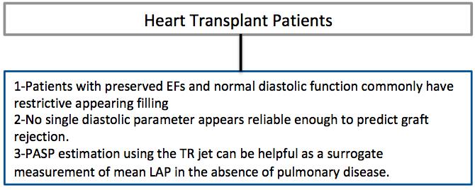
6 6 CONCLUSIONS ON DIASTOLIC FUNCTION IN THE CLINICAL REPORT (For full recommendation refer to the Left Ventricular Diastolic Function Guideline p. 288) Conclusions about diastolic function should be included in the report, particularly if the indication is for dyspnea or HF including: o Filling pressures o Grade of diastolic dysfunction o Comparison to prior studies Consider diastolic stress test in borderline cases Consider right heart catheterization if there is a concern for discordance between RV and LV filling pressures suggesting pulmonary vascular disease ESTIMATION OF FILLING PRESSURES IN SPECIFIC CARDIOVASCULAR DISEASES (For full recommendation refer to the Left Ventricular Diastolic Function Guideline p.288) General Considerations Many disease states, valvular and other anatomic abnormalities, as well as dysrhythmias may modify the relationship between indices of diastolic function and LV filling pressure The algorithm for assessing diastolic function and estimating LV filling pressures presented in the prior discussion has several limitations in these circumstances o The TR jet remains a valid marker of LAP, assuming there is no evidence of pulmonary vascular or parenchymal disease that may result in increased right ventricular systolic pressure (RVSP) o LA enlargement usually reflects an elevated LAP, except in the setting of atrial fibrillation / flutter, significant mitral valve disease, high flow states or cardiac transplantation In the absence of these conditions, an enlarged LA with a normal RA strongly suggests elevated LAP LA enlargement may persist in patients with well treated congestive heart failure and normal filling pressures. If the TR jet is > 2.8 m/s, an elevated LAP is suggested o Additional parameters such as pulmonary vein (PV) flow, Isovolumic Relaxation Time (IVRT), A r -A wave duration (time difference between the pulmonary vein atrial reversal wave duration and transmitral A wave duration), T E-e (time difference from onset of E wave and e ) and the IVRT / T E-e ratio may need to be assessed In patients with pulmonary hypertension, the lateral E/e may be helpful in differentiating whether or not a cardiac etiology is the underlying reason for an elevated RVSP
7 7 o With non-cardiac pulmonary hypertension, the lateral E/e is < 8 o In the presence of a cardiac etiology, the lateral E/e is > 13 Specific Conditions in Which the Standard Algorithm (Figure 8) May Not Apply (The algorithms presented in the Appendix on Page 9 may be used) Include: Hypertrophic Cardiomyopathy Restrictive Cardiomyopathy Valvular Heart Disease Cardiac Transplantation Atrial Fibrillation AV Block and Pacing DIASTOLIC STRESS TEST (For full recommendation refer to the Left Ventricular Diastolic Function Guideline p. 298) Indications Diastolic stress testing is indicated when resting echocardiography does not explain the symptoms of heart failure or dyspnea, especially with exertion Diastolic stress testing is most appropriate in patients with dyspnea and grade 1 diastolic dysfunction at rest Performance of Diastolic Stress Testing Diastolic stress testing should be performed using supine bike or treadmill stress testing (not pharmacologic stress testing) At rest, mitral E and annular (TDI) e velocities should be acquired, along with the peak velocity TR jet from multiple windows The above parameters are acquired during exercise at each stage (if a bike) or 1 to 2 minutes after termination of treadmill exercise with the expectation that the E and A velocities will become unmerged. Increased filling pressures usually persist for a few minutes post exercise which makes this information clinically relevant Pitfalls to Diastolic Stress Testing Higher level of MD/sonographer experience is needed for acquisition and interpretation of diastolic stress tests Acquisition of the indices can be a challenge to due body habitus and exercise/ post stress conditions One needs to be cautious in drawing conclusions with discrepant indices (when all three conditions are not met) Interpretation The test is considered positive when all of the following three conditions are met during exercise: o average E/e > 14 or septal E/e ratio > 15 o peak TR velocity > 2.8 m/sec o septal e velocity < 7 cm/sec
8 8 NOVEL INDICES OF LV DIASTOLIC FUNCTION (For full recommendation refer to the Left Ventricular Diastolic Function Guideline p. 301) A variety of new indices of LA and LV systolic and diastolic function (e.g. global longitudinal systolic strain, global longitudinal diastolic strain, LV untwisting rate) have been proposed as markers of LV and LA filling pressures. For the most part, these parameters have not been incorporated in to daily practice. PROGNOSTIC INFORMATION OBTAINED FROM DIASTOLIC FUNCTION ASSESSMENT Prognosis in Patients with HFrEF In patients with left ventricular systolic dysfunction, classic simple measures of diastolic function including E/A ratio and deceleration time carry important prognostic significance Short Mitral E deceleration time (DT) (< ms) is associated with heart failure symptoms, death and hospitalization in patients presenting with acute myocardial infarction DT provides incremental prognostic information to clinical parameters of wall motion score and LVEF in patients with HFrEF Grade II or grade III diastolic dysfunction that does not improve despite adequate medical therapy is highly predictive of worse outcomes in this patient population (see guidelines for references) A pseudonormal mitral Doppler inflow pattern has also been associated with poorer outcomes in patients with heart failure and coronary disease in one meta-analysis Recent studies assessing the prognostic power of e and the E/e ratio demonstrate that these variables are predictive of adverse events after acute MI and in patients with and without heart failure (see guidelines for references) Low values of mitral annular tissue Doppler s velocity, in addition to e, were found to be predictors of death after myocardial infarction in one study Increased left atrial and right atrial volume indices have been shown to be predictive of adverse prognosis with myocardial infarction and heart failure Lastly, there is growing literature showing LV global strain, diastolic strain rate, as well as LA strain provide incremental prognostic information in several disease states including patients presenting with acute myocardial infarction, AF, and HFrEF Prognosis in Patients with HFpEF RV dysfunction has been noted in a subset of HFpEF patients and is associated with worse outcomes Other echo parameters associated with worse outcomes in HFpEF include LV hypertrophy, LA volumes, E/e ratio, peak velocity of TR jet, and GLS Arterial function with its resistive and pulsatile aspects further refines the prognostic ev aluation of patients with HFpEF
9 9 APPENDIX Flow Charts Filling Pressures in Specific Cardiovascular Diseases flow charts

10 10
Μαρία Μπόνου Διευθύντρια ΕΣΥ, ΓΝΑ Λαϊκό
 Μαρία Μπόνου Διευθύντρια ΕΣΥ, ΓΝΑ Λαϊκό Diastolic HF DD: Diastolic Dysfunction DHF: Diastolic HF HFpEF: HF with preserved EF DD Pathophysiologic condition: impaired relaxation, LV compliance, LV filling
Diastolic Heart Function: Applying the New Guidelines Case Studies
 Diastolic Heart Function: Applying the New Guidelines Case Studies Mitral Regurgitation The New ASE William Guidelines: A. Zoghbi Role MD, of FASE, 2D/3D MACCand CMR Professor and Chairman, Department
Diastology State of The Art Assessment
 Diastology State of The Art Assessment Dr. Mohammad AlGhamdi Assistant professor, KSAU-HS Consultant Cardiologist King AbdulAziz Cardiac Center Ministry of National Guard Health Affairs Diagnostic Clinical
Left ventricular diastolic function and filling pressure in patients with dilated cardiomyopathy
 Left ventricular diastolic function and filling pressure in patients with dilated cardiomyopathy Bogdan A. Popescu University of Medicine and Pharmacy Bucharest, Romania My conflicts of interest: I have
Diastolic Function Assessment Practical Ways to Incorporate into Every Echo
 Diastolic Function Assessment Practical Ways to Incorporate into Every Echo Jae K. Oh, MD Echo Hawaii 2018 2018 MFMER 3712003-1 Learning Objectives My presentation will help you to Appreciate the importance
Diastolic Function: What the Sonographer Needs to Know. Echocardiographic Assessment of Diastolic Function: Basic Concepts 2/8/2012
 Diastolic Function: What the Sonographer Needs to Know Pat Bailey, RDCS, FASE Technical Director Beaumont Health System Echocardiographic Assessment of Diastolic Function: Basic Concepts Practical Hints
Diastolic Function Assessment New Guideline Update Practical Approach
 Mayo Clinic Department of Cardiovascular Diseases Mayo Clinic Echocardiography Review Course for Boards and Recertification Diastolic Function Assessment New Guideline Update Practical Approach Jae K.
An Integrated Approach to Study LV Diastolic Function
 An Integrated Approach to Study LV Diastolic Function Assoc. Prof. Adriana Ilieşiu, FESC University of Medicine Carol Davila Bucharest, Romania LV Diastolic Dysfunction impaired relaxation (early diastole)
OPTIMIZING ECHO ACQUISTION FOR STRAIN AND DIASTOLOGY
 OPTIMIZING ECHO ACQUISTION FOR STRAIN AND DIASTOLOGY October 8, 2017 Deborah Agler, ACS, RDCS, FASE Coordinator of Education and Training Cleveland Clinic General Principles Diastology Clinical Data Heart
Evalua&on)of)Le-)Ventricular)Diastolic) Dysfunc&on)by)Echocardiography:) Role)of)Ejec&on)Frac&on)
of)Le-)Ventricular)Diastolic) Dysfunc&on)by)Echocardiography:) Role)of)Ejec&on)Frac&on)") Evalua&on)of)Le-)Ventricular)Diastolic) Dysfunc&on)by)Echocardiography:) Role)of)Ejec&on)Frac&on) N.Koutsogiannis) Department)of)Cardiology) University)Hospital)of)Patras)! I have no conflicts of interest
Evalua&on)of)Le-)Ventricular)Diastolic) Dysfunc&on)by)Echocardiography:) Role)of)Ejec&on)Frac&on) N.Koutsogiannis) Department)of)Cardiology) University)Hospital)of)Patras)! I have no conflicts of interest
Diastolic Function Overview
 Diastolic Function Overview Richard Palma BS, RDCS, RCS, APS, FASE Director and Clinical Coordinator The Hoffman Heart and Vascular Institute School of Cardiac Ultrasound None Disclosures Learning Objectives
Choose the grading of diastolic function in 82 yo woman
 Question #1 Choose the grading of diastolic function in 82 yo woman E= 80 cm/s A= 70 cm/s LAVI < 34 ml/m 2 1= Grade 1 2= Grade 2 3= Grade 3 4= Normal 5= Indeterminate 2018 MFMER 3712003-1 Choose the grading
European Heart Journal - Cardiovascular Imaging Advance Access published July 18, 2016
 European Heart Journal - Cardiovascular Imaging Advance Access published July 18, 2016 European Heart Journal Cardiovascular Imaging doi:10.1093/ehjci/jew082 Recommendations for the Evaluation of Left
LV FUNCTION ASSESSMENT: WHAT IS BEYOND EJECTION FRACTION
 LV FUNCTION ASSESSMENT: WHAT IS BEYOND EJECTION FRACTION Jamilah S AlRahimi Assistant Professor, KSU-HS Consultant Noninvasive Cardiology KFCC, MNGHA-WR Introduction LV function assessment in Heart Failure:
HFpEF. April 26, 2018
 HFpEF April 26, 2018 (J Am Coll Cardiol 2017;70:2476 86) HFpEF 50% or more (40-71%) of patients with CHF have preserved LV systolic function. HFpEF is an increasingly frequent hospital discharge. Outcomes
Imaging in Heart Failure: A Multimodality Approach. Thomas Ryan, MD
 Imaging in Heart Failure: A Multimodality Approach Thomas Ryan, MD Heart Failure HFrEF HFpEF EF50% Lifetime risk 20% Prevalence 6M Americans Societal costs - $30B 50% 5-year survival 1 Systolic
Basic Approach to the Echocardiographic Evaluation of Ventricular Diastolic Function
 Basic Approach to the Echocardiographic Evaluation of Ventricular Diastolic Function J A F E R A L I, M D U N I V E R S I T Y H O S P I T A L S C A S E M E D I C A L C E N T E R S T A F F C A R D I O T
Echo Doppler Assessment of Right and Left Ventricular Hemodynamics.
 Echo Doppler Assessment of Right and Left Ventricular Hemodynamics. Itzhak Kronzon, MD, FASE, FACC, FESC, FAHA, FACP, FCCP Northwell, Lenox Hill Hospital, New York Professor of Cardiology Hofstra University
Value of echocardiography in chronic dyspnea
 Value of echocardiography in chronic dyspnea Jahrestagung Schweizerische Gesellschaft für /Schweizerische Gesellschaft für Pneumologie B. Kaufmann 16.06.2016 Chronic dyspnea Shortness of breath lasting
The Patient with Atrial Fibrilation
 Assessment of Diastolic Function The Patient with Atrial Fibrilation Assoc. Prof. Adriana Ilieşiu, FESC University of Medicine Carol Davila Bucharest, Romania Associated Conditions with Atrial Fibrillation
Heart Failure in Women: Dr Goh Ping Ping Cardiologist Asian Heart & Vascular Centre
 Heart Failure in Women: More than EF? Dr Goh Ping Ping Cardiologist Asian Heart & Vascular Centre Overview Review pathophysiology as it relates to diagnosis and management Rational approach to workup:
Effect of Heart Rate on Tissue Doppler Measures of E/E
 Cardiology Department of Bangkok Metropolitan Administration Medical College and Vajira Hospital, Bangkok, Thailand Abstract Background: Our aim was to study the independent effect of heart rate (HR) on
How to Assess Diastolic Dysfunction?
 How to Assess Diastolic Dysfunction? Fausto J Pinto, MD, PhD, FESC, FACC, FASE Lisbon University Dyastolic Dysfunction Impaired relaxation Elevated filling pressures Ischemic heart disease Cardiomyopathies
P = 4V 2. IVC Dimensions 10/20/2014. Comprehensive Hemodynamic Evaluation by Doppler Echocardiography. The Simplified Bernoulli Equation
 Comprehensive Hemodynamic Evaluation by Doppler Echocardiography Itzhak Kronzon, MD North Shore LIJ/ Lenox Hill Hospital New York, NY Disclosure: Philips Healthcare St. Jude Medical The Simplified Bernoulli
Diastole is Not a Single Entity Four Components of Diastolic Dysfunction
 Physiology of Diastolic Function Made Easy James D. Thomas, MD, FACC, FASE Director, Center for Heart Valve Disease Bluhm Cardiovascular Institute Professor of Medicine, Feinberg School of Medicine, Northwestern
Comprehensive Hemodynamics By Doppler Echocardiography. The Echocardiographic Swan-Ganz Catheter.
 Comprehensive Hemodynamics By Doppler Echocardiography. The Echocardiographic Swan-Ganz Catheter. Itzhak Kronzon, MD, FASE, FACC, FESC, FAHA, FACP, FCCP North Shore HS, LIJ/Lenox Hill Hospital, New York
DOPPLER HEMODYNAMICS (1) QUANTIFICATION OF PRESSURE GRADIENTS and INTRACARDIAC PRESSURES
 QUANTIFICATION OF PRESSURE GRADIENTS and INTRACARDIAC PRESSURES") THORAXCENTRE DOPPLER HEMODYNAMICS (1) QUANTIFICATION OF PRESSURE GRADIENTS and INTRACARDIAC PRESSURES J. Roelandt DOPPLER HEMODYNAMICS Intracardiac pressures and pressure gradients Volumetric measurement
THORAXCENTRE DOPPLER HEMODYNAMICS (1) QUANTIFICATION OF PRESSURE GRADIENTS and INTRACARDIAC PRESSURES J. Roelandt DOPPLER HEMODYNAMICS Intracardiac pressures and pressure gradients Volumetric measurement
RIGHT VENTRICULAR SIZE AND FUNCTION
 RIGHT VENTRICULAR SIZE AND FUNCTION Edwin S. Tucay, MD, FPCC, FPCC, FPSE Philippine Society of Echocardiography Quezon City, Philippines Echo Mission, BRTTH, Legaspi City, July 1-2, 2016 NO DISCLOSURE
The new Guidelines: Focus on Chronic Heart Failure
 The new Guidelines: Focus on Chronic Heart Failure Petros Nihoyannopoulos MD, FRCP, FESC Professor of Cardiology Imperial College London and National & Kapodistrian University of Athens 2 3 4 The principal
Chapter 52 Diastolic Stress Echocardiography
 Chapter 52 Diastolic Stress Echocardiography SATISH C. GOVIND AASHA S. GOPAL ANATOLI KIOTSEKOGLOU SAMIR K. SAHA PHYSIOLOGY OF DIASTOLE Left ventricular (LV) diastole can typically be defined as a phase
Objectives. Diastology: What the Radiologist Needs to Know. LV Diastolic Function: Introduction. LV Diastolic Function: Introduction
 Objectives Diastology: What the Radiologist Needs to Know. Jacobo Kirsch, MD Cardiopulmonary Imaging, Section Head Division of Radiology Cleveland Clinic Florida Weston, FL To review the physiology and
Noninvasive assessment of left ventricular (LV)
") Comparative Value of Tissue Doppler Imaging and M-Mode Color Doppler Mitral Flow Propagation Velocity for the Evaluation of Left Ventricular Filling Pressure* Michal Kidawa, MD; Lisa Coignard, MD; Gérard
Comparative Value of Tissue Doppler Imaging and M-Mode Color Doppler Mitral Flow Propagation Velocity for the Evaluation of Left Ventricular Filling Pressure* Michal Kidawa, MD; Lisa Coignard, MD; Gérard
Diastolic Functions: Evaluation & Clinical Applications
 Special Articles Diastolic Functions: Evaluation & Clinical Applications Senior Consultant Cardiologist, Metro Heart Institute, Delhi Immediate Past President, Cardiological Society of India (Cardiovasc.
Adel Hasanin Ahmed 1
 Adel Hasanin Ahmed 1 PERICARDIAL DISEASE The pericardial effusion ends anteriorly to the descending aorta and is best visualised in the PLAX. PSAX is actually very useful sometimes for looking at posterior
Diastology Disclosures: None. Dias2011:1
 Diastology 2011 James D. Thomas, M.D., F.A.C.C. Cardiovascular Imaging Center Department of Cardiology Cleveland Clinic Foundation Cleveland, Ohio, USA Disclosures: None Dias2011:1 Is EVERYBODY a member!?!
Evaluation of Left Ventricular Diastolic Dysfunction by Doppler and 2D Speckle-tracking Imaging in Patients with Primary Pulmonary Hypertension
 ESC Congress 2011.No 85975 Evaluation of Left Ventricular Diastolic Dysfunction by Doppler and 2D Speckle-tracking Imaging in Patients with Primary Pulmonary Hypertension Second Department of Internal
COMPLEX CONGENITAL HEART DISEASE: WHEN IS IT TOO LATE TO INTERVENE?
 COMPLEX CONGENITAL HEART DISEASE: WHEN IS IT TOO LATE TO INTERVENE? Aurora S. Gamponia, MD, FPPS, FPCC, FPSE OBJECTIVES Identify complex congenital heart disease at high risk or too late for intervention
Echocardiography for the Electrophysiologist: Day-to-day practice. Emmanuel Fares, MD
 Echocardiography for the Electrophysiologist: Day-to-day practice Emmanuel Fares, MD EP and pacing service, Department of Cardiovascular Medicine, Cairo University Agenda Role of echo in arrhythmia management:
HFNEF. Heart Failure is
 HFNEF Bijoy K. Khandheria, MD. FASE, FACP, FACC FESC Professor of Medicine University of Wisconsin Director. Echocardiography Services Aurora Health Care No conflicts or off label use CP1173868-1 Heart
NEW GUIDELINES. A Guideline Protocol for the Echocardiographic assessment of Diastolic Dysfunction
 NEW GUIDELINES A Guideline Protocol for the Echocardiographic assessment of Diastolic Dysfunction Echocardiography plays a central role in the non-invasive evaluation of diastole and should be interpreted
Echocardiographic Cardiovascular Risk Stratification: Beyond Ejection Fraction
 Echocardiographic Cardiovascular Risk Stratification: Beyond Ejection Fraction October 4, 2014 James S. Lee, M.D., F.A.C.C. Associates in Cardiology, P.A. Silver Spring, M.D. Disclosures Financial none
Atrial dyssynchrony syndrome: An overlooked cause of heart failure with normal ejection fraction
 Atrial dyssynchrony syndrome: An overlooked cause of heart failure with normal ejection fraction JC Eicher, G Laurent, O Barthez, A Mathé, G Bertaux, JE Wolf Heart Failure Treatment Unit, Rhythmology and
E/Ea is NOT an essential estimator of LV filling pressures
 Euroecho Kopenhagen Echo in Resynchronization in 2010 E/Ea is NOT an essential estimator of LV filling pressures Wilfried Mullens, MD, PhD December 10, 2010 Ziekenhuis Oost Limburg Genk University Hasselt
Diastolic Heart Failure
 Chronic Heart Failure Prevalence overall = 2-3 % Diastolic Heart Failure Patrick Wouters University Hospital Ghent Belgium (Heart Failure + Asymptomatic Ventricular Dysfunction) Prevalence > 70 y = 10-20
LV geometric and functional changes in VHD: How to assess? Mi-Seung Shin M.D., Ph.D. Gachon University Gil Hospital
 LV geometric and functional changes in VHD: How to assess? Mi-Seung Shin M.D., Ph.D. Gachon University Gil Hospital LV inflow across MV LV LV outflow across AV LV LV geometric changes Pressure overload
Prognostic Value of Left Atrial Size and Function
 Prognostic Value of Left Atrial Size and Function James D. Thomas, M.D., F.A.C.C. Cardiovascular Imaging Center Department of Cardiology Cleveland Clinic Foundation Cleveland, Ohio, USA Conflicts: None
Jong-Won Ha*, Jeong-Ah Ahn, Jae-Yun Moon, Hye-Sun Suh, Seok-Min Kang, Se-Joong Rim, Yangsoo Jang, Namsik Chung, Won-Heum Shim, Seung-Yun Cho
 Eur J Echocardiography (2006) 7, 16e21 CLINICAL/ORIGINAL PAPERS Triphasic mitral inflow velocity with mid-diastolic flow: The presence of mid-diastolic mitral annular velocity indicates advanced diastolic
Nancy Goldman Cutler, MD Beaumont Children s Hospital Royal Oak, Mi
 Nancy Goldman Cutler, MD Beaumont Children s Hospital Royal Oak, Mi Identify increased LV wall thickness (WT) Understand increased WT in athletes Understand hypertrophic cardiomyopathy (HCM) Enhance understanding
Global left ventricular circumferential strain is a marker for both systolic and diastolic myocardial function
 Global left ventricular circumferential strain is a marker for both systolic and diastolic myocardial function Toshinari Onishi 1, Samir K. Saha 2, Daniel Ludwig 1, Erik B. Schelbert 1, David Schwartzman
MAKING SENSE OF MODERATE GRADIENTS IN PATIENTS WITH SYMPTOMATIC AORTIC STENOSIS
 MAKING SENSE OF MODERATE GRADIENTS IN PATIENTS WITH SYMPTOMATIC AORTIC STENOSIS David A. Orsinelli, MD, FACC, FASE Professor, Internal Medicine Director, Structural Heart Imaging The Ohio State University
Pulmonary Hypertension: Echocardiographic Evaluation of Pulmonary Hypertension and Right Ventricular Function. Irmina Gradus-Pizlo, MD
 Pulmonary Hypertension: Echocardiographic Evaluation of Pulmonary Hypertension and Right Ventricular Function Irmina Gradus-Pizlo, MD Disclosures: Nothing to disclose Overview Is pulmonary hypertension
ECHO HAWAII. Role of Stress Echo in Valvular Heart Disease. Not only ischemia! Cardiomyopathy. Prosthetic Valve. Diastolic Dysfunction
 Role of Stress Echo in Valvular Heart Disease ECHO HAWAII January 15 19, 2018 Kenya Kusunose, MD, PhD, FASE Tokushima University Hospital Japan Not only ischemia! Cardiomyopathy Prosthetic Valve Diastolic
Echocardiographic Evaluation of the Cardiomyopathies. Stephanie Coulter, MD, FACC, FASE April, 2016
 Echocardiographic Evaluation of the Cardiomyopathies Stephanie Coulter, MD, FACC, FASE April, 2016 Cardiomyopathies (CMP) primary disease intrinsic to cardiac muscle Dilated CMP Hypertrophic CMP Infiltrative
Ivana Nedeljkovic, M Ostojic, V Giga, V Stojanov, J Stepanovic, A Djordjevic Dikic, B Beleslin, M Nikolic, M Petrovic, D Popovic
 Combined cardiopulmonary exercise stress echocardiography test: New test for assessment of diastolic dysfunction in patients with hypertension Ivana Nedeljkovic, M Ostojic, V Giga, V Stojanov, J Stepanovic,
Hemodynamic Assessment. Assessment of Systolic Function Doppler Hemodynamics
 Hemodynamic Assessment Matt M. Umland, RDCS, FASE Aurora Medical Group Milwaukee, WI Assessment of Systolic Function Doppler Hemodynamics Stroke Volume Cardiac Output Cardiac Index Tei Index/Index of myocardial
ΚΑΡΔΙΑΚΗ ΑΝΕΠΑΡΚΕΙΑ ΜΕ ΔΙΑΤΗΡΗΜΕΝΟ ΚΛΑΣΜΑ ΕΞΩΘΗΣΗΣ
 ΚΑΡΔΙΑΚΗ ΑΝΕΠΑΡΚΕΙΑ ΜΕ ΔΙΑΤΗΡΗΜΕΝΟ ΚΛΑΣΜΑ ΕΞΩΘΗΣΗΣ ΙΓΝΑΤΙΟΣ ΟΙΚΟΝΟΜΙΔΗΣ MD, PHD, FESC ΑΝΑΠΛΗΡΩΤΗΣ ΚΑΘΗΓΗΤΗΣ ΚΑΡΔΙΟΛΟΓΙΑΣ Β ΚΑΡΔΙΟΛΟΓΙΚΗ ΚΛΙΝΙΚΗ, ΕΘΝΙΚΌ ΚΑΙ ΚΑΠΟΔΙΣΤΡΙΑΚΌ ΠΑΝΕΠΙΣΤΗΜΙΟ ΑΘΗΝΩΝ ΝΟΣΟΚΟΜΕΙΟ
Tissue Doppler and Strain Imaging. Steven J. Lester MD, FRCP(C), FACC, FASE
, FACC, FASE") Tissue Doppler and Strain Imaging Steven J. Lester MD, FRCP(C), FACC, FASE Relevant Financial Relationship(s) None Off Label Usage None a. Turn the wall filters on and turn down the receiver gain. b. Turn
Tissue Doppler and Strain Imaging Steven J. Lester MD, FRCP(C), FACC, FASE Relevant Financial Relationship(s) None Off Label Usage None a. Turn the wall filters on and turn down the receiver gain. b. Turn
LA Function analysis Marcia Barbosa Vice Presidente - Brazilian Soc of Cardiology President-elect - Interamerican Soc of Cardiology
 LA Function analysis Marcia Barbosa Vice Presidente - Brazilian Soc of Cardiology President-elect - Interamerican Soc of Cardiology Belo Horizonte Brazil DECLARATION OF CONFLICT OF INTEREST Nothing to
Constriction vs Restriction Case-based Discussion
 Mayo Clinic Department of Cardiovascular Diseases Mayo Clinic Echocardiography Review Course for Boards and Recertification Constriction vs Restriction Case-based Discussion Jae K. Oh, MD Samsung Professor
Highlights from EuroEcho 2009 Echo in cardiomyopathies
 Highlights from EuroEcho 2009 Echo in cardiomyopathies Bogdan A. Popescu University of Medicine and Pharmacy, Bucharest, Romania ESC Congress 2010 Hypertrophic cardiomyopathy To determine the differences
Clinical implication of exercise pulmonary hypertension: when should we measure it?
 Clinical implication of exercise pulmonary hypertension: when should we measure it? Jang-Young, Kim Wonju College of Medicine, Yonsei Univ. Exercise pulmonary hypertension (EPH) Introduction of pulmonary
Elevated LV filling pressure is a major determinant of cardiac symptoms and
 LEFT VENTRICULAR FILLING PRESSURE, DIASTOLIC FUNCTION, AND HEART RATE PATRIZIO LANCELLOTTI, MD, PhD, FESC PERSPECTIVES Author affiliations: University of Liège hospital, GIGA Cardiovascular Science, Heart
Echocardiography: Guidelines for Valve Quantification
 Echocardiography: Guidelines for Echocardiography: Guidelines for Chamber Quantification British Society of Echocardiography Education Committee Richard Steeds (Chair), Gill Wharton (Lead Author), Jane
22 nd Annual Conference of the Saudi Heart Association Riyadh, Saudi Arabia
 22 nd Annual Conference of the Saudi Heart Association Riyadh, Saudi Arabia New Echocardiographic Modalities to Evaluate Ventricular Function in Congenital Heart Disease: Tissue Doppler & Strain Rate Imaging
Bogdan A. Popescu. University of Medicine and Pharmacy Bucharest, Romania. EAE Course, Bucharest, April 2010
 Bogdan A. Popescu University of Medicine and Pharmacy Bucharest, Romania EAE Course, Bucharest, April 2010 This is how it started Mitral stenosis at a glance 2D echo narrow diastolic opening of MV leaflets
i n d i a n h e a r t j o u r n a l 6 8 ( ) Available online at ScienceDirect
 Available online at ScienceDirect") i n d i a n h e a r t j o u r n a l 6 8 ( 2 0 1 6 ) 8 3 8 7 Available online at www.sciencedirect.com ScienceDirect journal homepage: www.elsevier.com/locate/ihj Original Article Myocardial Performance
i n d i a n h e a r t j o u r n a l 6 8 ( 2 0 1 6 ) 8 3 8 7 Available online at www.sciencedirect.com ScienceDirect journal homepage: www.elsevier.com/locate/ihj Original Article Myocardial Performance
Tissue Doppler Imaging
 Cronicon OPEN ACCESS Hesham Rashid* Tissue Doppler Imaging CARDIOLOGY Editorial Department of Cardiology, Benha University, Egypt *Corresponding Author: Hesham Rashid, Department of Cardiology, Benha University,
Conflicts of interest: GE, Abbott, Edwards (honoraria)
") Understanding Diastole and Its Contribution to Heart Failure: State of the Art in 2016 James D. Thomas, MD, FACC, FASE Director, Center for Heart Valve Disease Bluhm Cardiovascular Institute Professor
Understanding Diastole and Its Contribution to Heart Failure: State of the Art in 2016 James D. Thomas, MD, FACC, FASE Director, Center for Heart Valve Disease Bluhm Cardiovascular Institute Professor
10/7/2013. Systolic Function How to Measure, How Accurate is Echo, Role of Contrast. Thanks to our Course Director: Neil J.
 Systolic Function How to Measure, How Accurate is Echo, Role of Contrast Neil J. Weissman, MD MedStar Health Research Institute & Professor of Medicine Georgetown University Washington, D.C. No Disclosures
ECHO HAWAII. My home. Pulmonary Hypertension and Pulmonary Embolism: Role of Echo U.S.A. Japan. Hawaii Island 1/9/2018
 Pulmonary Hypertension and Pulmonary Embolism: Role of Echo ECHO HAWAII January 15 19, 2018 Kenya Kusunose, MD, PhD, FASE Tokushima University Hospital Japan My home Japan U.S.A Hawaii Island 1 Economy
Echo-Doppler evaluation of left ventricular diastolic function. Michel Slama Amiens France
 Echo-Doppler evaluation of left ventricular diastolic function Michel Slama Amiens France Left ventricular pressure Pressure A wave [ LVEDP LVEDP préa Congestive cardiac failure with preserved systolic
3/27/2014. Introduction.
 Introduction. Myocardial perfusion & contractility becomes abnormal immediately after the onset of ischaemia, even before the development of the symptoms & ST segment changes. 1 Myocardial Wall Motion
Incorporating the New Echo Guidelines Into Everyday Practice
 Incorporating the New Echo Guidelines Into Everyday Practice Clinical Case RIGHT VENTRICULAR FAILURE Gustavo Restrepo MD President Elect Interamerican Society of Cardiology Director Fellowship Training
The importance of left atrium in LV diastolic function
 II Baltic Heart Failure Meeting and Congress of Latvian Society of Cardiology The importance of left atrium in LV diastolic function Dr. Artem Kalinin Eastern Clinical University Hospital Riga 30.09.2010.
Echocardiographic and Doppler Assessment of Cardiac Functions in Patients of Non-Insulin Dependent Diabetes Mellitus
 ORIGINAL ARTICLE JIACM 2002; 3(2): 164-8 Echocardiographic and Doppler Assessment of Cardiac Functions in Patients of Non-Insulin Dependent Diabetes Mellitus Rajesh Rajput*, Jagdish**, SB Siwach***, A
Pediatric Echocardiographic Normal values. SIEC Firenze Febbraio 2016
 Pediatric Echocardiographic Normal values Massimiliano Cantinotti MD Fondazione Toscana G. Monasterio and Institute of Clinical Physiology (CNR) Massa and Pisa SIEC Firenze 18-20 Febbraio 2016 Background
Diagnosis is it really Heart Failure?
 ESC Congress Munich - 25-29 August 2012 Heart Failure with Preserved Ejection Fraction From Bench to Bedside Diagnosis is it really Heart Failure? Prof. Burkert Pieske Department of Cardiology Med.University
ΚΑΡΔΙΟΛΟΓΟΣ EUROPEAN ACCREDITATION IN TRANSTHORACIC AND TRANSESOPHAGEAL ECHOCARDIOGRAPHY
 1 ΚΑΡΔΙΟΛΟΓΟΣ EUROPEAN ACCREDITATION IN TRANSTHORACIC AND TRANSESOPHAGEAL ECHOCARDIOGRAPHY 2 Constrictive pericarditis (CP) is characterized by impaired ventricular filling due to a stiffened or noncompliant
Echocardiographic Correlates of Pulmonary Artery Systolic Pressure
 Echocardiographic Correlates of Pulmonary Artery Systolic Pressure The Role of Left Ventricular Diastolic Function Yoram Agmon MD, Shemy Carasso MD, Diab Mutlak MD, Jonathan Lessick MD Dsc, Izhak Kehat
Echocardiography. Guidelines for Valve and Chamber Quantification. In partnership with
 Echocardiography Guidelines for Valve and Chamber Quantification In partnership with Explanatory note & references These guidelines have been developed by the Education Committee of the British Society
Dr.Fayez EL Shaer Consultant cardiologist Assistant professor of cardiology KKUH
 Pulmonary Hypertension in patients with Heart Failure with Preserved Ejection Fraction Dr.Fayez EL Shaer Consultant cardiologist Assistant professor of cardiology KKUH Recent evaluation of available data
Role of echocardiography in the assessment of ischemic heart disease 분당서울대학교병원윤연이
 Role of echocardiography in the assessment of ischemic heart disease 분당서울대학교병원윤연이 Outline Evaluation of Chest pain Evaluation of MI complications Prediction of Outcomes Evaluation of Chest pain Evaluation
Swan Song: Echocardiography as a Pulmonary Artery Catheter? Interdepartmental Division of Critical Care Medicine
 Swan Song: Echocardiography as a Pulmonary Artery Catheter? The swan is without spot, and it sings sweetly as it dies, that song ending its life Leonardo Da Vinci Curr Opin Anesthesiol 2016, 29:36 45 Circulation.
Abstract nr AHA, Chicago November European Heart Journal Cardiovascular Imaging, in press. Nr Peter Blomstrand
 Left Ventricular Diastolic Function Assessed by Echocardiography and Tissue Doppler Imaging is a strong Predictor of Cardiovascular Events in Patients with Diabetes Mellitus Type 2 Peter Blomstrand, Martin
PRESENTER DISCLOSURE INFORMATION. There are no potential conflicts of interest regarding current presentation
 PRESENTER DISCLOSURE INFORMATION There are no potential conflicts of interest regarding current presentation Better synchrony and diastolic function for septal versus apical right ventricular permanent
좌심실수축기능평가 Cardiac Function
 Basic Echo Review Course 좌심실수축기능평가 Cardiac Function Seonghoon Choi Cardiology Hallym university LV systolic function Systolic function 좌심실수축기능 - 심근의수축으로심실에서혈액을대동맥으로박출하는기능 실제임상에서 LV function 의의미 1Diagnosis
imagine 2018 Echocardiography Today - Do, Learn, Do More, Learn More November 3-4, 2018 Georgia Technology Hotel and Conference Center
 Piedmont Heart presents imagine 2018 Echocardiography Today - Do, Learn, Do More, Learn More November 3-4, 2018 Georgia Technology Hotel and Conference Center Program Co-Directors Mani A. Vannan MBBS FASE
Stephen Glen ISCHAEMIC HEART DISEASE AND LEFT VENTRICULAR FUNCTION
 Stephen Glen ISCHAEMIC HEART DISEASE AND LEFT VENTRICULAR FUNCTION Overview Coronary arteries Terminology to describe contractility Measuring ventricular function Systolic dysfunction Practice cases- LV
Tissue Doppler and Strain Imaging
 Tissue Doppler and Strain Imaging Steven J. Lester MD, FRCP(C), FACC, FASE Relevant Financial Relationship(s) None Off Label Usage None 1 Objective way with which to quantify the minor amplitude and temporal
Adult Echocardiography Examination Content Outline
 Adult Echocardiography Examination Content Outline (Outline Summary) # Domain Subdomain Percentage 1 2 3 4 5 Anatomy and Physiology Pathology Clinical Care and Safety Measurement Techniques, Maneuvers,
Echo in Pulmonary HTN
 Echo in Pulmonary HTN Steven A. Goldstein MD FACC FASE Professor of Medicine Georgetown University Medical Center MedStar Heart Institute Washington Hospital Center Monday, October 10, 2017 Pulmonary Artery
Athlete s Heart vs. Cardiomyopathy
 Athlete s Heart vs. Cardiomyopathy Linda D. Gillam, MD, MPH, FASE Chair, Department of Cardiovascular Medicine Medical Director, Cardiovascular Service Line Former Team Cardiologist to the New York Jets
Pericardial Disease: Case Examples. Echo Fiesta 2017
 Pericardial Disease: Case Examples Echo Fiesta 2017 2014 2014 MFMER MFMER 3346252-1 slide-1 Objectives Have a systematic approach to evaluation of constriction 2014 MFMER 3346252-2 CASE 1 2013 MFMER 3248567-3
Evaluation of the Right Ventricle and Risk Stratification for Sudden Cardiac Death
 Evaluation of the Right Ventricle and Risk Stratification for Sudden Cardiac Death Presenters: Sabrina Phillips, MD FACC FASE Director, Adult Congenital Heart Disease Services The University of Oklahoma
Right Heart Evaluation ASE Guidelines Review. Chris Mann RDCS, RCS, FASE Faculty, Echocardiography Pitt Community College Greenville, NC
 Right Heart Evaluation ASE Guidelines Review Chris Mann RDCS, RCS, FASE Faculty, Echocardiography Pitt Community College Greenville, NC Objectives Briefly review right atrial and right ventricular anatomy
Velocity, strain and strain rate: Doppler and Non-Doppler methods. Thoraxcentre, Erasmus MC,Rotterdam
 Velocity, strain and strain rate: Doppler and Non-Doppler methods J Roelandt J. Roelandt Thoraxcentre, Erasmus MC,Rotterdam Basics of tissue Doppler imaging Instantaneous annular velocity profiles IVCT
Three-dimensional Wall Motion Tracking:
 Three-dimensional Wall Motion Tracking: A Novel Echocardiographic Method for the Assessment of Ventricular Volumes, Strain and Dyssynchrony Jeffrey C. Hill, BS, RDCS, FASE Jennifer L. Kane, RCS Gerard
Mechanisms of False Positive Exercise Electrocardiography: Is False Positive Test Truly False?
 Mechanisms of False Positive Exercise Electrocardiography: Is False Positive Test Truly False? Masaki Izumo a, Kengo Suzuki b, Hidekazu Kikuchi b, Seisyo Kou b, Keisuke Kida b, Yu Eguchi b, Nobuyuki Azuma
Dobutamine Stress testing In Low Flow, Low EF, Low Gradient Aortic Stenosis Case Studies
 Dobutamine Stress testing In Low Flow, Low EF, Low Gradient Aortic Stenosis Case Studies Mitral Regurgitation The New ASE Guidelines: Role of 2D/3D and CMR William A. Zoghbi MD, FASE, MACC Professor and
Right Ventricular Systolic Dysfunction is common in Hypertensive Heart Failure: A Prospective Study in Sub-Saharan Africa
 Right Ventricular Systolic Dysfunction is common in Hypertensive Heart Failure: A Prospective Study in Sub-Saharan Africa 1 Ojji Dike B, Lecour Sandrine, Atherton John J, Blauwet Lori A, Alfa Jacob, Sliwa