Bony ypelvis. Composition: formed by coccyx, and their articulations Two portions
|
|
|
- Harry Gibson
- 5 years ago
- Views:
Transcription
1 Pelvis
2
3
4 Bony ypelvis Composition: formed by paired hip bones, sacrum, coccyx, and their articulations Two portions Greater pelvis Lesser pelvis Terminal line ( pelvic inlet): formed by promontory of sacrum, arcuate line, pectin of pubis, pubic tubercle, upper border of pubic symphysis Pelvic outlet: t formed by tip of coccyx, sacrotuberous ligament, ischial tuberosity, ramus of ischium, inferior ramus of pubic symphysis
5
6
7
8 Anterior wall of the pelvis (posterior view)
9 Posterior wall of the pelvis
10 Lateral wall of the pelvis
11 Inferior wall or floor of the pelvis


12 Muscles of pelvic wall Piriform m. Obturator internus m.


13 Muscles of floor of pelvis and pelivic diaphragm Muscles of floor of pelvis Levator ani 肛提肌 Levateo prostate 前列腺提肌 (pubovaginalis 耻骨阴道肌 ) Puborectalis 耻骨直肠肌 Pubococcygeus 耻尾肌 and iliococcygeus 髂尾肌 yg Coccygeus 尾骨肌
14
15 Pelivic diaphragm 盆膈 Superior fascia of pelvic diaphragm 盆膈上筋膜 Levator ani 肛提肌 Coccygeus 尾骨肌 Inferior fascia of pelvic diaphragm 盆膈下筋膜
16 Pelvic fascia Parietal pelvic fascia A continuation of the transverse fascia into the pelvis. It covers the piriformis and obturator internus Attaches to the arcuate line of the pubis and ilium, thickens over the obturator internus to form the arcus tendineus, the origin of portions of the levator ani muscle At the tendinous arch of levator ani it splits to cover both superior and inferior surfaces of the levator ani as superior and inferior fascia of pelvic diaphragm


17 Visceral pelvic fascia Lies between the peritoneum and the pelvic viscera It is a continuation of the extraperitoneal connective tissue Ensheathes retroperitoneal viscera and forms septa between retroperitoneal organs Rectovesical septum 直肠膀胱隔 Rectovaginal septum 直肠阴道隔


18 Retropubic space 耻骨后隙 Lies between pubic symphysis and urinary bladder Pararectal space 直肠周间隙 Lies around the rectus Retrorectal space 直肠后隙
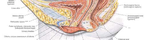
19
20 Sagittal section of the male pelvis


21
22
23
24
25
26 Sagittal section of the female pelvis
27


28
29

30
31 Common iliac artery 髂总动脉 Common iliac artery Continuation of abdominal aorta at level of L4 vertebra Terminates in front of sacroiliac joint by dividing idi into internal and external iliac arteries
32 Internal iliac artery 髂内动脉 Parietal iliac artery Obturator a. 闭孔动脉 Iliolumber a. 髂腰动脉 Lateral sacral a. 骶外侧动脉 Superior gluteal a. 臀上动脉 Inferior gluteal a. 臀下动脉
33 Visceral branches Umbilical a. 脐动脉 superior vesical a. 膀胱上动脉 Inferior vesical a. 膀胱下动脉 Uterine a. 子宫动脉 Inferior rectal a. 直肠下动脉 Internal pudendal a. 阴部内动脉
34
35 Uterine a. 子宫动脉 about 2cm from neck of uterus it crosses above and in front of ureter
36 Internal pudendal artery 阴部内动脉 Perineal artery Anal artery artery of penis (clitoris)
37 External iliac artery Inferior epigastric artery Deep iliac circumflex artery
38 Veins of pelvis Internal iliac vein 髂内静脉 Parietal tributaries: accompany with arteries Visceral tributaries superior rectal vein inferior mesenteric v. 1Rectal venous plexus inferior rectal vein internal iliac v. 直肠静脉丛 anal vein internal pudendal v. 2Vesical venous plexus 膀胱静脉丛 vesical v. 3Uterine venous plexus 子宫静脉丛 uterine v.


39 External iliac vein 髂外静脉 accompany the artery Common iliac vein 髂总静脉 formed by union of internal and external iliac veins in front of sacroiliac joint, end upon L4~L5 by uniting each other to form inferior vena cava


40 The lymphatic drainage of pelvis Internal iliac lymph node Surround internal iliac vessels Receive afferents from pelvic viscera, perineum, buttock and back of thigh External iliac lymph nodes Lie along external iliac artery Receive afferents from lower limb and some parts of pelvic viscera Sacral lymph node Common iliac lymph node Lie along common iliac artery Receive afferents from all the above nodes Efferents pass to lumbar lymph node
41 Sacral plexus 骶丛 Formation: formed by anterior rami of L4 and L5 spinal nerves (the lumbrosacral trunk) and anterior rami of sacral and coccygeal nerves Position: lies in pelvic cavity, anterior to sacrum and piriformis
42
43 Branches Superior gluteal Inferior gluteal Pudendal Posterior femoral cutaneou Sciatic Common


44 Pudendal nerve block
45 Autonomic plexuses of plevic Hypogastric plexus Superior hypogastric plexus 上腹下丛 : lies in front of L5 between common iliac ateries Inferior hypogastric plexus 下腹下丛 (pelvic plexus): lie on each side of rectum Sacral sympathetic trunk Is continuos above with the abdominal part Has 4 or 5 ganglion


46 Relationships of rectus Anteriorly In male Fundus of bladder Seminal vesicle Prostate Ampulla ductus deferentis In female neck of uterus Vagina
47 Posteriorly Sacrum and coccyx Piriformisif i Lateral sacral vessels Anterior branches of sacral and coccygeal nerves Sacral sympathetic trunk Laterally Pelvic plexus Superior and inferior rectal vessels Levator ani


48 Vessels and lymphatics of rectum Arteries Superior rectal a. Inferior rectal a. Median sacral a. Lymphatic drainge Lymphatics follow arterial blood supply to following nodes: Superior rectal ln Superior rectal ln. Inferior mesenteric ln. Pararectal ln. Internal iliac ln. Sacral ln.
Inferior Pelvic Border
 Pelvis + Perineum Pelvic Cavity Enclosed by bony, ligamentous and muscular wall Contains the urinary bladder, ureters, pelvic genital organs, rectum, blood vessels, lymphatics and nerves Pelvic inlet (superior
Dana Alrafaiah. - Amani Nofal. - Ahmad Alsalman. 1 P a g e
 - 2 - Dana Alrafaiah - Amani Nofal - Ahmad Alsalman 1 P a g e This lecture will discuss five topics as follows: 1- Arrangement of pelvic viscera. 2- Muscles of Pelvis. 3- Blood Supply of pelvis. 4- Nerve
NOTES FROM GUTMAN LECTURE 10/26 Use this outline to study from. As you go through Gutman s lecture, fill in the topics.
 NOTES FROM GUTMAN LECTURE 10/26 Use this outline to study from. As you go through Gutman s lecture, fill in the topics. Anatomy above the arcuate line Skin Camper s fascia Scarpa s fascia External oblique
Pelvis MCQs. Block 1. B. Reproductive organs. C. The liver. D. Urinary bladder. 1. The pelvic diaphragm includes the following muscles: E.
 Pelvis MCQs Block 1 1. The pelvic diaphragm includes the following muscles: A. The obturator internus B. The levator ani C. The coccygeus D. The external urethral sphincter E. The internal urethral sphincter
ischium Ischial tuberosities Sacrotuberous ligament The coccyx
 Perineum General lfeatures Region of below pelvic diaphragm A diamond shape space whose boundaries are those of the pelvic outlet Lower border of symphysis pubis Rami of pubis and ischium Ischial tuberosities
Slide Read the tables it is about the difference between male & female pelvis.
 I didn t include the slides, this is only what the doctor read or said because he skipped a lot of things because we took it previously, very important to go back to the slides (*there is an edited version)
Perineum. done by : zaid al-ghnaneem
 Perineum done by : zaid al-ghnaneem Hello everyone, this sheet will talk about 2 nd Lecture which is Perineum but there are some slides and info from 1 st Lecture. Everything included Slides + Pics Let
Table 2. First Generated List of Expert Responses. Likert-Type Scale. Category or Criterion. Rationale or Comments (1) (2) (3) (4)
 (2) (3) (4)") Table 2. First Generated List of Expert Responses. Likert-Type Scale Category or Criterion Anatomical Structures and Features Skeletal Structures and Features (1) (2) (3) (4) Rationale or Comments 1. Bones
Table 2. First Generated List of Expert Responses. Likert-Type Scale Category or Criterion Anatomical Structures and Features Skeletal Structures and Features (1) (2) (3) (4) Rationale or Comments 1. Bones
ORIENTING TO BISECTED SPECIMENS ON THE PELVIS PRACTICAL
 ORIENTING TO BISECTED SPECIMENS ON THE PELVIS PRACTICAL The Pelvis is just about as complicated as head and neck and considerably more mysterious. You have to be able to visualize (imagine) the underlying
Anatomy & Physiology Pelvic Girdles 10.1 General Information
 Anatomy & Physiology Pelvic Girdles 10.1 General Information ICan2Ed, Inc. In human anatomy, the pelvis (plural pelves or pelvises) is the lower part of. The area of the body that is between the abdomen
UROGENITAL SYSTEM By Dr.Ahmed Salman
 The University Of Jordan Faculty Of Medicine Anatomy Department UROGENITAL SYSTEM By Dr.Ahmed Salman Assistance Professor of Anatomy &embryology PELVIS Learning Objectives 1. Bony pelvis, its joints and
Pelvis Perineum MCQs. Block 1.1. A. Urinary bladder. B. Rectum. C. Reproductive organs. D. The thigh
 Pelvis Perineum MCQs Block 1.1 1. The pelvic diaphragm includes the following muscles: A. The coccygeus B. The levator ani C. The external urethral sphincter D. The internal urethral sphincter E. The obturator
THE PELVIS VASCULAR AND NERVOUS SYSTEM SOMATIC AND AUTONOMIC NERVES
 THE PELVIS VASCULAR AND NERVOUS SYSTEM SOMATIC AND AUTONOMIC NERVES THE ABDOMINAL AORTA The abdominal aorta begins at the aor9c hiatus in the diaphragm at the level of the T12 vertebra and ends at the
REPRODUCTIVE SYSTEM By Dr.Ahmed Salman
 The University Of Jordan Faculty Of Medicine Anatomy Department REPRODUCTIVE SYSTEM By Dr.Ahmed Salman Assistant Professor of Anatomy &embryology Perineum It is the diamond-shaped lower end of the trunk
2. List the 8 pelvic spaces: list one procedure or dissection which involves entering that space.
 Name: Anatomy Quiz: Pre / Post 1. In making a pfannensteil incision you would traverse through the following layers: a) Skin, Camper s fascia, Scarpa s fascia, external oblique aponeurosis, internal oblique
Abdomen. Retroperitoneal space
 Abdomen. Retroperitoneal space Abdominal cavity The space bounded by: Anterolateral abdominal wall Posterior abdominal wall Diaphragm Pelvic walls and pelvic floor. Subdivided into: True abdominal cavity
B) cervix of uterus C) vagina D) rectum. 1. What number illustrates the adnexal area? (Fig. 4-64) A) 4 B) 5 C) 8 D) 9
 cervix of uterus C) vagina D) rectum. 1. What number illustrates the adnexal area? (Fig. 4-64) A) 4 B) 5 C) 8 D) 9") Pelvis Practice Problems 1. What number illustrates the adnexal area? (Fig. 4-64) A) 4 B) 5 C) 8 D) 9 2. What number illustrates the cervix? (Fig. 4-64) A) 4 B) 8 C) 5 D) 6 3. Which of the following is
Pelvis Practice Problems 1. What number illustrates the adnexal area? (Fig. 4-64) A) 4 B) 5 C) 8 D) 9 2. What number illustrates the cervix? (Fig. 4-64) A) 4 B) 8 C) 5 D) 6 3. Which of the following is
-15. -Alaa Albandi. -Dr. Mohammad Almohtasib. 0 P a g e
 -15 -Alaa Albandi - -Dr. Mohammad Almohtasib 0 P a g e In this last lecture, we will talk about the sigmoid colon, rectum, and anal canal. Sigmoid colon It has a mesentery called pelvic mesocolon or sigmoidal
Femoral Triangle and Adductor Canal. Dr. Heba Kalbouneh Associate Professor of Anatomy and Histology
 Femoral Triangle and Adductor Canal Dr. Heba Kalbouneh Associate Professor of Anatomy and Histology Femoral Triangle and Adductor Canal Femoral triangle Is a triangular depressed area located in the upper
Skeletal System Module 13: The Pelvic Girdle and Pelvis
 OpenStax-CNX module: m47993 1 Skeletal System Module 13: The Pelvic Girdle and Pelvis Donna Browne Based on The Pelvic Girdle and Pelvis by OpenStax College This work is produced by OpenStax-CNX and licensed
ANATYOMY OF The thigh
 ANATYOMY OF The thigh 1- Lateral cutaneous nerve of the thigh Ι) Skin of the thigh Anterior view 2- Femoral branch of the genitofemoral nerve 5- Intermediate cutaneous nerve of the thigh 1, 2 and 3 are
Urinary Bladder. Prof. Imran Qureshi
 Urinary Bladder Prof. Imran Qureshi Urinary Bladder It develops from the upper end of the urogenital sinus, which is continuous with the allantois. The allantois degenerates and forms a fibrous cord in
ANATYOMY OF The thigh
 ANATYOMY OF The thigh 1- Lateral cutaneous nerve of the thigh Ι) Skin of the thigh Anterior view 2- Femoral branch of the genitofemoral nerve 5- Intermediate cutaneous nerve of the thigh 1, 2 and 3 are
The front of the thigh. Dr.Amjad shatarat
 The front of the thigh Femoral triangle (Scarpa s triangle) Is a triangular depressed area located in the upper part of the medial aspect of the thigh immediately below the inguinal ligament. Superiorly:
LAB Notes#1. Ahmad Ar'ar. Eslam
 LAB Notes#1 Ahmad Ar'ar Eslam 1 P a g e Anatomy lab Notes Lower limb bones :- Pelvic girdle: It's the connection between the axial skeleton and the lower limb; it's made up of one bone called the HIP BONE
STRUCTURAL BASIS OF MEDICAL PRACTICE EXAMINATION 3. October 16, 2015
 STRUCTURAL BASIS OF MEDICAL PRACTICE EXAMINATION 3 October 16, 2015 PART l. Answer in the space provided. (12 pts) 1. Identify the structures. (2 pts) A. B. A B C. D. C D 2. Identify the structures. (2
PELVIS & SACRUM Dr. Jamila El-Medany Dr. Essam Eldin Salama
 PELVIS & SACRUM Dr. Jamila El-Medany Dr. Essam Eldin Salama Learning Objectives At the end of the lecture, the students should be able to : Describe the bony structures of the pelvis. Describe in detail
STRUCTURAL BASIS OF MEDICAL PRACTICE EXAMINATION 3. October 17, 2014
 STRUCTURAL BASIS OF MEDICAL PRACTICE EXAMINATION 3 October 17, 2014 PART l. Answer in the space provided. (12 pts) 1. Identify the structures. (2 pts) A. B. A B C. D. C D 2. Identify the structures. (2
Femoral Artery. Its entrance to the thigh Position Midway between ASIS and pubic symphysis
 Lower Limb Vessels Lecture Objectives Describe the major arteries of the lower limb. Describe the deep and superficial veins of the lower limb. Describe the topographical relationships of the arteries
THE ABDOMEN SUPRARENAL GLANDS KIDNEY URETERS URINARY BLADDER
 THE ABDOMEN SUPRARENAL GLANDS KIDNEY URETERS URINARY BLADDER THE SUPRARENAL GLANDS The suprarenal (adrenal) glands lie immediately superior and slightly anterior to the upper pole of either kidney. Golden
GI module Lecture: 9 د. عصام طارق. Objectives:
 GI module Lecture: 9 د. عصام طارق Objectives: To list structures forming posterior abdominal wall. To follow aorta & its main branches. To describe IVC & its main tributaries. To list nerves of posterior
Gluteal region DR. GITANJALI KHORWAL
 Gluteal region DR. GITANJALI KHORWAL Gluteal region The transitional area between the trunk and the lower extremity. The gluteal region includes the rounded, posterior buttocks and the laterally placed
Lecture 10 Arteries and veins of the upper limb
 Lecture 10 Arteries and veins of the upper limb 1. Identify the Subclavian, axillary, brachial (deep and superficial), radial and ulnar arteries and superficial/deep palmar arches 2. Describe the major
Learning objectives. SGD on Functions of Testosterone. Class
 Learning objectives SGD on Functions of Testosterone Class 2016 14-1-2013 Discuss o Process of spermatogenesis. o Sex determination. o Process of maturation of sperms. o Physiology of mature sperms. Discuss
The thigh. Prof. Oluwadiya KS
 The thigh Prof. Oluwadiya KS www.oluwadiya.com The Thigh: Boundaries The thigh is the region of the lower limb that is approximately between the hip and knee joints Anteriorly, it is separated from the
أحمد رواجبة- محمود الحربي- أحمد السالمان-
 -6 أحمد رواجبة- محمود الحربي- أحمد السالمان- 1 P a g e The Male Reproductive System The male genital system structures are divided into: Internal structures: 1- Prostate 3-Ejaculatory ducts External structures:
Gross Anatomy of the Urinary System
 Gross Anatomy of the Urinary System Lecture Objectives Overview of the urinary system. Describe the external and internal anatomical structure of the kidney. Describe the anatomical structure of the ureter
First practical session. Bones of the gluteal region
 First practical session 2017 Bones of the gluteal region The Hip bone The hip bone is made of: 1 The ilium: superior in position 2 The ischium:postero-inferior in position 3 The pubis: antero-inferior
The posterior abdominal wall. Prof. Oluwadiya KS
 The posterior abdominal wall Prof. Oluwadiya KS www.oluwadiya.sitesled.com Posterior Abdominal Wall Lumbar vertebrae and discs. Muscles opsoas, quadratus lumborum, iliacus, transverse, abdominal wall
Posterior Abdominal wall-
 Structures of posterior abdominal wall: o Bony boundaries: 5 lumber vertebra and their intervertebral disc, iliac fossa and iliac crest. o Muscles: psoas major, quadrates lumborum, transversus abdominis,
Abdomen: Introduction. Prof. Oluwadiya KS
 Abdomen: Introduction Prof. Oluwadiya KS www.oluwadiya.com Abdominopelvic Cavity Abdominal Cavity Pelvic Cavity Extends from the inferior margin of the thorax to the superior margin of the pelvis and the
ANATOMY (7). INTRODUCTION TO THE PELVIS. MOHAMMAD ALLOUH. BNB. 1 P a g e
. INTRODUCTION TO THE PELVIS. MOHAMMAD ALLOUH. BNB. 1 P a g e") ANATOMY (7). INTRODUCTION TO THE PELVIS. MOHAMMAD ALLOUH. BNB 1 P a g e Introduction to the Pelvis: Pelvic Wall & Pelvic Cavity pelvis (L, basin, sink ), is The region of the trunk that lies inferoposterior
ANATOMY (7). INTRODUCTION TO THE PELVIS. MOHAMMAD ALLOUH. BNB 1 P a g e Introduction to the Pelvis: Pelvic Wall & Pelvic Cavity pelvis (L, basin, sink ), is The region of the trunk that lies inferoposterior
Anatomy of the Pelvis
 Anatomy of the Pelvis Please view our Editing File before studying this lecture to check for any changes. Color Code Important Doctors Notes Notes/Extra explanation Objectives At the end of the lecture,
Omran Saeed. Mohammad Al-muhtaseb. 1 P a g e
 13 Omran Saeed Mohammad Al-muhtaseb 1 P a g e Posterior abdominal wall - The diaphragm separates between thoracic cavity and abdominal cavity. Structures of posterior abdominal wall: (below diaphragm)
Benha University. Faculty of Medicine. Anatomy Department Course code (MED 0701) Model answer of Anatomy examination. (Abdomen,Pelvis and Thorax)
 Model answer of Anatomy examination. (Abdomen,Pelvis and Thorax)") 1 Benha University Faculty of Medicine Anatomy Department Course code (MED 0701) Model answer of Anatomy examination (Abdomen,Pelvis and Thorax) 1 st year 2 nd term Date :18 /5 /2013 2 I-Short account
1 Benha University Faculty of Medicine Anatomy Department Course code (MED 0701) Model answer of Anatomy examination (Abdomen,Pelvis and Thorax) 1 st year 2 nd term Date :18 /5 /2013 2 I-Short account
Nerves on the Posterior Abdominal Wall
 Nerves on the Posterior Abdominal Wall Lumbar Plexus The lumbar plexus, which is one of the main nervous pathways supplying the lower limb, is formed in the psoasmuscle from the anterior ramiof the upper
Lumbar Plexus. Ventral rami L1 L4 Supplies: Major nerves.. Abdominal wall External genitalia Anteromedial thigh
 Lower Limb Nerves Lectures Objectives Describe the structure and relationships of the plexuses of the lower limb. Describe the course, relationships and structures supplied for the major nerves of the
The Hip (Iliofemoral) Joint. Presented by: Rob, Rachel, Alina and Lisa
 Joint. Presented by: Rob, Rachel, Alina and Lisa") The Hip (Iliofemoral) Joint Presented by: Rob, Rachel, Alina and Lisa Surface Anatomy: Posterior Surface Anatomy: Anterior Bones: Os Coxae Consists of 3 Portions: Ilium Ischium Pubis Bones: Pubis Portion
The Hip (Iliofemoral) Joint Presented by: Rob, Rachel, Alina and Lisa Surface Anatomy: Posterior Surface Anatomy: Anterior Bones: Os Coxae Consists of 3 Portions: Ilium Ischium Pubis Bones: Pubis Portion
Yes, cranially with ovarian, caudally with vaginal. Yes, with uterine artery (collateral circulation between abdominal +pelvic source)
") Blood supply to internal female genitalia: uterine Internal iliac Sup. large branch: uterus, inf. Small branch: cervix+ sup. Vagina Yes, cranially with ovarian, caudally with vaginal Medially in base of
Blood supply to internal female genitalia: uterine Internal iliac Sup. large branch: uterus, inf. Small branch: cervix+ sup. Vagina Yes, cranially with ovarian, caudally with vaginal Medially in base of
Pelvic Angiogram - Male
 Pelvic Angiogram - Male Common iliac artery Internal iliac artery Lateral sacral artery Iliolumbar artery Posterior trunk of internal iliac artery Superior gluteal artery Internal pudendal artery External
Lectures of Human Anatomy
 Lectures of Human Anatomy Lower Limb Gluteal Region and Hip Joint By DR. ABDEL-MONEM AWAD HEGAZY M.B. with honor 1983, Dipl."Gynecology and Obstetrics "1989, Master "Anatomy and Embryology" 1994, M.D.
Pathogenesis of Chronic Pelvic Pain
 Pathogenesis of Chronic Pelvic Pain Yong-Chul Kim Department of anesthesia and pain medicine, Seoul National University College of Medicine 1 Overview Anatomy Nerve innervation CPP by pathology CPP by
The os coxae or hip bone consists of three flat bones, ilium, ischium and pubis, which fuse together to form the acetabulum.
 The os coxae The os coxae or hip bone consists of three flat bones, ilium, ischium and pubis, which fuse together to form the acetabulum. The ilium extends from the acetabulum upwards forming the lateral
Academic Year 2017/2018. FALL SEMESTER DDS Program Monday: 5:45pm 7:15pm Thursday: 5:30pm- 7:00pm
 CURRICULUM FOR HUMAN ANATOMY COURSE LAB SESSIONS Academic Year 2017/2018 FALL SEMESTER DDS Program Monday: 5:45pm 7:15pm Thursday: 5:30pm- 7:00pm Category 1: Bones name, type, structure Joints: type, articular
The University Of Jordan Faculty Of Medicine THE LOWER LIMB. Dr.Ahmed Salman Assistant Prof. of Anatomy. The University Of Jordan
 The University Of Jordan Faculty Of Medicine THE LOWER LIMB Dr.Ahmed Salman Assistant Prof. of Anatomy. The University Of Jordan Gluteal Region Cutaneous nerve supply of (Gluteal region) 1. Lateral cutaneous
Copyright 2003 Pearson Education, Inc. publishing as Benjamin Cummings. Dr. Nabil Khouri MD, MSc, Ph.D
 Dr. Nabil Khouri MD, MSc, Ph.D Pelvic Girdle (Hip) Organization of the Lower Limb It is divided into: The Gluteal region The thigh The knee The leg The ankle The foot The thigh and the leg have compartments
Infracolic Compartments
 模块 8- 局部解剖学 Infracolic Compartments JING LI Ph.D Department of Human Anatomy School of Medicine, Zhejiang University derfreiefall@zju.edu.cn June 20 2014 Jejunum and ileum 空肠和回肠 Jejunum and ileum 空肠和回肠
Lumbar and Sacral Plexuses. Dr. Heba Kalbouneh Associate Professor of Anatomy and Histology
 Lumbar and Sacral Plexuses Dr. Heba Kalbouneh Associate Professor of Anatomy and Histology Structure of Spinal Nerves: Somatic Pathways dorsal root CNS interneuron spinal nerve dorsal ramus somatic sensory
TABLE OF CONTENTS. 1. Introduction I. 2. Lecturers I. 3. Timetable I. 4. Assessment I. 5. Study material II. 6. General information II.
 TABLE OF CONTENTS Page A. ORGANISATIONAL COMPONENT 1. Introduction I 2. Lecturers I 3. Timetable I 4. Assessment I 5. Study material II 6. General information II B. STUDY COMPONENT 1. Block XI: Session
Lab Monitor Images Dissection of the Abdominal Vasculature + Lower Digestive System
 Lab Monitor Images Dissection of the Abdominal Vasculature + Lower Digestive System Stomach & Duodenum Frontal (AP) View Nasogastric tube 2 1 3 4 Stomach Pylorus Duodenum 1 Duodenum 2 Duodenum 3 Duodenum
rotation of the hip Flexion of the knee Iliac fossa of iliac Lesser trochanter Femoral nerve Flexion of the thigh at the hip shaft of tibia
 Anatomy of the lower limb Anterior & medial compartments of the thigh Dr. Hayder The fascia lata encloses the entire thigh like a sleeve/stocking. Three intramuscular fascial septa (lateral, medial, and
ANATOMY OF PELVICAYCEAL SYSTEM -DR. RAHUL BEVARA
 1 ANATOMY OF PELVICAYCEAL SYSTEM -DR. RAHUL BEVARA 2 KIDNEY:ANATOMY OVERVIEW Kidneys are retroperitoneal, in posterior abdominal region, extending from T12 L3 Bean-shaped Right kidney is lower than left
ANATOMY TEAM GLUTEAL REGION & BACK OF THIGH
 ANATOMY TEAM GLUTEAL REGION & BACK OF THIGH OBJECTIVES By the end of this lecture, the student should be able to identify and discuss: Contents of gluteal region: Groups of Glutei muscles and small muscles
[ANATOMY #12] April 28, 2013
![[ANATOMY #12] April 28, 2013](/thumbs/86/93473883.jpg "[ANATOMY #12] April 28, 2013") Sympathetic chain : Sympathetic chain is each of the pair of ganglionated longitudinal cords of the sympathetic nervous system; extend from level of atlas (base of skull) till coccyx. It is paravertebral
Sympathetic chain : Sympathetic chain is each of the pair of ganglionated longitudinal cords of the sympathetic nervous system; extend from level of atlas (base of skull) till coccyx. It is paravertebral
The Lower Limb. Anatomy RHS 241 Lecture 2 Dr. Einas Al-Eisa
 The Lower Limb Anatomy RHS 241 Lecture 2 Dr. Einas Al-Eisa The bony pelvis Protective osseofibrous ring for the pelvic viscera Transfer of forces to: acetabulum & head of femur (when standing) ischial
ANATYOMY OF The thigh
 ANATYOMY OF The thigh 1- Lateral cutaneous nerve of the thigh Ι) Skin of the thigh Anterior view 2- Femoral branch of the genitofemoral nerve 1, 2 and 3 are From the lumber plexus 5- Intermediate cutaneous
TABLE OF CONTENTS A. ORGANISATIONAL COMPONENT B. STUDY COMPONENT. Page. 1. Introduction I. 2. Lecturers I. 3. Timetable I. 4.
 TABLE OF CONTENTS Page A. ORGANISATIONAL COMPONENT 1. Introduction I 2. Lecturers I 3. Timetable I 4. Assessment I 5. Study material II 6. General information II B. STUDY COMPONENT 1. Block XI: Session
Bones of Lower Limb. Dr. Heba Kalbouneh Associate Professor of Anatomy and Histology
 Bones of Lower Limb Dr. Heba Kalbouneh Associate Professor of Anatomy and Histology Bones of the lower limb Hip Bone Made up of 3 bones: 1) Ilium (flat), superior in position 2) Ischium (L), postero-inferior
ABDOMINAL WALL & RECTUS SHEATH
 ABDOMINAL WALL & RECTUS SHEATH Learning Objectives Describe the anatomy, innervation and functions of the muscles of the anterior, lateral and posterior abdominal walls. Discuss their functional relations
SUBJECTS 2nd year, 1st semester I. 1. Primitive gut - limits, derivatives 2. Foregut -limits, evolution, derivatives 3. Midgut -limits, evolution,
 SUBJECTS 2nd year, 1st semester I. 1. Primitive gut - limits, derivatives 2. Foregut -limits, evolution, derivatives 3. Midgut -limits, evolution, derivatives 4. Hindgut- limits, evolution, derivatives
Identify the muscles associated with the medial compartment of the thigh. Identify the attachment points of the medial thigh muscles.
 L 8 A B O R A T O R Y Thigh MEDIAL THIGH Identify the muscles associated with the medial compartment of the thigh. Identify the attachment points of the medial thigh muscles. Identify the actions of these
Muscles of the lower extremities. Dr. Nabil khouri MD, MSc, Ph.D
 Muscles of the lower extremities Dr. Nabil khouri MD, MSc, Ph.D Posterior leg Popliteal fossa Boundaries Biceps femoris (superior-lateral) Semitendinosis and semimembranosis (superior-medial) Gastrocnemius
Ureters, Urinary Bladder & Urethra
 Ureters, Urinary Bladder & Urethra Please check our Editing File هذا العمل ال يغني عن المصدر األساسي للمذاكرة Lecture 2 } و م ن ي ت و ك ع ل ا لل ه ف ه و ح س ب ه { Objectives o Describe the course of ureter
The Thoracic wall including the diaphragm. Prof Oluwadiya KS
 The Thoracic wall including the diaphragm Prof Oluwadiya KS www.oluwadiya.com Components of the thoracic wall Skin Superficial fascia Chest wall muscles (see upper limb slides) Skeletal framework Intercostal
Baraa Ayed حسام أبو عوض. Ahmad Salman. 1 P a g e
 4 Baraa Ayed حسام أبو عوض Ahmad Salman 1 P a g e Today we are going to cover these concepts: Iliotibial tract Anterior compartment of the thigh and the hip Medial compartment of the thigh Femoral triangle
大體老師無語良師 大體解剖學實驗 HUMAN DISSECTION POSTERIOR ABDOMINAL VISCERA & WALL 盧家鋒助理教授 臺北醫學大學醫學系解剖學暨細胞生物學科 臺北醫學大學醫學院轉譯影像研究中心.
 大體老師無語良師 大體解剖學實驗 HUMAN DISSECTION POSTERIOR ABDOMINAL VISCERA & WALL 盧家鋒助理教授 臺北醫學大學醫學系解剖學暨細胞生物學科 臺北醫學大學醫學院轉譯影像研究中心 http://www.ym.edu.tw/~cflu REFERENCES Dissector s guide [1] Dissection Guide for Gray's
Objectives. Pelvic Anatomy: Staying Out of Trouble. Disclosures. Anatomy 101. Anterior Abdominal Wall. Arcuate Line. Abheha Satkunaratnam MD, FRCS(C)
") Objectives Pelvic Anatomy: Staying Out of Trouble Abheha Satkunaratnam MD, FRCS(C) To focus on key anatomy for the gynaecologic surgeon advancing their minimally invasive gynaecologic skills To provide
Objectives Pelvic Anatomy: Staying Out of Trouble Abheha Satkunaratnam MD, FRCS(C) To focus on key anatomy for the gynaecologic surgeon advancing their minimally invasive gynaecologic skills To provide
The Male Reproductive System 男性生殖系
 The Male Reproductive System 男性生殖系 陈晓 Chenxiao.610@qq.com 浙江大学医学院干细胞与组织工程中心临床医学 运动医学博士 The Reproductive System The male reproductive organs The female reproductive organs 2 1 Structures of the reproductive
Anatomy. Contents Brain (Questions)
") Anatomy 12 Contents 12.1 Brain (Questions).................................................... 683 12.2 Head and Neck (Questions)............................................. 685 12.3 Thorax (Questions)...................................................
Anatomy 12 Contents 12.1 Brain (Questions).................................................... 683 12.2 Head and Neck (Questions)............................................. 685 12.3 Thorax (Questions)...................................................
Group of students. - Rawan almujabili د. محمد المحتسب - 1 P a g e
 - 14 - Group of students - Rawan almujabili د. محمد المحتسب - 1 P a g e Nerves of the posterior abdominal wall The spinal cord gives off spinal nerves between the vertebrae. In the abdomen, through the
Fig. A.1. Frontal. plane. Transverse. plane. Sagittal plane. Copyright McGraw-Hill Education. Permission required for reproduction or display.
 Fig. A.1 Frontal plane Transverse plane Sagittal plane McGraw-Hill Education/Joe DeGrandis Fig. A.2 (a) Sagittal section (b) Frontal section (c) Transverse section Table A.1 Fig. A.3 Cephalic r. (head)
Anatomy Review File اللهم ال سهل إال ماجعلته سهال وأنت تجعل الحزن
 Anatomy Review File اللهم ال سهل إال ماجعلته سهال وأنت تجعل الحزن إذا شئت سهال This work was done by: Alanoud Abuhaimed Dania Alkelabi Ghada Alothaim Jawaher Abanumy Rawan AlWadee Wejdan Alzeid Mohanad
Preview from Notesale.co.uk Page 1 of 34
 Abdominal viscera and digestive tract Digestive tract Abdominal viscera comprise majority of the alimentary system o Terminal oesophagus, stomach, pancreas, spleen, liver, gallbladder, kidneys, suprarenal
Lecture 08 THIGH MUSCLES ANTERIOR COMPARTMENT. Dr Farooq Khan Aurakzai. Dated:
 Lecture 08 THIGH MUSCLES ANTERIOR COMPARTMENT BY Dr Farooq Khan Aurakzai Dated: 11.02.2017 INTRODUCTION to the thigh Muscles. The musculature of the thigh can be split into three sections by intermuscular
Anatomy of the renal system. Professor Nawfal K. Al-Hadithi
 Anatomy of the renal system Professor Nawfal K. Al-Hadithi Objectives To describe the posterior abdominal wall To identify the main anatomical landmarks of the kidneys & ureters To describe the suprarenal
Lower limb summary. Anterior compartment of the thigh. Done By: Laith Qashou. Doctor_2016
 Lower limb summary Done By: Laith Qashou Doctor_2016 Anterior compartment of the thigh Sartorius Anterior superior iliac spine Upper medial surface of shaft of tibia 1. Flexes, abducts, laterally rotates
Basic Body Structure
 Basic Body Structure The Cell All life consists of microscopic living structures called cells. They perform various functions throughout the body. All cells are similar in structure, but not identical.
Adductor canal (Subsartorial) or Hunter s canal
 or Hunter s canal") Adductor canal (Subsartorial) or Hunter s canal John Hunter described the exposure and ligation of the femoral artery in this canal for aneurysm of the popliteal artery; this method has the advantage that
Adductor canal (Subsartorial) or Hunter s canal John Hunter described the exposure and ligation of the femoral artery in this canal for aneurysm of the popliteal artery; this method has the advantage that
The Anterolateral Abdominal Wall By Prof. Dr. Muhammad Imran Qureshi
 1 P age The Anterolateral Abdominal Wall By Prof. Dr. Muhammad Imran Qureshi Introduction The abdomen is the region of the trunk located between the thorax and the pelvis. It includes the anterolateral
C. Bones of the Pelvic Girdle
 C. Bones of the Pelvic Girdle 1. 2 coxal bones (a.k.a hip bones): -bony pelvis is made up of hip bones, sacrum, & coccyx -pelvic bones are large & heavy & attach to the axial skeleton via sacrum/coccyx
Perineum. Dept. of Human Anatomy Zhou Hong Ying
 Perineum Dept. of Human Anatomy Zhou Hong Ying OUTLINE Subdivision The Layers Urogenital Diaphragm Main Structures inside Superficial & Deep Perineal Spaces Ischioanal Fossa Perineum A narrow region Urogenital
The abdominal Esophagus, Stomach and the Duodenum. Prof. Oluwadiya KS
 The abdominal Esophagus, Stomach and the Duodenum Prof. Oluwadiya KS www.oluwadiya.com Viscera of the abdomen Abdominal esophagus: Terminal part of the esophagus The stomach Intestines: Small and Large
Borders of the Abdomen
 Abdominal wall Borders of the Abdomen Abdomen is the region of the trunk that lies between the diaphragm above and the inlet of the pelvis below Borders Superior: Costal cartilages 7-12. Xiphoid process:
THE DESCENDING THORACIC AORTA
 Intercostal Arteries and Veins Each intercostal space contains a large single posterior intercostal artery and two small anterior intercostal arteries. The anterior intercostal arteries of the lower spaces
ANATOMY. Schedule for 2014/2015 academic school year (2x15 weeks)
") ANATOMY Schedule for 2014/2015 academic school year (2x15 weeks) SEMESTER LECTURES LAB CLASSES SEMINARS TOTAL FIRST 4 hours (2+2) 4 hours (2+2) 1 hour 135 hours SECOND 3 hours 4 hours (2+2) 2 hours 135
ANATOMY Schedule for 2014/2015 academic school year (2x15 weeks) SEMESTER LECTURES LAB CLASSES SEMINARS TOTAL FIRST 4 hours (2+2) 4 hours (2+2) 1 hour 135 hours SECOND 3 hours 4 hours (2+2) 2 hours 135
Mohammad Ashraf. Abdulrahman Al-Hanbali. Ahmad Salman. 1 P a g e
 - 7 Mohammad Ashraf Abdulrahman Al-Hanbali Ahmad Salman 1 P a g e Structures under the cover of Gluteus Maximus: 1-Bones: Ileum, Femur (Head, greater trochanter and gluteal tuberosity), Ischium (ischial
Muscles of Gluteal Region
 1 The Gluteal Region In the gluteal region the skin is tough with many layers underneath. Directly under it is the superficial fascia followed by the deep fascia then the muscles and the bones of the thigh.
Netter's Anatomy Flash Cards Section 4 List 4 th Edition
 Netter's Anatomy Flash Cards Section 4 List 4 th Edition https://www.memrise.com/course/1577335/ Section 4 Abdomen (31 cards) Plate 4-1 Bony Framework of Abdomen 1.1 Costal cartilages 1.2 Iliac crest 1.3
Citation Acta medica Nagasakiensia. 1963, 8(
 NAOSITE: Nagasaki University's Ac Title Radical Operation For Prostatic Car Author(s) Kondoh, Atushi Citation Acta medica Nagasakiensia. 1963, 8( Issue Date 1963-12-25 URL http://hdl.handle.net/10069/15473
END-SEMESTER EXAM 2018 ANATOMY, HISTOLOGY AND EMBRYOLOGY FACULTY OF MEDICINE, 2 ND SEMESTER
 University of Szeged, Faculty of Medicine Department of Anatomy, Histology and Embryology Chairman: Prof. Antal Nógrádi MD, PhD, DSc Kossuth L. sgt. 40., H-6724 Szeged, Hungary Tel.: +36-62-545-665 P.