Borders of the Abdomen
|
|
|
- Adrian Skinner
- 6 years ago
- Views:
Transcription

1 Abdominal wall
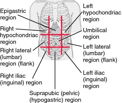
2 Borders of the Abdomen Abdomen is the region of the trunk that lies between the diaphragm above and the inlet of the pelvis below Borders Superior: Costal cartilages Xiphoid process: Inferior: Pubic bone and iliac crest: Level of L4. Umbilicus: Level of IV disc L3-L4

3 Abdominal Quadrants Formed by two intersecting lines: Vertical & Horizontal Intersect at umbilicus. Quadrants: Upper left. Upper right. Lower left. Lower right
4 Abdominal Regions Divided into 9 regions by two pairs of planes: 1- Vertical Planes: -Left and right lateral planes - Midclavicular planes -passes through the midpoint between the ant.sup.iliac spine and symphysis pupis 2- Horizontal Planes: -Subcostal plane - at level of L3 vertebra -Joins the lower end of costal cartilage on each side -Intertubercular plane: -- At the level of L5 vertebra - Through tubercles of iliac crests.
5

6 Abdominal wall divided into:- Anterior abdominal wall Posterior abdominal wall

7 What are the Layers of Anterior Abdominal Wall Skin Superficial Fascia - Above the umbilicus one layer - Below the umbilicus two layers Camper's fascia - fatty superficial layer. Scarp's fascia - deep membranous layer. Deep fascia : Thin layer of C.T covering the muscle may absent Muscular layer External oblique muscle Internal oblique muscle Transverse abdominal muscle Rectus abdominis Transversalis fascia Extraperitoneal fascia Parietal Peritoneum
8 Superficial Fascia Camper's fascia - fatty layer= dartos muscle in male Scarpa's fascia - membranous layer. Attachment of scarpa s fascia= membranous fascia INF: Fascia lata Sides: Pubic arch Post: Perineal body - Membranous layer in scrotum referred to as colle s fascia - Rupture of penile urethra lead to extravasations of urine into(scrotum, perineum, penis &abdomen)

9 Muscles Rectus abdominis External oblique muscle Internal oblique muscle Transverse abdominal muscle
10 External oblique muscle -Broad -Thin Direction: Downward forward medially Origin Insertion outer surface of lower 8 ribs. Xiphoid process, Linea alba, pubic crest, pubic tubercle, iliac crest(ant. Half). Nerve Supply 1- Lower 6 th thoracic nerves 2- L1( iliohypogastric n., ilioinguinal n.)

11 Muscles of the anterior abdominal wall
12 Aponeurosis of external oblique muscle Superficial inguinal ring. Inguinal ligament Lacunar ligament Pectineal ligament Boundaries of inguinal canal Formation of rectus sheath (


13 Inguinal ligament 1- folded back ward the lower border of aponeurosis of external muscle on it self 2- between ant.sup.iliac spine and the pupic tubercle
14 Superficial inguinal ring triangular shape - 2- Defect in external oblique aponeurosis - 3- lies immediately above and medial to the pupic tubercle - 4- Opening for passing the spermatic cord or ligament of uterus
15


16 Lacunar ligament 1- extension of aponeurosis of external muscle backward and upward to the pectineal line 2- on the superior ramus of the pupis 3- its sharp, free crecentric edge forms the medial margin of the femoral ring Pectineal ligament 1- Continuation of the lacunar ligment at pectineal line 2- Continuation with a thickeing of the periosteum

17 Internal Oblique Direction: upward forward medially Origin Lumbar Fascia, Ant 2/3 iliac crest, lateral two thirds of inguinal ligament. Insertion - Lower three ribs& costal cartilage, Xiphoid process, Linea alba, symphesis pubis. - Nerve Supply Lower 6 th thoracic nerves, iliohypogastric n & ilioinguinal n L1.
18 Internal oblique muscle..cont Conjoint tendon - The lowest tendinous fibers of internal oblique which joint with transversus abdominis - Attach medially to linea alba - Support the inguinal canal - Has lateral free border Cremastric fascia Internal oblique has free lower border arches over the spermatic cord or ligament of uterus - Cremastric muscle - Fascia - Int. abd.muscle assist in the formation of the Roof of the inguinal canal

19
20 Conjoint tendon & Cremastric fascia


21 Transversus Abdominis Direction - Its fibers run horizontally forward under the internal oblique Origin - Inner surface of lower six costal cartilage, lumbar fascia, anterior two thirds of iliac crest, lateral third of inguinal ligament. Insertion Xiphoid process, Linea alba, symphysis pubis. The lower part fuses with internal oblique to form conjoint tendon which attach to pupic crest and pectineal line Nerve Supply Lower six thoracic nerves, L1( iliohypogastric n.& ilioinguinal n.)
22 Transversus Abdominis cont Assist in the formation of Conjoint tendon Rectus sheath
23 RECTUS ABDOMINIS - Long strap muscle - Extends along the whole length of the anterior abdominal wall - In the rectus sheath Origin Symphsis pubis, pubic crest Insertion 5 th, 6 th and 7 th costal cartilage & xiphoid process. Nerve Supply Lower 6 th thoracic nerves
24 Rectus abdominis muscle cont - Linea semilunaris - Tendinous intersection:

25 Lines & Land marks of the Anterior Abdominal Wall Linea alba: - Located along the midline. -Between the xiphoid process & symphysis pupis - Formed by the fusion of aponeurosises of three abdominal wall( Ex.In,Tran. Abd.muscle) - Linea semilunaris - Lateral margins of rectus abd..muscle - Can be palpated - Extend from 9 th c.c to pupic tubercle
26 Tendinous intersection: = Linea transverses - 3 transverse fibrous bands - divide the rectus abdominis muscle into distinct segments 1- one at level of xiphoid process 2- one at level of umbilicus and 3- one half way between these two - They can be palpated as a transverse depressions

27 Pyramidalis muscle Origin Ant. Surface of the pupis Insertion: Linea alba -It lies in front of the lower part of the rectus abdominis muscle -Nerve supply 12 th subcostal nerve
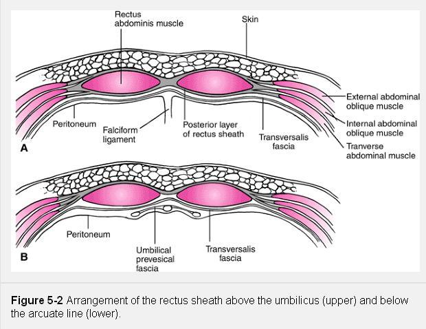
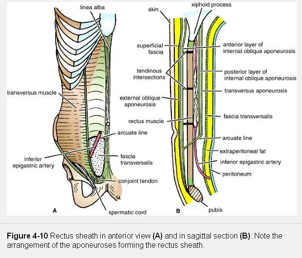
28 Rectus sheath
29 Rectus sheath.cont The rectus sheath is a long fibrous sheath Formed mainly by the aponeuroses of the three lateral abdominal muscles. Contents - Rectus abdominis muscle - Pyramidalis muscle (if present) - The anterior rami of the lower six thoracic nerves - The superior and inferior epigastric vessels - Lymphatic vessels.
30 Rectus sheath.cont Description the rectus sheath is considered at three levels. 1- Above the costal margin 2- Between the costal margin and the level of the anterior superior iliac spine 3- Between the level of the anterosuperior iliac spine and the pubis the anterior wall
31

32 ABOVE THE COSTAL MARGIN, - ANTERIOR WALL # APONEUROSIS OF THE EXTERNAL OBLIQUE. - POSTERIOR WALL # THORACIC WALL THAT IS, THE FIFTH, SIXTH, AND SEVENTH COSTAL CARTILAGES AND THE INTERCOSTAL SPACES.

33 Between the costal margin and the level of the anterior superior iliac spine - The aponeurosis of the internal oblique splits to enclose the rectus muscle - the external oblique aponeurosis is directed in front of the muscle - the transversus aponeurosis is directed behind the muscle.

34 Between the level of the anterosuperior iliac spine and the pubis the anterior wall : the aponeurosis of all three muscles form. The posterior wall is absent, and the rectus muscle lies in contact with the fascia transversalis.
35 Rectus sheath cont The posterior wall of the rectus sheath is not attached to the rectus abdominis muscle. The anterior wall is firmly attached to it by the muscle's tendinous intersections Linea semicircularis (arcuate line) Is a crescent-shaped line marking the inferior limit of the posterior layer of the rectus sheath just below the level of the iliac crest.
36
37 . Others fascia in the ant. abd.ominal wall Transversalis fascia - a thin layer of fascia that lines the Transversus Abdominis muscle - continue to diaphragm, iliac muscle & pelvis fascia - contribute to femoral sheath Extraperitoneal Fascia The thin layer of C.T and adipose tissue between the peritoneum and fascia transversalis. Parietal peritoneum It is a thin serous membrane Continuous below with the parietal peritoneum lining the pelvis..
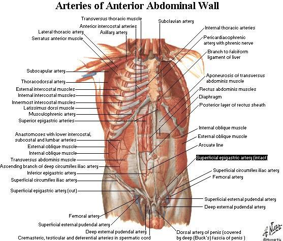
38

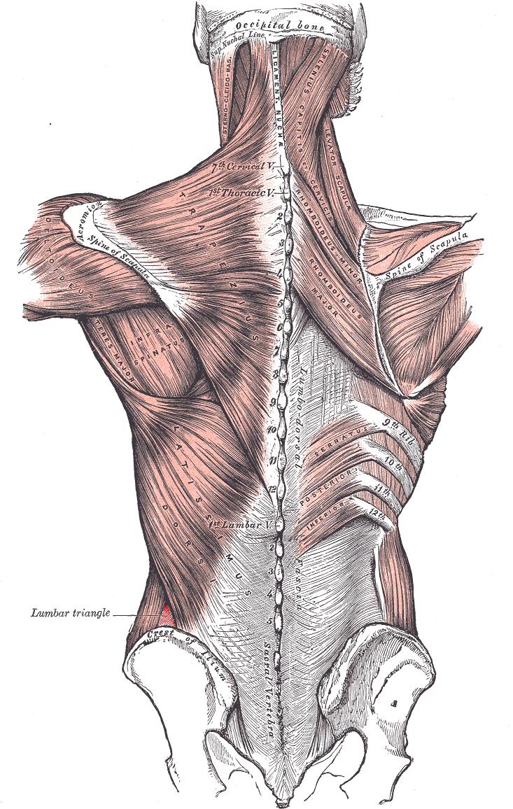
39 Lumbar triangle
40 lumbar triangle 1- the inferior lumbar (Petit) triangle, which lies superficially 2- the superior lumbar (Grynfeltt) triangle, which is deep and superior to the inferior triangle. -Of the two, the superior triangle is the more consistently found in cadavers,and is more commonly the site of herniation - however, the inferior lumbar triangle is often simply called the lumbar triangle, perhaps owing to its more superficial location and ease in demonstration.
41 Lumber triangle(petitis) The inferior lumbar (Petit) triangle is formed - Medially by the latissimus dorsi muscle - laterally by the external abdominal oblique muscle - Inferiorly by the iliac crest - The floor internal abdominal oblique muscle. - The fact that herniation occasionally occur here is of clinical importance.
42 Superior lumbar (Grynfeltt-Lesshaft) triangle Medially: by the quadratus lumborum muscle laterally :by the internal abdominal oblique muscle Superiorly: by the 12th rib. The floor : transversalis fascia Roof: is the external abdominal oblique muscle
43
44 Action of the Ant. Abdominal muscle Deep expiration Increase the intra abdominal pressure in - Vomiting - Cough - Defecation - Labour Protect viscera keep viscera in position Rectus abdominis bends trunk forward
45 Blood supply of the ant. Abdominal wall Arteries Sup. Epigastric artery Inf. Epigastric artery Intercostal arteries Lumbar arteries Deep circumflex artery
46
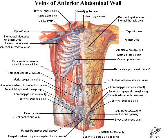
47 Blood supply cont Veins 1- Above the umbilicus - Lat. Thoracic. vein. Axillary vein 2- Below the umbilicus - Inf. Epigastric Femoral vein 3- Paraumbilica veins - Ligamentum teres portal vein( Porto- systemic anastomosis)
48
49 Nerve supply of the ant. Abdominal wall Thoracoabdominal nerve: Lower 6 th thoracic nerves & 12 th subcostal nerve Dermatomes (Anterior, lateral cutaneous nerve terminal branches of Thoracoabdominal nerve T7 to skin superior to umbilicus below xiphoid process T10 to skin surrounding umbilicus L1 to skin inferior to umbilicus above sym.pubis LI nerve - Iliohypogastric nerve - Ilioinguinal nerve

50
51 Lymphatic drainage of ant. Abdominal wall Above the umbilicus Ant.axillary L.N Below the umbilicus Sup. Inguinal L.N Above the iliac crest Post.axillary.L.N Below the iliac crest Sup.inguinal L.N
52 Clinical notes Abdominal stab wounds Surgical incision
53 Abdominal stab wounds Lateral to rectus sheath Ant. To rectus sheath In the midline= Linea alba - Structures in the various layers through which an abdominal stab wound depend on the anatomical location
54 Surgical incision - The length and direction of surgical incision through the ant. Abdominal wall to expose the underlying viscera are largely controlled by 1- position & direction of nerves 2- direction of muscle fibers 3- arrangement of the apponeurosis forming the rectus sheath - The incision should be mad In the direction of the line of cleavage in the skin so that the hairline scare is produced
55 Incision through the rectus sheath Widely used The rectus abdominis muscle and its nerve supply are kept intact On closure the ant & post wall of the sheath are sutured separately and the rectus muscle back into position between the suture lines
56 Common types of incisions Paramedian incision Pararectus incsion Midline incision Transrectus incision Transverse incision Muscle splitting Abdominothoracic incision
ABDOMINAL WALL & RECTUS SHEATH
 ABDOMINAL WALL & RECTUS SHEATH Learning Objectives Describe the anatomy, innervation and functions of the muscles of the anterior, lateral and posterior abdominal walls. Discuss their functional relations
-2 ة يمجع وبأ اه م - - Dr Muhtaseb Al - 1
 -2 م ها أبو عجمي ة - - Dr Al - Muhtaseb 1 Refer to Snell for clinical notes (as the doctor said in his first lecture O_O) and to the slides for illustrations. This sheet is about abdomen, there are anterior
Abdomen: Introduction. Prof. Oluwadiya KS
 Abdomen: Introduction Prof. Oluwadiya KS www.oluwadiya.com Abdominopelvic Cavity Abdominal Cavity Pelvic Cavity Extends from the inferior margin of the thorax to the superior margin of the pelvis and the
Gross Anatomy ABDOMEN/SESSION 1 Dr. Firas M. Ghazi
 Anterior Abdominal Wall Structure, muscles and surface anatomy Curricular Objectives By the end of this session students are expected to: Practical 1. Identify the hip and distinguish the three bones forming
The Anterolateral Abdominal Wall By Prof. Dr. Muhammad Imran Qureshi
 1 P age The Anterolateral Abdominal Wall By Prof. Dr. Muhammad Imran Qureshi Introduction The abdomen is the region of the trunk located between the thorax and the pelvis. It includes the anterolateral
GI anatomy Lecture: 2 د. عصام طارق
 GI anatomy Lecture: 2 د. عصام طارق Objectives: To define rectus sheath. To describe anatomy of inguinal canal. To relates types of inguinal hernia to the region. To explore spermatic cord. Rectus Abdominis
This presentation will discuss the anatomy of the anterior abdominal wall as it pertains to gynaecological and obstetric surgery.
 This presentation will discuss the anatomy of the anterior abdominal wall as it pertains to gynaecological and obstetric surgery. 1 The border of the anterior abdominal wall is defined superiorly by the
Abdominal muscles. Subinguinal hiatus and ingiunal canal. Femoral and adductor canals. Neurovascular system of the lower limb. Sándor Katz M.D.,Ph.D.
 Abdominal muscles. Subinguinal hiatus and ingiunal canal. Femoral and adductor canals. Neurovascular system of the lower limb. Sándor Katz M.D.,Ph.D. External oblique muscle Origin: outer surface of the
Anatomy of the Thorax
 Anatomy of the Thorax A) THE THORACIC WALL Boundaries Posteriorly by the thoracic part of the vertebral column Anteriorly by the sternum and costal cartilages Laterally by the ribs and intercostal spaces
THE THORACIC WALL. Boundaries Posteriorly by the thoracic part of the vertebral column. Anteriorly by the sternum and costal cartilages
 THE THORACIC WALL Boundaries Posteriorly by the thoracic part of the vertebral column Anteriorly by the sternum and costal cartilages Laterally by the ribs and intercostal spaces Superiorly by the suprapleural
STERNUM. Lies in the midline of the anterior chest wall It is a flat bone Divides into three parts:
 STERNUM Lies in the midline of the anterior chest wall It is a flat bone Divides into three parts: 1-Manubrium sterni 2-Body of the sternum 3- Xiphoid process The body of the sternum articulates above
Inguinal Canal. It is an oblique passage through the lower part of the anterior abdominal wall. Present in both sexes
 Inguinal canal Inguinal Canal It is an oblique passage through the lower part of the anterior abdominal wall Present in both sexes It allows structures to pass to and from the testis to the abdomen in
The front of the thigh. Dr.Amjad shatarat
 The front of the thigh Femoral triangle (Scarpa s triangle) Is a triangular depressed area located in the upper part of the medial aspect of the thigh immediately below the inguinal ligament. Superiorly:
Breasts (mammae) In female breast:
 In female breast:") اهداف جلسه ا شناي ی با ساختمان پستان عضلات قفسه سينه ا شناي ی با ديافراگم ا شناي ی با عضلات شکم ا شناي ي با Breasts (mammae) In female breast: Modified sweat glands a secondary sexual Source of nutrition
اهداف جلسه ا شناي ی با ساختمان پستان عضلات قفسه سينه ا شناي ی با ديافراگم ا شناي ی با عضلات شکم ا شناي ي با Breasts (mammae) In female breast: Modified sweat glands a secondary sexual Source of nutrition
The Thoracic wall including the diaphragm. Prof Oluwadiya KS
 The Thoracic wall including the diaphragm Prof Oluwadiya KS www.oluwadiya.com Components of the thoracic wall Skin Superficial fascia Chest wall muscles (see upper limb slides) Skeletal framework Intercostal
Thoracolumbar Anatomy Eric Shamus Catherine Patla Objectives
 1 2 Thoracolumbar Anatomy Eric Shamus Catherine Patla Objectives List the muscular and ligamentous attachments of the thoracic and lumbar spine Describe how the muscles affect the spine and upper extremity
Femoral Triangle and Adductor Canal. Dr. Heba Kalbouneh Associate Professor of Anatomy and Histology
 Femoral Triangle and Adductor Canal Dr. Heba Kalbouneh Associate Professor of Anatomy and Histology Femoral Triangle and Adductor Canal Femoral triangle Is a triangular depressed area located in the upper
ANATYOMY OF The thigh
 ANATYOMY OF The thigh 1- Lateral cutaneous nerve of the thigh Ι) Skin of the thigh Anterior view 2- Femoral branch of the genitofemoral nerve 5- Intermediate cutaneous nerve of the thigh 1, 2 and 3 are
REPRODUCTIVE SYSTEM By Dr.Ahmed Salman
 The University Of Jordan Faculty Of Medicine Anatomy Department REPRODUCTIVE SYSTEM By Dr.Ahmed Salman Assistant Professor of Anatomy &embryology Perineum It is the diamond-shaped lower end of the trunk
The posterior abdominal wall. Prof. Oluwadiya KS
 The posterior abdominal wall Prof. Oluwadiya KS www.oluwadiya.sitesled.com Posterior Abdominal Wall Lumbar vertebrae and discs. Muscles opsoas, quadratus lumborum, iliacus, transverse, abdominal wall
musculoskeletal system anatomy nerves of the lower limb 1 done by: dina sawadha & mohammad abukabeer
 musculoskeletal system anatomy nerves of the lower limb 1 done by: dina sawadha & mohammad abukabeer What is the importance of plexuses? plexuses provides us the advantage of a phenomenon called convergence
Diaphragm and intercostal muscles. Dr. Heba Kalbouneh Associate Professor of Anatomy and Histology
 Diaphragm and intercostal muscles Dr. Heba Kalbouneh Associate Professor of Anatomy and Histology Skeletal System Adult Human contains 206 Bones 2 parts: Axial skeleton (axis): Skull, Vertebral column,
ANATYOMY OF The thigh
 ANATYOMY OF The thigh 1- Lateral cutaneous nerve of the thigh Ι) Skin of the thigh Anterior view 2- Femoral branch of the genitofemoral nerve 5- Intermediate cutaneous nerve of the thigh 1, 2 and 3 are
GI module Lecture: 9 د. عصام طارق. Objectives:
 GI module Lecture: 9 د. عصام طارق Objectives: To list structures forming posterior abdominal wall. To follow aorta & its main branches. To describe IVC & its main tributaries. To list nerves of posterior
Lecture 01 Internal surface of anterolateral abdominal wall. BY Dr Farooq Khan Aurakzai
 Lecture 01 Internal surface of anterolateral abdominal wall BY Dr Farooq Khan Aurakzai Dated: 21.12.2017 Internal surface of the anterolateral abdominal wall The internal ( posterior ) surface of the anterolateral
1TRUNK: BODY WALL AND SPINE
 TRUNK: BODY WALL AND SPINE SURFACE ANATOMY SKELETON JOINTS & LIGAMENTS MUSCLES VASCULATURE NERVES SPINAL CORD & VERTEBRAL CANAL ANTERIOR BODY WALL & MAMMARY GLAND LATERAL BODY WALL INGUINAL REGION SUPERFICIAL
A A M J Anveshana Ayurveda Medical Journal
 A A M J Anveshana Ayurveda Medical Journal www.aamj.in ISSN: 2395-4159 Case Report Variation in Pattern of Rectus Sheath and Rectus Abdominis muscle w.s.r. to Diastasis Recti Teena Jain 1 Sunil Kumar Yadav
3 Movements of the Trunk. Flexion Rotation Extension
 3 Movements of the Trunk Flexion Rotation Extension 1 TRUNK FLEXION 2 TRUNK FLEXION: Rectus Abdominalis O: Crest of Pubis & ligaments covering front of symphysis pubis. I: By «3 portions into cartilages
Lab Activity 11: Group I
 Lab Activity 11: Group I Muscles Martini Chapter 11 Portland Community College BI 231 Origin and Insertion Origin: The place where the fixed end attaches to a bone, cartilage, or connective tissue. Insertion:
LECTURE -I. Intercostal Spaces & Its Content. BY Dr Farooq Khan Aurakzai. Date:
 LECTURE -I Intercostal Spaces & Its Content BY Dr Farooq Khan Aurakzai Date: 18.04.18 Layers of IC space: Following are the layers of the thoracic region: Skin Subcutaneous CT External IC muscle and membrane
Abdomen... PART ONE. Anterolateral abdominal muscles. Anterior abdominal wall. External oblique
 Abdomen... PART ONE Anterior abdominal wall The skin and subcutaneous tissues of the anterior abdominal wall have been dealt with as part of the body wall (see p. 185). For clinical purposes, such as the
Internal abdominal wall and inguinal region. Mathew Wedel, 2015
 Internal abdominal wall and inguinal region Mathew Wedel, 2015 gut tube umbilicus gut tube dorsal mesentery visceral peritoneum gut tube FOREGUT dorsal mesentery parietal peritoneum MIDGUT & HINDGUT gut
Copy Right- Hongqi ZHANG-Department of Anatomy-Fudan University. Systematic Anatomy. Locomotor system - Part 6
 Systematic Anatomy Locomotor system - Part 6 Muscles of abdomen Muscles of the upper limb Dr.Hongqi Zhang ( 张红旗 ) Email: zhanghq58@126.com 1 Muscles of abdomen Muscles of the upper limb Muscles of abdomen
_Ch04_Drake 4/14/04 3:28 PM Page 217. Abdomen. Conceptual overview 218. Regional anatomy 240. Surface anatomy 342. Clinical cases 351
 217-360_Ch04_Drake 4/14/04 3:28 PM Page 217 4 Conceptual overview 218 Regional anatomy 240 Surface anatomy 342 Clinical cases 351 217-360_Ch04_Drake 4/14/04 3:28 PM Page 218 Conceptual overview GENERAL
Lecture 08 THIGH MUSCLES ANTERIOR COMPARTMENT. Dr Farooq Khan Aurakzai. Dated:
 Lecture 08 THIGH MUSCLES ANTERIOR COMPARTMENT BY Dr Farooq Khan Aurakzai Dated: 11.02.2017 INTRODUCTION to the thigh Muscles. The musculature of the thigh can be split into three sections by intermuscular
ANATYOMY OF The thigh
 ANATYOMY OF The thigh 1- Lateral cutaneous nerve of the thigh Ι) Skin of the thigh Anterior view 2- Femoral branch of the genitofemoral nerve 1, 2 and 3 are From the lumber plexus 5- Intermediate cutaneous
Lab 9 Abdomen MUSCLES
 Lab 9 Abdomen MUSCLES External abdominal oblique continuous with the external intercostal muscle; its fibers point in a caudal direction as it moves anteriorly until it inserts on the linea alba via its
CHAPTER 5. Abdomen GENERAL TERMINOLOGY
 114 CHAPTER 5 Abdomen GENERAL TERMINOLOGY WALLS OF THE ABDOMINAL CAVITY Subcutaneous Layer of the Anterolateral Abdominal Wall Bony Components Muscular Components Anterolateral Muscles Rectus Abdominis
STRUCTURAL BASIS OF MEDICAL PRACTICE EXAMINATION 3. October 16, 2015
 STRUCTURAL BASIS OF MEDICAL PRACTICE EXAMINATION 3 October 16, 2015 PART l. Answer in the space provided. (12 pts) 1. Identify the structures. (2 pts) A. B. A B C. D. C D 2. Identify the structures. (2
Abdominal wall dehiscence: current status in emergency settings
 Abdominal wall dehiscence: current status in emergency settings Submitted for partial fulfillment for the requirements of M.Sc. in General Surgery By Mahmoud Abdelmonem Ameen Biomy M.B.B.Ch. Under supervision
STRUCTURAL BASIS OF MEDICAL PRACTICE EXAMINATION 3. October 17, 2014
 STRUCTURAL BASIS OF MEDICAL PRACTICE EXAMINATION 3 October 17, 2014 PART l. Answer in the space provided. (12 pts) 1. Identify the structures. (2 pts) A. B. A B C. D. C D 2. Identify the structures. (2
Anatomy of thoracic wall
 Anatomy of thoracic wall Topographic Anatomy of the Thorax 1 Bones of Thoracic wall ribs 1-7"true" ribs -those which attach directly to the sternum true ribs actually attach to the sternum by means of
Hernias of the Abdominal Wall:
 Hernias of the Abdominal Wall: Inguinal Anatomy in the Male Bob Caruthers. CST. PhD The surgical repair of an inguinal hernia, although one of the most common of surgical procedures, presents a special
Perineum. done by : zaid al-ghnaneem
 Perineum done by : zaid al-ghnaneem Hello everyone, this sheet will talk about 2 nd Lecture which is Perineum but there are some slides and info from 1 st Lecture. Everything included Slides + Pics Let
Baraa Ayed حسام أبو عوض. Ahmad Salman. 1 P a g e
 4 Baraa Ayed حسام أبو عوض Ahmad Salman 1 P a g e Today we are going to cover these concepts: Iliotibial tract Anterior compartment of the thigh and the hip Medial compartment of the thigh Femoral triangle
rotation of the hip Flexion of the knee Iliac fossa of iliac Lesser trochanter Femoral nerve Flexion of the thigh at the hip shaft of tibia
 Anatomy of the lower limb Anterior & medial compartments of the thigh Dr. Hayder The fascia lata encloses the entire thigh like a sleeve/stocking. Three intramuscular fascial septa (lateral, medial, and
Sports Medicine Part II : ANATOMY OF THE SPINE, ABDOMEN AND SHOULDER COMPLEX
 Sports Medicine 25 1.1 Part II : ANATOMY OF THE SPINE, ABDOMEN AND SHOULDER COMPLEX c.w.p. Wagner High School, Sports Medicine, A. Morgan, T. Morgan & A. Eastlake, 2008 Muscles of the Upper Limbs In this
Muscles of the Core. PSK 4U Mr. S. Kelly North Grenville DHS
 Muscles of the Core PSK 4U Mr. S. Kelly North Grenville DHS Intercostal Muscles Run between the ribs Provide shape and movement for chest wall External intercostals: aid in both quiet (passive) and forced
Group of students. - Rawan almujabili د. محمد المحتسب - 1 P a g e
 - 14 - Group of students - Rawan almujabili د. محمد المحتسب - 1 P a g e Nerves of the posterior abdominal wall The spinal cord gives off spinal nerves between the vertebrae. In the abdomen, through the
The thigh. Prof. Oluwadiya KS
 The thigh Prof. Oluwadiya KS www.oluwadiya.com The Thigh: Boundaries The thigh is the region of the lower limb that is approximately between the hip and knee joints Anteriorly, it is separated from the
Inguinal Hernia. Dr. Budi Irwan, SpB-KBD. Department of Surgery Faculty of Medicine University of North Sumatera Adam Malik National Hospital
 Inguinal Hernia Dr. Budi Irwan, SpB-KBD Division of Digestive Surgery Department of Surgery Faculty of Medicine University of North Sumatera Adam Malik National Hospital Definition Abnormal protrusion
LECTURE 6 MUSCLES OF TRUNK
 LECTURE 6 MUSCLES OF TRUNK Forma tion of somi Formation of somite Formation of somis Formation of somite Sources of muscle development Preotic myotomes Mesoderm of branchial arches Occipital myotomes Trunk
Inferior Pelvic Border
 Pelvis + Perineum Pelvic Cavity Enclosed by bony, ligamentous and muscular wall Contains the urinary bladder, ureters, pelvic genital organs, rectum, blood vessels, lymphatics and nerves Pelvic inlet (superior
CHAPTER 5. Abdomen, Pelvis and Perineum
 106 CHAPTER 5 Abdomen, Pelvis and Perineum GENERAL TERMINOLOGY WALLS OF THE ABDOMINAL CAVITY Bony Components Muscular Components Psoas Major and Iliacus Quadratus Lumborum More About the Abdominal Diaphragm
Thoracic Muscles Origin Insertion Action Innervation
 MUSCLES OF THE THORAX, BACK & ABDOMEN Muscles of the Thorax Thoracic Muscles Origin Insertion Action Innervation M. pectoralis major pars clavicularis clavicula (medial ½ ) M. pectoralis major pars sternocostalis
M. Al-Mohtaseb. Tala Saleh. Faisal Nimri
 4 5 M. Al-Mohtaseb Tala Saleh Faisal Nimri Inguinal Hernia - An abdominal hernia is the protrusion of part of the abdominal content beyond the normal confines of the abdominal wall through weak points
Abdominal Hernia Omar alnoubani MD,MRCS
 Abdominal Hernia Omar alnoubani MD,MRCS Definition of hernia Anatomical landmarks Overview of types of hernia Presentation and Management of common types of hernia What is the definition of a hernia? An
Note : I put the sheet's info within the slides to easily understand this lecture Done by : Zaid Al-Ghnaneem
 Note : I put the sheet's info within the slides to easily understand this lecture Done by : Zaid Al-Ghnaneem Thoracic Wall Lecture Objectives Describe the shape and outline of the thoracic cage including
Tor Chiu. Deep Inferior Epigastric Artery Perforator Flap 161
 18 Deep Inferior Epigastric Artery Perforator Flap Tor Chiu Deep Inferior Epigastric Artery Perforator Flap 161 Deep Inferior Epigastric Artery Perforator Flap FLAP TERRITORY The deep inferior epigastric
thoracic cage inlet and outlet landmarks of the anterior chest wall muscles of the thoracic wall sternum joints ribs intercostal spaces diaphragm
 Thoracic Wall Lecture Objectives Describe the shape and outline of the thoracic cage including inlet and outlet. Describe the anatomical landmarks of the anterior chest wall. List various structures making
大體老師無語良師 大體解剖學實驗 HUMAN DISSECTION ANTERIOR ABDOMINAL WALL & INGUINAL REGION 盧家鋒助理教授 臺北醫學大學醫學系解剖學暨細胞生物學科 臺北醫學大學醫學院轉譯影像研究中心.
 大體老師無語良師 大體解剖學實驗 HUMAN DISSECTION ANTERIOR ABDOMINAL WALL & INGUINAL REGION 盧家鋒助理教授 臺北醫學大學醫學系解剖學暨細胞生物學科 臺北醫學大學醫學院轉譯影像研究中心 http://www.ym.edu.tw/~cflu REFERENCES Dissector s guide [1] Dissection Guide for
Regional anesthesia of the trunk and abdominal wall has traditionally
 REGIONAL ANESTHESIA AND ACUTE PAIN SPECIAL ARTICLE Essentials of Our Current Understanding: Abdominal Wall Blocks Ki Jinn Chin, FRCPC,* John G. McDonnell, MD, FCARCSI, Brendan Carvalho, MD, Aidan Sharkey,
Clarification of Terms
 Clarification of Terms The Spine, Spinal Column, and Vertebral Column are synonymous terms referring to the bony components housing the spinal cord Spinal Cord = made of nervous tissue Facet = a small,
Slide Read the tables it is about the difference between male & female pelvis.
 I didn t include the slides, this is only what the doctor read or said because he skipped a lot of things because we took it previously, very important to go back to the slides (*there is an edited version)
Objective Evaluation Of Modified Abdominal Wall Component Separation
 Objective Evaluation Of Modified Abdominal Wall Component Separation A thesis submitted for the partial fulfillment of the MD Degree in General surgery By AHMED MOHAMED TALAAT HASSANIN (M.B.B.CH, MSc.)
Clarification of Terms
 Clarification of Terms The Spine, Spinal Column, and Vertebral Column are synonymous terms referring to the bony components housing the spinal cord Spinal Cord = made of nervous tissue Facet = a small,
Clarification of Terms
 Clarification of Terms The Spine, Spinal Column, and Vertebral Column are synonymous terms referring to the bony components housing the spinal cord Spinal Cord = made of nervous tissue Facet = a small,
Adductor canal (Subsartorial) or Hunter s canal
 or Hunter s canal") Adductor canal (Subsartorial) or Hunter s canal John Hunter described the exposure and ligation of the femoral artery in this canal for aneurysm of the popliteal artery; this method has the advantage that
Adductor canal (Subsartorial) or Hunter s canal John Hunter described the exposure and ligation of the femoral artery in this canal for aneurysm of the popliteal artery; this method has the advantage that
Chapter 8: The abdomen and perineum
 Chapter 8: The abdomen and perineum The abdomen is that part of the trunk between the diaphragm and pelvis. For descriptive purposes the anterior abdominal wall is divided into nine regions by two horizontal
Intercostal Muscles LO4
 Intercostal Muscles LO4 4 List the structures, from superficial to deep, in an intercostal space. Describe their relationships to each other, to the associated neurovascular bundle and to the pleural cavity.
Copyright 2010 Pearson Education, Inc.
 E. VERTEBRAL COLUMN 1. The vertebral column extends from the skull to the pelvis and forms the vertical axis of the skeleton. 2. The vertebral column is composed of vertebrae that are separated by intervertebral
NBME Anatomy Review. Sylvia Nelsen, Ph.D. March 19, 2015
 NBME Anatomy Review Sylvia Nelsen, Ph.D. March 19, 2015 UPPER & LOWER LIMBS 1. What is the most likely diagnosis in this case? A. Rotator cuff tendinitis: pain w/o weakness B. Adhesive capsulitis: absolute
2. List the 8 pelvic spaces: list one procedure or dissection which involves entering that space.
 Name: Anatomy Quiz: Pre / Post 1. In making a pfannensteil incision you would traverse through the following layers: a) Skin, Camper s fascia, Scarpa s fascia, external oblique aponeurosis, internal oblique
Axial Muscles of the Abdominal Wall, and Thorax *
 OpenStax-CNX module: m46485 1 Axial Muscles of the Abdominal Wall, and Thorax * OpenStax This work is produced by OpenStax-CNX and licensed under the Creative Commons Attribution License 4.0 By the end
Large veins of the thorax Brachiocephalic veins
 Large veins of the thorax Brachiocephalic veins Right brachiocephalic vein: formed at the root of the neck by the union of the right subclavian & the right internal jugular veins. Left brachiocephalic
Netter's Anatomy Flash Cards Section 4 List 4 th Edition
 Netter's Anatomy Flash Cards Section 4 List 4 th Edition https://www.memrise.com/course/1577335/ Section 4 Abdomen (31 cards) Plate 4-1 Bony Framework of Abdomen 1.1 Costal cartilages 1.2 Iliac crest 1.3
Anatomy of the Large Intestine
 Large intestine Anatomy of the Large Intestine 2 Large Intestine Extends from ileocecal valve to anus Length = 1.5-2.5m = 5 feet Regions Cecum = 2.5-3 inch Appendix= 3-5 inch Colon Ascending= 5 inch Transverse=
HERNIAS .(A) .(B) 5. .(A) 7..( (Lumbar hernia),
 .(B) 5. .(A) 7..( (Lumbar hernia),") HERNIAS ysms91@wonju.yonsei.ac.kr 1..(B) 2..(B) 3..(A) 4. (Hesselbach's striangle).(b) 5.,.(A) 6. (Sliding hernia).(a) 7..( (Lumbar hernia), (Obturator hernia), (Sciatica hernia)).(b) Hernia = rupture
HERNIAS ysms91@wonju.yonsei.ac.kr 1..(B) 2..(B) 3..(A) 4. (Hesselbach's striangle).(b) 5.,.(A) 6. (Sliding hernia).(a) 7..( (Lumbar hernia), (Obturator hernia), (Sciatica hernia)).(b) Hernia = rupture
Anterior and Medial compartments of the thigh. Dr. Heba Kalbouneh Associate Professor of Anatomy and Histology
 Anterior and Medial compartments of the thigh Dr. Heba Kalbouneh Associate Professor of Anatomy and Histology Terms Related to Movements Movement Flexion Extension Abduction Adduction Medial (internal)
Gluteal region DR. GITANJALI KHORWAL
 Gluteal region DR. GITANJALI KHORWAL Gluteal region The transitional area between the trunk and the lower extremity. The gluteal region includes the rounded, posterior buttocks and the laterally placed
NOTES FROM GUTMAN LECTURE 10/26 Use this outline to study from. As you go through Gutman s lecture, fill in the topics.
 NOTES FROM GUTMAN LECTURE 10/26 Use this outline to study from. As you go through Gutman s lecture, fill in the topics. Anatomy above the arcuate line Skin Camper s fascia Scarpa s fascia External oblique
Lumbar Plexus. Ventral rami L1 L4 Supplies: Major nerves.. Abdominal wall External genitalia Anteromedial thigh
 Lower Limb Nerves Lectures Objectives Describe the structure and relationships of the plexuses of the lower limb. Describe the course, relationships and structures supplied for the major nerves of the
Dana Alrafaiah. - Amani Nofal. - Ahmad Alsalman. 1 P a g e
 - 2 - Dana Alrafaiah - Amani Nofal - Ahmad Alsalman 1 P a g e This lecture will discuss five topics as follows: 1- Arrangement of pelvic viscera. 2- Muscles of Pelvis. 3- Blood Supply of pelvis. 4- Nerve
3 Mohammad Al-Mohtasib Areej Mosleh
 3 Mohammad Al-Mohtasib Areej Mosleh ***Muscles Connecting the Upper Limb to the Vertebral Column 1.Trapezius Muscle ***The first muscle on the back is trapezius muscle, it s called so according
Surgical Physiopathology of the Inguinal Region
 Surgical Physiopathology of the Inguinal Region The myriad of procedures for the treatment of hernias raises the suspicion that some unknown element conditions the not always perfect outcome of surgery;
Benha University. Faculty of Medicine. Anatomy Department Course code (MED 0701) Model answer of Anatomy examination. (Abdomen,Pelvis and Thorax)
 Model answer of Anatomy examination. (Abdomen,Pelvis and Thorax)") 1 Benha University Faculty of Medicine Anatomy Department Course code (MED 0701) Model answer of Anatomy examination (Abdomen,Pelvis and Thorax) 1 st year 2 nd term Date :18 /5 /2013 2 I-Short account
1 Benha University Faculty of Medicine Anatomy Department Course code (MED 0701) Model answer of Anatomy examination (Abdomen,Pelvis and Thorax) 1 st year 2 nd term Date :18 /5 /2013 2 I-Short account
LAB Notes#1. Ahmad Ar'ar. Eslam
 LAB Notes#1 Ahmad Ar'ar Eslam 1 P a g e Anatomy lab Notes Lower limb bones :- Pelvic girdle: It's the connection between the axial skeleton and the lower limb; it's made up of one bone called the HIP BONE
Biology Human Anatomy Abdominal and Pelvic Cavities
 Biology 351 - Human Anatomy Abdominal and Pelvic Cavities Please place your name and I.D. number on the back of the last page of this exam. You must answer all questions on this exam. Because statistics
Table 2. First Generated List of Expert Responses. Likert-Type Scale. Category or Criterion. Rationale or Comments (1) (2) (3) (4)
 (2) (3) (4)") Table 2. First Generated List of Expert Responses. Likert-Type Scale Category or Criterion Anatomical Structures and Features Skeletal Structures and Features (1) (2) (3) (4) Rationale or Comments 1. Bones
Table 2. First Generated List of Expert Responses. Likert-Type Scale Category or Criterion Anatomical Structures and Features Skeletal Structures and Features (1) (2) (3) (4) Rationale or Comments 1. Bones
PLEURAE and PLEURAL RECESSES
 PLEURAE and PLEURAL RECESSES By Dr Farooq Aman Ullah Khan PMC 26 th April 2018 Introduction When sectioned transversely, it is apparent that the thoracic cavity is kidney shaped: a transversely ovoid space
VERTEBRAL COLUMN VERTEBRAL COLUMN
 VERTEBRAL COLUMN FUNCTIONS: 1) Support weight - transmits weight to pelvis and lower limbs 2) Houses and protects spinal cord - spinal nerves leave cord between vertebrae 3) Permits movements - *clinical
Nerves on the Posterior Abdominal Wall
 Nerves on the Posterior Abdominal Wall Lumbar Plexus The lumbar plexus, which is one of the main nervous pathways supplying the lower limb, is formed in the psoasmuscle from the anterior ramiof the upper
The os coxae or hip bone consists of three flat bones, ilium, ischium and pubis, which fuse together to form the acetabulum.
 The os coxae The os coxae or hip bone consists of three flat bones, ilium, ischium and pubis, which fuse together to form the acetabulum. The ilium extends from the acetabulum upwards forming the lateral
The trunk and spinal column. Functions of Spine. Bones 6/5/2017. Chapter 10. Consider the complexity of functions. 33 bones of the spine
 The trunk and spinal column Chapter 10 Functions of Spine Consider the complexity of functions provides stability to a cylinder permits movement in all directions supports structures of considerable weight
OPEN ACCESS ATLAS OF OTOLARYNGOLOGY, HEAD & NECK OPERATIVE SURGERY
 OPEN ACCESS ATLAS OF OTOLARYNGOLOGY, HEAD & NECK OPERATIVE SURGERY RECTUS ABDOMINIS FLAP FOR HEAD & NECK RECONSTRUCTION Patrik Pipkorn, Brian Nussenbaum The rectus abdominis flap is based on the deep inferior
THIEME. Anterior and Medial Compartments of the Thigh
 CHAPTER Anterior and Medial Compartments of the Thigh Learning Objectives 2 At the end of the dissection, you should be able to identify the following: Cutaneous nerves innervating the skin of the anterior
KEYWORDS Vitap Marma, Inguinal Canal, Spermatic Cord, Round Ligament of Uterus, Impotency, Sterility
 Int J Ayu Pharm Chem REVIEW ARTICLE www.ijapc.com e-issn 2350-0204 A Dissection Based Study on Medico Surgical Importance of Vitapa Marma in Impotency & Sterility Prashant Nishad 1 *, Varun Rajpuria 2
DESCRIPTION: This is the part of the trunk, which is located between the root of the neck and the superior border of the abdominal region.
 1 THE THORACIC REGION DESCRIPTION: This is the part of the trunk, which is located between the root of the neck and the superior border of the abdominal region. SHAPE : T It has the shape of a truncated
THE DESCENDING THORACIC AORTA
 Intercostal Arteries and Veins Each intercostal space contains a large single posterior intercostal artery and two small anterior intercostal arteries. The anterior intercostal arteries of the lower spaces
Muscles of the Thigh. 6.1 Identify, describe the attachments of and deduce the actions of the muscles of the thigh: Anterior group
 Muscles of the Thigh 6.1 Identify, describe the attachments of and deduce the actions of the muscles of the thigh: Anterior group Sartorius: This is a long strap like muscle with flattened tendons at each