Femoral Triangle and Adductor Canal. Dr. Heba Kalbouneh Associate Professor of Anatomy and Histology
|
|
|
- Coleen Shelton
- 5 years ago
- Views:
Transcription


1 Femoral Triangle and Adductor Canal Dr. Heba Kalbouneh Associate Professor of Anatomy and Histology

2 Femoral Triangle and Adductor Canal


3 Femoral triangle Is a triangular depressed area located in the upper part of the medial aspect of the thigh immediately below the inguinal ligament. Superiorly: Inguinal ligament (the base of the triangle) Laterally: Medial border of sartorius Medially: Medial border of adductor longus The apex: directed downwards and is formed by the meeting point of sartorius and adductor longus


4 A B C Floor: Gutter shaped from lateral to medial is made by: A. Iliacus B. Psoas C. Pectineus D. Adductor longus D
 Great saphenous vein Ilioinguinal")
5 Roof: Formed by 1-Skin 2-Superficial fascia and its contents 3-Deep fascia containing the saphenous opening Superficial inguinal lymph nodes Genitofemoral nerve (femoral branch) Great saphenous vein Ilioinguinal nerve

6 Lateral cutaneous nerve of the thigh Femoral vein Contents of the femoral triangle 1-Femoral nerve and its branches 2-Femoral artery and its branches 3-Femoral vein and its tributaries 4-Deep inguinal lymph nodes 5- Femoral sheath!!! 6-Femoral branch of genitofemoral nerve 7-Lateral cutaneous nerve of the thigh Femoral nerve Femoral artery Genitofemoral nerve (femoral branch) Deep inguinal lymph nodes


7 Nerve Artery Vein Inguinal lymph nodes NAVI gate The femoral triangle from lateral to medial
8 Femoral sheath

9 The femoral sheath The femoral sheath is a funnel shaped sheath of fascia surrounding the upper 1.5 inches of femoral vessels

10 The femoral sheath is formed by a downwards extension of the abdominal fascia Anterior wall: is a downward prolongation of the fascia transversalis of the anterior abdominal wall Posterior wall: is a downward prolongation of the fascia iliaca of the posterior abdominal wall Two Anterio-posterior septa divide the sheath into 3 compartments:
 occupied by the lymph vessels and one of the deep inguinal lymph nodes (femoral")
11 1-Lateral compartment (arterial) occupied by the femoral artery and femoral branch of the genitofemoral nerve 2-Intermediate compartment (venous) occupied by the femoral vein 3-Medial compartment (lymphatic) occupied by the lymph vessels and one of the deep inguinal lymph nodes (femoral canal)
12 Femoral canal Is the small medial compartment of the femoral sheath. 0.5 inch in length (just admits the tip of the little finger) Base: directed upwards, towards the abdominal cavity called the femoral ring The femoral ring is closed by a condensation of extraperitoneal tissue called femoral septum Apex: directed downwards, its closed by fusion of its wall

13 The Femoral canal contains: 1-a plug of fat 2-a constant lymph node the node of the femoral canal or Cloquet s gland, and lymph vessels The canal has two functions: first, as a dead space for expansion of the distended femoral vein and, second, as a lymphatic pathway from the lower limb to the external iliac nodes


14 The femoral ring is the base of the femoral canal. Part of the intestine can sometimes pass through the femoral ring into the femoral canal pushing the femoral septum before it and causing a femoral hernia Note: the femoral ring is wider in females because of their wider pelvis and therefore, femoral hernia is commoner in females than in males
15 Inguinal ligament Ilium Pectineal ligament The boundaries of the femoral ring are: Anteriorly: Inguinal ligament Medially: Lacunar ligament (the sharp free edge that connects the inguinal ligament to the pectineal ligament) Laterally: Femoral vein Posteriorly: Pectineal ligament (the thickened periosteum along the pectineal line of the superior pubic ramus and continues medially with the lacunar ligament) Lacunar ligament
.")
16 Inguinal ligament Ilium Pectineal ligament Lacunar ligament Because of these anatomic structures, the neck of the sac is unable to expand. Once an abdominal viscus has passed through the neck into the body of the sac, it may be difficult to push it up and return it to the abdominal cavity (irreducible hernia). Furthermore, after the patient strains or coughs, a piece of bowel may be forced through the neck, and its blood vessels may be compressed by the femoral ring, seriously impairing its blood supply (strangulated hernia). A femoral hernia is a dangerous condition and should always be treated surgically

17 Pectineal ligament Inguinal ligament Lacunar ligament

18 A protrusion of abdominal parietal peritoneum down through the femoral canal to form hernial sac The neck of the hernial sac is located below and lateral to the pubic tubercle in femoral hernia While in the inguinal hernia The neck of the hernial sac is located above and medial to the pubic tubercle

19 Inguinal canal
20 Differential diagnosis of a femoral hernia Consider diseases that may involve other anatomic structures close to the inguinal ligament. Inguinal hernia: The swelling lies above and medial to the pubic tubercle. Superficial inguinal lymph nodes: Usually, more than one lymph node is enlarged. In patients with inflammation of the nodes (lymphadenitis), carefully examine the entire area of the body that drains its lymph into these nodes. Great saphenous vein: A localized dilatation of the terminal part of the great saphenous vein (saphenous varix) Psoas sheath: Tuberculous infection of a lumbar vertebra can result in the extravasation of pus down the psoas sheath into the thigh. Femoral artery: Aneurysm of the femoral artery

21 Femoral triangle Surface anatomy

22 Adductor canal (Subsartorial) or Hunter s canal John Hunter described the exposure and ligation of the femoral artery in this canal for aneurysm of the popliteal artery; this method has the advantage that the artery at this site is healthy and will not tear when tied, as may happen if ligation is attempted immediately above the aneurysm
.")
23 Adductor (Subsartorial) Canal Is an intermuscular canal in the middle 1/3 of the medial side of the thigh beneath the sartorius It begins above at the apex of the femoral triangle It ends below at adductor hiatus (the opening in the adductor magnus). In cross section it is triangular (has 3 walls) 1-Anteromedial wall 2-Anterolateral wall 3-Posterior wall (floor)
24
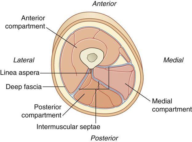
25 The Anteriomedial wall is formed by: Sartorius and fascia The Anterolateral wall is formed by: Vastus medialis The posterior wall (floor) is formed by: Adductor longus and magnus Transverse section through the middle of the left thigh as seen from above
26 Adductor longus Adductor magnus The posterior wall is formed by: Adductor longus and magnus

27 The adductor canal contains 1- Femoral artery 2- Femoral vein 3- Saphenous nerve 4- Nerve of vastus medialis 5- Obturator nerve 6- Subsartorial plexus of nerves Subsartorial plexus of nerves: Located on the fascia under the sartorius and is formed by branches from 1- Medial cutaneous nerve of the thigh 2- Saphenous nerve 3- Obturator nerve
28 The femoral artery 1-Enters the thigh from behind the inguinal ligament as a continuation of the external iliac artery. 2-Lies midway between the anterior superior iliac spine and the symphysis pubis Midinguinal point 3-Its upper half lies superficial in the femoral triangle while in the lower half it lies deep in the subsartorial (adductor) canal (what does this mean?) See next slides 4-Ends at adductor hiatus by entering the popliteal fossa as THE POPLITEAL ARTERY


29 External iliac artery Inguinal ligament Saphenous nerve Femoral artery Adductor hiatus Popliteal artery
 covered by skin")

30 In the upper part of its course, femoral artery lies in the femoral triangle (superficial) covered by skin and fascia 1-Exposed to injuries 2-Good approach for taking femoral pulse 3- Femoral Artery Catheterization

31 Branches of the femoral artery THREE superficial branches TWO deep branches
32 Superficial branches 1-Superficial circumflex iliac artery 2-Superficial epigastric artery 3-Superficial external pudendal artery They supply cutaneous regions of the upper thigh, lower abdomen, and perineum. Deep branches 1-Deep external pudendal artery 2-Profunda femoris artery Branches of the femoral artery in the adductor canal Descending genicular artery

33 Superficial epigastric artery Superficial circumflex iliac artery Superficial external pudendal artery Pudendal to be ashamed in latin. Refers to the external genitalia Deep external pudendal artery

34 The femoral vein lies medial to the artery in the upper part of the femoral triangle, then it lies posterior and finally lateral to the artery at the apex of the femoral triangle Saphenous nerve crosses femoral artery anteriorly Femoral artery Saphenous nerve Adductor hiatus Popliteal artery
 Medial femoral circumflex artery B) Lateral femoral circumflex artery C) Perforating")
35 The profunda femoris artery (Deep artery of thigh) arises from the posterolateral side of the femoral artery about (4 cm) below the inguinal ligament it gives off A) Medial femoral circumflex artery B) Lateral femoral circumflex artery C) Perforating arteries

36 External iliac artery Common iliac artery Internal iliac artery Obturator Artery Obturator Artery The obturator artery is a branch of the internal iliac artery It passes forward on the lateral wall of the pelvis and accompanies the obturator nerve It gives off muscular branches and an articular branch to the hip joint
37 Femoral Vein Profunda femoris artery Great saphenous vein Femoral vein Enters the thigh by passing through the adductor hiatus as a continuation of the popliteal vein Ascends through the thigh, lying at first on the lateral side of the artery, then posterior to it, and finally on its medial side Leaves the thigh in the intermediate compartment of the femoral sheath and passes behind the inguinal ligament to become the external iliac vein. Tributaries The tributaries of the femoral vein are the great saphenous vein and veins that correspond to the branches of the femoral artery drain into the great saphenous vein 1- Superficial circumflex iliac vein 2- Superficial epigastric vein 3- Superficial external pudendal vein

38 Superficial epigastric vein Superficial circumflex iliac vein Femoral vein Superficial external pudendal vein Great saphenous vein
39 Test Your Knowledge 1-Mention Branches of Femoral artery 2-Boundaries of adductor canal 3-Branches of femoral nerve
The front of the thigh. Dr.Amjad shatarat
 The front of the thigh Femoral triangle (Scarpa s triangle) Is a triangular depressed area located in the upper part of the medial aspect of the thigh immediately below the inguinal ligament. Superiorly:
Adductor canal (Subsartorial) or Hunter s canal
 or Hunter s canal") Adductor canal (Subsartorial) or Hunter s canal John Hunter described the exposure and ligation of the femoral artery in this canal for aneurysm of the popliteal artery; this method has the advantage that
Adductor canal (Subsartorial) or Hunter s canal John Hunter described the exposure and ligation of the femoral artery in this canal for aneurysm of the popliteal artery; this method has the advantage that
rotation of the hip Flexion of the knee Iliac fossa of iliac Lesser trochanter Femoral nerve Flexion of the thigh at the hip shaft of tibia
 Anatomy of the lower limb Anterior & medial compartments of the thigh Dr. Hayder The fascia lata encloses the entire thigh like a sleeve/stocking. Three intramuscular fascial septa (lateral, medial, and
ANATYOMY OF The thigh
 ANATYOMY OF The thigh 1- Lateral cutaneous nerve of the thigh Ι) Skin of the thigh Anterior view 2- Femoral branch of the genitofemoral nerve 5- Intermediate cutaneous nerve of the thigh 1, 2 and 3 are
The thigh. Prof. Oluwadiya KS
 The thigh Prof. Oluwadiya KS www.oluwadiya.com The Thigh: Boundaries The thigh is the region of the lower limb that is approximately between the hip and knee joints Anteriorly, it is separated from the
Lecture 08 THIGH MUSCLES ANTERIOR COMPARTMENT. Dr Farooq Khan Aurakzai. Dated:
 Lecture 08 THIGH MUSCLES ANTERIOR COMPARTMENT BY Dr Farooq Khan Aurakzai Dated: 11.02.2017 INTRODUCTION to the thigh Muscles. The musculature of the thigh can be split into three sections by intermuscular
ANATYOMY OF The thigh
 ANATYOMY OF The thigh 1- Lateral cutaneous nerve of the thigh Ι) Skin of the thigh Anterior view 2- Femoral branch of the genitofemoral nerve 5- Intermediate cutaneous nerve of the thigh 1, 2 and 3 are
THIEME. Anterior and Medial Compartments of the Thigh
 CHAPTER Anterior and Medial Compartments of the Thigh Learning Objectives 2 At the end of the dissection, you should be able to identify the following: Cutaneous nerves innervating the skin of the anterior
Ali Yaghi. Omar Eyad. Ahmad Salman. 1 P a g e
 5 Ali Yaghi Omar Eyad Ahmad Salman 1 P a g e **There are two types of groin hernia; the femoral hernia and the inguinal hernia. But how can we differentiate between the inguinal hernia and the femoral
Baraa Ayed حسام أبو عوض. Ahmad Salman. 1 P a g e
 4 Baraa Ayed حسام أبو عوض Ahmad Salman 1 P a g e Today we are going to cover these concepts: Iliotibial tract Anterior compartment of the thigh and the hip Medial compartment of the thigh Femoral triangle
ANATYOMY OF The thigh
 ANATYOMY OF The thigh 1- Lateral cutaneous nerve of the thigh Ι) Skin of the thigh Anterior view 2- Femoral branch of the genitofemoral nerve 1, 2 and 3 are From the lumber plexus 5- Intermediate cutaneous
Anterior and Medial compartments of the thigh. Dr. Heba Kalbouneh Associate Professor of Anatomy and Histology
 Anterior and Medial compartments of the thigh Dr. Heba Kalbouneh Associate Professor of Anatomy and Histology Terms Related to Movements Movement Flexion Extension Abduction Adduction Medial (internal)
Abdominal muscles. Subinguinal hiatus and ingiunal canal. Femoral and adductor canals. Neurovascular system of the lower limb. Sándor Katz M.D.,Ph.D.
 Abdominal muscles. Subinguinal hiatus and ingiunal canal. Femoral and adductor canals. Neurovascular system of the lower limb. Sándor Katz M.D.,Ph.D. External oblique muscle Origin: outer surface of the
musculoskeletal system anatomy nerves of the lower limb 1 done by: dina sawadha & mohammad abukabeer
 musculoskeletal system anatomy nerves of the lower limb 1 done by: dina sawadha & mohammad abukabeer What is the importance of plexuses? plexuses provides us the advantage of a phenomenon called convergence
lower limb Anterior Compartment: lecture 3 The deep fascia ( fascia lata) divides the thigh into 3 compartments:
 divides the thigh into 3 compartments:") lower limb lecture 3 The deep fascia ( fascia lata) divides the thigh into 3 compartments: 1. Anterior Extensor compartment 2. Medial Adductor compartment 3. Posterior Flexor compartment Anterior Compartment:
lower limb lecture 3 The deep fascia ( fascia lata) divides the thigh into 3 compartments: 1. Anterior Extensor compartment 2. Medial Adductor compartment 3. Posterior Flexor compartment Anterior Compartment:
Femoral Artery. Its entrance to the thigh Position Midway between ASIS and pubic symphysis
 Lower Limb Vessels Lecture Objectives Describe the major arteries of the lower limb. Describe the deep and superficial veins of the lower limb. Describe the topographical relationships of the arteries
Lumbar Plexus. Ventral rami L1 L4 Supplies: Major nerves.. Abdominal wall External genitalia Anteromedial thigh
 Lower Limb Nerves Lectures Objectives Describe the structure and relationships of the plexuses of the lower limb. Describe the course, relationships and structures supplied for the major nerves of the
MUSCULOSKELETAL LOWER LIMB
 MUSCULOSKELETAL LOWER LIMB Spinal Cord Lumbar and Sacral Regions Spinal cord Dorsal root ganglion Conus medullaris Cauda equina Dorsal root ganglion of the fifth lumbar nerve End of subarachnoid space
DISSECTION SCHEDULE. Session I - Hip (Front) & Thigh (Superficial)
 & Thigh (Superficial)") DISSECTION SCHEDULE Session I - Hip (Front) & Thigh (Superficial) Surface anatomy Inguinal region Gluteal region Thigh Leg Foot bones Hip bone Femur Superficial fascia Great saphenous vein Superficial
DISSECTION SCHEDULE Session I - Hip (Front) & Thigh (Superficial) Surface anatomy Inguinal region Gluteal region Thigh Leg Foot bones Hip bone Femur Superficial fascia Great saphenous vein Superficial
Identify the muscles associated with the medial compartment of the thigh. Identify the attachment points of the medial thigh muscles.
 L 8 A B O R A T O R Y Thigh MEDIAL THIGH Identify the muscles associated with the medial compartment of the thigh. Identify the attachment points of the medial thigh muscles. Identify the actions of these
1-Muscles: 2-Blood supply: Branches of the profunda femoris artery. 3-Nerve supply: Sciatic nerve
 1-Muscles: B i c e p s f e m o r i s S e m i t e n d i n o s u s S e m i m e m b r a n o s u s a small part of the adductor magnus (h a m s t r i n g p a r t o r i s c h i a l p a r t ) 2-Blood supply:
Lower Limb Nerves. Clinical Anatomy
 Lower Limb Nerves Clinical Anatomy Lumbar Plexus Ventral rami L1 L4 Supplies: Abdominal wall External genitalia Anteromedial thigh Major nerves.. Lumbar Plexus Nerves relation to psoas m. : Obturator n.
Abdomen: Introduction. Prof. Oluwadiya KS
 Abdomen: Introduction Prof. Oluwadiya KS www.oluwadiya.com Abdominopelvic Cavity Abdominal Cavity Pelvic Cavity Extends from the inferior margin of the thorax to the superior margin of the pelvis and the
THE LOWER LIMB NERVES VESSELS
 THE LOWER LIMB NERVES VESSELS LOWER LIMB: FEMORAL TRIANGLE FEMORAL TRIANGLE LOWER LIMB: FEMORAL TRIANGLE FEMORAL TRIANGLE is a triangular landmark useful in dissection and in understanding relationships
HUMAN BODY COURSE LOWER LIMB NERVES AND VESSELS
 HUMAN BODY COURSE LOWER LIMB NERVES AND VESSELS October 22, 2010 D. LOWER LIMB MUSCLES 2. Lower limb compartments ANTERIOR THIGH COMPARTMENT General lfunction: Hip flexion, knee extension, other motions
The posterior abdominal wall. Prof. Oluwadiya KS
 The posterior abdominal wall Prof. Oluwadiya KS www.oluwadiya.sitesled.com Posterior Abdominal Wall Lumbar vertebrae and discs. Muscles opsoas, quadratus lumborum, iliacus, transverse, abdominal wall
ABDOMINAL WALL & RECTUS SHEATH
 ABDOMINAL WALL & RECTUS SHEATH Learning Objectives Describe the anatomy, innervation and functions of the muscles of the anterior, lateral and posterior abdominal walls. Discuss their functional relations
Leg. Dr. Heba Kalbouneh Associate Professor of Anatomy and Histology
 Leg Dr. Heba Kalbouneh Associate Professor of Anatomy and Histology Skin of the Leg Cutaneous Nerves Medially: The saphenous nerve, a branch of the femoral nerve supplies the skin on the medial surface
The Hip (Iliofemoral) Joint. Presented by: Rob, Rachel, Alina and Lisa
 Joint. Presented by: Rob, Rachel, Alina and Lisa") The Hip (Iliofemoral) Joint Presented by: Rob, Rachel, Alina and Lisa Surface Anatomy: Posterior Surface Anatomy: Anterior Bones: Os Coxae Consists of 3 Portions: Ilium Ischium Pubis Bones: Pubis Portion
The Hip (Iliofemoral) Joint Presented by: Rob, Rachel, Alina and Lisa Surface Anatomy: Posterior Surface Anatomy: Anterior Bones: Os Coxae Consists of 3 Portions: Ilium Ischium Pubis Bones: Pubis Portion
Contents of the Posterior Fascial Compartment of the Thigh
 Contents of the Posterior Fascial Compartment of the Thigh 1-Muscles: B i c e p s f e m o r i s S e m i t e n d i n o s u s S e m i m e m b r a n o s u s a small part of the adductor magnus (h a m s t
Lecture 09. Popliteal Fossa. BY Dr Farooq Khan Aurakzai
 Lecture 09 Popliteal Fossa BY Dr Farooq Khan Aurakzai Dated: 14.02.2018 What is popliteus? Introduction Anything relating to, or near the part of the leg behind the knee. From New Latin popliteus the muscle
Abdominal Hernia Omar alnoubani MD,MRCS
 Abdominal Hernia Omar alnoubani MD,MRCS Definition of hernia Anatomical landmarks Overview of types of hernia Presentation and Management of common types of hernia What is the definition of a hernia? An
Inferior Pelvic Border
 Pelvis + Perineum Pelvic Cavity Enclosed by bony, ligamentous and muscular wall Contains the urinary bladder, ureters, pelvic genital organs, rectum, blood vessels, lymphatics and nerves Pelvic inlet (superior
VENOUS DRAINAGE OF THE LOWER LIMB
 Anatomy of the lower limb Superficial veins & nerve injuries Dr. Hayder VENOUS DRAINAGE OF THE LOWER LIMB The venous drainage of the lower limb is of huge clinical & surgical importance. Since the venous
GI anatomy Lecture: 2 د. عصام طارق
 GI anatomy Lecture: 2 د. عصام طارق Objectives: To define rectus sheath. To describe anatomy of inguinal canal. To relates types of inguinal hernia to the region. To explore spermatic cord. Rectus Abdominis
Inguinal Canal. It is an oblique passage through the lower part of the anterior abdominal wall. Present in both sexes
 Inguinal canal Inguinal Canal It is an oblique passage through the lower part of the anterior abdominal wall Present in both sexes It allows structures to pass to and from the testis to the abdomen in
The Thoracic wall including the diaphragm. Prof Oluwadiya KS
 The Thoracic wall including the diaphragm Prof Oluwadiya KS www.oluwadiya.com Components of the thoracic wall Skin Superficial fascia Chest wall muscles (see upper limb slides) Skeletal framework Intercostal
Regional Anaesthesia
 Regional Anaesthesia Lower limb anatomy and blocks Hip and Knee Joint Hip Joint: Nerve supply Lumbar plexus Femoral nerve through the nerve to the Rectus Femoris Ant division of the Obturator nerve The
Mohammad Ashraf. Abdulrahman Al-Hanbali. Ahmad Salman. 1 P a g e
 - 7 Mohammad Ashraf Abdulrahman Al-Hanbali Ahmad Salman 1 P a g e Structures under the cover of Gluteus Maximus: 1-Bones: Ileum, Femur (Head, greater trochanter and gluteal tuberosity), Ischium (ischial
Gluteal region DR. GITANJALI KHORWAL
 Gluteal region DR. GITANJALI KHORWAL Gluteal region The transitional area between the trunk and the lower extremity. The gluteal region includes the rounded, posterior buttocks and the laterally placed
2. List the 8 pelvic spaces: list one procedure or dissection which involves entering that space.
 Name: Anatomy Quiz: Pre / Post 1. In making a pfannensteil incision you would traverse through the following layers: a) Skin, Camper s fascia, Scarpa s fascia, external oblique aponeurosis, internal oblique
musculoskeletal system <lower limb vessle> <1> done by:renad abu ruman &rama alawamleh
 musculoskeletal system done by:renad abu ruman &rama alawamleh The entrance to the anterior compartment of the leg is through lingual lig. & superior ramus, & to the posterior compartment
LAB Notes#1. Ahmad Ar'ar. Eslam
 LAB Notes#1 Ahmad Ar'ar Eslam 1 P a g e Anatomy lab Notes Lower limb bones :- Pelvic girdle: It's the connection between the axial skeleton and the lower limb; it's made up of one bone called the HIP BONE
Borders of the Abdomen
 Abdominal wall Borders of the Abdomen Abdomen is the region of the trunk that lies between the diaphragm above and the inlet of the pelvis below Borders Superior: Costal cartilages 7-12. Xiphoid process:
JMSCR Vol 06 Issue 10 Page October 2018
 www.jmscr.igmpublication.org Impact Factor (SJIF): 6.379 Index Copernicus Value: 79.54 ISSN (e)-2347-176x ISSN (p) 2455-0450 DOI: https://dx.doi.org/10.18535/jmscr/v6i10.08 Original Article Study of Branching
Perineum. done by : zaid al-ghnaneem
 Perineum done by : zaid al-ghnaneem Hello everyone, this sheet will talk about 2 nd Lecture which is Perineum but there are some slides and info from 1 st Lecture. Everything included Slides + Pics Let
BOGOMOLETS NATIONAL MEDICAL UNIVERSITY. Department of Human Anatomy GUIDELINES. The theme of the lesson The vessels of the lower limb.
 BOGOMOLETS NATIONAL MEDICAL UNIVERSITY Department of Human Anatomy GUIDELINES Academic discipline HUMAN ANATOMY Module 2 The theme of the lesson The vessels of the lower limb. Course Faculties І Medical
Lumbar and Sacral Plexuses. Dr. Heba Kalbouneh Associate Professor of Anatomy and Histology
 Lumbar and Sacral Plexuses Dr. Heba Kalbouneh Associate Professor of Anatomy and Histology Structure of Spinal Nerves: Somatic Pathways dorsal root CNS interneuron spinal nerve dorsal ramus somatic sensory
REPRODUCTIVE SYSTEM By Dr.Ahmed Salman
 The University Of Jordan Faculty Of Medicine Anatomy Department REPRODUCTIVE SYSTEM By Dr.Ahmed Salman Assistant Professor of Anatomy &embryology Perineum It is the diamond-shaped lower end of the trunk
Misc Anatomy. Upper Limb! 2. Lower Limb! 5. Venous Drainage! Head & neck! 8
 Misc Anatomy Upper Limb! 2 Arteries!... 2 Veins!... 2 Spaces!... 4 Lower Limb! 5 Arteries!... 5 Venous Drainage!... 6 Spaces!... 7 Head & neck! 8 Artery!... 8 Ultrasound View for IJ CVL!... 8 Arteries
حسام أبو عوض. -Dr. Mohammad Muhtasib. 1 P a g e
 5 حسام أبو عوض - -Dr. Mohammad Muhtasib 1 P a g e There are two types of inguinal hernia: direct and indirect. Hernia: protrusion of the small intestine or the greater omentum of the intra-abdominal organs
Venous drainage of the lower limb
 Venous drainage of the lower limb INTRODUCTION It is of immense clinical and surgical importance. The venous blood against gravity. FACTORS HELPING THE VENOUS DRAINAGE OF THE LOWER LIMB The contraction
Bones of Lower Limb. Dr. Heba Kalbouneh Associate Professor of Anatomy and Histology
 Bones of Lower Limb Dr. Heba Kalbouneh Associate Professor of Anatomy and Histology Bones of the lower limb Hip Bone Made up of 3 bones: 1) Ilium (flat), superior in position 2) Ischium (L), postero-inferior
M. Al-Mohtaseb. Tala Saleh. Faisal Nimri
 4 5 M. Al-Mohtaseb Tala Saleh Faisal Nimri Inguinal Hernia - An abdominal hernia is the protrusion of part of the abdominal content beyond the normal confines of the abdominal wall through weak points
Fascial Compartments of the Upper Arm
 Fascial Compartments of the Upper Arm The upper arm is enclosed in a sheath of deep fascia and has two fascial septa: 1- Medial fascial septum (medial intermuscular septum): attached to the medial supracondylar
The Hay is in the Barn
 Anatomy 1 Practical 1 Review Made by Forrest Allen (nerd) Edited by TJ Williamson (not nerd) The Hay is in the Barn 2019 Thunderbringers Too much to handle https://www.youtube.com/watch?v=glii-kaza d8
Muscles of the Thigh. 6.1 Identify, describe the attachments of and deduce the actions of the muscles of the thigh: Anterior group
 Muscles of the Thigh 6.1 Identify, describe the attachments of and deduce the actions of the muscles of the thigh: Anterior group Sartorius: This is a long strap like muscle with flattened tendons at each
Lectures of Human Anatomy
 Lectures of Human Anatomy Lower Limb Gluteal Region and Hip Joint By DR. ABDEL-MONEM AWAD HEGAZY M.B. with honor 1983, Dipl."Gynecology and Obstetrics "1989, Master "Anatomy and Embryology" 1994, M.D.
GI module Lecture: 9 د. عصام طارق. Objectives:
 GI module Lecture: 9 د. عصام طارق Objectives: To list structures forming posterior abdominal wall. To follow aorta & its main branches. To describe IVC & its main tributaries. To list nerves of posterior
Muscles of the lower extremities. Dr. Nabil khouri MD, MSc, Ph.D
 Muscles of the lower extremities Dr. Nabil khouri MD, MSc, Ph.D Posterior leg Popliteal fossa Boundaries Biceps femoris (superior-lateral) Semitendinosis and semimembranosis (superior-medial) Gastrocnemius
Group of students. - Rawan almujabili د. محمد المحتسب - 1 P a g e
 - 14 - Group of students - Rawan almujabili د. محمد المحتسب - 1 P a g e Nerves of the posterior abdominal wall The spinal cord gives off spinal nerves between the vertebrae. In the abdomen, through the
Anatomy of the Large Intestine
 Large intestine Anatomy of the Large Intestine 2 Large Intestine Extends from ileocecal valve to anus Length = 1.5-2.5m = 5 feet Regions Cecum = 2.5-3 inch Appendix= 3-5 inch Colon Ascending= 5 inch Transverse=
Anatomage Table Instructors Guide- Lower Limb
 The Lower Limb Anatomage Table Instructors Guide- Lower Limb Table of Contents Lower Limb 1- The Skeletal System...3 1: Hip Bone...3 2: Hip Joint and Femur...4 3: Patella and Knee Joint...7 4: Tibia, Fibula,
Where should you palpate the pulse of different arteries in the lower limb?
 Where should you palpate the pulse of different arteries in the lower limb? The femoral artery In the femoral triangle, its pulse is easily felt just inferior to the inguinal ligament midway between the
Copyright 2003 Pearson Education, Inc. publishing as Benjamin Cummings. Dr. Nabil Khouri MD, MSc, Ph.D
 Dr. Nabil Khouri MD, MSc, Ph.D Pelvic Girdle (Hip) Organization of the Lower Limb It is divided into: The Gluteal region The thigh The knee The leg The ankle The foot The thigh and the leg have compartments
Bony ypelvis. Composition: formed by coccyx, and their articulations Two portions
 Pelvis Bony ypelvis Composition: formed by paired hip bones, sacrum, coccyx, and their articulations Two portions Greater pelvis Lesser pelvis Terminal line ( pelvic inlet): formed by promontory of sacrum,
Dana Alrafaiah. - Amani Nofal. - Ahmad Alsalman. 1 P a g e
 - 2 - Dana Alrafaiah - Amani Nofal - Ahmad Alsalman 1 P a g e This lecture will discuss five topics as follows: 1- Arrangement of pelvic viscera. 2- Muscles of Pelvis. 3- Blood Supply of pelvis. 4- Nerve
The arm: *For images refer back to the slides
 The arm: *For images refer back to the slides Muscles of the arm: deltoid, triceps (which is located at the back of the arm), biceps and brachialis (it lies under the biceps), brachioradialis (it lies
Part One Anatomy Practice MeQ's
 Part One natomy Practice MeQ's 1. Regarding the flexor retinaculum, which of the following statements is FLS?: o It is crossed by palmaris longus, the palmar branch of the median nerve, and the superficial
Functional anatomy and variability of the blood vessels of the upper and lower limbs. Anastasia Bendelic Human Anatomy Departament
 Functional anatomy and variability of the blood vessels of the upper and lower limbs Anastasia Bendelic Human Anatomy Departament Plan: 1. Variations of the branching pattern of the aortic arch 2. Arterial
Benha University. Faculty of Medicine. Anatomy Department Course code (MED 0701) Model answer of Anatomy examination. (Abdomen,Pelvis and Thorax)
 Model answer of Anatomy examination. (Abdomen,Pelvis and Thorax)") 1 Benha University Faculty of Medicine Anatomy Department Course code (MED 0701) Model answer of Anatomy examination (Abdomen,Pelvis and Thorax) 1 st year 2 nd term Date :18 /5 /2013 2 I-Short account
1 Benha University Faculty of Medicine Anatomy Department Course code (MED 0701) Model answer of Anatomy examination (Abdomen,Pelvis and Thorax) 1 st year 2 nd term Date :18 /5 /2013 2 I-Short account
Abdomen. Retroperitoneal space
 Abdomen. Retroperitoneal space Abdominal cavity The space bounded by: Anterolateral abdominal wall Posterior abdominal wall Diaphragm Pelvic walls and pelvic floor. Subdivided into: True abdominal cavity
Bony Anatomy. Femur. Femoral Head Femoral Neck Greater Trochanter Lesser Trochanter Intertrochanteric Crest Intertrochanteric Line Gluteal Tuberosity
 Hip Anatomy Bony Anatomy Femur Femoral Head Femoral Neck Greater Trochanter Lesser Trochanter Intertrochanteric Crest Intertrochanteric Line Gluteal Tuberosity Bony Anatomy Pelvic Girdle Acetabulum 3 bones
Anatomy of the renal system. Professor Nawfal K. Al-Hadithi
 Anatomy of the renal system Professor Nawfal K. Al-Hadithi Objectives To describe the posterior abdominal wall To identify the main anatomical landmarks of the kidneys & ureters To describe the suprarenal
Neck-2. Dr. Heba Kalbouneh Associate Professor of Anatomy and Histology
 Neck-2 ` Dr. Heba Kalbouneh Associate Professor of Anatomy and Histology Triangles of the neck Side of the neck Midline Lower border of mandible Line between angle of mandible and mastoid Superior nuchal
6/10/2016. Anatomy #7. Jamaledin Abu Ghaida Hussein Al-Sa di. The adductor canal & the popliteal fossa
 6/10/2016 Anatomy #7 The adductor canal & the popliteal fossa Jamaledin Abu Ghaida Hussein Al-Sa di Hello This lecture is short (9 pages) and easy. It includes the records of both groups; A and B. I included
Gross Anatomy Coloring Book Series. Lower Extremity Arteries
 Gross Anatomy Coloring Book Series Lower Extremity Arteries 1 Femoral Artery and Associated Branches For the life of the flesh is in the blood. Leviticus 17:11 Femoral Artery and Associated Branches After
STRUCTURAL BASIS OF MEDICAL PRACTICE EXAMINATION 3. October 17, 2014
 STRUCTURAL BASIS OF MEDICAL PRACTICE EXAMINATION 3 October 17, 2014 PART l. Answer in the space provided. (12 pts) 1. Identify the structures. (2 pts) A. B. A B C. D. C D 2. Identify the structures. (2
The Lower Limb II. Anatomy RHS 241 Lecture 3 Dr. Einas Al-Eisa
 The Lower Limb II Anatomy RHS 241 Lecture 3 Dr. Einas Al-Eisa Tibia The larger & medial bone of the leg Functions: Attachment of muscles Transfer of weight from femur to skeleton of the foot Articulations
ANATOMY TEAM GLUTEAL REGION & BACK OF THIGH
 ANATOMY TEAM GLUTEAL REGION & BACK OF THIGH OBJECTIVES By the end of this lecture, the student should be able to identify and discuss: Contents of gluteal region: Groups of Glutei muscles and small muscles
First practical session. Bones of the gluteal region
 First practical session 2017 Bones of the gluteal region The Hip bone The hip bone is made of: 1 The ilium: superior in position 2 The ischium:postero-inferior in position 3 The pubis: antero-inferior
Internal abdominal wall and inguinal region. Mathew Wedel, 2015
 Internal abdominal wall and inguinal region Mathew Wedel, 2015 gut tube umbilicus gut tube dorsal mesentery visceral peritoneum gut tube FOREGUT dorsal mesentery parietal peritoneum MIDGUT & HINDGUT gut
It is formed by fusion of 3 bones: I. Ilium (superior bone). II. Pubis (antero-inferior bone). III. Ischium (postero-inferior bone).
. II. Pubis (antero-inferior bone). III. Ischium (postero-inferior bone).") It is formed by fusion of 3 bones: I. Ilium (superior bone). II. Pubis (antero-inferior bone). III. Ischium (postero-inferior bone). Pubis Acetabulum Ana (242 ) The three constituent of bones of the hip
It is formed by fusion of 3 bones: I. Ilium (superior bone). II. Pubis (antero-inferior bone). III. Ischium (postero-inferior bone). Pubis Acetabulum Ana (242 ) The three constituent of bones of the hip
The femoral artery and its branches in the baboon Papio anubis
 O R I G I N A L A R T I C L E Folia Morphol. Vol. 66, No. 4, pp. 291 295 Copyright 2007 Via Medica ISSN 0015 5659 www.fm.viamedica.pl The femoral artery and its branches in the baboon Papio anubis Dyl
musculoskeletal system anatomy nerves of the lower limb 2 done by: Dina sawadha & mohammad abukabeer
 musculoskeletal system anatomy nerves of the lower limb 2 done by: Dina sawadha & mohammad abukabeer #Sacral plexus : emerges from the ventral rami of the spinal segments L4 - S4 and provides motor and
Lower Extremity Ultrasound-Guided Regional Anesthesia. Stephanie Duffy, CRNA Regional Anesthesia Faculty Acute Pain Service NMCSD
 Lower Extremity Ultrasound-Guided Regional Anesthesia Stephanie Duffy, CRNA Regional Anesthesia Faculty Acute Pain Service NMCSD Objectives Review anatomy of lumbosacral plexus Lumbar plexus blocks Psoas
The os coxae or hip bone consists of three flat bones, ilium, ischium and pubis, which fuse together to form the acetabulum.
 The os coxae The os coxae or hip bone consists of three flat bones, ilium, ischium and pubis, which fuse together to form the acetabulum. The ilium extends from the acetabulum upwards forming the lateral
STRUCTURAL BASIS OF MEDICAL PRACTICE EXAMINATION 3. October 16, 2015
 STRUCTURAL BASIS OF MEDICAL PRACTICE EXAMINATION 3 October 16, 2015 PART l. Answer in the space provided. (12 pts) 1. Identify the structures. (2 pts) A. B. A B C. D. C D 2. Identify the structures. (2
NOTES FROM GUTMAN LECTURE 10/26 Use this outline to study from. As you go through Gutman s lecture, fill in the topics.
 NOTES FROM GUTMAN LECTURE 10/26 Use this outline to study from. As you go through Gutman s lecture, fill in the topics. Anatomy above the arcuate line Skin Camper s fascia Scarpa s fascia External oblique
Anatomy of the Thorax
 Anatomy of the Thorax A) THE THORACIC WALL Boundaries Posteriorly by the thoracic part of the vertebral column Anteriorly by the sternum and costal cartilages Laterally by the ribs and intercostal spaces
Table 2. First Generated List of Expert Responses. Likert-Type Scale. Category or Criterion. Rationale or Comments (1) (2) (3) (4)
 (2) (3) (4)") Table 2. First Generated List of Expert Responses. Likert-Type Scale Category or Criterion Anatomical Structures and Features Skeletal Structures and Features (1) (2) (3) (4) Rationale or Comments 1. Bones
Table 2. First Generated List of Expert Responses. Likert-Type Scale Category or Criterion Anatomical Structures and Features Skeletal Structures and Features (1) (2) (3) (4) Rationale or Comments 1. Bones
NBME Anatomy Review. Sylvia Nelsen, Ph.D. March 19, 2015
 NBME Anatomy Review Sylvia Nelsen, Ph.D. March 19, 2015 UPPER & LOWER LIMBS 1. What is the most likely diagnosis in this case? A. Rotator cuff tendinitis: pain w/o weakness B. Adhesive capsulitis: absolute
The peritoneum. Prof. Oluwadiya KS, MBBS, FMCS(Orthop) Website:
 Website:") The peritoneum Prof. Oluwadiya KS, MBBS, FMCS(Orthop) Website: http://oluwadiya.com The peritoneum Serous membrane that lines the abdominopelvic cavity and invests the viscera The largest serous membrane
The peritoneum Prof. Oluwadiya KS, MBBS, FMCS(Orthop) Website: http://oluwadiya.com The peritoneum Serous membrane that lines the abdominopelvic cavity and invests the viscera The largest serous membrane
Figure 1 - Hip and Pelvis
 Hip Figure 1 - Hip and Pelvis The terms hip and pelvis are frequently used interchangeably, but strictly speaking, the pelvis is a girdle of bones and the hip is a joint. The pelvis consists of The sacrum
A cadaveric study on the variation of branching pattern of femoral artery
 Original article: A cadaveric study on the variation of branching pattern of femoral artery Dr Banani Kundu, Dr Nabanita Chakraborty, Dr Soham Chakraborty, DrAlpana De Name of the Institute/college: Department
Year 2004 Paper one: Questions supplied by Megan
 QUESTION 47 A 58yo man is noted to have a right foot drop three days following a right total hip replacement. On examination there is weakness of right ankle dorsiflexion and toe extension (grade 4/5).
Slide Read the tables it is about the difference between male & female pelvis.
 I didn t include the slides, this is only what the doctor read or said because he skipped a lot of things because we took it previously, very important to go back to the slides (*there is an edited version)
Acland's DVD Atlas of Human Anatomy. Transcript for Volume Robert D Acland
 Acland's DVD Atlas of Human Anatomy Transcript for Volume 2 2007 Robert D Acland This free downloadable pdf file is to be used for individual study only. It is not to be reproduced in any form without
Medial Groin and Hernia: Sonographic Evaluation. Adam M. Pourcho DO Swedish Sports Medicine
 Medial Groin and Hernia: Sonographic Evaluation Adam M. Pourcho DO Swedish Sports Medicine Disclosures Hernia Eval Takes Practice: Fake it till you make it Objectives Understand anatomy of medial hip and