High Ankle Sprains: Diagnosis & Treatment
|
|
|
- Ann Fisher
- 5 years ago
- Views:
Transcription
1 High Ankle Sprains: Diagnosis & Treatment Mark J. Mendeszoon, DPM, FACFAS, FACFAOM Precision Orthopaedic Specialties University Regional Hospitals Advanced Foot & Ankle Fellowship- Director
2
3
4

5

6 It Is Only an Ankle Sprain

7 Evaluate Degree of Ecchymosis & Edema

8 If Not Properly Treated Chronic Pain & Ankle Instability
9 Epidemiology Waterman et al. JBJS 2010 states: 2 million ankle sprains per year = 2 billion in health care cost Injury results in time lost and disability in 60% of patients 30% of all sport injury

10 Epidemiology Syndesmotic Injuries: 1% to 18% of all ankle sprains 32% develop calcification and chronic pain High incidence of post traumatic arthritis Greater source of impairment than the typical lateral ankle sprain
11


12 Anatomy Inferior Tibiofibular Joint: defined as a syndesmotic articulation which consists of five separate portions Motion in all three planes
13 Anatomy
14 Syndesmotic Ligaments: Anterior Inferior Tibio Fibular Ligament Posterior Inferior Tibio Fibular Ligament Transverse Tibio Fibular Ligament Interosseous Ligament Interosseous Membrane

15 Deltoid Ligament The deep portion of the deltoid ligament also contributes to syndesmotic stability Acting as a restraint against lateral shift of the talus
16 Biomechanics of Syndesmosis RELEVANT ASPECTS OFANKLE: A considerable clearance takes place between the talus and the distal fibula, which is limited by the tibiofibular syndesmosis With normal stance, almost no twisting and shearing forces act on the ankle joint= static tibfib tension Axial loading tensions AITF and PITF with increase of 10-17% of body weight intact syndesmosis, the intermalleolar distance increases with dorsiflexion of the talus by 1.0 to 1.25 mm
17 Haraguchi et al Intact syndesmosis Fibula ROTATES 2 * externally Equals ~ 2.4 mm distally mm Anterior -posteriorly THUS Fibula moves in 3 D

18 Ogilvie & Harris 1994 Study on Individual Ligaments for Syndesmotic Stability 35% ATIFL 33% TRANSVERSE LIG. 22% IOL 9% PTIFL

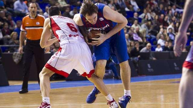
19 MECHANISM OF INJURY HIGH VELOCITY INJURIES COLLISION SPORTS SKIIING X GAMES


20 FORCED EXTERNAL ROTATION SEVERE INVERSION & PLANTARFLEXION
21 CLASSIFICATION ACFAS 1997 EDWARDS & DELEE 1984 Gerber et al 1998
22 Perform Thorough History & Physical Underlying Etiological Conditions ( Ligamentous Laxity) Understand Mechanism of Injury Establish Realistic Goals & Time Table

23 Inversion Ankle Sprain Examination Proximal Fibula Distal Fibula Peroneal Tendons Sinus Tarsi / Anterior Process Calcaneus Calcaneal - Cuboid Joint Base of Fifth Metatarsal Deltoid Ligaments

24 SQUEEZE TEST


25 Other Clinical Testing For High Ankle Sprains Dorsiflexion Compression Dorsiflexion External Rotation Crossed Leg Test Heel Thump
26
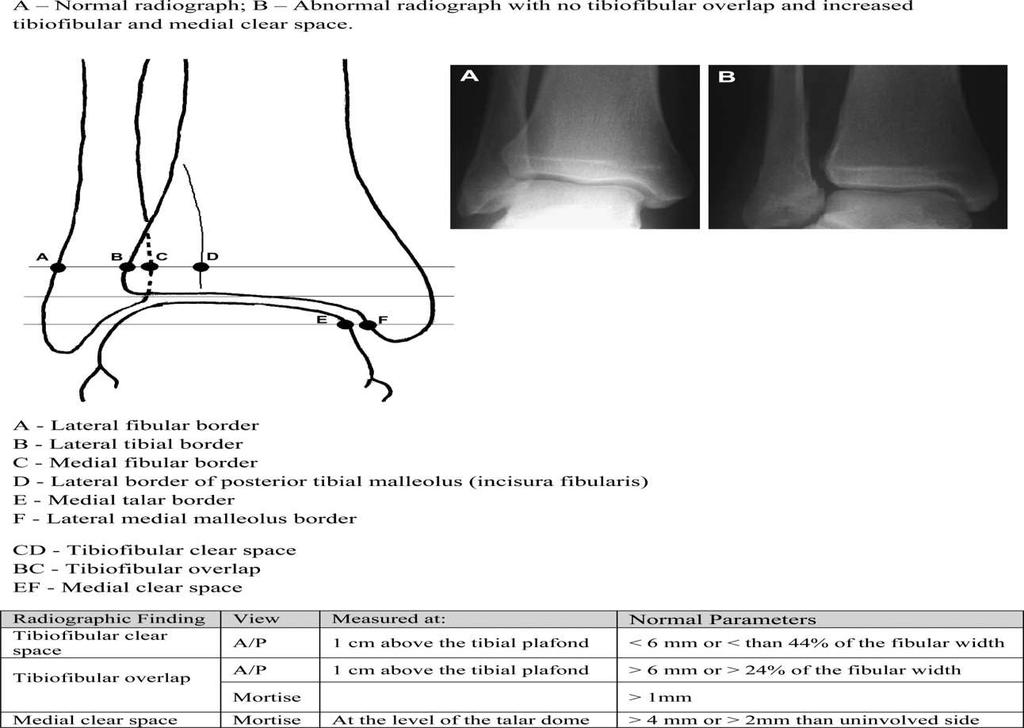
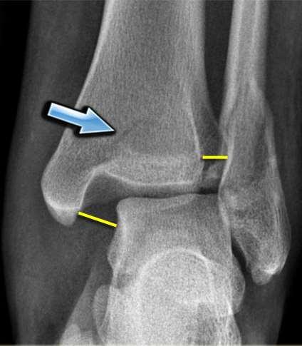
27 Radiographic Exam 3 views: AP, Mortise, Lateral Contralateral X-Rays Tibiofibular Clear Space Medial Clear Space Tibiofibular Overlap
28

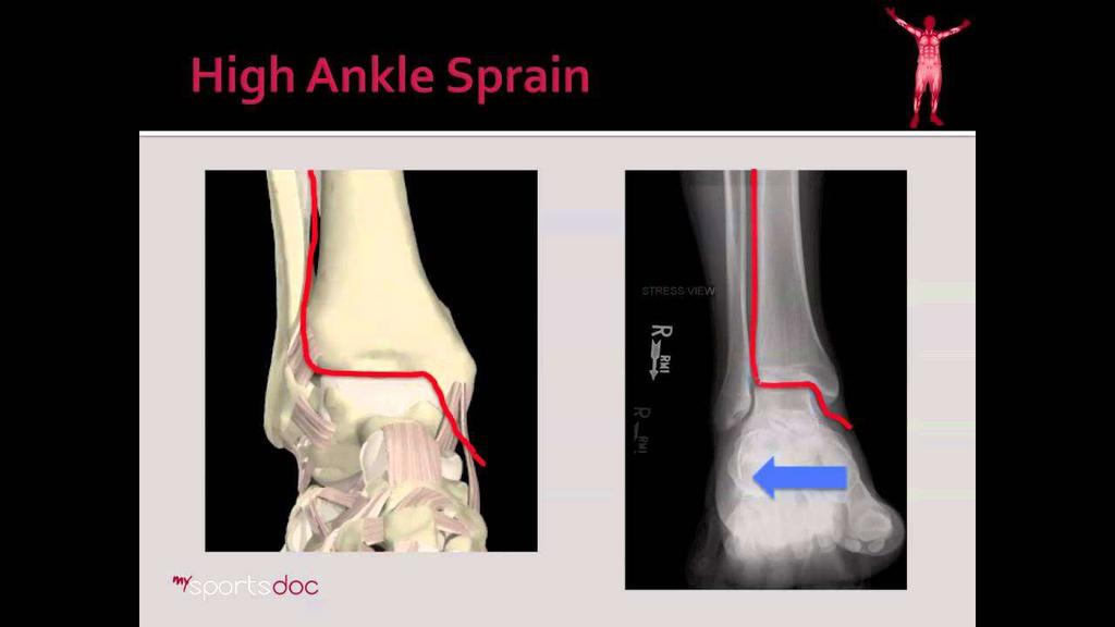
29 Radiographic Exam External Rotational Stress Test As static radiograph or intra operatively Shows widening of tib-fib clear space and can show deltoid disruption


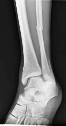
30 Associated Injuries Ankle Fractures: PER 3, SER 4, PAB Maisonneuve Posterior Malleolar Fractures Tillaux Chaput Fractures
31 Associated Injuries Cont.
32 Diagnostic Testing


33 CT Scan Accurate Detect Diastasis of 2-3 mm Bilateral observes: fibular shift, rotation, shortening avulsions


34 Diagnostic Ultrasound Perform in Office Quick & Inexpensive No Radiation Learning Curve**** Mei-Dan et al AM.Journ. Sports Med % specificity/sensitivity AITFL 0.4 mm
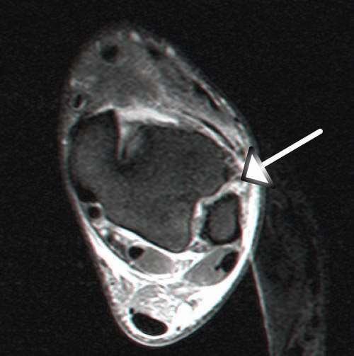
35 MRI Gold Standard Shown to effectively display the components of the syndesmotic complex wit 93% specificity and 100% sensitivity for injury of the AITFL, and 100% specificity and sensitivity for inju the PITFL compared with arthroscopy in acute injuries
36

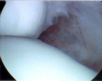
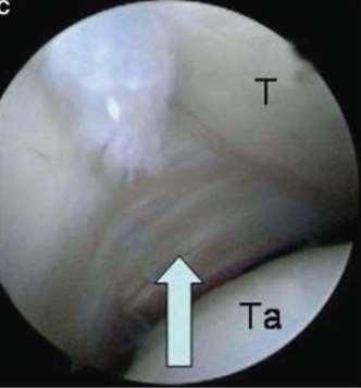
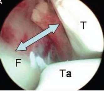
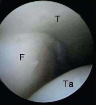
37 Diagnostic Arthroscopy Direct visualization of AITFL and PITFL Must use both anterior and posterior portals Intraoperative Dx User dependent, learning curve Can perform complete direct repair
38
39 Treatment Protocol
40 Conservative Treatment RICE NSAIDS NWB Boot Physical Therapy Favorable results: % Good to Excellent Outcomes
41 Surgical Indications Diastasis > 2mm isolated or with Fractures REQUIRES SURGERY

42 Surgical Algorithm
43 Surgical Treatment Percutaneous repair: Single or double screws with or w/out plate or washers Absorbable screws Suture button (single or double) Direct repair: Arthroscopic debridement with direct ligament repair Open with tendon graft

44 Surgical Treatment Approach: Anterolateral linear over distal fibula Allows complete debridement of avulsed ligaments tissue or debris that may block proper reduction
 reduction clamp The anterior rim of the fibula should align with Chaput s")
45 urgical Treatment Reduction: position the fibula properly into the incisura fibularis of the tibia, which is best achieved with a bimalleolar (pelvic) reduction clamp The anterior rim of the fibula should align with Chaput s tubercule

46 Surgical Treatment In cases of malreduction the medial aspect of the ankle and the deltoid ligament should be explored via arthrotomy All ligamentous or capsular debris is removed After proper reduction, the position of the fibula may be secured temporarily with a Kirschner wire Ensure that a proper tibiofibular distance is obtained in Neutral Ankle Position

47
48
49
50

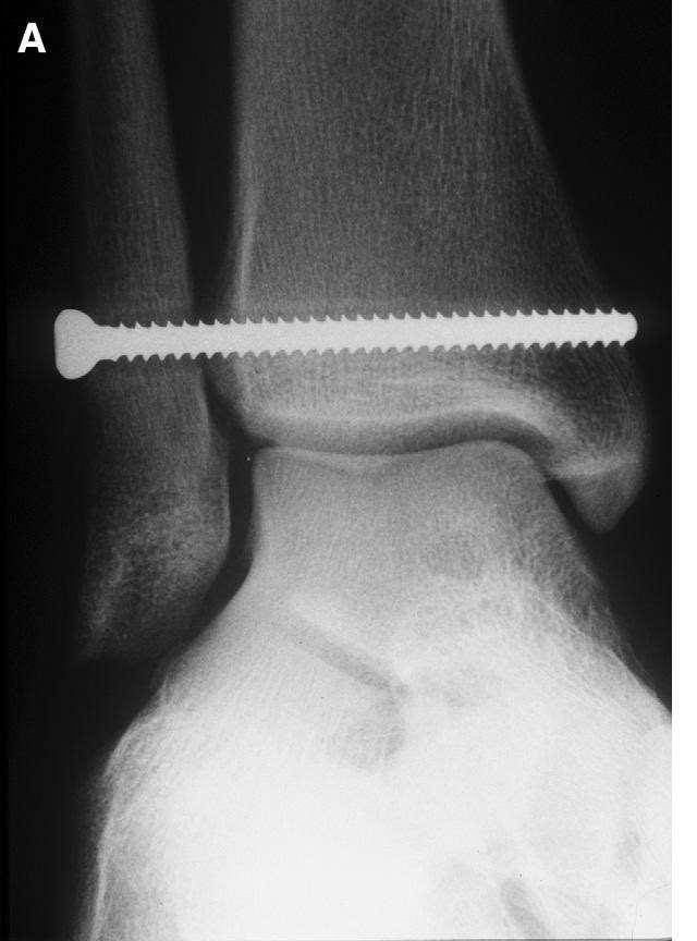
51 Pearls for Screw Placement screws applied 30 degrees posterolateral-anteromedial screws placed 2 cm -4.5 cm above joint line obtain minimum 3-4 cortices Full Threaded Screws Washers vs. Plates
52 Outcomes of screw treatment: Leeds and Ehrlich + Fritschy no recurrence after open reduction, screw fixation, and suture of the AITFL Edwards and DeLee 4-year results of 34 patients = adequacy of syndesmosis reduction and arthritis at followu Proper syndesmosis reduction is key


53 Surgical Treatment Suture button: Follows same principles as screw fixation Faster rehabilitation? 1 vs. 2 Clinical studies show relatively equal rigidity as compared with screws Allows more normal motion of joint


54 Surgical Treatment Open with Tendon Graft Well suited more for chronic diastasis
55 Post Operative Care screws removed weeks NWB 2-4 weeks cast WB as tolerated 4-6 weeks Physical Therapy after screws removed

56 Post Op Complications Typical Post Op Complications Heterotopic Ossifications ~ 32% Tib- Fib Synostosis with persistent pain CHRONIC INSTABILTY DUE TO MISSED DIAGNOSIS OR MALREDUCTION
57 Review Syndesmotic Complex provides a dynamic support to the ankle for normal motion Understanding Anatomy, Biomechanics & Mechanism of Injury is Paramount Appreciated Clinical Exam & Special Testing Recognize Diagnostic Testing Appreciate Surgical Indications & Techniques

58
Disclosures. Syndesmosis Injury. Syndesmosis Ligaments. Objectives. Mark M. Casillas, M.D.
 Disclosures Syndesmosis Injury No relevant disclosures Mark M. Casillas, M.D. 1 Objectives Syndesmosis Ligaments Understand the syndesmosis anatomy and function Classify syndesmosis injuries Describe treatment
Ankle Ligament Injury: Don t Worry- It s Only a Sprain Wes Jackson MD Orthopaedic Foot & Ankle
 Ankle Ligament Injury: Don t Worry- It s Only a Sprain Wes Jackson MD Orthopaedic Foot & Ankle Outline I. Epidemiology II. Classification and Types of Sprains III. Anatomy IV. Clinical Assessment and Imaging
Clinical evaluation where no obvious fracture a. Squeeze test
 7:43 am The Syndesmotic Injury: From Subtle to Severe Robert B. Anderson, MD Chief, Foot and Ankle Carolinas Medical Center OrthoCarolina (Charlotte, North Carolina) 7:30-8:25 am Symposium 1: Management
Burwood Road, Concord Dora Street, Hurstville Lethbridge Street, Penrith 160 Belmore Road, Randwick
 www.orthosports.com.au 47 49 Burwood Road, Concord 29 31 Dora Street, Hurstville 119 121 Lethbridge Street, Penrith 160 Belmore Road, Randwick Update on Syndesmosis Ankle Sprains By Todd Gothelf Foot,
BIOMECHANICS OF ANKLE FRACTURES
 BIOMECHANICS OF ANKLE FRACTURES William R Reinus, MD MBA FACR Significance of Ankle Fractures Most common weight-bearing Fx 70% of all Fxs Incidence is increasing Bimodal distribution Men 15-24 Women over
X-Ray Rounds: (Plain) Radiographic Evaluation of the Ankle.
 Radiographic Evaluation of the Ankle.") X-Ray Rounds: (Plain) Radiographic Evaluation of the Ankle www.fisiokinesiterapia.biz Anatomy Complex hinge joint Articulations among: Fibula Tibia Talus Tibial plafond Distal tibial articular surface
X-Ray Rounds: (Plain) Radiographic Evaluation of the Ankle www.fisiokinesiterapia.biz Anatomy Complex hinge joint Articulations among: Fibula Tibia Talus Tibial plafond Distal tibial articular surface
Syndesmotic Ankle Injuries: Diagnosis and Treatment
 Syndesmotic Ankle Injuries: Diagnosis and Treatment John A. Scolaro, M.D., M.A. Assistant Professor of Orthopaedic Surgery University of California, Irvine California Orthopaedic Association - 2016 Disclosures
Donald Stewart, MD. Lateral ligament injuries Chronic lateral ligament instability Syndesmosis Injuries
 Donald Stewart, MD Arlington Orthopedic Associates Lateral ligament injuries Chronic lateral ligament instability Syndesmosis Injuries Anatomy Mechanism of Injury Classification Diagnostic Tests Management
Objective. Reducing a displaced Syndesmosis 2/11/2016. Ankle Fractures Common Misconceptions. Common Myths in ankle fracture management
 Ankle Fractures Common Misconceptions Jackson Lee, MD Associate Professor Clinical Orthopedics Keck School of Medicine of the University of Southern California Objective Common Myths in ankle fracture
FIBULAR & SYNDESMOSIS MALUNIONS
 FIBULAR & SYNDESMOSIS MALUNIONS MICHAEL P. CLARE, MD FLORIDA ORTHOPAEDIC INSTITUTE TAMPA, FL USA MORTISE INHERENTLY UNSTABLE LATERAL MALLEOLUS ACTS AS BUTTRESS / POST RESIST LATERAL TRANSLATION OF TALUS
Outline. Ankle/Foot Anatomy Ankle Sprains Ottawa Ankle Rules DDx: The Sprain That Wasn t
 Ankle Injuries Outline Ankle/Foot Anatomy Ankle Sprains Ottawa Ankle Rules DDx: The Sprain That Wasn t Anatomy: Ankle Mortise Bony Anatomy Lateral Ligament Complex Medial Ligament Complex Ankle Sprains
Isolated Syndesmotic Instability The High Ankle Sprain Robert B. Anderson, MD
 Isolated Syndesmotic Instability The High Ankle Sprain Robert B. Anderson, MD Chief, Foot & Ankle Service Carolinas Medical Center OrthoCarolina Team Orthopaedist, Carolina Panthers Charlotte, North Carolina
Ankle Sprains and Their Imitators
 Ankle Sprains and Their Imitators Mark Halstead, MD Dr. Mark Halstead is the Associate Professor of the Departments of Orthopedics and Pediatrics at Washington University School of Medicine; Director of
PRONATION-ABDUCTION FRACTURES
 C H A P T E R 1 2 PRONATION-ABDUCTION FRACTURES George S. Gumann, DPM (The opinions of the author should not be considered as reflecting official policy of the US Army Medical Department.) Pronation-abduction
1/27/2016. Background. Background. Seth R. Yarboro University of Virginia January 29, Distal tibio fibular joint
 Seth R. Yarboro January 29, 2015 Background Distal tibio fibular joint maintains ankle stability while allowing motion Dorsiflexion/external rotation mechanism Poor alignment ankle arthritis Background
The Syndesmosis. Syndesmosis: How to Reduce and How Perfect? Boston Medical Center. Indications. Technique 11/19/2018.
 Syndesmosis: How to Reduce and How Perfect? Paul Tornetta III Professor Boston Medical Center Boston Medical Center The Syndesmosis Indications Subluxation Instability Technique Fluoroscopic Open 1 Weber
Peggers Super Summaries: Foot Injuries
 Lisfranc Injury ANATOMY Roman arch with recessed 2 nd MT base AP medial side of intermediate cuneiform to 2 nd MT base Oblique medial side of lateral cuneiform with 3 rd MT base and 4 th with medial boarder
Disclosures! The Syndesmosis. Syndesmosis: How and When to Reduce. Boston Medical Center. Indications. Technique.
 Syndesmosis: How and When to Reduce Paul Tornetta III Professor Boston Medical Center Boston Medical Center Publications: Disclosures! Rockwood and Green, Tornetta and Einhorn; Subspecialty series, Court-Brown,
Surgery-Ortho. Fractures of the tibia and fibula. Management. Treatment of low energy fractures. Fifth stage. Lec-6 د.
 Fifth stage Lec-6 د. مثنى Surgery-Ortho 28/4/2016 Indirect force: (low energy) Fractures of the tibia and fibula Twisting: spiral fractures of both bones Angulatory: oblique fractures with butterfly segment.
5 COMMON INJURIES IN THE FOOT & ANKLE
 5 COMMON INJURIES IN THE FOOT & ANKLE MICHAEL P. CLARE, MD FLORIDA ORTHOPAEDIC INSTITUTE TAMPA, FL USA MECHANISM OF INJURY HOW DID IT HAPPEN? HIGH ENERGY VS LOW ENERGY DIRECTION OF FORCES INVOLVED LIVING
Deltoid and Syndesmosis Ligament Injury of the Ankle Without Fracture
 Deltoid and Syndesmosis Ligament Injury of the Ankle Without Fracture Chris D. Miller, MD, Walter R. Shelton,* MD, Gene R. Barrett, MD, F. H. Savoie, MD, and Andrea D. Dukes, MS From the Mississippi Sports
Arthroscopy Of the Ankle.
 Arthroscopy Of the Ankle www.fisiokinesiterapia.biz Ankle Arthroscopy Anatomy Patient setup Portal placement Procedures Complications Anatomy Portals Anterior Anteromedial Anterolateral Anterocentral Posterior
Ankle Fracture: Tips and Tricks
 Ankle Fracture: Tips and Tricks Christiaan N. Mamczak, DO LCDR, MC, USN Naval Medical Center Portsmouth Department of Orthopaedic Surgery Assistant Professor Uniformed Services University of the Health
SURGICAL AND APPLIED ANATOMY
 Página 1 de 9 Copyright 2001 Lippincott Williams & Wilkins Bucholz, Robert W., Heckman, James D. Rockwood & Green's Fractures in Adults, 5th Edition SURGICAL AND APPLIED ANATOMY Part of "47 - ANKLE FRACTURES"
V E R I TAS MGH 1811 MGH 1811 V E R I TAS. *Gerber JP. Persistent disability with ankle sprains. Foot Ankle Int 19: , 1998.
 MGH 1811 Management of Ankle Instability Richard J. de Asla, M.D. V E R I TAS MGH 1811 I have no potential conflicts with this presentation. V E R I TAS It s just a sprain Lateral Ankle Sprains Most common
Paul Alley MD,DPM,MS,FACS,FAAOS,BFD Eby Orthopaedics,Jasper,Indiana
 Paul Alley MD,DPM,MS,FACS,FAAOS,BFD Eby Orthopaedics,Jasper,Indiana Very common Bone=fractures Description (cracked,broke,busted,or smashed) A=anatomic area of bone eg: head,neck,shaft B=bone involved
A Patient s Guide to Ankle Syndesmosis Injuries
 A Patient s Guide to Ankle Syndesmosis Injuries Introduction An ankle injury common to athletes is the ankle syndesmosis injury. This type of injury is sometimes called a high ankle sprain because it involves
Ankle Fractures in the Elderly: How to Deal with Poor Bone Quality
 : How to Deal with Poor Bone Quality Richard T. Laughlin, MD Professor of Orthopaedic Surgery University of Cincinnati College of Medicine No disclosures relative to this presentation acknowledgement Some
CURRENT TREATMENT OPTIONS
 CURRENT TREATMENT OPTIONS Fix single column or both: Always fix both. A study by Svend-Hansen corroborated the poor results associated with isolated medial malleolar fixation in bimalleolar ankle fractures.
Ankle and Foot Orthopaedic Tests Orthopedics and Neurology DX 612
 Ankle and Foot Orthopaedic Tests Orthopedics and Neurology DX 612 James J. Lehman, DC, MBA, DABCO University of Bridgeport College of Chiropractic Ankle & Foot Anatomy Stability of the ankle is dependent
Disclosures. OTA Resident Advanced Trauma Techniques Course: Ankle Fractures. No relevant disclosures. William H. Harvin, MD Dallas, TX
 OTA Resident Advanced Trauma Techniques Course: Ankle Fractures William H. Harvin, MD Dallas, TX January 31, 2017 Disclosures No relevant disclosures 1 Ankle Anatomy: Lateral ankle ligaments Ankle Anatomy:
Competence of the Deltoid Ligament in Bimalleolar Ankle Fractures After Medial Malleolar Fixation *
 Competence of the Deltoid Ligament in Bimalleolar Ankle Fractures After Medial Malleolar Fixation * BY PAUL TORNETTA, III, M.D. Investigation performed at Kings County Hospital, New York, N.Y. Abstract
11/2/17. Lateral Collateral Complex Medial Collateral Complex Distal Tibiofibular Syndesmosis Spring Ligament
 Andrew J Grainger Leeds, UK Lateral Collateral Complex ial Collateral Complex Distal Tibiofibular Syndesmosis Spring Ligament Brief anatomy review Scan tips and tricks Pathological appearances andrewgrainger@nhs.net
Radiographic Evaluation of Calcaneal Fractures. Kali Luker, PGY-1
 Radiographic Evaluation of Calcaneal Fractures Kali Luker, PGY-1 Anatomy Extraarticular Fractures Involve body, anterior process or tuberosity Treated with immobilization and NWB x 6 wks UNLESS Displaced
Craig S. Radnay, M.D. 1/27/2016. Access to the Talus for Treatment of Osteochondral Lesions. Epidemiology of OLT. Treatment of OLT
 Access to the Talus for Treatment of Osteochondral Lesions Craig S. Radnay, MD, MPH ISK Institute for Orthopaedics and Sports Medicine NYU/Hospital for Joint Diseases Tampa, FL January 23, 2016 Epidemiology
Commonly Missed Foot and Ankle Conditions. David Miller, DPM AMG Podiatry
 Commonly Missed Foot and Ankle Conditions David Miller, DPM AMG Podiatry Lisfranc Injuries Wide spectrum of injuries High energy Subtle subluxation which could be easily missed injuries Men are 2-4x s
Clin Podiatr Med Surg 19 (2002) Index
 Index") Clin Podiatr Med Surg 19 (2002) 335 344 Index Note: Page numbers of article titles are in bold face type. A Accessory soleus muscle, magnetic resonance imaging of, 300 Achilles tendon injury of, magnetic
Clin Podiatr Med Surg 19 (2002) 335 344 Index Note: Page numbers of article titles are in bold face type. A Accessory soleus muscle, magnetic resonance imaging of, 300 Achilles tendon injury of, magnetic
Ligament lesions of the ankle. Marc C. Attinger
 Ligament lesions of the ankle Marc C. Attinger Anatomy Mechanism of injury Each lig with its function during ROM in dorsiflexion/er ATFL slack, CFL tight in plantarflexion/ir CFL slack, ATFL tight Acute
MIDFOOT INJURIES-ARE WE UNDERTREATING IT? Mr Rajiv Limaye Mr Prasad Karpe University Hospital of North Tees 3 rd Foot and Ankle Symposium
 MIDFOOT INJURIES-ARE WE UNDERTREATING IT? Mr Rajiv Limaye Mr Prasad Karpe University Hospital of North Tees 3 rd Foot and Ankle Symposium Introduction Increasing sports injuries RTA and traumatic injuries
Ankle Injuries. Ankle Sprain. Range of Motion. The most likely diagnosis is lateral ligament sprain. Dorsiflexion Plantarflexion Inversion
 Ankle Injuries Dr Peter Brukner, OAM Sports Physician Associate Professor Centre for Sports Medicine Research & Education The University of Melbourne Adjunct Professor School of Human Movement Studies
.org. Ankle Fractures (Broken Ankle) Anatomy
 Anatomy") Ankle Fractures (Broken Ankle) Page ( 1 ) A broken ankle is also known as an ankle fracture. This means that one or more of the bones that make up the ankle joint are broken. A fractured ankle can range
Ankle Fractures (Broken Ankle) Page ( 1 ) A broken ankle is also known as an ankle fracture. This means that one or more of the bones that make up the ankle joint are broken. A fractured ankle can range
Mary Lloyd Ireland, M.D. Associate Professor University of Kentucky Dept. of Orthopaedic Surgery and Sports Medicine Lexington, Kentucky
 Common Ankle Injuries: Diagnosis and Treatment Mary Lloyd Ireland, M.D. Associate Professor University of Kentucky Dept. of Orthopaedic Surgery and Sports Medicine Lexington, Kentucky Disclaimer Slide
Surgical Technique. Foot and Ankle Technique Guide Ankle Syndesmosis Repair, Operative Technique
 Surgical Technique Foot and Ankle Technique Guide Ankle Syndesmosis Repair, Operative Technique INVISIKNOT Ankle Syndesmosis Repair Surgical Technique The following technique guide was prepared under close
Ankle Syndesmosis Injuries
 A Patient s Guide to Ankle Syndesmosis Injuries 1436 Exchange Street Middlebury, VT 05753 Phone: 802-388-3194 Fax: 802-388-4881 cvo@champlainvalleyortho.com DISCLAIMER: The information in this booklet
Anterior Impingement
 Anterior Impingement Ziali Sivardeen BMedSci, (MRCS), AFRCS, FRCS (Tr & Orth) Consultant Trauma and Orthopaedic Surgeon (Shoulder, Knee and Sports Injuries) Aims Causes of Anterior Ankle Pain Ankle Impingement
Introduction Introduction Ankle Sprains Ankle Sprains ankl nkle
 s/ Syndesmotic Injuries 21% of all athletic injuries are to the ankle 25% of NFL injuries are foot and ankle related Vast majority are simple inversion twisting types Classic sprains involve the lower
No disclosures relevant to this topic Acknowledgement: some clinical pictures were obtained from the OTA fracture lecture series and AO fracture
 CALCANEUS FRACTURES No disclosures relevant to this topic Acknowledgement: some clinical pictures were obtained from the OTA fracture lecture series and AO fracture lecture series INCIDENCE 2% of all fractures
Ankle Pain After a Sprain.
 Ankle Pain After a Sprain www.fisiokinesiterapia.biz Anterior Drawer Stress Test Talar Tilt Talar Tilt (CFL) Difficult to isolate from subtalar ROM Slight plantar flexion (dorsi = relative subtalar isolation)
ANKLE ARTHRODESIS Discussion, technical tips, your problems?
 ANKLE ARTHRODESIS Discussion, technical tips, your problems? Integra TM Ankle Days Ankle and HindfootTraining May 09th & 10th 2014 Brussels, Belgium J. de Halleux Ankle arthrodesis - Indications Arthritis
 Perry Julien, D.P.M. Past President, American Academy of Podiatric Sports Medicine Podiatry Coordinator, 1996 Summer Olympic Games Atlanta Georgia Private Practice, Atlanta Foot and Ankle Center, Atlanta,
The Lower Limb VII: The Ankle & Foot. Anatomy RHS 241 Lecture 7 Dr. Einas Al-Eisa
 The Lower Limb VII: The Ankle & Foot Anatomy RHS 241 Lecture 7 Dr. Einas Al-Eisa Ankle joint Synovial, hinge joint Allow movement of the foot in the sagittal plane only (1 degree of freedom): dorsiflexion:
Physical Examination of the Foot & Ankle
 Inspection Standing, feet straight forward facing toward examiner Swelling Deformity Flatfoot (pes planus and hindfoot valgus) High arch (pes cavus and hindfoot varus) Peek-a-boo heel Varus Too many toes
Management of Chronic Lateral Ligament Instability
 Management of Chronic Lateral Ligament Instability Bony Anatomy Curved trochlear surface of talus produces a cone-shaped articulation whose apex is directed medially; thus the fan-shaped deltoid is all
Sports Injuries of the Foot and Ankle Dominic Nielsen. Parkside Hospital Ashtead Hospital St George s
 Sports Injuries of the Foot and Ankle Dominic Nielsen Parkside Hospital Ashtead Hospital St George s Themes Ankle instability Ankle impingement Stress fractures 5 th MT fractures Peroneal subluxation Ankle
Key Words: ankle injury, ligaments, lower-leg injury, sprains, tibiofibular diastasis
 Ankle Syndesmosis Injuries: Anatomy, Biomechanics, Mechanism of Injury, and Clinical Guidelines for Diagnosis and Intervention Cheng-Feng Lin, MS 1 Michael T. Gross, PT, PhD 2 Paul Weinhold, PhD 3 Journal
Review relevant anatomy of the foot and ankle. Learn the approach to examining the foot and ankle
 Objectives Review relevant anatomy of the foot and ankle Learn the approach to examining the foot and ankle Learn the basics of diagnosis and treatment of ankle sprains Overview of other common causes
Anatomy and evaluation of the ankle.
 Anatomy and evaluation of the ankle www.fisiokinesiterapia.biz Ankle Anatomical Structures Tibia Fibular Talus Tibia This is the strongest largest bone of the lower leg. It bears weight and the bone creates
Saudi Journal of Medicine (SJM)
") Saudi Journal of Medicine (SJM) Scholars Middle East Publishers Dubai, United Arab Emirates Website: http://scholarsmepub.com/ ISSN 2518-3389 (Print) ISSN 2518-3397 (Online) Surgical Management of Bimalleolar
Saudi Journal of Medicine (SJM) Scholars Middle East Publishers Dubai, United Arab Emirates Website: http://scholarsmepub.com/ ISSN 2518-3389 (Print) ISSN 2518-3397 (Online) Surgical Management of Bimalleolar
Copyright 2004, Yoshiyuki Shiratori. All right reserved.
 Ankle and Leg Evaluation 1. History Chief Complaint: A. What happened? B. Is it a sharp or dull pain? C. How long have you had the pain? D. Can you pinpoint the pain? E. Do you have any numbness or tingling?
OTA Resident Core Curriculum Lecture Series Updated November 2010 Matt Graves, M.D. University of Mississippi Medical Center
 Ankle Fracture Update OTA Resident Core Curriculum Lecture Series Updated November 2010 Matt Graves, M.D. University of Mississippi Medical Center Objectives Following this session, you should be able
RADIOGRAPHY OF THE ANKLE and LOWER LEG
 RADIOGRAPHY OF THE ANKLE and LOWER LEG Patient Position: ANKLE AP Projection Part Position: True Slight to place foot s long axis Center to Central Ray: to IR Midway Note: Ankle joint is to tips of malleoli
Sports Injuries of the Ankle and Ankle Arthritis. Mr Amit Amin Consultant Foot and Ankle Surgeon Parkside Hospital
 Sports Injuries of the Ankle and Ankle Arthritis Mr Amit Amin Consultant Foot and Ankle Surgeon Parkside Hospital Impingement Painful mechanical limitation of full ankle movement secondary to osseous
OTM Lecture Gait and Somatic Dysfunction of the Lower Extremity
 OTM Lecture Gait and Somatic Dysfunction of the Lower Extremity Somatic Dysfunction Tenderness Asymmetry Range of Motion Tissue Texture Changes Any one of which must be present to diagnosis somatic dysfunction.
Foot and Ankle Complaints.
 Foot and Ankle Complaints www.fisiokinesiterapia.biz INTRODUCTION Anatomy and Function Foot Ankle Common complaints Common diagnoses FOOT AND ANKLE ANATOMY 26 bones and 2 sesamoids Forefoot Metatarsals
Sequalae of Ankle Sprains: Peri Articular Fractures of the Ankle in Sports Medicine.
 Sequalae of Ankle Sprains: Peri Articular Fractures of the Ankle in Sports Medicine www.fisiokinesiterapia.biz Chronic Ankle Pain The most common cause of chronic pain following an ankle sprain is a missed
Syndesmosis injuries in the pediatric and adolescent athlete: an analysis of risk factors related to operative intervention
 Syndesmosis injuries in the pediatric and adolescent athlete: an analysis of risk factors related to operative intervention The Harvard community has made this article openly available. Please share how
SUBTLE CAVUS IN SPORTS INJURIES
 SUBTLE CAVUS IN SPORTS INJURIES MICHAEL P. CLARE, MD FLORIDA ORTHOPAEDIC INSTITUTE TAMPA, FL USA NON-NEUROMUSCULAR NORMAL VARIANT: 20-25% INCIDENCE LEDOUX, ET AL. FAI 24, 2003 FOREFOOT-DRIVEN / MORE SUBTLE
Talus Fractures: When and Why on Screws and Plates
 Talus Fractures: When and Why on Screws and Plates Frank A. Liporace, MD Associate Professor Director of Orthopaedic Research New York University / Hospital for Joint Diseases, NY, NY Director Orthopaedic
Ankle Injuries. Resident Guidebook. Achilles tendon sprain/tear. Peroneal tendinopathy Peroneal subluxation. Extensor Hallucis Longus Tenosynovitis
 Ankle Injuries Achilles tendon sprain/tear Peroneal tendinopathy Peroneal subluxation Extensor Hallucis Longus Tenosynovitis Weber Fracture Stress fracture Calcaneal bursitis Calcaneal fracture Base of
Daniël Haverkamp. Disclosure 7/3/2015. Syndesmotic Instability Physical Exam & Imaging
 7/3/2015 Daniël Haverkamp Syndesmotic Instability Physical Exam & Imaging Disclosure Research Support from: Implantcast Mathys Medical Imove Medical Cotera Carbylan Consultancy agreement IMove Medical
DEPARTMENT OF TRAUMATOLOGY AND HAND SURGERY INSTITUTE OF MUSCULOSKELETAL SURGERY ANKLE AND FOOT INJURIES
 DEPARTMENT OF TRAUMATOLOGY AND HAND SURGERY INSTITUTE OF MUSCULOSKELETAL SURGERY ANKLE AND FOOT INJURIES Presenter: Dr George Ayerh ENGLISH PROGRAM LECTURES EN_11/A - 2018 TOPICS I. Part: Ankle & Foot
Low-Profile Knotless Suture and Button Fixation Device for Ankle Syndesmosis Repair: A Study of Creep
 Low-Profile Knotless Suture and Button Fixation Device for Ankle Syndesmosis Repair: A Study of Creep Christopher F. Hyer, DPM, MS Gregory C. Berlet, MD Kyle S. Peterson, DPM W. Drew Chapman, DPM Jeffrey
Operative Treatment of Syndesmotic Injuries With Assisted Arthroscopic Reduction
 SPECIAL FOCUS Operative Treatment of Syndesmotic Injuries With Assisted Arthroscopic Reduction Taylor N. Cabe, BA, Kaitlyn A. Rodriguez, BA, and Mark C. Drakos, MD Downloaded from https://journals.lww.com/techfootankle
Impingement Syndromes of the Ankle. Noaman W Siddiqi MD 5/4/2006
 Impingement Syndromes of the Ankle Noaman W Siddiqi MD 5/4/2006 Ankle Impingement Overview Clinical DX Increasingly recognized cause of chronic ankle pain Etiology can be soft tissue or osseous Professional
Treatment of malunited fractures of the ankle
 Treatment of malunited fractures of the ankle A LONG-TERM FOLLOW-UP OF RECONSTRUCTIVE SURGERY I. I. Reidsma, P. A. Nolte, R. K. Marti, E. L. F. B. Raaymakers From Academic Medical Center, Amsterdam, Netherlands
Complexities surrounding Lisfranc injuries
 Complexities surrounding Lisfranc injuries Lisfranc injuries are commonly associated with sporting injuries and are easily diagnosed with severe midfoot pain, swelling, deformity and inability to bear
Pilon fractures. Pat Yoon, MD Minneapolis Veterans Affairs Medical Center Associate Professor, University of Minnesota
 Pilon fractures Pat Yoon, MD Minneapolis Veterans Affairs Medical Center Associate Professor, University of Minnesota Disclosures Reviewer Foot and Ankle International Journal of the American Academy of
Foot and Ankle Update
 Foot and Ankle Update 2019 Instructional Course Hiro Tanaka It s your on-call weekend Objectives We are going to apply evidence based treatment for 2 patients who are admitted under your care 1. Dislocated
CASE REPORT RARE CASE OF DELTOID LIGAMENT AVULSION WITH MEDIAL MALLEOLUS FRACTURE OF ANKLE JOINT: CASE REPORT
 RARE CASE OF DELTOID LIGAMENT AVULSION WITH MEDIAL MALLEOLUS FRACTURE OF ANKLE JOINT: CASE REPORT Maruthi C.V 1, Roshan Pais 2 HOW TO CITE THIS ARTICLE: Maruthi CV, Roshan Pais. Rare case of deltoid ligament
Ankle Tendons in Athletes. Laura W. Bancroft, M.D.
 Ankle Tendons in Athletes Laura W. Bancroft, M.D. Outline Protocols Normal Anatomy Tendinopathy, partial and complete tears Posterior tibial, Flexor Hallucis Longus, Achilles, Peroneal and Anterior Tibial
The pilon tibiale fracture
 The pilon tibiale fracture Thomas Beck Spitalzentrum Oberwallis OTC Trauma course september 2017 xxx I have no financial relationships with commercial entities that produce healthcare related products.
Traumatic Injuries to the Foot and Ankle
 Traumatic Injuries to the Foot and Ankle Dr. Joseph N. Daniel Clinical Associate Professor of Orthopaedic Surgery Foot and Ankle Service, The Rothman Institute Thomas Jefferson University Hospital Philadelphia,
Sports Injuries of the Foot and Ankle. Mark McEleney, MD University of Iowa College of Medicine Refresher Course for the Family Physician 4/4/2018
 Sports Injuries of the Foot and Ankle Mark McEleney, MD University of Iowa College of Medicine Refresher Course for the Family Physician 4/4/2018 I. Objectives A. By the end of the lecture attendees will
Clarification of Terms
 Clarification of Terms The plantar aspect of the foot refers to the role or its bottom The dorsal aspect refers to the top or its superior portion The ankle and foot perform three main functions: 1. shock
Main Menu. Ankle and Foot Joints click here. The Power is in Your Hands
 1 The Ankle and Foot Joints click here Main Menu Copyright HandsOn Therapy Schools 2009 K.8 http://www.handsonlineeducation.com/classes/k8/k8entry.htm[3/27/18, 1:40:03 PM] Ankle and Foot Joint 26 bones
Radiographic assessment. Functional. Paul Tornetta III Professor 11/21/2016. Fracture not in coronal plane May need CT to evaluate
 The Posterior Malleolus Paul Tornetta III Professor Boston Medical Center Publications: Disclosures! Rockwood and Green, Tornetta and Einhorn; Subspecialty series, Court-Brown, Tornetta; Trauma, AAOS;
LCP Anterior Ankle Arthrodesis Plates. Part of the Synthes Locking Compression Plate (LCP) System.
 System.") LCP Anterior Ankle Arthrodesis Plates. Part of the Synthes Locking Compression Plate (LCP) System. Technique Guide Instruments and implants approved by the AO Foundation Table of Contents Introduction
LCP Anterior Ankle Arthrodesis Plates. Part of the Synthes Locking Compression Plate (LCP) System. Technique Guide Instruments and implants approved by the AO Foundation Table of Contents Introduction
Incidence, ultrasound evaluation and outcome of syndesmotic injuries in patients with ankle sprain
 2017; 3(4): 758-764 ISSN: 2395-1958 IJOS 2017; 3(4): 758-764 2017 IJOS www.orthopaper.com Received: 15-08-2017 Accepted: 16-09-2017 Dr. Ajoy SM Assistant Professor, Department of Orthopaedics, Ramaiah
5 COMMON CONDITIONS IN THE FOOT & ANKLE
 5 COMMON CONDITIONS IN THE FOOT & ANKLE MICHAEL P. CLARE, MD FLORIDA ORTHOPAEDIC INSTITUTE TAMPA, FL USA IN A NUTSHELL ~ ALL ANATOMY & BIOMECHANICS >90% OF CONDITIONS IN FOOT & ANKLE DIAGNISED FROM GOOD
Patrick B Ebeling, MD Minnesota Sports Medicine & Twin Cities Orthopedics Adjunct Associate Professor, University of Minnesota, Minneapolis
 Page 32 / SA ORTHOPAEDIC JOURNAL Autumn 2009 CLINICAL ARTICLE C LINICAL A RTICLE Treatment of syndesmoses disruptions: A prospective, randomized study comparing conventional screw fixation vs TightRope
Evidence-Based Examination of the Foot Presented by Alexis Wright, PT, PhD, DPT, FAAOMPT Practice Sessions/Skill Check-offs
 Evidence-Based Examination of the Foot Presented by Alexis Wright, PT, PhD, DPT, FAAOMPT Practice Sessions/Skill Check-offs Module Five: Movement Assessment of the Foot/Ankle (1 hour CEU Time) Skilled
OTA Speciality Day New Orleans Subtle Syndesmotic Injuries: How I diagnose them and How to Fix. Kenneth A Egol MD
 OTA Speciality Day 2018- New Orleans Subtle Syndesmotic Injuries: How I diagnose them and How to Fix Kenneth A Egol MD 1. Due to their inherent instability, it is well established that syndesmotic fixation
Patellofemoral Pathology
 Patellofemoral Pathology Matthew Murray, MD UT Health Science Center/UT Medicine Sports Medicine and Arthroscopic Surgery I have disclosed that I am a consultant for Biomet Orthopaedics. Anterior Knee
ROTATIONAL PILON FRACTURES
 CHAPTER 31 ROTATIONAL PILON FRACTURES George S. Gumann, DPM The opinions and commentary of the author should not be construed as refl ecting offi cial U.S. Army Medical Department policy. Pilon injuries
Midfoot - Reduction & Fixation - ORIF - screw fixation - AO Surgery Reference. ORIF - screw fixation
 Midfoot - TMT (Lisfranc) injury 1. Diagnosis ORIF - screw fixation Authors Mechanism of the injury Tarso-metatarsal (Lisfranc) injuries may be caused by direct or indirect forces. Direct forces include
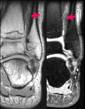
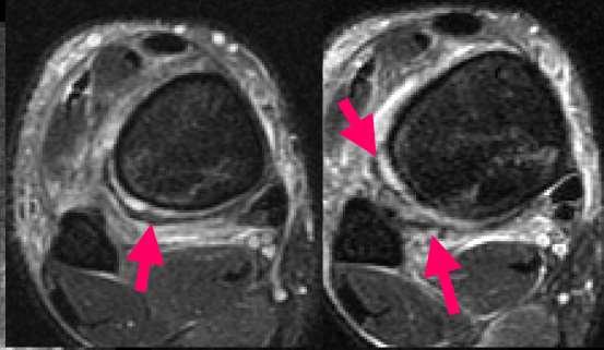
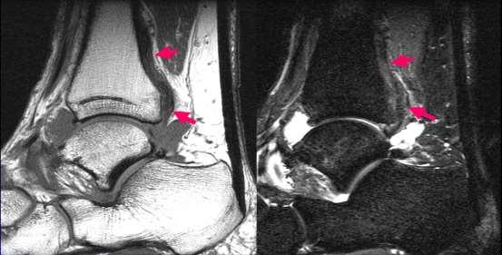
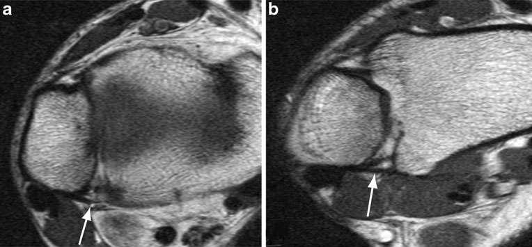
The fibular incisura of the tibia with recurrent sprained ankle on magnetic resonance imaging
 The fibular incisura of the tibia with recurrent sprained ankle on magnetic resonance imaging Ayfer Mavi, PhD, Hanifi Yildirim, MD, Hasan Gunes, MD, Turan Pestamalci, MD, Erdem Gumusburun, PhD. ABSTRACT
Back to sport after foot and ankle injury
 Back to sport after foot and ankle injury Mohammad Razi MD Rasoul Akram University Hospital Tehran One of the biggest challenges in a sports medicine practice is deciding when an athlete has sufficiently
2/23/2018. Syndesmosis Fixation: Screws Vs. Suture Button CSFA Tampa Feb Disclosures. Learning Objectives
 Syndesmosis Fixation: Screws Vs. Suture Button CSFA Tampa Feb. 2018 STEVEN STEINLAUF, MD THE ORTHOPAEDIC FOOT AND ANKLE INSTITUTE OF SOUTH FLORIDA THE UNIVERSITY OF MIAMI DEPARTMENT OF ORTHOPEDICS AND