The Muscular System. Chapter 10 Part C. PowerPoint Lecture Slides prepared by Karen Dunbar Kareiva Ivy Tech Community College
|
|
|
- Shana Stone
- 5 years ago
- Views:
Transcription

1 Chapter 10 Part C The Muscular System Annie Leibovitz/Contact Press Images PowerPoint Lecture Slides prepared by Karen Dunbar Kareiva Ivy Tech Community College
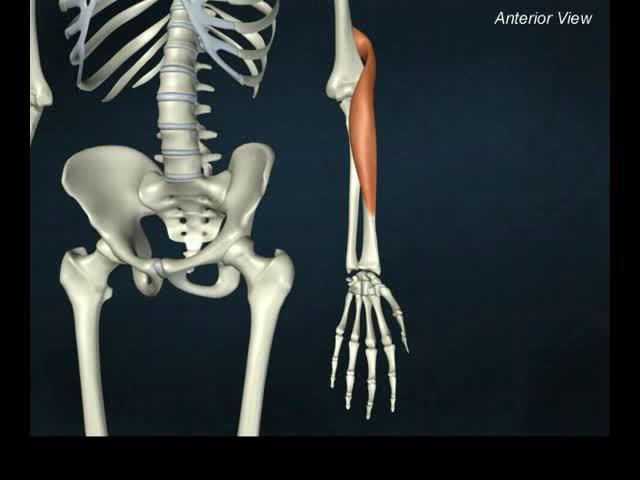
2 Table 10.9: Muscles Crossing the Shoulder Joint: Movements of the Arm (Humerus) Nine muscles cross shoulder joint Insert on and move humerus Some originate from scapula, others from axial skeleton Actions include flexion, extension, adduction Three prime movers of arm Pectoralis major Latissimus dorsi Deltoid
3 Table 10.9: Muscles Crossing the Shoulder Joint: Movements of the Arm (Humerus) (cont.) Rotator cuff muscles act as synergists and fixators; originate on scapula; reinforce shoulder capsule; prevent dislocation Supraspinatus Infraspinatus Teres minor Subscapularis Coracobrachialis and teres major: synergists
4 Table Muscles Crossing the Shoulder Joint: Movements of the Arm (Humerus)
5 A&P Flix : Pectoralis major
6 Table Muscles Crossing the Shoulder Joint: Movements of the Arm (Humerus)

7 A&P Flix : Deltoid
8 Figure 10.15a Muscles crossing the shoulder and elbow joints, causing movements of the arm and forearm, respectively. Clavicle Deltoid Sternum Pectoralis major Coracobrachialis Triceps brachii: Lateral head Long head Medial head Biceps brachii Brachialis Brachioradialis Anterior view
9 Table Muscles Crossing the Shoulder Joint: Movements of the Arm (Humerus) (continued)

10 A&P Flix : Latissimus dorsi

11 Table Muscles Crossing the Shoulder Joint: Movements of the Arm (Humerus) (continued)
12 Table Muscles Crossing the Shoulder Joint: Movements of the Arm (Humerus) (continued)
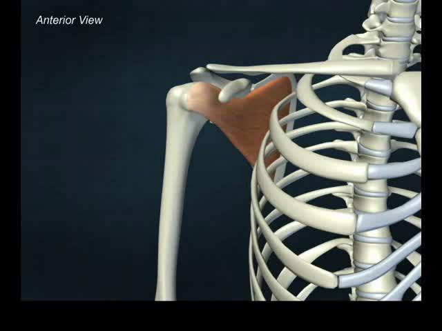
13 A&P Flix : Subscapularis
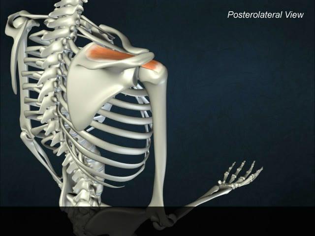
14 A&P Flix : Supraspinatus
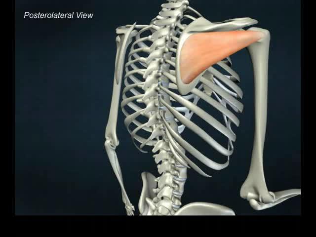
15 A&P Flix : Infraspinatus
16 Table Muscles Crossing the Shoulder Joint: Movements of the Arm (Humerus) (continued)

17 A&P Flix : Teres minor
18 A&P Flix : Teres major
19 Figure 10.15b Muscles crossing the shoulder and elbow joints, causing movements of the arm and forearm, respectively. Supraspinatus* Spine of scapula Deltoid (cut) Greater tubercle of humerus Infraspinatus* Teres minor* Teres major Triceps brachii: Lateral head Long head Latissimus dorsi Humerus Olecranon of ulna Anconeus Posterior view *Rotator cuff muscles

20 Figure 10.15c-d Muscles crossing the shoulder and elbow joints, causing movements of the arm and forearm, respectively. O O Biceps brachii Long head Short head O = origin I = insertion I O Subscapularis* O I I Coracobrachialis Brachialis *Rotator cuff muscles

21 A&P Flix : Muscles That Act on the Shoulder Joint and Humerus: An Overview (a)

22 A&P Flix : Muscles That Act on the Shoulder Joint and Humerus: An Overview (b)
23 A&P Flix : Muscles That Cross the Glenohumeral Joint
24 A&P Flix : Movement from the Rotator Cuff Muscles (a)

25 A&P Flix : Movement from the Rotator Cuff Muscles (b)

26 A&P Flix : Scapular Muscles of the Glenohumeral Joint (a)
27 A&P Flix : Axial Muscles of the Glenohumeral Joint (a)
28 A&P Flix : Movement at the Glenohumeral Joint: An Overview

29 A&P Flix : Movement at the Glenohumeral Joint (a)

30 A&P Flix : Movement at the Glenohumeral Joint (b)
31 Table 10.10: Muscles Crossing the Elbow Joint: Flexion and Extension of the Forearm Walls of fascia divide arm into two compartments Anterior muscles Posterior muscles

32 Table Muscles Crossing the Elbow Joint: Flexion and Extension of the Forearm
 Greater tubercle of humerus Infraspinatus* Teres minor* Teres major Triceps brachii: Lateral head Long head Latissimus dorsi Humerus")
33 Figure 10.15b Muscles crossing the shoulder and elbow joints, causing movements of the arm and forearm, respectively. Supraspinatus* Spine of scapula Deltoid (cut) Greater tubercle of humerus Infraspinatus* Teres minor* Teres major Triceps brachii: Lateral head Long head Latissimus dorsi Humerus Olecranon of ulna Anconeus Posterior view *Rotator cuff muscles

34 A&P Flix : Triceps brachii

35 Table Muscles Crossing the Elbow Joint: Flexion and Extension of the Forearm (continued)

36 Figure 10.15a Muscles crossing the shoulder and elbow joints, causing movements of the arm and forearm, respectively. Clavicle Deltoid Sternum Pectoralis major Coracobrachialis Triceps brachii: Lateral head Long head Medial head Biceps brachii Brachialis Brachioradialis Anterior view

37 A&P Flix : Biceps brachii

38 A&P Flix : Brachialis
39 A&P Flix : Brachioradialis

40 A&P Flix : The Elbow Joint and Forearm: An Overview
41 A&P Flix : Muscles of the Elbow Joint (a)

42 A&P Flix : Muscles of the Elbow Joint (b)
43 A&P Flix : Muscles of the Elbow Joint (c)
44 Table 10.11: Muscles of the Forearm: Movements of the Wrist, Hand, and Fingers Divided into anterior and posterior muscles Most anterior muscles are flexors; insert via flexor retinaculum Most posterior muscles are extensors; insert via extensor retinaculum Further divided into superficial and deep muscles
45 Table 10.11: Muscles of the Forearm: Movements of the Wrist, Hand, and Fingers (cont.) Actions: movements of wrist, fingers, thumb, as well as pronation and supination of forearm Pronator teres and pronator quadratus pronate forearm Supinator: synergist with biceps brachii in forearm supination
46 Table 10.11: Muscles of the Forearm: Movements of the Wrist, Hand, and Fingers (cont.) Anterior muscles Consist of five superficial and three deep muscles Most arise from common flexor tendon attached to medial epicondyle of humerus Most tendons of insertion held in place at wrist by flexor retinaculum
47 Table Muscles of the Forearm: Movements of the Wrist, Hand, and Fingers

48 A&P Flix : Pronator teres
49 A&P Flix : Flexor carpi radialis

50 Table Muscles of the Forearm: Movements of the Wrist, Hand, and Fingers

51 A&P Flix : Flexor carpi ulnaris
52 A&P Flix : Flexor digitorum superficialis

53 Table Muscles of the Forearm: Movements of the Wrist, Hand, and Fingers (continued)

54 Figure 10.16a Muscles of the anterior fascial compartment of the forearm acting on the right wrist and fingers. Superficial transverse ligament of palm Palmar aponeurosis Flexor digitorum superficialis Flexor carpi ulnaris Palmaris longus Flexor carpi radialis Medial epicondyle of humerus Medial head of triceps brachii Flexor retinaculum Pronator quadratus Flexor pollicis longus Extensor carpi radialis longus Brachioradialis Pronator teres Tendon of biceps brachii Biceps brachii
55 Figure 10.16b Muscles of the anterior fascial compartment of the forearm acting on the right wrist and fingers. Tendon of flexor digitorum superficialis Pronator quadratus Flexor pollicis longus Tendon of brachioradialis (cut) Tendon of flexor carpi ulnaris (cut) Tendon of flexor carpi radialis (cut) Flexor digitorum superficialis Extensor carpi radialis longus Supinator Tendon of biceps brachii (cut)
 Lumbricals Tendon of flexor carpi ulnaris (cut) Tendon of flexor pollicis longus Thenar muscles of thumb")
56 Figure 10.16c Muscles of the anterior fascial compartment of the forearm acting on the right wrist and fingers. Tendon of flexor digitorum profundus Tendon of flexor digitorum superficialis (cut) Lumbricals Tendon of flexor carpi ulnaris (cut) Tendon of flexor pollicis longus Thenar muscles of thumb Pronator quadratus Flexor pollicis longus Flexor digitorum profundus Supinator

57 A&P Flix : Anterior Muscles of the Wrist and Fingers (a)
58 A&P Flix : Anterior Muscles of the Wrist and Fingers (b)
59 Table 10.11: Muscles of the Forearm: Movements of the Wrist, Hand, and Fingers (cont.) Posterior muscles Consists of four superficial and four deep muscles All are innervated by the radial nerve or its branches Most arise from common flexor tendon attached to lateral epicondyle of humerus Most tendons of insertion held in place at wrist by extensor retinaculum

60 Table Muscles of the Forearm: Movements of the Wrist, Hand, and Fingers (continued)
")
61 A&P Flix : Posterior Muscles of the Wrist and Fingers (a)
")
62 A&P Flix : Posterior Muscles of the Wrist and Fingers (b)
63 A&P Flix : Extensor carpi radialis longus
64 A&P Flix : Extensor carpi radialis brevis

65 A&P Flix : Extensor digitorum

66 A&P Flix : Extensor carpi ulnaris

67 Table Muscles of the Forearm: Movements of the Wrist, Hand, and Fingers (continued)

68 Figure 10.17a Muscles of the posterior fascial compartment of the right forearm acting on the wrist and fingers. Extensor expansion Tendons of extensor digitorum Extensor pollicis longus Extensor pollicis brevis Abductor pollicis longus Extensor digitorum Extensor carpi radialis brevis Extensor carpi radialis longus Tendons of extensor carpi radialis brevis and longus Extensor indicis Extensor digiti minimi Extensor carpi ulnaris Flexor carpi ulnaris Anconeus Insertion of triceps brachii Brachioradialis

69 Figure 10.17b Muscles of the posterior fascial compartment of the right forearm acting on the wrist and fingers. Interossei Extensor indicis Extensor pollicis brevis Extensor pollicis longus Abductor pollicis longus Supinator Anconeus Olecranon of ulna

70 A&P Flix : Supinator
71 A&P Flix : Muscles of the Forearm (a)
")
72 A&P Flix : Muscles of the Forearm (b)
")
73 A&P Flix : Muscles of the Forearm (c)

74 Table Summary: Actions of Muscles Acting on the Arm, Forearm, and Hand

75 Table Summary: Actions of Muscles Acting on the Arm, Forearm, and Hand (continued)

76 Table Summary: Actions of Muscles Acting on the Arm, Forearm, and Hand (continued)
; innervated by radial nerve Humerus Brachialis Short head Long")
77 Figure 10.18a Summary: Actions of muscles of the right arm and forearm. Key: Extensors Flexors Others Triceps brachii Lateral head Long head Medial head Posterior compartment of arm (extends elbow); innervated by radial nerve Humerus Brachialis Short head Long head Biceps brachii Muscles of the arm Anterior compartment of arm (flexes elbow); innervated by musculocutaneous nerve
78 Figure 10.18b Summary: Actions of muscles of the right arm and forearm. Key: Extensors Flexors Others Posterior compartment of forearm (extends wrist and fingers); innervated by radial nerve Others Radius Abductor pollicis longus Pronator teres Brachioradialis (elbow flexor) Extensors Ulna Flexors Muscles of the forearm Anterior compartment of forearm (flexes wrist and fingers); innervated by median or ulnar nerve
79 A&P Flix : Muscles That Act on the Wrist and Fingers: An Overview

80 A&P Flix : Carpal Tunnel

81 Animation: Rotating Hand
82 Table 10.13: Intrinsic Muscles of the Hand: Fine Movements of the Fingers Small, weak muscles that lie entirely within palm of hand control precise movements of metacarpals and fingers (example: threading a needle) Abductors and adductors of fingers produce opposition move thumb toward little finger Flexion: thumb bends medially along palm, and fingers bend anteriorly Extension: thumb points laterally, and fingers move posteriorly
83 Table 10.13: Intrinsic Muscles of the Hand: Fine Movements of the Fingers (cont.) Flexion and extension
84 A&P Flix : Movements of the Wrist and Fingers (a)
85 Table 10.13: Intrinsic Muscles of the Hand: Fine Movements of the Fingers (cont.) Abduction and adduction
86 A&P Flix : Movements of the Wrist and Fingers (b)
87 Table 10.13: Intrinsic Muscles of the Hand: Fine Movements of the Fingers (cont.) Three muscle groups Thenar eminence (ball of thumb) Hypothenar eminence (ball of the little finger) Each of above groups has flexor, abductor, and opponens muscle Midpalmar muscles: lumbricals and interossei extend fingers Interossei muscles also abduct and adduct fingers
88 Table Intrinsic Muscles of the Hand: Fine Movements of the Fingers

89 Table Intrinsic Muscles of the Hand: Fine Movements of the Fingers (continued)
90 Table Intrinsic Muscles of the Hand: Fine Movements of the Fingers (continued)
91 Figure 10.19a Hand muscles, ventral views of right hand. Tendons of: Flexor digitorum profundus Flexor digitorum superficialis Third lumbrical Fourth lumbrical Opponens digiti minimi Flexor digiti minimi brevis Abductor digiti minimi Pisiform bone Flexor carpi ulnaris tendon Flexor digitorum superficialis tendons First superficial layer Fibrous sheath Second lumbrical Dorsal interossei First lumbrical Adductor pollicis Flexor pollicis brevis Abductor pollicis brevis Opponens pollicis Flexor retinaculum Abductor pollicis longus Tendons of: Palmaris longus Flexor carpi radialis Flexor pollicis longus
 Abductor digiti minimi (cut) Second layer")
92 Figure 10.19b Hand muscles, ventral views of right hand. Flexor digitorum profundus tendon Flexor digitorum superficialis tendon Palmar interossei Opponens digiti minimi Flexor digiti minimi brevis (cut) Abductor digiti minimi (cut) Second layer Opponens pollicis Dorsal interossei Adductor pollicis Flexor pollicis brevis Abductor pollicis brevis Flexor pollicis longus tendon

93 Figure 10.19c Hand muscles, ventral views of right hand. Palmar interossei Palmar interossei (isolated)
94 Figure 10.19d Hand muscles, ventral views of right hand. Dorsal interossei Dorsal interossei (isolated)
Muscular Nomenclature and Kinesiology - One
 Chapter 16 Muscular Nomenclature and Kinesiology - One Lessons 1-3 (with lesson 4) 1 Introduction 122 major muscles covered in this chapter Chapter divided into nine lessons Kinesiology study of human
Lab Activity 11: Group II
 Lab Activity 11: Group II Muscles Martini Chapter 11 Portland Community College BI 231 Origin and Insertion Origin: The place where the fixed end attaches to a bone, cartilage, or connective tissue. Insertion:
REFERENCE DIAGRAMS OF UPPER LIMB MUSCLES: NAMES, LOCATIONS, ATTACHMENTS, FUNCTIONS MUSCLES CONNECTING THE UPPER LIMB TO THE AXIAL SKELETON
 REFERENCE DIAGRAMS OF UPPER LIMB MUSCLES: NAMES, LOCATIONS, ATTACHMENTS, FUNCTIONS MUSCLES CONNECTING THE UPPER LIMB TO THE AXIAL SKELETON A25LAB EXERCISES: UPPER LIMB MUSCLES Page 1 MUSCLES CONNECTING
ARM Brachium Musculature
 ARM Brachium Musculature Coracobrachialis coracoid process of the scapula medial shaft of the humerus at about its middle 1. flexes the humerus 2. assists to adduct the humerus Blood: muscular branches
Muscles of the Upper Limb
 Muscles of the Upper Limb anterior surface of ribs 3 5 coracoid process Pectoralis minor pectoral nerves protracts / depresses scapula Serratus anterior Subclavius ribs 1-8 long thoracic nerve rib 1 ----------------
The Clavicle Right clavicle Deltoid tubercle: Conoid tubercle, conoid ligamen Impression for the
 The Clavicle Muscle Attachment Sites in the Upper Limb Pectoralis major Right clavicle Smooth superior surface of the shaft, under the platysma muscle tubercle: attachment of the deltoid Acromial facet
Key Relationships in the Upper Limb
 Key Relationships in the Upper Limb This list contains some of the key relationships that will help you identify structures in the lab. They are organized by dissection assignment as defined in the syllabus.
Human Anatomy Biology 351
 1 Human Anatomy Biology 351 Upper Limb Exam Please place your name on the back of the last page of this exam. You must answer all questions on this exam. Because statistics demonstrate that, on average,
medial half of clavicle; Sternum; upper six costal cartilages External surfaces of ribs 3-5
 MUSCLE ORIGIN INSERTION ACTION NERVE Pectoralis Major medial half of clavicle; Sternum; upper six costal cartilages Lateral lip of intertubercular groove of horizontal adduction Medial and lateral pectoral
Netter's Anatomy Flash Cards Section 6 List 4 th Edition
 Netter's Anatomy Flash Cards Section 6 List 4 th Edition https://www.memrise.com/course/1577581/ Section 6 Upper Limb (66 cards) Plate 6-1 Humerus and Scapula: Anterior View 1.1 Acromion 1.2 Greater tubercle
Practical 2 Worksheet
 Practical 2 Worksheet Upper Extremity BONES 1. Which end of the clavicle is on the lateral side (acromial or sternal)? 2. Describe the difference in the appearance of the acromial and sternal ends of the
Systematic Anatomy (For international students)
") Systematic Anatomy (For international students) Department of Anatomy,Fudan University Teaching contents Muscles of abdomen & upper limbs Dr.Hongqi Zhang ( 张红旗 ) Email: zhanghq58@126.com 1 Muscles of abdomen
Systematic Anatomy (For international students) Department of Anatomy,Fudan University Teaching contents Muscles of abdomen & upper limbs Dr.Hongqi Zhang ( 张红旗 ) Email: zhanghq58@126.com 1 Muscles of abdomen
MCQWeek2. All arise from the common flexor origin. The posterior aspect of the medial epicondyle is the common flexor origin.
 MCQWeek2. 1. Regarding superficial muscles of anterior compartment of the forearm: All arise from the common flexor origin. The posterior aspect of the medial epicondyle is the common flexor origin. Flexor
Al-Balqa Applied University
 Al-Balqa Applied University Faculty Of Medicine *You can use this checklist as a guide to you for the lab. the items on this checklist represent the main features of the models that you have to know for
Biceps Brachii. Muscles of the Arm and Hand 4/4/2017 MR. S. KELLY
 Muscles of the Arm and Hand PSK 4U MR. S. KELLY NORTH GRENVILLE DHS Biceps Brachii Origin: scapula Insertion: radius, fascia of forearm (bicipital aponeurosis) Action: supination and elbow flexion Innervation:
Copy Right- Hongqi ZHANG-Department of Anatomy-Fudan University. Systematic Anatomy. Locomotor system - Part 6
 Systematic Anatomy Locomotor system - Part 6 Muscles of abdomen Muscles of the upper limb Dr.Hongqi Zhang ( 张红旗 ) Email: zhanghq58@126.com 1 Muscles of abdomen Muscles of the upper limb Muscles of abdomen
Peripheral Nervous Sytem: Upper Body
 Peripheral Nervous Sytem: Upper Body MSTN121 - Neurophysiology Session 10 Department of Myotherapy Cervical Plexus Accessory nerve (CN11 + C1-5) Motor: trapezius and sternocleidomastoid Greater auricular
Muscles of the hand Prof. Abdulameer Al-Nuaimi
 Muscles of the hand Prof. Abdulameer Al-Nuaimi a.alnuaimi@sheffield.ac.uk abdulameerh@yahoo.com Thenar Muscles Thenar muscles are three short muscles located at base of the thumb. All are innervated by
Abduction of arm until your hand rich your head. Flexion of forearm at elbow joint. Extension of arm at elbow joint. Flexion of fingers 10.
 Num. answer 1. Medialy With the manubrium ( sternum ), and laterally with the acromion of the scapula 2. 1. Trapezius 2. Levator scapulae 3. Rhomboids 3. 1. Pectoralis major 2. Pectoralis minor 3. Latissiumus
The hand is full with sweat glands, activated at times of stress. In Slide #2 there was a mistake where the doctor mentioned lateral septum twice.
 We should only know: Name, action & nerve supply Layers - Skin - Superficial fascia - Deep fascia The hand is full with sweat glands, activated at times of stress. Deep fascia In Slide #2 there was a mistake
Main Menu. Wrist and Hand Joints click here. The Power is in Your Hands
 1 The Wrist and Hand Joints click here Main Menu K.5 http://www.handsonlineeducation.com/classes/k5/k5entry.htm[3/23/18, 1:40:40 PM] Bones 29 bones, including radius and ulna 8 carpal bones in 2 rows of
Supplied in part by the musculocutaneous nerve. Forms the axis of rotation in movements of pronation and supination
 Anatomy: Upper limb (15 questions) 1. Latissimus Dorsi: Is innervated by the dorsal scapular nerve Lies above feres major muscle Medially rotates the humerus All of the above 2. Supinator muscle is: Deep
LIST OF STRUCTURES TO BE IDENTIFIED IN LAB: UPPER EXTREMITY REVIEW 2016
 LIST OF STRUCTURES TO BE IDENTIFIED IN LAB: UPPER EXTREMITY REVIEW 2016 BONES Ribs, sternum, clavicle Humerus: Head, greater tubercle, lesser tubercle, intertubercular sulcus, surgical neck, anatomical
Nerves of the upper limb Prof. Abdulameer Al-Nuaimi. E. mail:
 Nerves of the upper limb Prof. Abdulameer Al-Nuaimi E-mail: a.al-nuaimi@sheffield.ac.uk E. mail: abdulameerh@yahoo.com Brachial plexus Median nerve After originating from the brachial plexus in the axilla,
Nerves of Upper limb. Dr. Brijendra Singh Professor & Head Department of Anatomy AIIMS Rishikesh
 Nerves of Upper limb Dr. Brijendra Singh Professor & Head Department of Anatomy AIIMS Rishikesh 1 Objectives Origin, course & relation of median & ulnar nerves. Motor & sensory distribution Carpal tunnel
Kinesiology of The Wrist and Hand. Cuneyt Mirzanli Istanbul Gelisim University
 Kinesiology of The Wrist and Hand Cuneyt Mirzanli Istanbul Gelisim University Bones The wrist and hand contain 29 bones including the radius and ulna. There are eight carpal bones in two rows of four to
MLT Muscle(s) Patient Position Therapist position Stabilization Limb Position Picture Put biceps on slack by bending elbow.
 Patient Position Therapist position Stabilization Limb Position Picture Put biceps on slack by bending elbow.") MLT Muscle(s) Patient Position Therapist position Stabilization Limb Position Picture Put biceps on slack by bending elbow. Pectoralis Minor Supine, arm at side, elbows extended, supinated Head of Table
MLT Muscle(s) Patient Position Therapist position Stabilization Limb Position Picture Put biceps on slack by bending elbow. Pectoralis Minor Supine, arm at side, elbows extended, supinated Head of Table
Connects arm to thorax 3 joints. Glenohumeral joint Acromioclavicular joint Sternoclavicular joint
 Connects arm to thorax 3 joints Glenohumeral joint Acromioclavicular joint Sternoclavicular joint Scapula Elevation Depression Protraction (abduction) Retraction (adduction) Downward Rotation Upward Rotation
Anatomy and Physiology II. Review Shoulder Girdle New Material Upper Extremities - Bones
 Anatomy and Physiology II Review Shoulder Girdle New Material Upper Extremities - Bones Anatomy and Physiology II Shoulder Girdle Review Questions From Last Lecture Can you identify the following muscles?
MUSCLES OF THE ELBOW REGION
 MUSCLES OF THE ELBOW REGION Dr Bronwen Ackermann COMMONWEALTH OF AUSTRALIA Copyright Regulation WARNING This material has been reproduced and communicated to you by or on behalf of the University of Sydney
The Forearm 2. Extensor & lateral Compartments of the Forearm
 The Forearm 2 Extensor & lateral Compartments of the Forearm 1-Lateral Fascial Compartment (at the lateral side of the forearm ) *Some books mention the lateral compartment contain just the Brachioradialis
STRUCTURAL BASIS OF MEDICAL PRACTICE EXAMINATION 5 October 6, 2006
 STRUCTURAL BASIS OF MEDICAL PRACTICE EXAMINATION 5 October 6, 2006 PART l. Answer in the space provided. (8 pts) 1. Identify the structures. (2 pts) B C A. _pisiform B. _ulnar artery A C. _flexor carpi
Lecture 9: Forearm bones and muscles
 Lecture 9: Forearm bones and muscles Remember, the region between the shoulder and the elbow = brachium/arm, between elbow and wrist = antebrachium/forearm. Forearm bones : Humerus (distal ends) Radius
Due in Lab weeks because of Thanksgiving Prelab #10. Homework #8. Both sides! Both sides!
 Lab 8 MUSCLES Due in Lab 10 2 weeks because of Thanksgiving Prelab #10 Both sides! Homework #8 Both sides! Refer to Muscles 22-23 Naming of muscles Origin Site of muscle attachment that doesn t move during
Levels of the anatomical cuts of the upper extremity RADIUS AND ULNA right
 11 CHAPTER 2 Levels of the anatomical cuts of the upper extremity AND right CUT 1 CUT 4 1 2 3 4 5 6 Isolated fixation of the radius is difficult at this level because of the anterolateral vessels and the
Nerve Injury. 1) Upper Lesions of the Brachial Plexus called Erb- Duchene Palsy or syndrome.
 Upper Lesions of the Brachial Plexus called Erb- Duchene Palsy or syndrome.") Nerve Injury - Every nerve goes to muscle or skin so if the nerve is injured this will cause paralysis in the muscle supplied from that nerve (paralysis means loss of function) then other muscles and other
Nerve Injury - Every nerve goes to muscle or skin so if the nerve is injured this will cause paralysis in the muscle supplied from that nerve (paralysis means loss of function) then other muscles and other
Fascial Compartments of the Upper Arm
 Fascial Compartments of the Upper Arm The upper arm is enclosed in a sheath of deep fascia and has two fascial septa: 1- Medial fascial septum (medial intermuscular septum): attached to the medial supracondylar
Muscle Anatomy Review Chart
 Muscle Anatomy Review Chart BACK Superficial (5) Trapezius Transverse cervical a. Latissimus dorsi Thoracodorsal a. Rhomboideus major Dorsal scapular a. Rhomboideus minor Levator scapulae Intermediate
Anatomy of the Upper Limb
 Anatomy of the Upper Limb Figure 53: The thenar & midpalmar spaces. The synovial (tendon) sheaths of the long flexors [Figure.54] These sheaths surround the tendons of the long flexors; flexor digitorum
Forearm and Wrist Regions Neumann Chapter 7
 Forearm and Wrist Regions Neumann Chapter 7 REVIEW AND HIGHLIGHTS OF OSTEOLOGY & ARTHROLOGY Radius dorsal radial tubercle radial styloid process Ulna ulnar styloid process ulnar head Carpals Proximal Row
divided by the bones ( redius and ulna ) and interosseous membrane into :
 and interosseous membrane into :") fossa Cubital Has: * floor. * roof : - Skin - superficial fasica - deep fascia ( include bicipital aponeurosis ) Structures within the roof : -cephalic and basilic veins -and between them median cubital
fossa Cubital Has: * floor. * roof : - Skin - superficial fasica - deep fascia ( include bicipital aponeurosis ) Structures within the roof : -cephalic and basilic veins -and between them median cubital
Prime movers provide the major force for producing a specific movement Antagonists oppose or reverse a particular movement Synergists
 Dr. Gary Mumaugh Prime movers provide the major force for producing a specific movement Antagonists oppose or reverse a particular movement Synergists Add force to a movement Reduce undesirable or unnecessary
11/15/2018. Temporalis Elevates & retracts mandible. Masseter = Prime mover of jaw closure. Levator scapulae Supraspinatus Clavicle.
 Due in Lab 10 Lab 8 MUSCLES 2 weeks because of Thanksgiving Prelab #10 Both sides! Homework #8 Both sides! Refer to Muscles 22-23 Examples of Origin & Insertion Naming of muscles Origin Site of muscle
compartments of the forearm
 " forearm posterior compartment " compartments of the forearm Posterior Fascial compartment Muscles: ** The superficial group 1. Extensor carpi radialis brevis 2. Ex. digitorum 3. Ex. digiti minimi 4.
forearm posterior compartment
 Quick revision: The anterior compartment of the forearm contains of 8 muscles... -4 superficial -1 intermediate -3 deep *All supplied by median nerve except 1 and 1/2 muscle (by ulnar N.) forearm posterior
Deep dry needling of the arm and hand muscles
 Deep dry needling of the arm and hand s 8 César Fernández-de-las-Peñas Javier González Iglesias Christian Gröbli Ricky Weissmann CHAPTER CONTENT Introduction................... 107 Clinical relevance of
Hand and Wrist Editing file. Color Code Important Doctors Notes Notes/Extra explanation
 Hand and Wrist Editing file Color Code Important Doctors Notes Notes/Extra explanation Objectives Describe the anatomy of the deep fascia of the wrist & hand (flexor & extensor retinacula & palmar aponeurosis).
Elbow, Wrist & Hand Evaluation.
 Elbow, Wrist & Hand Evaluation www.fisiokinesiterapia.biz Common Injuries to the Elbow, Wrist, Hand & Fingers Lateral epicondylitis tennis elbow Medial epicondylitis golfer s s elbow, little league elbow
Thank You for Your Support! Hosford Muscle Tables
 Thank You for Your Support! This PDF document has been placed online for your enjoyment and I hope you find it useful. These tables are both a teaching tool, and a study / review tool. I created these
The Elbow and the cubital fossa. Prof Oluwadiya Kehinde
 The Elbow and the cubital fossa Prof Oluwadiya Kehinde www.oluwadiya.com Elbow and Forearm Anatomy The elbow joint is formed by the humerus, radius, and the ulna Bony anatomy of the elbow Distal Humerus
# Anatomy. Upper Extremities Muscles and anatomy of axilla. Tiba Al-Ani 9/10/2015 Nabil. Page 0 of 16
 #10 25 Anatomy Upper Extremities Muscles and anatomy of axilla Tiba Al-Ani 9/10/2015 Nabil Page 0 of 16 Salam AWN Today s lecture is divided into two parts, the first part is the continuation of the upper
Module 7 - The Muscular System Muscles of the Arm and Trunk
 Module 7 - The Muscular System Muscles of the Arm and Trunk This Module will cover the muscle anatomy of the arms and trunk. We have already seen the muscles that move the humerus, so this module will
Anatomy of the Forearm
 Anatomy of the Forearm Musculoskeletal block- Anatomy-lecture 8 Editing file Objectives List the names of the Flexors Group of Forearm (superficial & deep muscles). Identify the common flexor origin of
The arm: *For images refer back to the slides
 The arm: *For images refer back to the slides Muscles of the arm: deltoid, triceps (which is located at the back of the arm), biceps and brachialis (it lies under the biceps), brachioradialis (it lies
Axilla and Brachial Region
 L 4 A B O R A T O R Y Axilla and Brachial Region BRACHIAL PLEXUS 5 Roots/Rami (ventral rami C5 T1) 3 Trunks Superior (C5, C6) Middle (C7) Inferior (C8, T1) 3 Cords Lateral Cord (Anterior Superior and Anterior
BLUE SKY SCHOOL OF PROFESSIONAL MASSAGE AND THERAPEUTIC BODYWORK. Musculoskeletal Anatomy & Kinesiology II REVIEW
 BLUE SKY SCHOOL OF PROFESSIONAL MASSAGE AND THERAPEUTIC BODYWORK Musculoskeletal Anatomy & Kinesiology II REVIEW MSAK101-II Session 4 LEARNING OBJECTIVES: By the end of this session, the student will be
Anatomage Table Instructors Guide- Upper Limb
 The Upper Limb Anatomage Table Instructors Guide- Upper Limb Table of Contents Upper Limb 1- The Skeletal System...3 1: Clavicle...3 2: Scapula...5 3: Shoulder (Glenohumeral) and Proximal Humerus...7 4:
BRACHIAL PLEXUS. DORSAL SCAPULAR NERVE (C5) supraclavicular branch innervates rhomboids (major and minor) and levator scapulae
 supraclavicular branch innervates rhomboids (major and minor) and levator scapulae") THE BRACHIAL PLEXUS DORSAL SCAPULAR NERVE (C5) supraclavicular branch innervates rhomboids (major and minor) and levator scapulae SCHEMA OF THE BRACHIAL PLEXUS THE BRACHIAL PLEXUS PHRENIC NERVE supraclavicular
THE BRACHIAL PLEXUS DORSAL SCAPULAR NERVE (C5) supraclavicular branch innervates rhomboids (major and minor) and levator scapulae SCHEMA OF THE BRACHIAL PLEXUS THE BRACHIAL PLEXUS PHRENIC NERVE supraclavicular
Figure 27: The synovial membrane of the shoulder joint (anterior view)
") The coracoacromial ligament; is an accessory ligament that protects the superior aspect of the joint extending from the coracoid process to the acromion over the tendon of supraspinatus. The synovial membrane
The coracoacromial ligament; is an accessory ligament that protects the superior aspect of the joint extending from the coracoid process to the acromion over the tendon of supraspinatus. The synovial membrane
LECTURE 8 HANDS: BONES AND MUSCLES
 LECTURE 8 HANDS: BONES AND MUSCLES WRIST AND HAND - Human hand can do power grip and precision grip - Thumb is 90 to the rest of the hand can do fine actions - Often able to do power actions o Take tools
region of the upper limb between the shoulder and the elbow Superiorly communicates with the axilla.
 1 region of the upper limb between the shoulder and the elbow Superiorly communicates with the axilla. Inferiorly, a number of important structures pass between arm & forearm through cubital fossa. 2 medial
13 13/3/2012. Adel Muhanna
 13 13/3/2012 Adel Muhanna بسم هللا الرحمن الرحيم The Hand Extensor retinaculum: Deep fascia of anterior compartment of the wrist is thickened to form flexor retinaculum : a bridge that have 6 structures
10/10/2014. Structure and Function of the Hand. The Hand. Osteology of the Hand
 Structure and Function of the Hand 19 bones and 19 joints are necessary to produce all the motions of the hand The Hand Dorsal aspect Palmar aspect The digits are numbered 1-5 Thumb = #1 Little finger
Muscles in the Shoulder, Chest, Arm, Stomach, and Back
 Muscles in the Shoulder, Chest, Arm, Stomach, and Back Shoulder Muscles Deltoid Supraspinatus Infraspinatus Teres Major Teres Minor Subscapularis Deltoid (Delts) Function: Raises the upper arm Origin:
The Human Muscular System Required reading before beginning this lab: Saladin, KS: Human Anatomy 5th ed (2017) Chapters 10, 11, 12 INTRODUCTION
 Chapters 10, 11, 12 INTRODUCTION") Biology 322: Human Anatomy The Human Muscular System Required reading before beginning this lab: Saladin, KS: Human Anatomy 5 th ed (2017) Chapters 10, 11, 12 INTRODUCTION We will use a number of lab periods
Biology 322: Human Anatomy The Human Muscular System Required reading before beginning this lab: Saladin, KS: Human Anatomy 5 th ed (2017) Chapters 10, 11, 12 INTRODUCTION We will use a number of lab periods
MUSCLES. Anconeus Muscle
 LAB 7 UPPER LIMBS MUSCLES Anconeus Muscle anconeus origin: distal end of dorsal surface of humerus insertion: lateral surface of ulna from distal margin of the semilunar notch to proximal end of the olecranon
Dr. Mahir Alhadidi Anatomy Lecture #9 Feb,28 th 2012
 Quick Revision: Upper arm is divided into two compartments: 1. Anterior Compartment: Contains three muscles (Biceps brachii, Coracobrachialis, Brachialis). Innervated by Musculocutaneous nerve. 2. Posterior
TABLES OF MUSCLE ACTIONS, INNERVATIONS, AND ATTACHMENTS
 TABLES OF MUSCLE ACTIONS, INNERVATIONS, AND ATTACHMENTS Table 1-1 ERECTOR SPINAE MUSCLES Intrinsic muscles producing extension and/or lateral of the spine Muscle Joint and Action Innervation Inferior Attachment
Wrist and Hand Anatomy
 Wrist and Hand Anatomy Bone Anatomy Scapoid Lunate Triquetrium Pisiform Trapeziod Trapezium Capitate Hamate Wrist Articulations Radiocarpal Joint Proximal portion Distal portion Most surface contact found
Clinical examination of the wrist, thumb and hand
 Clinical examination of the wrist, thumb and hand 20 CHAPTER CONTENTS Referred pain 319 History 319 Inspection 320 Functional examination 320 The distal radioulnar joint.............. 320 The wrist.......................
Functional Anatomy of the Elbow
 Functional Anatomy of the Elbow Orthopedic Institute Daryl C. Osbahr, M.D. Chief of Sports Medicine, Orlando Health Chief Medical Officer, Orlando City Soccer Club Orthopedic Consultant, Washington Nationals
Human Anatomy Lab #7: Muscles of the Cadaver
 Human Anatomy Lab #7: Muscles of the Cadaver Table of Contents: Expected Learning Outcomes.... 1 Introduction...... 1 Identifying Muscles on Yourself.... 2 Muscles of the Anterior Trunk and Arm.. 2 Muscles
Upper limb Arm & Cubital region 黃敏銓
 Upper limb Arm & Cubital region 黃敏銓 1 Arm Lateral intermuscular septum Anterior (flexor) compartment: stronger Medial intermuscular septum Posterior (extensor) compartment 2 Coracobrachialis Origin: coracoid
BOGOMOLETS NATIONAL MEDICAL UNIVERSITY. Department of human anatomy. GUIDELINES Student's independent work during the preparation to practical lesson
 BOGOMOLETS NATIONAL MEDICAL UNIVERSITY Department of human anatomy GUIDELINES Student's independent work during the preparation to practical lesson Academic discipline HUMAN ANATOMY Module 1 Content module
The Elbow and Radioulnar Joints Kinesiology. Dr Cüneyt Mirzanli Istanbul Gelisim University
 The Elbow and Radioulnar Joints Kinesiology Dr Cüneyt Mirzanli Istanbul Gelisim University 1 The Elbow & Radioulnar Joints Most upper extremity movements involve the elbow & radioulnar joints. Usually
The Free Upper Limb. Bone of the Arm. aus: Platzer, Locomotor System (ISBN ), 2009 Georg Thieme Verlag KG
, 2009 Georg Thieme Verlag KG") : ones, Ligaments, Joints The Free The bones of the free upper limb are The humerus The radius and ulna The carpal bones The metacarpal bones The phalanges one of the Arm Humerus (A H) The humerus articulates
: ones, Ligaments, Joints The Free The bones of the free upper limb are The humerus The radius and ulna The carpal bones The metacarpal bones The phalanges one of the Arm Humerus (A H) The humerus articulates
[[Sally Leaning Towards Peter To Take Cold Hand]]
![[[Sally Leaning Towards Peter To Take Cold Hand]]](/thumbs/84/91174469.jpg "[[Sally Leaning Towards Peter To Take Cold Hand]]") In this lecture we will talk about the bones of the hand, and the muscles and contents of the forearm. *The hand bones are: - Carpal bones. -Metacarpals. -Phalanges. *The carpal bones (wrist bones): They
In this lecture we will talk about the bones of the hand, and the muscles and contents of the forearm. *The hand bones are: - Carpal bones. -Metacarpals. -Phalanges. *The carpal bones (wrist bones): They
Synergist Muscles. Shoulder (glenohumeral joint) Flexion Deltoid (anterior fibers) Pectoralis major (upper fibers) Biceps Brachii Coracobrachialis
 Flexion Deltoid (anterior fibers) Pectoralis major (upper fibers) Biceps Brachii Coracobrachialis") Synergist Muscles Dr Gene Desepoli DrGeneLMT@gmail.com Shoulder (glenohumeral joint) Deltoid (anterior fibers) Pectoralis major (upper fibers) Biceps Brachii Coracobrachialis Deltoid (posterior fibers)
Synergist Muscles Dr Gene Desepoli DrGeneLMT@gmail.com Shoulder (glenohumeral joint) Deltoid (anterior fibers) Pectoralis major (upper fibers) Biceps Brachii Coracobrachialis Deltoid (posterior fibers)
The Muscular System. Chapter 10 Part D. PowerPoint Lecture Slides prepared by Karen Dunbar Kareiva Ivy Tech Community College
 Chapter 10 Part D The Muscular System Annie Leibovitz/Contact Press Images PowerPoint Lecture Slides prepared by Karen Dunbar Kareiva Ivy Tech Community College Table 10.14: Muscles Crossing the Hip and
BIOL 4260 Human Evolu3onary Anatomy Lecture 12: Limb Development. Lecture 2: Fossil Record
 BIOL 4260 Human Evolu3onary Anatomy Lecture 12: Limb Development Lecture 2: Fossil Record Outline Limb Evolution Limb Development Limb Function A Few Definitions Appendicular skeleton girdles & limbs Pectoral
Small muscles of the hand
 By the name of Allah Small muscles of the hand Revision: The palmar aponeurosis is triangular in shape with apex and base. It is divided into 4 bands that radiate to the medial four fingers. Dupuytren
In which arm muscle are intramuscular injections most often given? (not in text)
") AP1 Lab 9 - Muscles of the Arms and Legs Locate the following muscles on the models and on yourself. Recall anatomical position. Directional terms such as anterior, posterior, lateral, etc. all assume
AP1 Lab 9 - Muscles of the Arms and Legs Locate the following muscles on the models and on yourself. Recall anatomical position. Directional terms such as anterior, posterior, lateral, etc. all assume
e- Lateral pectoral nerve
 1. All of the following muscles have double innervations except: a- Brachialis b- Flexor digitorum profundus c- Trapezius d- Pectoralis major e- Subscapularis 2. You can t put your hand over your head
Upper Limb Muscles Muscles of Axilla & Arm
 Done By : Saleh Salahat Upper Limb Muscles Muscles of Axilla & Arm 1) Muscles around the axilla A- Muscles connecting the upper to thoracic wall (4) 1- pectoralis major Origin:- from the medial half of
STRUCTURAL BASIS OF MEDICAL PRACTICE EXAMINATION 5. September 30, 2011
 STRUCTURAL BASIS OF MEDICAL PRACTICE EXAMINATION 5 September 30, 2011 PART l. Answer in the space provided. (12 pts) 1. Identify the structures. (2 pts) EXAM NUMBER A. Suprascapular nerve B. Axillary nerve
BIOH111. o Cell Module o Tissue Module o Skeletal system o Integumentary system o Muscle system o Nervous system o Endocrine system
 BIOH111 o Cell Module o Tissue Module o Skeletal system o Integumentary system o Muscle system o Nervous system o Endocrine system TEXTBOOK AND REQUIRED/RECOMMENDED READINGS o Principles of anatomy and
In the name of Allah, Most gracious, Most merciful
 In the name of Allah, Most gracious, Most merciful This lecture includes the following: The Palmer Oponeurosis. The Carpel tunnel. The palmaris brevis muscle. The anatomical snuffbox. The Fibrous flexor
Location Terms. Anterior and posterior. Proximal and Distal The term proximal (Latin proximus; nearest) describes where the appendage joins the body.
 describes where the appendage joins the body.") HUMAN ANAT OMY Location Terms Anterior and posterior In human anatomical usage, anterior refers to the front of the individual. Similarly, posterior refers to the back of the subject. In standard anatomical
HUMAN ANAT OMY Location Terms Anterior and posterior In human anatomical usage, anterior refers to the front of the individual. Similarly, posterior refers to the back of the subject. In standard anatomical
Motion of Left Upper Extremity During A Right- Handed Golf Swing
 Motion of Left Upper Extremity During A Right- Handed Golf Swing Description of Movement While the movement required for a golf swing requires many muscles, joints, & ligaments throughout the body, the
The hand. it's the most important subject of the upper limb because it has a clinical importance. the palm of the hand**
 Today at 12:48 AM The hand it's the most important subject of the upper limb because it has a clinical importance. the palm of the hand** -the palmar aponeurosis located in the palm of the hand which is
Region of upper limb attachment to the trunk Proximal segment of limb overlaps parts of the trunk (thorax and back) and lower lateral neck.
 and lower lateral neck.") Region of upper limb attachment to the trunk Proximal segment of limb overlaps parts of the trunk (thorax and back) and lower lateral neck. includes Pectoral Scapular Deltoid regions of the upper limb
Region of upper limb attachment to the trunk Proximal segment of limb overlaps parts of the trunk (thorax and back) and lower lateral neck. includes Pectoral Scapular Deltoid regions of the upper limb
Acknowledgement. Here are some flash cards all set up in a "pdf" format for you! Thanks to Laura H. (spring 08)
") Acknowledgement Here are some flash cards all set up in a "pdf" format for you! Thanks to Laura H. (spring 08) for her donation to all my anatomy students! t Here is her suggestion for making flashcards
Acknowledgement Here are some flash cards all set up in a "pdf" format for you! Thanks to Laura H. (spring 08) for her donation to all my anatomy students! t Here is her suggestion for making flashcards
Key Points for Success:
 SELF WRIST & HAND 1 2 All of the stretches described in this chapter are detailed to stretch the right side. Key Points for Success: Sit comfortably in a position where you can straighten or fully extend
DENTISTRY 2017 UNIVERSITY OF JORDAN Midterm. Collected by by.. Farah Saadeh. Corrected by.. Rahaf Al-Jafari. Doctor.. Dr.
 DENTISTRY 2017 UNIVERSITY OF JORDAN Midterm Collected by by.. Farah Saadeh Corrected by.. Rahaf Al-Jafari Doctor.. Dr. Maher Al-Hadidi 1- Wrong statement: Answer: Bone deposition is a result of pressure
Chapter 6 part 2. Skeletal Muscles of the Body
 Chapter 6 part 2 Skeletal Muscles of the Body Basic Principles 600 + muscles in the human body (you are required to learn 45, lucky kids)! Skeletal Muscles pull on bones Origin of a muscle = point of attachment
Cubital fossa and forearm
 Cubital fossa and forearm Cubital fossa is the triangular space in front of elbow joint. - The Cubital fossa has boundaries: apex, base, roof and floor and it has contents. The base: an imaginary horizontal
3/27/2012. Muscle Classification: Functional Groups. Interactions of Skeletal Muscles. Naming Skeletal Muscles. Naming Skeletal Muscles
 Interactions of Skeletal Muscles Skeletal muscles work together or in opposition Muscles only pull (never push) As muscles shorten, the insertion generally moves toward the origin Whatever a muscle (or
Human Anatomy and Physiology I Laboratory
 Human Anatomy and Physiology I Laboratory Gross Anatomy of the Muscular System (Two weeks) 1 This lab involves study of the laboratory exercise Gross Anatomy of the Muscular System. Complete the Review
Anatomy Upper Limb Muscles
 Anatomy Upper Limb Muscles Rotator cuff/scapulohumeral muscles 4 muscles (SITS) form musculotendinous rotator cuff around glenohumeral joint, provide stability of joint Supraspinatus Course: med 2/3 supraspinatous
Epicranius (frontal belly) Zygomaticus minor. Zygomaticus major Buccinator
 Zygomaticus minor. Zygomaticus major Buccinator") Epicranius (frontal belly) Zygomaticus minor Zygomaticus major Buccinator Masseter Digastric (posterior belly) Stylohyoid Sternocleidomastoid Trapezius Scalenus Omohyoid (inferior belly) Orbicularis oris
Epicranius (frontal belly) Zygomaticus minor Zygomaticus major Buccinator Masseter Digastric (posterior belly) Stylohyoid Sternocleidomastoid Trapezius Scalenus Omohyoid (inferior belly) Orbicularis oris