Riding with the ants. A.P.M. Duarte 1, D. Attili-Angelis 1,2, N.C. Baron 1, J.Z. Groenewald 3, P.W. Crous 3,4,5, F.C. Pagnocca 1 INTRODUCTION
|
|
|
- Felicia Logan
- 6 years ago
- Views:
Transcription
1 Persoonia 38, 2017: ISSN (Online) RESEARCH ARTICLE Riding with the ants A.P.M. Duarte 1, D. Attili-Angelis 1,2, N.C. Baron 1, J.Z. Groenewald 3, P.W. Crous 3,4,5, F.C. Pagnocca 1 Key words Attini tribe leaf-cutting ants multi-gene analyses systematics Xenopenidiella Abstract Isolates of Teratosphaeriaceae have frequently been found in the integument of attine ants, proving to be common and diverse in this microenvironment. The LSU phylogeny of the ant-isolated strains studied revealed that they cluster in two main lineages. The first was associated with the genus Xenopenidiella whereas the other represented two ant-isolated lineages sister to the taxa Penidiella aggregata and P. drakensbergensis, which are allocated to the new genus Penidiellomyces. The genus Penidiella is limited to the lineage containing P. columbiana, which is not congeneric with Penidiellomyces or Penidiellopsis, nor with Simplicidiella, a novel genus introduced here to accommodate a strain isolated from ants. For species level analysis, the final 26 aligned sequences of the ITS (498 characters), cmda (389 characters), tef1 (342 characters) and tub2 (446 characters) gene regions lead to the introduction of six new species in Xenopenidiella, and one in respectively Penidiellopsis and Simplicidiella. The species described in this study were distinguished by the combination of morphological and phylogenetic data. Novelties on the integument of leaf-cutting ants from Brazil include: Penidiellopsis ramosus, Xenopenidiella clavata, X. formica, X. inflata, X. laevigata, X. nigrescens, X. tarda spp. nov., and Simplicidiella nigra gen. & sp. nov. Beta-tubulin is recommended as primary barcode for the distinction of species in Penidiellopsis, whereas ITS was sufficient to distinguish species of Xenopenidiella. Article info Received: 21 June 2016; Accepted: 5 September 2016; Published: 5 October INTRODUCTION 1 Institute of Biosciences, UNESP Univ. Estadual Paulista, Rio Claro, Center for the Study of Social Insects; corresponding author derlene@cpqba.unicamp.br. 2 Division of Microbial Resources, CPQBA University of Campinas, SP, Brazil. 3 CBS-KNAW Fungal Biodiversity Centre, Uppsalalaan 8, 3584 CT Utrecht, The Netherlands. 4 Department of Microbiology and Plant Pathology, Forestry and Agricultural Biotechnology Institute (FABI), University of Pretoria, Pretoria 0002, South Africa. 5 Microbiology, Department of Biology, Utrecht University, Padualaan 8, 3584 CH Utrecht, The Netherlands. Seen as important symbionts, fungi interact with a wide range of organisms, including insects (McLaughlin et al. 2009). According to Hawksworth (2012) it is estimated that at least M species of fungi are living on Earth, of which only around are presently known (Crous et al. 2015). Since the symbiotic relationships of attine ants with fungi are not fully understood and comprise a wide fungal diversity (Pagnocca et al. 2008, Rodrigues et al. 2011, Duarte et al. 2014), nests of ants are favourable environments to investigate for undescribed taxa. The symbiosis between attine ants and their mutualistic fungi (Leucocoprineae and Pterulaceae), nourished as food source (Mueller & Rabeling 2008) exists for approximately 50 M years (Schultz & Brady 2008). In order to maintain the symbiont, attine ants utilise different agricultural systems (Schultz & Brady 2008). The leaf-cutter agriculture performed by genera Atta and Acromyrmex is the most common in Brazil and consists of cutting fresh plant material to be incorporated into the fungal garden and used as energy source for the growth of the mutualistic fungus Leucoagaricus gongylophorus (Weber 1972). Recent studies of melanised fungi carried in the integument of leaf-cutting ants have revealed the existence of a wide diversity of undescribed fungal species (Duarte et al. 2014). Little & Currie (2007) were the first to show an association between a black yeast (phialophora-like), classified by the authors as the fifth symbiont, and Apterostigma ants (tribe Attini). Thus far, three new black fungal species isolated from leaf-cutting ants have been described: Phialophora attae, P. capiguarae (Attili-Angelis et al. 2014) and Ochroconis globalis (Samerpitak et al. 2015). In addition, Duarte et al. (2014) isolated several unknown species affiliated to Teratosphaeriaceae, which are the subject of the present study. The Teratosphaeriaceae was originally separated from Mycosphaerellaceae (Crous et al. 2007) and circumscribed to include saprobic, extremophilic, human opportunistic and plant pathogenic fungi (Quaedvlieg et al. 2014). Although often linked to diseases in plants, especially Myrtaceae and Proteaceae hosts (Pérez et al. 2009, Carnegie et al. 2011, Crous & Groenewald 2011, Hunter et al. 2011), Teratosphaeriaceae species do not have reports of specific associations with ants. However, this family is also rich in genera and species associated with rocks and other extreme environments (Egidi et al. 2014), and its ecology remains insufficiently known. In a recent revision to phylogenetically and morphologically classified lineages in Teratosphaeriaceae, Quaedvlieg et al. (2014) generated a multi-locus DNA sequence dataset and introduced 17 new genera within Teratosphaeriaceae, including Xenopenidiella, a saprobic fungus on leaf litter. Xenopenidiella was introduced based on a single species, X. rigidophora, which was represented by a single isolate (Quaedvlieg et al. 2014). The discovery of more species of Xenopenidiella will thus allow for a more robust generic circumscription of the genus and facilitate a better understanding of its ecology. The present study aims to provide a taxonomic study of Teratosphaeriaceae species obtained from the integument of leafcutting ants from Brazil. Here, we describe a novel genus Naturalis Biodiversity Center & Centraalbureau voor Schimmelcultures You are free to share - to copy, distribute and transmit the work, under the following conditions: Attribution: You must attribute the work in the manner specified by the author or licensor (but not in any way that suggests that they endorse you or your use of the work). Non-commercial: You may not use this work for commercial purposes. No derivative works: You may not alter, transform, or build upon this work. For any reuse or distribution, you must make clear to others the license terms of this work, which can be found at Any of the above conditions can be waived if you get permission from the copyright holder. Nothing in this license impairs or restricts the author s moral rights.
2 82 Persoonia Volume 38, 2017 isolated from ants (Simplicidiella), a new species of Penidiellopsis (P. ramosus), and six new species of Xenopenidiella (X. clavata, X. formica, X. inflata, X. laevigata, X. nigrescens, X. tarda). A third genus, Penidiellomyces, is also introduced for two species of Penidiella s.lat. MATERIALS AND METHODS Isolation techniques In order to isolate the fungi present in the integument of attine ants, three different methods were used as described below. For the oil flotation technique (Satow et al. 2008) the bodies of sampled ants were immersed in 25 ml of saline solution (NaCl 0.85 %) with antibiotics (200 U penicillin, 200 μg/ml chloramphenicol, 200 μg/ml streptomycin and 500 μg/ml cycloheximide). Tubes were vortexed and after 30 min of incubation at room temperature, approximately 20 % sterile mineral oil was added, followed by vortexing for 5 min. After 20 min of settling, 100 μl aliquots were removed from the oil-solution interface and plated on Mycosel agar using a Drigalsky rod. Plates were incubated in the dark at 25 C and monitored daily until the appearance of fungal colonies. For the nitrogen-free cultivation, ants were immersed in distilled water and sonicated for 15 min. Then, 150 µl of the solution containing fungal cells in suspension were plated in a freenitrogen medium described by Thanh (2006). Plates were incubated in the dark at 25 C for 3 wk. In the third method, a dextrose 50 % enrichment technique was performed by immersing the ants in tubes containing 50 % dextrose and 0.5 % yeast extract. After 10 d of incubation in the dark at 28 C, tubes were shaken and 150 µl of the mixed solution was plated on malt-dextrose agar (1 % malt, 1 % dextrose, 0.01 % chloramphenicol, 2 % agar) and again incubated in the dark at 28 C for 10 d (Thanh et al. 2013). Fungal isolates The 25 isolates analysed are listed in Table 1 and were all obtained from the integument of Atta capiguara and A. laevigata leaf-cutting ant gynes (primary reproductive female caste) and drones (male caste which role is to mate with a gyne) from Brazil. All isolates are maintained in the culture collection of the Microbial Resource Centre (CRM-UNESP, Brazil). Ex-type strains were deposited in the CBS-KNAW Fungal Biodiversity Centre (The Netherlands) and/or in the Brazilian Collection of Environmental and Industrial Microorganisms (CBMAI, Brazil). Sequences derived in this study were lodged at GenBank and taxonomic novelties with MycoBank (Crous et al. 2004). DNA extraction Isolates were transferred to fresh 2 % MA (Malt Agar) plates and incubated in the dark at 25 C for d. Approximately 1 cm 2 of mycelium was transferred to a 2 ml Eppendorf tube containing 500 μl of lysis buffer (50 mmol/l Tris-HCl; 250 mmol/l NaCl; 50 mmol/l EDTA; 0.3 % w/v SDS; ph 8) and the equivalent to 100 µl of glass beads (SIGMA-ALDRICH catalogue G8893). After vortexing for 4 min, the microtubes were incubated for 1 h at 65 C (two replicates). After centrifugation for 15 min at rpm the aqueous phase with DNA was transferred into new microtubes. DNA extracts were stored at -20 C prior to use. Table 1 Fungal isolates details and GenBank accession numbers of isolates included in this study. Species Isolate number 1 Source Isolation GenBank Accession numbers 2 technique tub2 cmda tef1 ITS LSU Penidiellopsis radicularis AP386 = CBS = A. capiguara gyne Oil flotation KU KU KU KT KU CBMAI 1938 AP387 = CBS = A. capiguara gyne Oil flotation KU KU KU KT KU CBMAI 1947 AP389 A. capiguara gyne Oil flotation KU KU KU KT KU AP410 = CBS A. capiguara gyne Oil flotation KU KU KU KT KU AP418 A. capiguara gyne Oil flotation KU KU KU KT KU AP440 = CBS = A. capiguara gyne Oil flotation KU KU KU KT KU CBMAI 1951 DOC350 = CBMAI 1952 A. capiguara drone Nitrogen-free KU KU KT KU DOC363 = CBMAI 1953 A. capiguara drone Nitrogen-free KU KU KU KT KU Penidiellopsis ramosus AP391 = CBMAI 1937 ET A. capiguara gyne Oil flotation KU KU KU KT KU AP392 = CBMAI 1948 A. capiguara gyne Oil flotation KU KU KU KT KU AP420 = CBMAI 1949 A. capiguara gyne Oil flotation KU KU KU KT KU AP421 = CBMAI 1950 A. capiguara gyne Oil flotation KU KU KU KT KU Simplicidiella nigra AP 416 = CBMAI 1939 ET A. capiguara gyne Oil flotation KU KU KU KT KU Xenopenidiella clavata DOC354 = CBMAI 1942 ET A. capiguara drone Nitrogen-free KU KT KU Xenopenidiella formica 67N = CBMAI 1954 A. laevigata drone Oil flotation KU KU KU KT KU AP380 = CBMAI 1946 A. capiguara gyne Oil flotation KU KU KU KT KU DOC323 = CBMAI 1941 ET A. capiguara drone Nitrogen-free KU KU KU KT KU DOC349 A. capiguara drone Nitrogen-free KU KU KU KT KU DOC362 A. capiguara drone Nitrogen-free KU KU KU KT KU Xenopenidiella inflata 87N = CBMAI 1945 ET A. laevigata drone Oil flotation KU KU KU KT KU Xenopenidiella laevigata 89N = CBMAI 1944 ET A. laevigata gyne Oil flotation KU KT KU Xenopenidiella nigrescens DOC356 = CBMAI 1943 ET A. capiguara drone Nitrogen-free KU KU KT KU Xenopenidiella tarda DOC248 = CBMAI 1940 ET A. capiguara drone Dextrose 50% KU KU KU KT KU DOC317 A. capiguara drone Nitrogen-free KU KU KU KT KU DOC343 A. capiguara drone Nitrogen-free KU KU KU KT KU CBS: CBS-KNAW Fungal Biodiversity Centre, Utrecht, The Netherlands; CBMAI: Brazilian Collection of Environmental and Industrial Microorganisms; all other codes are placed in the culture collection of the Microbial Resource Centre (UNESP, Brazil). 2 cmda: partial calmodulin gene; ITS: internal transcribed spacers and intervening 5.8S nrdna; LSU: partial 28S nrrna gene; tef1: partial translation elongation factor 1-alpha; tub2: partial betatubulin gene. ET ex-type.
3 A.P.M. Duarte et al.: Riding with the ants 83 Table 2 Primer combinations used for amplification and sequencing. Locus Primer Primer sequence 5 to 3 Annealing Orientation Reference temperature ( C) β-tubulin (tub2) T1 AACATGCGTGAGATTGTAAGT 52 Forward O Donnell & Cigelnik (1997) β-sandy-r GCRCGNGGVACRTACTTGTT Reverse Stukenbrock et al. (2012) Calmodulin (cmda) CAL-235F TTCAAGGAGGCCTTCTCCCTCTT 50 Forward Quaedvlieg et al. (2012) CAL2Rd TGRTCNGCCTCDCGGATCATCTC Reverse Groenewald et al. (2013) ITS ITS1 TCCGTAGGTGAACCTGCGG 52 Forward White et al. (1990) ITS4 TCCTCCGCTTATTGATATGC Reverse White et al. (1990) LSU (28S nrdna) LSU1Fd GRATCAGGTAGGRATACCCG 52 Forward Crous et al. (2009a) LR5 TCCTGAGGGAAACTTCG Reverse Vilgalys & Hester (1990) Translation elongation factor-1α (tef1) EF1-728F CATCGAGAAGTTCGAGAAGG 52 Forward Carbone & Kohn (1999) EF-2 GGARGTACCAGTSATCATGTT Reverse O Donnell et al. (1998) Multi-locus PCR amplification and sequencing Isolates were screened for five loci (ITS, LSU, cmda, tef1 and tub2) using the primer sets listed in Table 2. Amplification reactions were performed in a total volume of 25 μl containing 1 µl of 50 mm MgCl 2, 4 µl of 1.25 mm of dntp Mix, 2.5 µl of 10 PCR buffer, 2 µl of 10 µm of each primer (100 μm stock concentration), 0.2 µl of 5 U/µL of Taq polymerase (Invitrogen), 5 µl of DNA template (diluted 1 : 100) and 8.3 µl PCR water. PCR conditions were set as follows: an initial denaturation temperature of 96 C for 2 min, followed by 35 cycles of denaturation at 96 C for 45 s, primer annealing at the temperature stated in Table 2, primer extension at 72 C for 90 s and a final extension step at 72 C for 2 min. PCR products were analysed on a 1 % agarose gel and a negative control (DNA free) was also included to check for possible contaminations. Amplicons were purified using a NucleoSpin Gel and PCR Clean-up kit (Macherey-Nagel), according to the manufacturer s instructions. The PCR products were sequenced in both directions using the BigDye Terminator v. 3.1 Cycle Sequencing Kit (Applied Biosystems Life Technologies, Carlsbad, CA, USA) and an ABI Prism 3130 Genetic Analyzer (Applied Biosystems). Consensus sequences were generated using the BioEdit Sequence Alignment Editor v (Hall 1999). Alignment and phylogenetic reconstruction A preliminary identification of the isolates to genus level was made by comparing the consensus LSU sequences against NCBIs GenBank nucleotide database using the megablast algorithm. The most similar sequences were downloaded in FAS- TA format and subsequently combined with the Teratosphaeriaceae alignment of Quaedvlieg et al. (2014) (TreeBASE study S16145). A draft phylogeny was created from this alignment (data not shown), after which a reduced dataset was used to generate the final overview phylogeny presented here. The species level phylogeny was created from a concatenated alignment consisting of ITS, cmda, tub2 and tef1 sequences. All loci were aligned individually using the MAFFT v. 7 online portal ( Katoh & Standley 2013), after which they were manually checked and improved in MEGA v (Tamura et al. 2013). The phylogenetic reconstructions were conducted using the command line versions of MrBayes v (Ronquist & Huelsenbeck 2003) and PAUP v. 4.0b10 (Swofford 2003), while MrModeltest v. 2.2 (Nylander 2004) was used to determine the best nucleotide substitution model settings for MrBayes. A parsimony analysis was performed on the overview LSU alignment and the combined 4-locus alignment; prior to concatenation of the four loci, individual gene trees derived from each locus were examined for topological conflict compared to the trees from the other loci. Gaps were treated as a new state, missing data were indicated as missing data in the analyses and statistics such as tree length, consistency index, retention index and rescaled consistency index (TL, CI, RI and RC, respectively) were calculated. For the Bayesian analysis of the overview LSU phylogeny, the heating parameter was set to 0.2 and the search was stopped when convergence was reached (stopval = 0.01). Trees were saved every 100 generations and the Markov Chain Monte Carlo (MCMC) analysis of 4 chains started in parallel from a random tree topology. The optimal substitution model under the Akaike s Information Criterion was the GTR model with dirichlet (1,1,1,1) state frequency distribution and inverse gamma-shaped rate variation across sites. Sequences derived from this study were deposited in GenBank ( nlm.nih.gov/genbank) (Table 1), and the alignments and trees in TreeBASE ( The resulting phylogenetic trees were imported into and printed with Geneious v ( Kearse et al. 2012), and the layout of the tree for publication was done using Adobe Illustrator v. CS6. Morphology Colony characters and pigment production were observed on 2 % MA (Malt Agar), PDA (Potato Dextrose Agar, Acumedia ) and CMA (Corn Meal Agar, Himedia ) at 25 C in the dark for 21 d. Microscopic observations were based on slide culture techniques using CMA due to the rapid induction of sporulation. Agar blocks of ~1 cm 2 were placed on a sterile Petri dish and inoculated at the four edges. The block was subsequently covered with a sterile cover slip (~2 cm 2 ). Plates were incubated at 25 C in the dark for d. Slides were made in distilled water. Micrographs were taken using a phase-contrast microscope (DM 750, Leica; software Leica Application Suite v , Leica). RESULTS Phylogeny The LSU alignment consisted of 75 sequences (including the outgroup sequence, Toxicocladosporium rubrigenum GenBank FJ790305) and 776 characters were included in the analysis. Of the 776 analysed characters, 174 were parsimony-informative, 85 variable characters were parsimony-uninformative and 517 characters were constant. A maximum of equally most parsimonious trees were saved, one of which is shown in Fig. 1 (TL = 799 steps; CI = 0.458; RI = 0.777; RC = 0.356). The Bayesian analysis sampled 255 unique site patterns and lasted generations, after which convergence was achieved. The posterior probability values mapped unto Fig. 1 were calculated from the trees sampled from the trees generated; the remainder of the trees were discarded as burnin. A similar topology was observed between the Baye sian and parsimony analyses. The LSU phylogeny revealed that the strains from ants are present in two main clades, one of which is associated with the genus Xenopenidiella, whereas the other
4 84 Persoonia Volume 38, 2017 has as closest neighbour Penidiella aggregata (Gen Bank JF499862), Penidiella drakensbergensis (GenBank KC005792) and the recently described genus Penidiellopsis (GenBank LN834445). As the genus Penidiella is restricted to the lineage containing P. columbiana (represented here by GenBank EU019274, obtained from the ex-type culture CBS ), which is not congeneric with the lineage containing P. aggregata, P. drakensbergensis and the ant strains, one novel genus, Simplicidiella, is described below to accommodate one of the strains isolated from ants and another new genus. Penidiellomyces, is proposed for the two Penidiella species. For the species level analysis, a concatenated sequence alignment from 26 strains (including the outgroup species Parapenidiella pseudotasmaniensis strain CBS ) was subjected to maximum parsimony analyses. The final aligned sequences of the ITS (498 characters), cmda (389 characters), tef1 (342 characters) and tub2 (446 characters) gene regions had a total length of characters (including alignment gaps) which were included in the analyses. The gaps in the alignment were treated as fifth base for the parsimony analyses. From the analysed characters, 902 were constant (ITS: 293; cmda: 257; tef1: 152; tub2: 200), 255 were variable and parsimony- Toxicocladosporium rubrigenum FJ /100 Schizothyrium pomi KF /- Schizothyrium pomi KF /100 Uwebraunia commune DQ Uwebraunia dekkeri GQ x 1/60 Neopenidiella nectandrae EU / /91 Zasmidium anthuriicola FJ /100 Zasmidium citri KF /- 1/100 Zasmidium lonicericola KF Zasmidium nocoxi KF /100 Ramulispora sorghi GQ /59 Sphaerulina myriadea KF /86 Xenomycosphaerella elongata KF Xenomycosphaerella yunnanensis KF /80 Zymoseptoria verkleyi KC Ramularia endophylla AY /100 -/53 Ramularia pratensis var. pratensis KF /59 Apenidiella strumelloidea EU /69 Queenslandipenidiella kurandae KF /100 Parapenidiella pseudotasmaniensis GQ Parapenidiella tasmaniensis GU Stenella araguata KF /- Xenoteratosphaeria jonkershoekensis KF Eupenidiella venezuelensis EU /- 1/100 Myrtapenidiella eucalypti KF x 1/92 Myrtapenidiella tenuiramis GQ /- 1/86 Penidiella columbiana EU Xenophacidiella pseudocatenata KF /100 Suberoteratosphaeria pseudosuberosa KF Suberoteratosphaeria suberosa KF /- 0.98/71 1/100 Readeriella angustia KF Readeriella dimorphospora KF /66 Readeriella eucalyptigena CPC /95 Readeriella mirabilis KF /- Teratosphaeria cryptica KF Teratosphaeria aurantia KF Teratosphaeria miniata GQ /61 Teratosphaeria hortaea KF Teratosphaeria juvenalis KF /- 1/98 Teratosphaeria destructans EU Teratosphaeria eucalypti KF /- Teratosphaeria fibrillosa KF /58 Teratosphaeria fimbriata KF Teratosphaeria corymbiae KF Teratosphaeria considenianae KF Teratosphaeria gauchensis KF Teratosphaeria zuluensis KF Xenopenidiella rigidophora KF /87 Xenopenidiella tarda DOC248 Xenopenidiella tarda DOC317 1/100 Xenopenidiella tarda DOC343 Xenopenidiella laevigata 89N Xenopenidiella formica 67N Xenopenidiella formica AP380 Xenopenidiella formica DOC349 1/59 Xenopenidiella formica DOC362 Xenopenidiella formica DOC /- Xenopenidiella clavata DOC354 Xenopenidiella nigrescens DOC356 Xenopenidiella inflata 87N Penidiellomyces aggregatus JF Penidiellomyces drakensbergensis KC Simplicidiella nigra AP416 1/ /66 Penidiellopsis radicularis LN /- Penidiellopsis radicularis DOC /63 Penidiellopsis ramosus AP391 Penidiellopsis ramosus AP392 Penidiellopsis radicularis AP410 Penidiellopsis radicularis AP440 1/85 Penidiellopsis radicularis AP386 Penidiellopsis radicularis AP387 Penidiellopsis radicularis AP418 Penidiellopsis ramosus AP420 Penidiellopsis ramosus AP421 Penidiellopsis radicularis DOC changes Schizothyriaceae Dissoconiaceae Mycosphaerellaceae Teratosphaeriaceae Fig. 1 One of equally most parsimonious trees obtained from a maximum parsimony analysis of the LSU sequence alignment. The scale bar shows 10 changes, and posterior probability values from a Bayesian analysis (PP) and parsimony bootstrap support (PBS) values from replicates are shown at the nodes (PP/PBS). Thickened lines represent those branches also present in the strict consensus tree and families are indicated with coloured square blocks and the clades with ant isolates with light blue-coloured blocks with rounded corners. The darker blue block with rounded corners represents the novel genus introduced here to accommodate P. aggregata and P. drakensbergensis. The lengths of some branches were halved to facilitate easier layout. The tree was rooted to Toxicocladosporium rubrigenum (GenBank accession number FJ790305).
5 A.P.M. Duarte et al.: Riding with the ants changes Parapenidiella pseudotasmaniensis CBS DOC DOC N 89N DOC248 DOC317 DOC343 DOC323 DOC349 AP380 67N DOC362 AP416 AP AP AP AP AP AP389 AP DOC363 AP AP410 DOC350 AP440 Xenopenidiella nigrescens Xenopenidiella clavata Xenopenidiella inflata Xenopenidiella laevigata Xenopenidiella tarda Xenopenidiella formica Simplicidiella nigra Penidiellopsis ramosus Penidiellopsis radicularis Xenopenidiella Simplicidiella Penidiellopsis Fig. 2 One of 59 equally most parsimonious trees obtained from a maximum parsimony analysis of the combined ITS, cmda, tub2 and tef1 sequence alignment. The scale bar shows 100 changes, and bootstrap support values from replicates are shown at the nodes. Thickened lines represent those branches also present in the strict consensus tree and species are indicated with coloured square blocks with rounded corners and the genera with coloured blocks. The tree was rooted to Parapenidiella pseudotasmaniensis strain CBS (GenBank accession numbers KF901522, KF902589, KF902855, KF903152, respectively). uninformative (ITS: 93; cmda: 44; tef1: 65; tub2: 53) and 518 were parsimony-informative (ITS: 112; cmda: 88; tef1: 125; tub2: 193). A total of 59 equally most parsimonious trees (TL = steps; CI = 0.832; RI = 0.944; RC = 0.786) were saved from the parsimony analysis, one of which is shown in Fig. 2. Rerunning the analysis without the tef1 sequences (the least complete dataset) did not result in an overall topology that was much different from the topology depicted in Fig. 2; only some internal rearrangements were observed between the isolates representing each of the two multi-strain species in Penidiellopsis (data not shown). In addition, a comparison of the individual gene trees revealed the same generic and terminal species clades, with some rearrangements in the order of the species clades within the different genera (data not shown). The only exception was the genus Penidiellopsis, where the most support for the two species recognised in Fig. 2 was obtained from the tub2 sequences. The ITS sequence of strain DOC 350 was 100 % identical to Penidiellopsis radicularis (Gen- Bank LN834441); unfortunately no other loci were available to include the ex-type culture of this species in the multi-gene phylogeny. Several unique lineages are present in the obtained phylogeny (Fig. 2), and these are described as novel species in the Taxonomy section below. Taxonomy Penidiellomyces Crous, Attili-Angelis, A.P.M. Duarte, Pagnocca & J.Z. Groenew., gen. nov. MycoBank MB Etymology. Named after its morphological similarity to the genus Penidiella. Type species. Penidiellomyces aggregatus (Crous) Crous & A.P.M. Duarte. Hyphomycetous. Mycelium consisting of branched, septate, smooth, pale brown hyphae. Conidiophores solitary, arising from superficial mycelium, erect, brown, smooth, septate, straight to irregularly geniculate-sinuous. Conidiogenous cells terminal, subcylindrical, unbranched, medium brown, smooth, tapering to a flattened or rounded apical region, scars unthickened, aggregated, somewhat darkened, not refractive. Ramoconidia 0 1-septate, medium brown, smooth, ellipsoidal to obclavate, obovoid or subcylindrical, with 1 3-apical hila. Intermediate and terminal conidia subcylindrical to ellipsoid, 0 1-septate, brown, in chains of up to 6; hila truncate, unthickened, somewhat darkened. Notes Penidiellomyces resembles Penidiella in having solitary conidiophores with an apical apparatus that can appear penicillate, giving rise to branched chains of conidia. It differs, however, by having a terminal conidiogenous cell with a more well-developed apical rachis that gives rise to ramoconidia. The recent introduction of several penidiella-like genera in this complex (Quaedvlieg et al. 2014), however, will make it difficult to identify species of Penidiellomyces without the aid of molecular data. Ecologically, both known species share a foliicolous habitat on plants known from South Africa (Crous et al. 2007, 2012, Crous & Groenewald 2011), and are presumed to be saprobic. Penidiellomyces aggregatus (Crous) Crous & A.P.M. Duarte, comb. nov. MycoBank MB Basionym. Penidiella aggregata Crous, Persoonia 26: Description and illustration See Crous & Groenewald (2011).


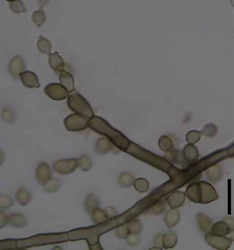
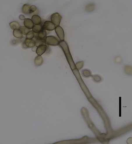

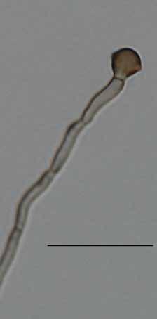
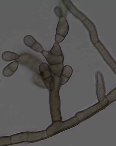
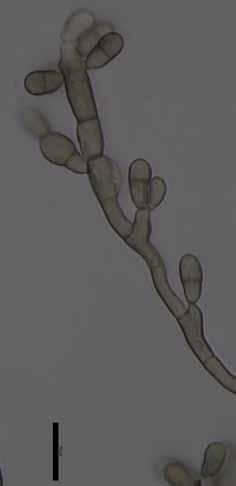
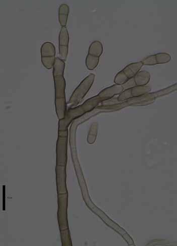
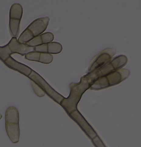
6 86 Persoonia Volume 38, 2017 a b c d e f g h i j k l m Fig. 3 Penidiellopsis radicularis. a, d. Colony on CMA; b, e. colony on MA; c, f. colony on PDA; g k. conidiophores with conidia in chains; l m. chlamydospores. Scale bars = 10 µm.
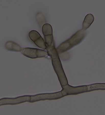
7 A.P.M. Duarte et al.: Riding with the ants 87 Penidiellomyces drakensbergensis (Crous) Crous & Attili- Angelis, comb. nov. MycoBank MB Basionym. Penidiella drakensbergensis Crous, Persoonia 29: Description and illustration See Crous et al. (2012). Penidiellopsis Sandoval-Denis et al., Persoonia 36: Type species. Penidiellopsis radicularis Sandoval-Denis et al. Conidiophores differentiated, solitary, erect, straight to geniculate-sinuous, rarely branched, pale to medium brown, smooth- and thick-walled. Conidiogenous cells integrated, terminal or intercalary, pale to medium brown, smooth, mono- and polyblastic, giving rise to one or more sets of ramoconidia, scars truncate, slightly darkened, unthickened and not refractive. Ramoconidia 0 1-septate, obovoid, ellipsoid or slightly clavate, pale to medium brown, smooth- and thick-walled, apical part with denticle-like loci, basal scar flattened, slightly darkened, unthickened and not refractive. Conidia in branched acropetal chains, 0-septate, obovoid, ellipsoid or limoniform, pale to medium brown, smooth, thick-walled, with conidial scars truncate or protuberant, somewhat darkened, unthickened and not refractive. Sexual morph unknown. Notes Penidiellopsis was introduced as a new genus to accommodate an isolate from a human nail, collected in South Carolina, USA (Crous et al. 2016). Phylogenetically it is closely related to Penidiellomyces aggregatus and P. drakens bergensis. Conidiophores in Penidiella are penicillate with welldeveloped apical branches, characteristics that were absent in Penidiellopsis. Morphological data are only known from slow-growing colonies in artificial media. Penidiellopsis radicularis Sandoval-Denis et al., Persoonia 36: Fig. 3 Isolated from gynes and drones of Atta capiguara (Myrmicinae, Attini tribe). Mycelium consisting of septate, smooth-walled, pale brown, µm wide hyphae. Conidiophores erect, solitary, from superficial mycelium, branched or not, µm. Conidiogenous cells mostly elongate, aseptate, smooth-walled, µm. Conidia catenate in branched chains, 0 1-septate, smooth, lemon-shaped with prominent hila, not thickened, slightly darkened, µm. Chlamydospores terminal and intercalary. Hila present but neither thickened nor darkened. Culture characteristics Colonies were grown at 25 C for 21 d. On CMA, colonies flat with velutinous central portion, dark olivaceous to black, lobed margin, reaching 10 mm diam. Colonies on MA greyish green with darker mycelium in the centre and margin, crenate surface, reaching 11.5 mm diam. On PDA, dark olivaceous folded colonies, with a raised, velutinous central portion, reaching 11 mm diam. Colonies sporulating on all media, with dark mycelium and olivaceous black in reverse on all three media. Specimen examined. Brazil, São Paulo (S " W ", elev. 798 m), isolated from the integument of Atta capiguara gyne, Nov. 2009, F.C. Pagnocca, CBMAI Notes Similar to P. ramosus although colonies are smaller. Penidiellopsis radicularis (originally isolated and described from an infection of a human nail) was found on gynes and drones, while P. ramosus only occurs on gynes of Atta capiguara. Penidiellopsis ramosus Attili-Angelis & A.P.M. Duarte, sp. nov. MycoBank MB817886; Fig. 4 Etymology. Name refers to its apically branched conidiophores. Isolate from gynes of Atta capiguara (Myrmicinae, Attini tribe). Mycelium consisting of septate, smooth-walled, pale olivaceous-brown, µm wide hyphae. Conidiophores ascending to erect, apically branched, bearing conidia in short chains, irregularly geniculate-sinuous, µm. Conidio g enous cells aseptate, smooth-walled, µm; scars not thickened, slightly darkened. Conidia catenate in branched chains, aseptate, smooth, limoniform to fusiform, µm. Chlamydospores not observed. Hila present but neither thickened nor darkened. Culture characteristics Colonies were grown at 25 C for 21 d. On CMA, colonies flat with velutinous central portion, dark olivaceous to black, reaching 13 mm diam. Colonies on MA greyish green with darker mycelium in the centre and margin, sulcate surface, reaching 14 mm diam. On PDA, dark olivaceous folded colonies, raised with an entire edge, velutinous central portion, reaching 12 mm diam. Colonies sporulating on all media, with dark mycelium and olivaceous black in reverse on all three media. Specimens examined. Brazil, São Paulo (S " W ", elev. 798 m), isolated from the integument of Atta capiguara gyne, Nov. 2009, F.C. Pagnocca (holotype and culture ex-type CBMAI 1937, preserved as metabolically inactive). Notes Similar to P. radicularis, but chlamydospores were not observed. Although conidia are similar in shape and size, those of P. ramosus are aseptate. Simplicidiella Crous, Attili-Angelis, A.P.M. Duarte, Pagnocca & J.Z. Groenew., gen. nov. MycoBank MB Etymology. Named after the presence of simple and poorly differentiated conidiophores (Simplici-, Latin = simple). Type species. Simplicidiella nigra A.P.M. Duarte & Attili-Angelis. Hyphomycetous. Mycelium consisting of septate, smooth-walled, pale brown hyphae. Conidiophores erect, solitary, poorly branched or unbranched. Conidiogenous cells elongate, aseptate, smooth-walled; scars not thickened, slightly darkened. Conidia catenate, in branched chains, 0 1-septate, smooth, ellipsoidal to broadly ellipsoidal with prominent hila. Sexual morph unknown. Notes The genus Simplicidiella is described to accommodate ant-isolated melanised fungi in Brazil, with simple and poorly differentiated reproductive structures. Its few phenotypic characteristics underly the need of molecular tools for identification. The genus is presumed here to be saprobic. Simplicidiella is distinct from Penidiella and other penidiella-like genera for not sharing the following characteristics: penicillate conidiophores with branches (as in Penidiella s.str. and Queenslandipenidiella); being described as foliicolous (as in Neopenidiella and Myrtapenidiella); associated with opportunistic human infections with dimorphic conidiophores (as reported for Eupenidiella); production of ramoconidia (as in Apenidiella), and mycelium strongly branched (as observed in Xenopenidiella). Simplicidiella nigra A.P.M. Duarte & Attili-Angelis, sp. nov. MycoBank MB817415; Fig. 5 Etymology. Name refers to very dark colonies on CMA. Isolated from gyne of Atta capiguara (Myrmicinae, Attini tribe). Mycelium consisting of septate, smooth-walled, pale brown, µm wide hyphae. Conidiophores erect, solitary, poorly branched or unbranched, µm. Conidiogenous cells elongate, aseptate, smooth-walled, µm; scars not thickened, slightly darkened. Conidia catenate in


.")
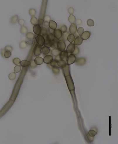
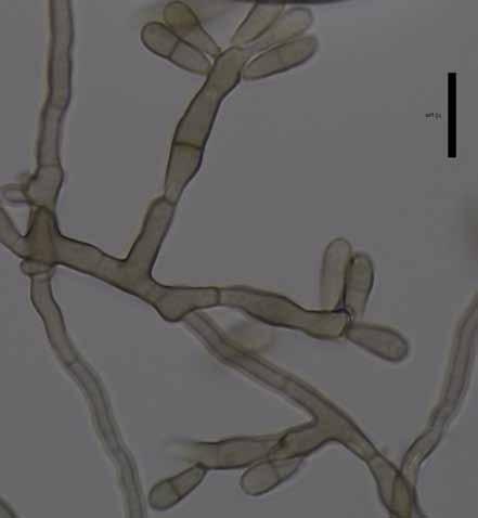
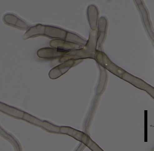
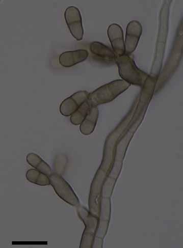
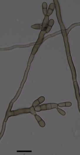
8 88 Persoonia Volume 38, 2017 a b c d e f g h i j k l Fig. 4 Penidiellopsis ramosus. a, d. Colony on CMA; b, e. colony on MA; c, f. colony on PDA; g l. conidiophores with conidia in chains. Scale bars = 10 µm. branched chains, 0 1-septate, smooth, ellipsoidal to broadly ellipsoidal with prominent hila, µm. Chlamydospores not observed. Culture characteristics Colonies were grown at 25 C for 21 d. On CMA, colonies flat with velutinous central portion, colony margin dark olivaceous to black, reaching 14 mm diam. Colonies on MA flat with raised velutinous central portion, intermediate section with olivaceous green mycelium, and darker margins, reaching 11 mm diam. Exudate produced. On PDA, colonies with raised greyish central portion producing exudate, black mycelium and darker margins, reaching 11 mm diam. Sporulation and olivaceous black reverse were present in all media. Specimen examined. Brazil, São Paulo (S " W ", elev. 798 m), isolated from the integument of Atta capiguara gyne, Nov. 2009, F.C. Pagnocca (holotype and culture ex-type CBMAI 1939, preserved as metabolically inactive). Notes Due to few phenotypic characteristics available, identification of S. nigra requires molecular data. This species is presumed to be saprobic and a potential hydrocarbon-degrader, based on the selective method used for isolation and due to the known presence of a complex mixture of hydrocarbons on the


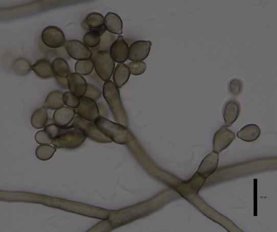
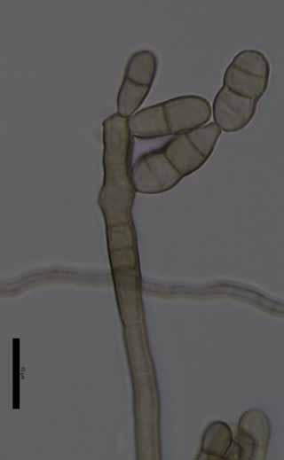
9 A.P.M. Duarte et al.: Riding with the ants 89 a b c d e f g h i k j l m Fig. 5 Simplicidiella nigra. a, d. Colony on CMA; b, e. colony on MA; c, f. colony on PDA; g k. conidiophores with conidia in chains; l m. conidiogenous cells. Scale bars = 10 µm.
.")


.")

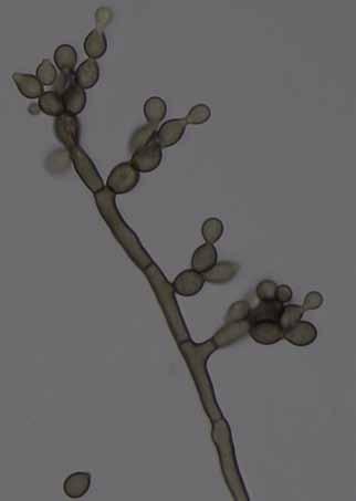
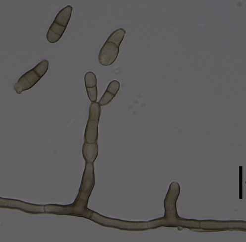
10 90 Persoonia Volume 38, 2017 cuticle of insects (Howard & Blomquist 2005). The species is described based on a single isolate, although its phylogenetic placement was supported by all loci and analyses. Xenopenidiella Quaedvlieg & Crous, Persoonia 33: Type species. Xenopenidiella rigidophora (Crous et al.) Quaedvlieg & Crous. Xenopenidiella clavata Attili-Angelis, A.P.M. Duarte & Pagnocca, sp. nov. MycoBank MB817416; Fig. 6 Etymology. Named after its clavate conidia. Isolated from drone of Atta capiguara (Myrmicinae, Attini tribe). Mycelium consisting of septate, smooth-walled, pale brown, 3 4 µm wide hyphae. Conidiophores mostly reduced to conidiogenous cells arising directly from hyphae, rarely 1 5-septate, a b c d e f g h i j k l m Fig. 6 Xenopenidiella clavata. a, d. Colony on CMA; b, e. colony on MA; c, f. colony on PDA; g m. conidiophores with conidia. Scale bars = 10 µm.
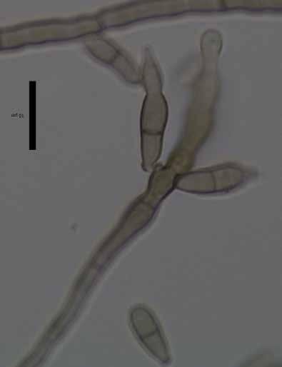
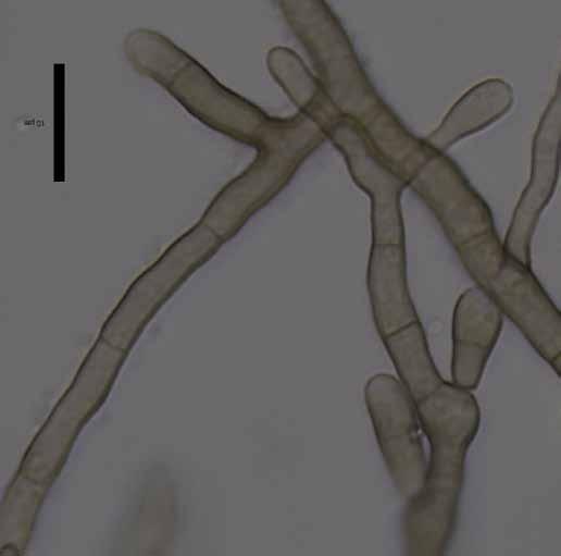
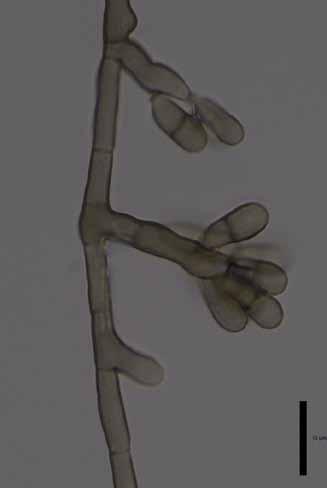
11 A.P.M. Duarte et al.: Riding with the ants 91 brown, smooth-walled, straight to geniculate-sinuous, µm. Conidiogenous cells intercalary or terminal, brown, smooth-walled, µm, with several aggregated terminal scars, not thickened nor darkened. Conidia 2 3-celled, smooth-walled to finely verrucose, clavate, apex obtuse, base truncate, constricted at both septa, µm; hila not thickened, slightly darkened, no conidial scars observed. Chlamydospores not observed. Culture characteristics Colonies were grown at 25 C for 21 d. Colonies on CMA flat and dark olivaceous to brown, with a velutinous central portion, reaching 10 mm diam. Colonies on MA with a slightly elevated velutinous central portion, greenish mycelium and black margins, reaching 11 mm diam. On PDA, flat velutinous dark olivaceous to brown colonies, reaching 10 mm diam. Colonies with low to moderate sporulation and olivaceous black in reverse on all media. Specimen examined. Brazil, São Paulo (S " W ", elev. 798 m), isolated from the integument of Atta capiguara drone, Nov. 2013, F.C. Pagnocca (holotype and culture ex-type CBMAI 1942, preserved as metabolically inactive). Notes Xenopenidiella clavata is ecologically distinct from X. rigidophora (type species) in its association to the leaf-cutting ants, which represents a new niche for the genus. Morphologically, conidiophores of X. clavata are not strongly branched, hyphae are not guttulate, and its mycelium is not strongly branched. Dimorphic conidiophores are present in both species, but in X. clavata conidiogenous cells are predominantly intercalary and conidia are only finely verrucose. Xenopenidiella formica A.P.M. Duarte, Attili-Angelis & N.C. Baron, sp. nov. MycoBank MB817417; Fig. 7 Etymology. Named after the isolation source (ants). Isolated from gynes and drones of Atta capiguara and A. laevigata (Myrmicinae, Attini tribe). Mycelium consisting of septate, smooth-walled, pale olivaceous, 2 3 µm wide hyphae, producing poorly differentiated conidiophores. Conidiophores erect, solitary, poorly branched or unbranched, smooth, brown, straight to geniculate-sinuous, µm. Conidiogenous cells smooth-walled, bearing conidia from flat to semi-denticulate scars, µm. Conidia 1-septate, smooth-walled, clavate, apex obtuse, base truncate, constricted at septum, µm. Chlamydospores not observed. Culture characteristics Colonies were grown at 25 C for 21 d. Colonies on CMA were dark olivaceous and flat, velutinous, reaching 12 mm diam. On MA, flat colonies with velutinous central portion, greenish mycelium and darker margins, reaching 12.5 mm diam. On PDA, velutinous colonies with raised olivaceous green central portion, reaching 11 mm diam. Poor sporulation and olivaceous black in reverse on all media. Specimen examined. Brazil, São Paulo (S " W ", elev. 798 m), isolated from the integument of Atta capiguara drone, Nov. 2013, F.C. Pagnocca (holotype and culture ex-type CBMAI 1941, preserved as metabolically inactive). Notes Phylogenetically, X. formica is closely related to X. tarda, but with a faster growth rate in culture, and smaller conidia. It is represented by five isolates from gynes and drones of A. capiguara and A. laevigata, while X. tarda was found only on A. capiguara. In contrast to X. rigidophora, X. formica does not exhibit strongly branched hyphae, and its conidia are not verrucose. Xenopenidiella inflata A.P.M. Duarte, N.C. Baron, Pagnocca & Attili-Angelis, sp. nov. MycoBank MB817418; Fig. 8 Etymology. Named after its swollen conidiogenous cells (Inflatus, Latin = swollen). Isolated from drone of A. laevigata (Myrmicinae, Attini tribe). Mycelium consisting of septate, smooth, pale brown, µm wide hyphae. Conidiophores reduced to conidiogenous cells, or subcylindrical, smooth, brown, up to 3-septate, straight to geniculous-flexuous, µm. Conidiogenous cells swollen or elongated, arising from mycelium or terminal on conidiophores, sometimes rejuvenating and elongating to have lateral and terminal conidiogenous cells, smooth-walled, µm; scars not thickened nor darkened. Conidia 1-septate, smooth-walled, clavate, constricted at septum, truncate hila, µm. Chlamydospores not observed. Culture characteristics Colonies were grown at 25 C for 21 d. On CMA, colonies with black velutinous mycelium and pronounced central portion, reaching 12.5 mm diam. Colonies on MA greyish green, velutinous and sulcate, black margins, reaching 12 mm diam. On PDA colonies were greenish and raised at the centre, olivaceous green towards the sulcate and darker margin, reaching 12.5 mm diam. Poor to moderate sporulation and olivaceous black reverse on all media. Specimen examined. Brazil, São Paulo (S " W ", elev. 798 m), isolated from the integument of Atta laevigata drone, Nov. 2011, F.C. Pagnocca (holotype and culture ex-type CBMAI 1945, preserved as metabolically inactive). Notes Xenopenidiella inflata is described based on a single isolate from an A. laevigata drone. Although phylogenetically closely related to X. laevigata, it differs based on its swollen conidiogenous cells and slower growth rate on cultural media. Xenopenidiella inflata differs from the X. rigidophora in the following morphological aspects: pale brown mycelium present (not pale olivaceous to medium brown); thinner hyphae (1.5 3 µm wide, not up to 6 µm diam); conidiogenous cells terminal and intercalary; conidia not verrucose (not appearing like small spines under light microscope); thicker hila. Xenopenidiella laevigata N.C. Baron, A.P.M. Duarte, Pagnocca & Attili-Angelis, sp. nov. MycoBank MB817419; Fig. 9 Etymology. Name refers to the first isolation source: the ant species Atta laevigata. Isolated from gyne of A. laevigata (Myrmicinae, Attini tribe). Mycelium consisting of septate, olivaceous-brown, smooth and thick-walled, µm wide hyphae. Conidiophores brown, smooth to finely verrucose, thick-walled erect, straight to geniculous-sinuous, 1 10-septate, µm. Conidiogenous cells in loose branches, smooth-walled to finely verrucose, terminal or lateral, µm; scars unthickened and not darkened. Conidia 1-septate, smooth-walled to finely verrucose, clavate, constricted at somewhat darker septum, µm; hila unthickened and not darkened. Chlamydospores not observed. Culture characteristics Colonies were grown at 25 C for 21 d. On CMA, colonies with black velutinous mycelium and aerial central portion, reaching 18 mm diam. Colonies on MA greyish green, velutinous and sulcate, black margins, reaching 19 mm diam. On PDA colonies were dark olivaceous, sulcate and folded, with raised central portion and velutinous mycelium, reaching 18 mm diam. Poor sporulation and olivaceous black reverse on all three media. Specimen examined. Brazil, São Paulo (S " W ", elev. 798 m), isolated from the integument of Atta laevigata gyne, Nov. 2011, F.C. Pagnocca (holotype and culture ex-type CBMAI 1944, preserved as metabolically inactive). Notes In this study both species X. laevigata and X. inflata are known from single strains isolated from A. laevigata via the oil flotation method. The parsimony tree shows that they are closely related, but the former differs in the melanisation


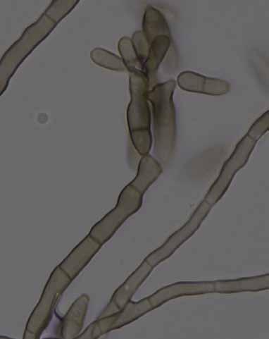
12 92 Persoonia Volume 38, 2017 a b c d e f g h i j k l Fig. 7 Xenopenidiella formica. a, d. Colony on CMA; b, e. colony on MA; c, f. colony on PDA; g l. conidiophores with conidia. Scale bars = 10 µm.


13 A.P.M. Duarte et al.: Riding with the ants 93 a b c d e f g h i j k l m n Fig. 8 Xenopenidiella inflata. a, d. Colony on CMA; b, e. colony on MA; c, f. colony on PDA; g n. conidiophores with conidia. Scale bars = 10 µm.
, growth rate and conidial size. A comparison with X.")


.")


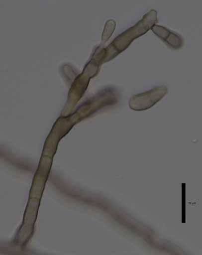
14 94 Persoonia Volume 38, 2017 of the mycelium (colonies are darker and hyphae are thicker), growth rate and conidial size. A comparison with X. rigidophora shows differences such as less-branched and thinner hyphae, rare macronematous conidiophores that are loosely penicillate, and conidiogenous cells frequently intercalary. Xenopenidiella nigrescens Attili-Angelis, A.P.M. Duarte & Pagnocca, sp. nov. MycoBank MB817420; Fig. 10 Etymology. Named after its dark brown mycelium. Isolated from drone of Atta capiguara (Myrmicinae, Attini tribe). Mycelium consisting of septate, olivaceous-brown, smooth and thick-walled 3 4 µm wide hyphae. Conidiophores brown, smooth to finely verrucose, thick-walled, erect, straight to flexuous, 0 6-septate, µm. Conidiogenous cells arising from mycelium, loosely branched, smooth-walled to verrucose, terminal and lateral, µm. Conidia 1-septate, smooth-walled to finely verrucose, dark brown, clavate and constricted, µm; hila unthickened and not darkened. Chlamydospores not observed. a b c d e f g h i j k l Fig. 9 Xenopenidiella laevigata. a, d. Colony on CMA; b, e. colony on MA; c, f. colony on PDA; g l. conidiophores with conidia. Scale bars = 10 µm.


15 A.P.M. Duarte et al.: Riding with the ants 95 Culture characteristics Colonies were grown at 25 C for 21 d. Colonies on CMA were flat and dark olivaceous to brown, velutinous with raised central portion, irregular margins, reaching 14 mm diam. Colonies on MA with velutinous and slightly folded surface, raised central portion, olivaceous mycelium, reaching 13 mm diam. On PDA, dark olivaceous highly folded colonies, raised velutinous central portion, reaching 13 mm diam. Colonies presented low to moderate sporulation and olivaceous black reverse on all media. Specimen examined. Brazil, São Paulo (S " W ", elev. 798 m), isolated from the integument of Atta capiguara drone, Nov. 2013, F.C. Pagnocca (holotype and culture ex-type CBMAI 1943, preserved as metabolically inactive). Notes Xenopenidiella nigrescens was found to consistently have the darkest colonies on all three media tested when compared to other Xenopenidiella species. A combination of previously mentioned characteristics is also found in this species: slow growth rate (as in X. tarda), clavate conidia (as in X. clavata), 1-septate, smooth-walled to finely verrucose conidia a b c d e f g h i j k l Fig. 10 Xenopenidiella nigrescens. a, d. Colony on CMA; b, e. colony on MA; c, f. colony on PDA; g l. conidiophores with conidia. Scale bars = 10 µm.


16 96 Persoonia Volume 38, 2017 a b c d e f g h i j k l m Fig. 11 Xenopenidiella tarda. a, d. Colony on CMA; b, e. colony on MA; c, f. colony on PDA; g m. conidiophores with conidia. Scale bars = 10 µm.
17 A.P.M. Duarte et al.: Riding with the ants 97 (resembling X. laevigata), conidiophores poorly differentiated (as in X. formica). In comparison to X. rigidophora, X. nigrescens differs in having darker mycelium, conidiogenous cells smooth to verrucose, and conidia rarely occurring in branched chains. Molecular data are required to confirm its identification. Xenopenidiella tarda Pagnocca, A.P.M. Duarte & Attili-Angelis, sp. nov. MycoBank MB817421; Fig. 11 Etymology. Named due to very slow growth rate (Tardus, Latin = slow). Isolated from drones of Atta capiguara (Myrmicinae, Attini tribe). Mycelium consisting of septate, smooth-walled, pale brown, 2 3 µm wide hyphae. Conidiophores ascending to erect, solitary, poorly branched or unbranched, 0 4-septate, µm. Conidiogenous cells elongate, aseptate, smoothwalled, µm; scars unthickened and not darkened. Conidia 1-septate, smooth-walled to finely verrucose, clavate, constricted at septum, µm; hila truncate, unthickened and not darkened. Chlamydospores not observed. Culture characteristics Colonies were grown at 25 C for 21 d. On CMA, colonies dark olivaceous, velutinous, flat and small, reaching 9 mm diam. Colonies on MA flat with aerial velutinous central portion, olivaceous green mycelium and black margin, reaching 9 mm diam. On PDA colonies with raised greyish green central portion, and black margins, reaching 9 mm diam. Moderate sporulation and olivaceous black reverse present on all three media. Specimen examined. Brazil, São Paulo (S " W ", elev. 798 m), isolated from the integument of Atta capiguara drone, Nov. 2013, F.C. Pagnocca (holotype and culture ex-type CBMAI 1940, preserved as metabolically inactive). Notes Xenopenidiella tarda is represented by two isolates from drones of A. capiguara. It is morphologically similar to X. formica, although colonies have a slower growth rate in culture. This species can be distinguished from X. rigidophora because mycelium of X. tarda does not show to be strongly branched, not necessarily constricted at septa and hyphae are not so wide. Furthermore, conidiogenous cells are less wide and pale brown; conidia mostly smooth-walled or finely verrucose. DISCUSSION Leaf-cutting ants from the Attini tribe are social insects limited to the New World, widely distributed from Argentina to Southern USA, with a range of latitude from N10 to S25 (Mayhé-Nunes & Jaffé 1998). These ants have been investigated for the huge economic losses they can cause by damaging plant leaves, and for their unique ability for fungiculture, which has become a model system for coevolutionary studies (Chapela et al. 1994, Currie et al. 2003). These insects were found to harbour a large and unknown diversity of microorganisms (Pagnocca et al. 2008, Rodrigues et al. 2008, 2011, Guedes et al. 2012, Duarte et al. 2014), and some novel species of filamentous fungi and yeasts were already described from the ants microenvironment (Middelhoven et al. 2003, Carreiro et al. 2004, Pagnocca et al. 2010, Augustin et al. 2013, Attili-Angelis et al. 2014, Melo et al. 2014, Masiulionis et al. 2015, Meirelles et al. 2015, Samerpitak et al. 2015, Montoya et al. 2016). However, the occurrence of species of Teratosphaeriaceae in the ants environment has never been documented. The family Teratosphaeriaceae was established based on molecular phylogenetic analyses, which strongly supported its separation from Mycosphaerellaceae. Teratosphaeria and related genera were initially associated with leaf diseases of Eucalyptus (Myrtaceae) and Proteaceae, but their ecological habitat vary from saprobic, human opportunistic and plant pathogen to extremophilic species (Crous et al. 2007, Ruibal et al. 2009, Teodoro et al. 2012, Egidi et al. 2014, Quaedvlieg et al. 2014). Investigators in Brazil have isolated plant pathogenic fungi (Guedes et al. 2012) and opportunistic melanised representatives (Duarte et al. 2014) from workers of Atta laevigata and gynes of Atta spp., respectively, which were shown here to belong to the Teratosphaeriaceae. One genus and eight new species of Teratosphaeriaceae were isolated when investigating the diversity of fungi occurring on the integument of pre-nuptial flight gynes and drones. The aim of these investigations was to further explore and better understand the primary microbial community that is dispersed into a young ant colony by these ants. This is therefore the first study to provide a taxonomic treatment of Teratosphaeriaceae from ants, which has significantly expanded the number of Xenopenidiella species in addition to just the type species, X. rigidophora, thereby allowing for a more robust amended description of the genus. Previous reports on filamentous fungi from ant niches characterised similar isolates as found in the soil community (Pagnocca et al. 2008, Rodrigues et al. 2008), which is different from the results found in this study, where the fungi were isolated directly from the insects themselves. It can be hypothesized that the isolation methods used in the other studies may have contributed to this difference. The lack of knowledge about ant nest microenvironments encouraged studies with a focus on this microenvironment, but even less is known about the ant integument from which the new species were obtained. One interesting ecological aspect of the Teratosphaeriaceae is that the family harbours isolates from extreme environments. However, the present scientific information is still insufficient to answer several questions about the studied substrate, for instance, if it should be treated as extreme or not. It is known that the ph of fungal gardens ranges from 4.35 to 5 (Powell & Stradling 1986), but nothing has been published about the ph of the integument itself. On the other hand, the presence of cuticular hydrocarbons and alkaloids on the integument was already reported (Roux et al. 2009). The main lineages in the Teratosphaeriaceae remained obscured until the extensive revision published by Quaedvlieg et al. (2014). In this publication, a previous discussion point regarding the phylogenetic position of Piedraiaceae was once again raised as this fungal family appears to cluster within the Teratosphaeriaceae (Crous et al. 2009a). Other reports studying rock-inhabiting fungi (RIF) showed that these are phylogenetically highly diverse, and highlighted the importance of larger taxon samplings to define questions about generic boundaries (Ruibal et al. 2009, 2011). All these results demonstrate the importance of dedicated projects on fungal isolation from diverse niches. Although this study still under-represents the total biodiversity on leaf-cutting ants, it does reveal yet another source of Teratosphaeriaceae species. It is known that the morphology of many genera of saprobic and plant pathogenic fungi have evolved several times independently (Crous et al. 2009b), which explains how these penidiella-like isolates could adapt to such an unlikely environment such as gynes and drones of attine ants. The species phylogeny presented in Fig. 2 is based on four genomic loci (ITS, cmda, tef1 and tub2 gene regions). For the species of Xenopenidiella, all loci for which data was available could distinguish the included species (data not shown, trees for individual loci were deposited in TreeBASE). However, the amplification success rate of tef1 and cmda for this genus was not as high as for ITS and tub2. The ex-type culture of X. formica, CBMAI 1941, clusters slightly distant to the rest of
Two new species of Corynespora from Uttar Pradesh, India
 Two new species of Corynespora from Uttar Pradesh, India Kumar S 1,2*, Singh R 2, Gond DK 1 and Saini DC 1 1 Birbal Sahni Institute of Palaeobotany, 53, University Road, Lucknow 226007(U.P.), India. 2
First Report of Penicillium adametzioides from Decayed Grapes (Vitis vinifera) in Pakistan
 in Pakistan") International Journal of Current Microbiology and Applied Sciences ISSN: 2319-7706 Volume 5 Number 12 (2016) pp. 316-320 Journal homepage: http://www.ijcmas.com Original Research Article http://dx.doi.org/10.20546/ijcmas.2016.512.034
International Journal of Current Microbiology and Applied Sciences ISSN: 2319-7706 Volume 5 Number 12 (2016) pp. 316-320 Journal homepage: http://www.ijcmas.com Original Research Article http://dx.doi.org/10.20546/ijcmas.2016.512.034
Goosiomyces bambusicola - A new cheirosporous anamorphic species from Western Ghats, India.
 Current Research in Environmental & Applied Mycology 4 (2): 211 216 (2014) ISSN 2229-2225 www.creamjournal.org Article CREAM Copyright 2014 Doi 10.5943/cream/4/2/8 Online Edition Goosiomyces bambusicola
Cercosporoid leaf pathogens from whorled milkweed and spineless safflower in California
 doi:10.5598/imafungus.2011.02.01.02 IMA Fungus volume 2 no 1: 7 12 Cercosporoid leaf pathogens from whorled milkweed and spineless safflower in California Steven T. Koike 1, Aziz Baameur 2, Johannes Z.
ITS accuracy at GenBank. Conrad Schoch Barbara Robbertse
 ITS accuracy at GenBank Conrad Schoch Barbara Robbertse Improving accuracy Barcode tag in GenBank Barcode submission tool Standards RefSeq Targeted Loci Well validated sequences already in GenBank Bacteria
INTRODUCTION PRODUCT DESCRIPTION
 INTRODUCTION Mycoplasma are known as important contaminants of biological products derived from cell lines in the biopharmaceutical industry affecting every parameter of a cell culture system. Contaminated
Journal of Chemical and Pharmaceutical Research, 2017, 9(1): Review Article. Graphium Salixicum: A New Species Explored from Salix Alba
: Review Article. Graphium Salixicum: A New Species Explored from Salix Alba") Available online www.jocpr.com Journal of Chemical and Pharmaceutical Research, 2017, 9(1):69-74 Review Article ISSN : 0975-7384 CODEN(USA) : JCPRC5 Graphium Salixicum: A New Species Explored from Salix
Available online www.jocpr.com Journal of Chemical and Pharmaceutical Research, 2017, 9(1):69-74 Review Article ISSN : 0975-7384 CODEN(USA) : JCPRC5 Graphium Salixicum: A New Species Explored from Salix
Braunomyces dictyosporus gen. sp. nov. from Vietnam ARTICLE. Introduction. Material and Methods
 doi:10.5598/imafungus.2014.05.01.01 IMA Fungus volume 5 no 1: 1 5 Braunomyces dictyosporus gen. sp. nov. from Vietnam Vadim A. Mel nik 1 and Pedro W. Crous 2 1 Laboratory of the Systematics and Geography
Principles of phylogenetic analysis
 Principles of phylogenetic analysis Arne Holst-Jensen, NVI, Norway. Fusarium course, Ås, Norway, June 22 nd 2008 Distance based methods Compare C OTUs and characters X A + D = Pairwise: A and B; X characters
World Journal of Microbiology Vol. 1(2), pp , September, ISSN: XXXX-XXXX
, pp , September, ISSN: XXXX-XXXX") World Journal of Microbiology Vol. 1(2), pp. 013-016, September, 2014. www.premierpublishers.org, ISSN: XXXX-XXXX WJM Research Article Study of Fungal Genus Gyrothrix Corda from the forest flora of Indian
World Journal of Microbiology Vol. 1(2), pp. 013-016, September, 2014. www.premierpublishers.org, ISSN: XXXX-XXXX WJM Research Article Study of Fungal Genus Gyrothrix Corda from the forest flora of Indian
The Journal of Animal & Plant Sciences, 26(6): 2016, Page: Javaidet al., The J. Anim. Plant Sci. 26(6):2016
: 2016, Page: Javaidet al., The J. Anim. Plant Sci. 26(6):2016") The Journal of Animal & Plant Sciences, 26(6): 2016, Page: 1894-1898 Javaidet al., The J. Anim. Plant Sci. 26(6):2016 ISSN: 1018-7081 Short Communication NEW HOST RECORD OF ALTERNARIA BRASSICICOLA INFECTING
The Journal of Animal & Plant Sciences, 26(6): 2016, Page: 1894-1898 Javaidet al., The J. Anim. Plant Sci. 26(6):2016 ISSN: 1018-7081 Short Communication NEW HOST RECORD OF ALTERNARIA BRASSICICOLA INFECTING
Key words wild passion-fruit, Mycosphaerellaceae, tropical fruits, cercosporoid fungi
 A NEW FUNGAL DISEASE CAUSED BY A PSEUDOCERCOSPORA SPECIES ON PASSIFLORA SETACEA IN PLANALTINA-DF, BRAZIL Alexei C. Dianese 1, Ana M. Costa 1 & José C. Dianese 2 ( 1 Embrapa Cerrados, Br-020, Km 18, 73310-970
Dokmaia monthadangii gen. et sp. nov., a synnematous anamorphic fungus on Manglietia garrettii
 Dokmaia monthadangii gen. et sp. nov., a synnematous anamorphic fungus on Manglietia garrettii I. Promputtha1, K. D. Hyde2, P. Lumyong3, E. H. C. McKenzie4 & S. Lumyong1 1 Department of Biology, Faculty
New species and records of Bipolaris and Curvularia from Thailand
 Mycosphere 8(9): 1556 1574 (2017) www.mycosphere.org ISSN 2077 7019 Article Doi 10.5943/mycosphere/8/9/11 Copyright Guizhou Academy of Agricultural Sciences New species and records of Bipolaris and Curvularia
LESSON ASSIGNMENT. Introduction to Medical Mycology. After completing this lesson, you should be able to:
 LESSON ASSIGNMENT LESSON 1 Introduction to Medical Mycology. TEXT ASSIGNMENT Paragraphs 1-1 through 1-7. TASKS OBJECTIVES After completing this lesson, you should be able to: 1-1. Select the statement
7-001b: Malt agar method for the detection of Alternaria dauci on Daucus carota (carrot)
") International Rules for Seed Testing Annexe to Chapter 7: Seed Health Testing Methods 7-001b: Malt agar method for the detection of Alternaria dauci on Daucus carota (carrot) Published by: International
International Rules for Seed Testing Annexe to Chapter 7: Seed Health Testing Methods 7-001b: Malt agar method for the detection of Alternaria dauci on Daucus carota (carrot) Published by: International
Doi /mycosphere/8/8/14 Copyright Guizhou Academy of Agricultural Sciences
 New Cylindrocladiella spp. from Thailand soils Lombard L 1*, Cheewangkoon R 2*, Crous PW 1,2 1 Westerdijk Fungal Biodiversity Institute, Uppsalalaan 8, 3584 CT Utrecht, The Netherlands 2 Department of
REINWARDTIA Published by Herbarium Bogoriense LBN, Bogor Vol. 10, Part 2, pp (1985) THE ANAMORPH OF SARAWAKUS SUCCISUS RIFAI
 THE ANAMORPH OF SARAWAKUS SUCCISUS RIFAI") REINWARDTIA Published by Herbarium Bogoriense LBN, Bogor Vol. 10, Part 2, pp. 265 270 (1985) THE ANAMORPH OF SARAWAKUS SUCCISUS RIFAI MIEN A. RIFAI, KARTINI KRAMADIBRATA Herbarium Bogorievnc LBN, Bogor,
REINWARDTIA Published by Herbarium Bogoriense LBN, Bogor Vol. 10, Part 2, pp. 265 270 (1985) THE ANAMORPH OF SARAWAKUS SUCCISUS RIFAI MIEN A. RIFAI, KARTINI KRAMADIBRATA Herbarium Bogorievnc LBN, Bogor,
Identification of Yeasts. Medical Mycology Training Network 15 November 2018 Dr Tan Ai Ling Department of Microbiology, Singapore General Hospital
 Identification of Yeasts Medical Mycology Training Network 15 November 2018 Dr Tan Ai Ling Department of Microbiology, Singapore General Hospital Definition of Yeasts Eukaryote cells have defined nucleus
Colletotrichum gloeosporioides, the causal agent of citrus anthracnose in Guizhou Province
 Colletotrichum gloeosporioides, the causal agent of citrus anthracnose in Guizhou Province Jiang YL, Tan P, Zhou XY, Hou XL and Wang Y* Department of Plant Pathology, Guizhou University, Guiyang, Guizhou,
Additions to helicoid fungi from India
 Current Research in Environmental & Applied Mycology 6 (4): 248 255 (2016) ISSN 2229-2225 www.creamjournal.org Article CREAM Copyright 2016 Doi 10.5943/cream/6/4/2 Online Edition Additions to helicoid
7-001a: Blotter method for the detection of Alternaria dauci on Daucus carota (carrot)
") International Rules for Seed Testing Annexe to Chapter 7: Seed Health Testing Methods 7-001a: Blotter method for the detection of Alternaria dauci on Daucus carota (carrot) Published by: International
International Rules for Seed Testing Annexe to Chapter 7: Seed Health Testing Methods 7-001a: Blotter method for the detection of Alternaria dauci on Daucus carota (carrot) Published by: International
Microorganisms as Freight Haulage Systems. Colin Ingham Wageningen University, NL
 Microorganisms as Freight Haulage Systems Colin Ingham Wageningen University, NL Swarming bacteria Surface, collective, flagellar-driven, motility common in soil bacteria. Often involves surfactants/lubricants
7-002b: Malt agar method for the detection of Alternaria radicina on Daucus carota (carrot)
") International Rules for Seed Testing Annexe to Chapter 7: Seed Health Testing Methods 7-002b: Malt agar method for the detection of Alternaria radicina on Daucus carota (carrot) Published by: International
International Rules for Seed Testing Annexe to Chapter 7: Seed Health Testing Methods 7-002b: Malt agar method for the detection of Alternaria radicina on Daucus carota (carrot) Published by: International
Evaluation of an alternative slide culture technique for the morphological identification of fungal species
 Research Article Evaluation of an alternative slide culture technique for the morphological identification of fungal species Abstract M H Wijedasa 1, L V C Liyanapathirana 1. Sri Lanka Journal of Infectious
Dactylella shizishanna sp. nov., from Shizi Mountain, China
 Dactylella shizishanna sp. nov., from Shizi Mountain, China XueFeng Liu 1,2 and KeQin Zhang 1 * 1 Laboratory for Conservation and Utilization of Bio-resource, Yunnan University, Kunming, Yunnan 650091,
Gonatophragmium lichenophilum sp. nov. a new lichenicolous hyphomycete from Austria
 MYCOBIOTA 5: 7 13 (2015) RESEARCH ARTICLE ISSN 1314-7129 (print) http://dx.doi.org/10.12664/mycobiota.2015.05.02 doi: ISSN 1314-7781 (online) www.mycobiota.com Gonatophragmium lichenophilum sp. nov. a
Colonial and Morphological Characteristics of various fungi Species Isolated from soil in Bangalore city
 Bulletin of Environment, Pharmacology and Life Sciences Bull. Env. Pharmacol. Life Sci., Vol 6[1] December 2016: 17-21 2016 Academy for Environment and Life Sciences, India Online ISSN 2277-1808 Journal
A novel method for the management of mealybug in Cotton.
 A novel method for the management of mealybug in Cotton. Gulsar Banu J Principal Scientist, Central Institute for Cotton Research, Regional Station, Coimbatore-641 003. Tamil Nadu Introduction Due to large
The BLAST search on NCBI ( and GISAID
 Supplemental materials and methods The BLAST search on NCBI (http:// www.ncbi.nlm.nih.gov) and GISAID (http://www.platform.gisaid.org) showed that hemagglutinin (HA) gene of North American H5N1, H5N2 and
Blotter method for the detection of Alternaria dauci on Daucus carota
 International Rules for Seed Testing Annexe to Chapter 7: Seed Health Testing Methods 7-001a: Blotter method for the detection of Alternaria dauci on Daucus carota Published by: International Seed Testing
Cytochalasins from an Australian marine sediment-derived Phomopsis sp. (CMB-M0042F): Acid-mediated intra-molecular cycloadditions enhance
: Acid-mediated intra-molecular cycloadditions enhance") SUPPORTING INFORMATION Cytochalasins from an Australian marine sediment-derived Phomopsis sp. (CMB-M42F): Acid-mediated intra-molecular cycloadditions enhance chemical diversity Zhuo Shang, Ritesh Raju,
SUPPORTING INFORMATION Cytochalasins from an Australian marine sediment-derived Phomopsis sp. (CMB-M42F): Acid-mediated intra-molecular cycloadditions enhance chemical diversity Zhuo Shang, Ritesh Raju,
Epidemiology of Gray Leaf Spot of Perennial Ryegrass Philip Harmon and Richard Latin. Objective
 Epidemiology of Gray Leaf Spot of Perennial Ryegrass Philip Harmon and Richard Latin Objective Rationale The continuing objective of this research is to investigate the environmental factors that influence
Fungal Systematics and Evolution
 Fungal Systematics and Evolution VOLUME 2 DECEMBER 208 PAGES 9 doi.org/0.34/fuse.208.02.0 Arthrinium species associated with bamboo and reed plants in China N. Jiang, J. Li 2, C.M. Tian * The Key Laboratory
Diversity, distribution and taxonomy of Cladosporium associated with Celastraceae
 Plant Pathology & Quarantine 6(1): 48 52 (2016) ISSN 2229-2217 www.ppqjournal.org Article PPQ Copyright 2016 Online Edition Doi 10.5943/ppq/6/1/7 Diversity, distribution and taxonomy of Cladosporium associated
Morphological and molecular characterization of Cercospora zebrina from black bindweed in Iran
 Morphological and molecular characterization of Cercospora zebrina from black bindweed in Iran Bakhshi M 1, Arzanlou M 2* and Babai-Ahari A 3 1 PhD Student of Plant Pathology, Plant Protection Department,
Subcutaneous Mycosis
 Subcutaneous Mycosis Fungal infections 1. Superficial mycosis. 2. Coetaneous mycosis: Dermatophytoses. 3. Subcutaneous mycosis. 4. Systemic mycosis. 5. Opportunistic mycosis. Subcutanus mycoses Fungal
Stilbella holubovae, a new synnematous hyphomycete species on driftwood from the Philippines and South Africa
 Stilbella holubovae, a new synnematous hyphomycete species on driftwood from the Philippines and South Africa Keith A. Seifert 1, Susan J. Stanley 2 and Kevin D. Hyde 2 Centre for Land and Biological Resources
Two new lichenicolous species of Hainesia (asexual Ascomycetes) growing on Cladonia
 growing on Cladonia") Two new lichenicolous species of Hainesia (asexual Ascomycetes) growing on Cladonia Paul Diederich 1 & Pieter van den Boom 2 1 Musée national d histoire naturelle, 25 rue Munster, L 2160 Luxembourg, Luxembourg
Two new lichenicolous species of Hainesia (asexual Ascomycetes) growing on Cladonia Paul Diederich 1 & Pieter van den Boom 2 1 Musée national d histoire naturelle, 25 rue Munster, L 2160 Luxembourg, Luxembourg
Identification of Botryosphaeriaceae associated with the die-back of ornamental trees in the Western Balkans
 COST action FP1401 A global network of nurseries as early warning system against alien tree pests- Global warning Training school Fungal taxonomy and fungal identification using traditional (i.e. not molecular)
A Cladosarum-like spontaneous mutant of Aspergillus aureolatus
 A Cladosarum-like spontaneous mutant of Aspergillus aureolatus By M. Muntanjola-Cvetkovic & J. Bata Institute for Biological Research Botanicki Zavod, Takovska 43, Belgrade, Yugoslavia Summary. A spontaneous
7-002a: Blotter method for the detection of Alternaria radicina on Daucus carota (carrot)
") International Rules for Seed Testing Annexe to Chapter 7: Seed Health Testing Methods 7-002a: Blotter method for the detection of Alternaria radicina on Daucus carota (carrot) Published by: International
International Rules for Seed Testing Annexe to Chapter 7: Seed Health Testing Methods 7-002a: Blotter method for the detection of Alternaria radicina on Daucus carota (carrot) Published by: International
Research & Reviews: Journal of Microbiology and Biotechnology
 e-issn:2320-3528 Research & Reviews: Journal of Microbiology and Biotechnology Two Novel Eupenicillium Species Isolated from Soil in China Hong-Kai Wang, Yi-Chen Cao, Fu-Cheng Lin* State Key Laboratory
Molecular phylogeny of Australian isolates of Sporothrix schenckii sensu lato. David New Microbiology Registrar, PathWest
 Molecular phylogeny of Australian isolates of Sporothrix schenckii sensu lato David New Microbiology Registrar, PathWest Background Sporothrix schenckii causes a cutaneous mycosis Sporotrichosis (aka rose
Isolation and identification of Mycoplasma gallisepticum in chickensbn from industrial farms in Kerman province
 Available online at http://www.ijabbr.com International journal of Advanced Biological and Biomedical Research Volume 2, Issue 1, 2014: 100-104 Isolation and identification of Mycoplasma gallisepticum
Introduction. Study of fungi called mycology.
 Fungi Introduction Study of fungi called mycology. Some fungi are beneficial: ex a) Important in production of some foods, ex: cheeses, bread. b) Important in production of some antibiotics, ex: penicillin
Foliicolous Mycosphaerella spp. and their anamorphs on Corymbia and Eucalyptus
 Foliicolous Mycosphaerella spp. and their anamorphs on Corymbia and Eucalyptus Pedro W. Crous 1*, Brett A. Summerell 2, Angus J. Carnegie 3, Caroline Mohammed 4, Winanda Himaman 5 and Johannes Z. Groenewald
Chromatin IP (Isw2) Fix soln: 11% formaldehyde, 0.1 M NaCl, 1 mm EDTA, 50 mm Hepes-KOH ph 7.6. Freshly prepared. Do not store in glass bottles.
 Fix soln: 11% formaldehyde, 0.1 M NaCl, 1 mm EDTA, 50 mm Hepes-KOH ph 7.6. Freshly prepared. Do not store in glass bottles.") Chromatin IP (Isw2) 7/01 Toshi last update: 06/15 Reagents Fix soln: 11% formaldehyde, 0.1 M NaCl, 1 mm EDTA, 50 mm Hepes-KOH ph 7.6. Freshly prepared. Do not store in glass bottles. 2.5 M glycine. TBS:
Chromatin IP (Isw2) 7/01 Toshi last update: 06/15 Reagents Fix soln: 11% formaldehyde, 0.1 M NaCl, 1 mm EDTA, 50 mm Hepes-KOH ph 7.6. Freshly prepared. Do not store in glass bottles. 2.5 M glycine. TBS:
Protocol for Gene Transfection & Western Blotting
 The schedule and the manual of basic techniques for cell culture Advanced Protocol for Gene Transfection & Western Blotting Schedule Day 1 26/07/2008 Transfection Day 3 28/07/2008 Cell lysis Immunoprecipitation
This is the written version of our Hot Topic video presentation available at: MayoMedicalLaboratories.com/hot-topics
 This is the written version of our Hot Topic video presentation available at: MayoMedicalLaboratories.com/hot-topics Welcome to Mayo Medical Laboratories hot topics. These presentations provide short discussion
The Schedule and the Manual of Basic Techniques for Cell Culture
 The Schedule and the Manual of Basic Techniques for Cell Culture 1 Materials Calcium Phosphate Transfection Kit: Invitrogen Cat.No.K2780-01 Falcon tube (Cat No.35-2054:12 x 75 mm, 5 ml tube) Cell: 293
Species of Phaeoacremonium Associated with Infections in Humans and Environmental Reservoirs in Infected Woody Plants
 JOURNAL OF CLINICAL MICROBIOLOGY, Apr. 2005, p. 1752 1767 Vol. 43, No. 4 0095-1137/05/$08.00 0 doi:10.1128/jcm.43.4.1752 1767.2005 Copyright 2005, American Society for Microbiology. All Rights Reserved.
Asian Journal of Agriculture and Rural Development. Screening of Mycotoxin Produced by Fusarium Verticillioides and F. Proliferatum in Culture Media
 Asian Journal of Agriculture and Rural Development journal homepage: http://www.aessweb.com/journals/5005 Screening of Mycotoxin Produced by Fusarium Verticillioides and F. Proliferatum in Culture Media
APPENDIX-I. The compositions of media used for the growth and differentiation of Pseudomonas aeruginosa are as follows:
 APPENDIX-I The compositions of media used for the growth and differentiation of Pseudomonas aeruginosa are as follows: COMPOSITION OF DIFFERENT MEDIA STOCK CULTURE AGAR (AYERS AND JOHNSON AGAR) Gms/Litre
STUDIES ON CULTURAL, MORPHOLOGICAL AND PATHOGENIC VARIABILITY AMONG THE ISOLATES OF FUSARIUM OXYSPORUM F. SP. CICERI CAUSING WILT OF CHICKPEA
 Volume-7, Issue-1 Jan-Mar-2017 Coden: IJPAJX-CAS-USA, Copyrights@2015ISSN-2231-4490 Received: 1 st Oct-2016 Revised: 26 th Nov-2016 Accepted: 27 th Nov-2016 DOI: 10.21276/Ijpaes http://dx.doi.org/10.21276/ijpaes
Aperto Cell Lysis and Protein Solubilization Users Manual
 Aperto Cell Lysis and Protein Solubilization Users Manual Revision 2 THIS MANUAL APPLIES TO THE FOLLOWING PRODUCTS: 3A8600 Aperto, 5X Cell Lysis Buffer. 20mL 3A8610 Aperto, 5X Cell Lysis Buffer. 100mL
Additions to the Mycosphaerella complex ARTICLE
 doi:10.5598/imafungus.2011.02.01.08 IMA Fungus volume 2 no 1: 49 64 Additions to the Mycosphaerella complex Pedro W. Crous 1, Kazuaki Tanaka 2, Brett A. Summerell 3 and Johannes Z. Groenewald 1 1 CBS-KNAW
Effect of Environmental Factors on the Growth of Aspergillus Species Associated with Stored Millet Grains in Sokoto.
 Available online at http://www.ajol.info/index.php/njbas/index Nigerian Journal of Basic and Applied Science (2011), 19(2):218-223 ISSN 0794-5698 ffect of nvironmental Factors on the Growth of Aspergillus
aP. Code assigned: Short title: Remove (abolish) the species Narcissus symptomless virus in the genus Carlavirus, family Betaflexiviridae
 the species Narcissus symptomless virus in the genus Carlavirus, family Betaflexiviridae") This form should be used for all taxonomic proposals. Please complete all those modules that are applicable (and then delete the unwanted sections). For guidance, see the notes written in blue and the
This form should be used for all taxonomic proposals. Please complete all those modules that are applicable (and then delete the unwanted sections). For guidance, see the notes written in blue and the
Blotter method for the detection of Alternaria radicina on Daucus carota
 International Rules for Seed Testing Annexe to Chapter 7: Seed Health Testing Methods 7-002a: Blotter method for the detection of Alternaria radicina on Daucus carota Published by: International Seed Testing
Accepted Manuscript. All that glitters is not Ramularia. S.I.R. Videira, J.Z. Groenewald, U. Braun, H.-D. Shin, P.W. Crous
 Accepted Manuscript All that glitters is not Ramularia S.I.R. Videira, J.Z. Groenewald, U. Braun, H.-D. Shin, P.W. Crous PII: S0166-0616(16)30001-X DOI: 10.1016/j.simyco.2016.06.001 Reference: SIMYCO 28
Product # Kit Components
 3430 Schmon Parkway Thorold, ON, Canada L2V 4Y6 Phone: (905) 227-8848 Fax: (905) 227-1061 Email: techsupport@norgenbiotek.com Pneumocystis jirovecii PCR Kit Product # 42820 Product Insert Background Information
MYCOTAXON. Volume 91, pp January March Dactylella coccinella sp. nov., an anamorphic species. 1
 MYCOTAXON Volume 91, pp. 127 132 January March 2005 Dactylella coccinella sp. nov., an anamorphic species YING YANG 1,2 & XING ZHONG LIU 1 liuxz@sun.im.ac.cn 1 Key Laboratory of Systematic Mycology & Lichenology
PRESENTER: DENNIS NYACHAE MOSE KENYATTA UNIVERSITY
 18/8/2016 SOURCES OF MICROBIAL CONTAMINANTS IN BIOSAFETY LABORATORIES IN KENYA PRESENTER: DENNIS NYACHAE MOSE KENYATTA UNIVERSITY 1 INTRODUCTION Contamination occurs through avoidable procedural errors
NEW FUNGAL RECORDS ON EUCALYPTUS SPP. FROM DISTRICT FAISALABAD PAKISTAN
 Pak. J. Bot., 42(5): 3317-3321, 2010. NEW FUNGAL RECORDS ON EUCALYPTUS SPP. FROM DISTRICT FAISALABAD PAKISTAN SYED QAISER ABBAS, TEHREEMA IFTIKHAR, MUBASHIR NIAZ AND NAILA SADAF Department of Botany, Government
Study of wilt producing Fusarium sp. from tomato (Lycopersicon esculentum Mill)
") ISSN: 2319-7706 Volume 3 Number 8 (2014) pp. 854-858 http://www.ijcmas.com Original Research Article Study of wilt producing Fusarium sp. from tomato (Lycopersicon esculentum Mill) Patel Harikrushana 1,
ISSN: 2319-7706 Volume 3 Number 8 (2014) pp. 854-858 http://www.ijcmas.com Original Research Article Study of wilt producing Fusarium sp. from tomato (Lycopersicon esculentum Mill) Patel Harikrushana 1,
OVERVIEW OF CURRENT IDENTIFICATION SYSTEMS AND DATABASES
 OVERVIEW OF CURRENT IDENTIFICATION SYSTEMS AND DATABASES EVERY STEP OF THE WAY 1 EVERY STEP OF THE WAY MICROBIAL IDENTIFICATION METHODS DNA RNA Genotypic Sequencing of ribosomal RNA regions of bacteria
CARIBBEAN FOOD CROPS SOCIETY 44 Forty Fourth Annual Meeting 2008
 CARIBBEAN FOOD CROPS SOCIETY 44 Forty Fourth Annual Meeting 2008 Miami, Florida, USA Vol. XLIV - Number 2 Continued Poster Session Abstracts With Some Posters Expanded as Full Papers UF MEETING HOST: UNIVERSITY
RAFI, M. AND SAJJAD-UR-RAHMAN Department of Veterinary Microbiology, University of Agriculture, Faisalabad 38040, Pakistan
 INTERNATIONAL JOURNAL OF AGRICULTURE & BIOLOGY 1560 8530/00/04 4 553 558 http://www.ijab.org Isolation and Identification of Indigenous Penicillium chrysogenum Series RAFI, M. AND SAJJAD-UR-RAHMAN Department
Incongruence between EF-1α Phylogeny and Morphology of Metarhizium majus and Metarhizium guizhouense in Japan
 34: 19-23 (2010) Incongruence between EF-1α Phylogeny and Morphology of Metarhizium majus and Metarhizium guizhouense in Japan OUMI NISHI, KAZUHIRO IIYAMA, CHISA YASUNAGA-AOKI and SUSUMU SHIMIZU * Laboratory
Molecular Identification of Lipase Producing Bacteria based on 16S rdna Sequencing
 International Journal of Current Microbiology and Applied Sciences ISSN: 2319-7706 Volume 6 Number 5 (2017) pp. 2067-2071 Journal homepage: http://www.ijcmas.com Original Research Article https://doi.org/10.20546/ijcmas.2017.605.230
Phylogenetic reassessment of Mycosphaerella spp. and their anamorphs occurring on Eucalyptus
 STUDIES IN MYCOLOGY 50: 195 214. 2004. Phylogenetic reassessment of Mycosphaerella spp. and their anamorphs occurring on Eucalyptus Pedro W. Crous 1*, Johannes Z. Groenewald 1, J. Pedro Mansilla 2, Gavin
For in vitro Veterinary Diagnostics only. Kylt Rotavirus A. Real-Time RT-PCR Detection.
 For in vitro Veterinary Diagnostics only. Kylt Rotavirus A Real-Time RT-PCR Detection www.kylt.eu DIRECTION FOR USE Kylt Rotavirus A Real-Time RT-PCR Detection A. General Kylt Rotavirus A products are
A Glomerella species phylogenetically related to Colletotrichum acutatum on Norway maple in Massachusetts
 Mycologia, 100(5), 2008, pp. 710 715. DOI: 10.3852/07-192 # 2008 by The Mycological Society of America, Lawrence, KS 66044-8897 A Glomerella species phylogenetically related to Colletotrichum acutatum
Four species of Zygophiala (Schizothyriaceae, Capnodiales) are associated with the sooty blotch and flyspeck complex on apple
 are associated with the sooty blotch and flyspeck complex on apple") Mycologia, 100(2), 2008, pp. 246 258. # 2008 by The Mycological Society of America, Lawrence, KS 66044-8897 Four species of Zygophiala (Schizothyriaceae, Capnodiales) are associated with the sooty blotch
Mycologia, 100(2), 2008, pp. 246 258. # 2008 by The Mycological Society of America, Lawrence, KS 66044-8897 Four species of Zygophiala (Schizothyriaceae, Capnodiales) are associated with the sooty blotch
Pinpoint Slide RNA Isolation System II Catalog No. R1007
 INSTRUCTION MANUAL Pinpoint Slide RNA Isolation System II Catalog No. R1007 Highlights Allows for the isolation of total RNA from paraffin-embedded tissue sections on glass slides Simple procedure combines
Mango Dieback and Gummosis in Sindh, Pakistan Caused by Lasiodiplodia theobromae
 2004 Plant Management Network. Accepted for publication 16 January 2004. Published 2 March 2004. Mango Dieback and Gummosis in Sindh, Pakistan Caused by Lasiodiplodia theobromae Muhammad Ali Khanzada,
Curviclavula, a new genus of anamorphic Helotiales (Leotiomycetes) isolated from air
 isolated from air") Mycol Progress (2015) 14: 3 DOI 10.1007/s11557-015-1025-3 ORIGINAL ARTICLE Curviclavula, a new genus of anamorphic Helotiales (Leotiomycetes) isolated from air Gregorio Delgado & Andrew N. Miller & Fernando
Mycol Progress (2015) 14: 3 DOI 10.1007/s11557-015-1025-3 ORIGINAL ARTICLE Curviclavula, a new genus of anamorphic Helotiales (Leotiomycetes) isolated from air Gregorio Delgado & Andrew N. Miller & Fernando
7-012: Detection of Alternaria padwickii on Oryza sativa (Rice)
") International Rules for Seed Testing Annexe to Chapter 7: Seed Health Testing Methods Published by: International Seed Testing Association (ISTA), Bassersdorf, Switzerland 2014 DISCLAIMER: whilst ISTA
International Rules for Seed Testing Annexe to Chapter 7: Seed Health Testing Methods Published by: International Seed Testing Association (ISTA), Bassersdorf, Switzerland 2014 DISCLAIMER: whilst ISTA
Scanning Electron Microscopy (SEM) of Seed Infected with Seed Borne Fungi
 of Seed Infected with Seed Borne Fungi") International Journal of Current Microbiology and Applied Sciences ISSN: 2319-7706 Volume 7 Number 07 (2018) Journal homepage: http://www.ijcmas.com Original Research Article https://doi.org/10.20546/ijcmas.2018.707.476
International Journal of Current Microbiology and Applied Sciences ISSN: 2319-7706 Volume 7 Number 07 (2018) Journal homepage: http://www.ijcmas.com Original Research Article https://doi.org/10.20546/ijcmas.2018.707.476
Bioprospecting of Neem for Antimicrobial Activity against Soil Microbes
 ISSN: 2454-132X Impact factor: 4.295 (Volume3, Issue1) Available online at: www.ijariit.com Bioprospecting of Neem for Antimicrobial Activity against Soil Microbes R. Prasanna PRIST University, Tamilnadu
ENG MYCO WELL D- ONE REV. 1.UN 29/09/2016 REF. MS01283 REF. MS01321 (COMPLETE KIT)
") ENG MYCO WELL D- ONE MYCO WELL D-ONE System for the presumptive identification and antimicrobial susceptibility test of urogenital mycoplasmas, Gardnerella vaginalis, Trichomonas vaginalis, Candida albicans
ENG MYCO WELL D- ONE MYCO WELL D-ONE System for the presumptive identification and antimicrobial susceptibility test of urogenital mycoplasmas, Gardnerella vaginalis, Trichomonas vaginalis, Candida albicans
GCSE BIOLOGY. Materials For this paper you must have: a ruler a scientific calculator. Please write clearly in block capitals. Surname.
 Please write clearly in block capitals. Centre number Candidate number Surname Forename(s) Candidate signature GCSE BIOLOGY Foundation Tier Paper 1F F Specimen 2018 (set 2) Time allowed: 1 hour 45 minutes
(ii) The effective population size may be lower than expected due to variability between individuals in infectiousness.
 The effective population size may be lower than expected due to variability between individuals in infectiousness.") Supplementary methods Details of timepoints Caió sequences were derived from: HIV-2 gag (n = 86) 16 sequences from 1996, 10 from 2003, 45 from 2006, 13 from 2007 and two from 2008. HIV-2 env (n = 70) 21
Supplementary methods Details of timepoints Caió sequences were derived from: HIV-2 gag (n = 86) 16 sequences from 1996, 10 from 2003, 45 from 2006, 13 from 2007 and two from 2008. HIV-2 env (n = 70) 21
MALDI Sepsityper Kit
 Instructions for Use MALDI Sepsityper Kit Kit for identification of microorganisms from positive blood cultures using the MALDI Biotyper system CARE products are designed to support our worldwide customers
TAXONOMY. Type species: Braunomyces dictyosporus Melnik & Crous 2014.
 doi:10.5598/imafungus.2014.05.01.01 IMA FUNGUS VOLUME 5 NO 1: 1 5 Braunomyces dictyosporus gen. sp. nov. from Vietnam Vadim A. Mel nik 1 and Pedro W. Crous 2 1 Laboratory of the Systematics and Geography
Hepatitis B Virus Genemer
 Product Manual Hepatitis B Virus Genemer Primer Pair for amplification of HBV Viral Specific Fragment Catalog No.: 60-2007-10 Store at 20 o C For research use only. Not for use in diagnostic procedures
Microbiological Methods V-A- 1 SALMONELLA SPECIES PRESUMPTIVE AND CONFIRMATION TESTS
 Microbiological Methods V-A- 1 PRESUMPTIVE AND CONFIRMATION TESTS PRINCIPLE SCOPE Enrichment and selective procedures are used to provide a reasonably sensitive, definitive and versatile means of qualitatively
Studies in Mycology 58:
 available online at www.studiesinmycology.org doi:10.3114/sim.2007.58.05 Studies in Mycology 58: 105 156. 2007. Biodiversity in the Cladosporium herbarum complex (Davidiellaceae, Capnodiales), with standardisation
SUPPLEMENTARY INFORMATION. Divergent TLR7/9 signaling and type I interferon production distinguish
 SUPPLEMENTARY INFOATION Divergent TLR7/9 signaling and type I interferon production distinguish pathogenic and non-pathogenic AIDS-virus infections Judith N. Mandl, Ashley P. Barry, Thomas H. Vanderford,
aM. Code assigned:
 This form should be used for all taxonomic proposals. Please complete all those modules that are applicable (and then delete the unwanted sections). For guidance, see the notes written in blue and the
In vitro antagonistic activity of Trichoderma species against Fusarium oxysporum f. sp. melongenae
 International Journal of Applied Agricultural Research ISSN 0973-2683 Volume 12, Number 1 (2017) pp. 87-95 Research India Publications http://www.ripublication.com In vitro antagonistic activity of Trichoderma
First report of Erysiphe baptisiae on Baptisia in UK with emended description
 Schlechtendalia 28 (2014) First report of Erysiphe baptisiae on Baptisia in UK with emended description Roger T. A. COOK, Alick HENRICI & Uwe BRAUN Abstract: Cook, R. T. A., Henrici, A. & Braun, U. 2014:
Chrysosporium leigongshanense sp. nov. from Guizhou Province, China
 Mycosphere 8(8): 1210 1216 (2017) www.mycosphere.org ISSN 2077 7019 Article Doi 10.5943/mycosphere/8/8/17 Copyright Guizhou Academy of Agricultural Sciences Chrysosporium leigongshanense sp. nov. from
A NOTE ON THE CONIDIAL SCAR IN THE XYLARIACEAE
 New PhytoL (1967) 66, 65-66. A NOTE ON THE CONIDIAL SCAR IN THE XYLARIACEAE BY G. N. GREENHALGH The Hartley Botanical Laboratories, University of Liverpool {Received 5 July 1966) The conidial scars produced
AP BIOLOGY. Investigation #7 Cell Division: Mitosis and Meiosis. Slide 1 / 35. Slide 2 / 35. Slide 3 / 35. Investigation #1: Artificial Selection
 New Jersey Center for Teaching and Learning Slide 1 / 35 Progressive Science Initiative This material is made freely available at www.njctl.org and is intended for the non-commercial use of students and
Forest Pest Management SD14 M CI Report 87-12
 Forest Pest Management SD14 M CI Report 87-12 A-3 27._ 3450 December 1987 MI)RTALITY OF BAREROOT COLORADO BLUE SPRUCE SEEDLINGS MONTANA STATE NURSERY, MISSOULA by R. L. James, Plant Pathologist INTRODUCTION
Multiple gene genealogies and phenotypic characters differentiate several novel species of Mycosphaerella and related anamorphs on banana
 Persoonia 20, 2008: 19 37 www.persoonia.org RESEARCH ARTICLE doi:10.3767/003158508x302212 Multiple gene genealogies and phenotypic characters differentiate several novel species of Mycosphaerella and related
Tween 40-based precipitate production observed on modified chromogenic agar and development of biological identification kit for Malassezia species
 Medical Mycology May 2006, 44, 227231 Tween 40-based precipitate production observed on modified chromogenic agar and development of biological identification kit for Malassezia species TAKAMASA KANEKO*$,
Histopathology Description:
 2013-2-1 CANINE HEART Ahmed M. Abubakar BOVINE PATHOLOGY CONTRIBUTING INSTITUTION : The Royal Veterinary college, Dept. of Pathology and Biology Signalment: 11-month-old male Border Collie dog (Canis familiaris)