What MRI has taught us about ultrasound. W. Michael Karlin DVM, MS, Dipl ACVS Mid-Atlantic Equine Medical Center
|
|
|
- Helen Powers
- 5 years ago
- Views:
Transcription

1 What MRI has taught us about ultrasound W. Michael Karlin DVM, MS, Dipl ACVS Mid-Atlantic Equine Medical Center
2 Overview Ultrasound uses MRI Experimentally induced injury How does understanding MRI help MRI sequences Changes over time Clinical Implications Cases

3 Tendinitis: Diagnosis Careful palpation & evaluation Thermography Ultrasonography MRI CT
4 Ultrasonography Primary modality for tendon injury Operator dependent Non-invasive In humans orthopedics MR imaging is the modality of choice Minimal operator dependence Image reproducibility


5 A knowledge of anatomy is vital Ultrasonography Establish normal to determine abnormal
6 Ultrasound Fundamentals used since the early 1980s high-frequency sound waves typically between 2-13MHz transducer reflected processed sound propagates differently depending on the density of the tissue gray scale


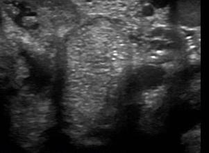
7 Ultrasound Appearance
8 MRI Gold standard for soft tissue injuries In human orthopedics, magnetic resonance (MR) is the imaging modality of choice because of image reproducibility and minimal operator dependence The fibrous nature and low water content of normal equine tendon generates a low signal intensity


9 MRI fundamentals

10 T1 weighted Good anatomic detail High SNR Low artifact susceptibility Used with contrast agents such as gadolinium Fluid is hypo-intense (gray to black)


11 T2 weighted Shows pathology Best for fluid and soft tissue contrast SNR lower than T1 Higher artifact susceptibility Fluid is hyper-intense (white)
 Fat suppression Lower")

12 STIR (Short Time Inversion Recovery) Fat suppression Lower SNR Separates fat from pathology Bone edema Fluid is hyper-intense (white)

13 Lesions

14 Goal 2 weeks 16 weeks

15 Materials and Methods Collagenase inj. In SDFT Time 0 Staggered injections of HL SDFTs Euthanasia 16 wks Ultrasonographic and MRI assessments carried out at the end of the study Statistical Analysis- 2 way repeated measure ANOVA
 LH at 8 weeks (N=2) Total x-sectional area in MIZ T1, T2, STIR Total x-sectional area Signal")
16 Materials and Methods RF and LF 16 weeks (N=8) LH at 3 weeks (N=2) LH at 4 weeks (N=2) LH at 6 weeks (N=2) LH at 8 weeks (N=2) Total x-sectional area in MIZ T1, T2, STIR Total x-sectional area Signal intensities

17 Lesion Example


18 Collagen Content and Alignment Normal Tendon Injured Tendon


19 Proteoglycan Content and Alignment Normal Tendon Injured Tendon

20 MR Cross-sectional Area MR Signal Intensity
21 Results: Signal Intensities (SI) * T1 SI levels were the highest, T2 SI levels were the lowest, and STIR SI was intermediate * There was no significant difference over time.
22 Results: T1-Weighted Signal Intensities (SI) * T1 SI levels were significantly increased over the normal deep digital flexor tendon at all time points.

23 Results: STIR Signal Intensities (SI) * STIR SI levels were significantly increased over the normal deep digital flexor tendon at all time points.
24 Results: T2-Weighted Signal Intensities * T2 SI levels were equivocal over the normal deep digital flexor tendon at all time points.


25 Results (x-sectional area) % TCSA in tendon injuries at < 4 weeks P=0.81 US T1 T2 STIR weeks
")


26 Results (x-sectional area) % TCSA in tendon injuries at 16 weeks P<0.001* US T1 T2 STIR * * * 16 weeks
27 Discussion In chronic injuries, ultrasonography and MRI indicated markedly different areas of injury during healing. US <<< MRI T1 sequence generated the highest signal intensity. The T1 and STIR signals remained elevated throughout the 16 weeks of the repair process. T2 data were equivocal. Subjectively, histological evidence of injury is more consistent with the MRI data.
28 Clinical Implications Ultrasonography still has a critical role in diagnosis and management of tendon injury. Interpret ultrasound findings during healing with caution. Off-angled views may enhance subtle lesions. Look for evidence of increased size in chronic injuries. Improvement in Fiber alignment score (60%)*, Type score (70%), Lesion size (50%), TCSA (50%) Genovese, unpublished data MRI guidelines? T2 weighted images should be normal or significantly improved
29 Case 1 4 yo, STB, Gelding 2/5 RF lameness Sore to palpation of SL and Inferior Check Lig Carpus injections helped for short duration Blocking PD- N/C Abaxial- 20% improvement Low and High 4 point- N/C
30 Case 1 Nuclear Scintigraphy IRU- R carpus, mild uptake L carpus U/S- MRI-
31 Case 1 Nuclear Scintigraphy IRU- R carpus, mild uptake L carpus U/S- irregularity to the fiber pattern of the check ligament MRI- Fiber disruption of the junction of the distal palmar ligament and the inferior check lig


32 Case 1


33 Case 1
34 Case 2 12yo, Quarter Horse, Gelding incr digital pulse 3/5 RF Distal limb flexion + Blocks to a PD
35 Case 2 U/S- core lesion in the medial lobe of the DDFT MRI- severe navicular changes and increased signal in the medial lobe of the DDFT


36 Case 2
37 Case 3 15 yo, Irish Cob, Gelding LH- grade 2/5- several month hx Very positive to flexion blocked to a low-4 point
38 Case 3 U/S- enlarged Lat SL Branch MRI-


39 Case 3
40 Case 4 12 yo, Oldenberg, Mare LH- grade 2+/5 Positive to digit flexion Improved with analgesia of digital tendon sheath Moderate to severe effusion
41 Case 4 U/S- adhesions and possible lesion of he DDFT MRI Thickening of the Manica Flexoria DDFT


42 Case 4
43 Case 5 10 yo, draft cross, mare 8 month history of LF lameness Grade 2/5 3+/5 circle to the left 70% improvement with abaxial Remainder with proximal suspensory Initially a Nuc Scan no abnormalities
44 Case 5 U/S- Desmopathy of the straight sesamoidean MRI- Desmopahty of the SSL Navicular bone edema


45 Case 5


46 Case 5 T2 STIR
47 Case 6 7 yo, Warmblood, Mare 1 year hx of RF injury to the digital flexor tendon sheath Tenoscopy and annular ligament resection Still lame RF- grade 2/5 Blocks to tendon sheath
48 Case 6 U/S Core lesion in the SDFT Scar tissue within the digital tendon sheath MRI SDFT lesion Scar tissue and adhesions

49 Case 6


50 Case 6
51 Case 7 15 yo, Warmblood, gelding Several months of RH lameness 3+/5 Severely positive to digit flexion Low-4 point- 80% improvement High suspensory- switched
52 Case 7 U/S- Enlargement of the medial SL Branch and disruption of the fiber pattern MRI-


53 Case 7


54 Case 7

55 Case 7


56 Case 7

57 Case 7 Dorsolateral distal MTIII condylar absorption secondary to villonodular synovitis?

58 Tear of the proximal straight sesamoidean ligament Case 7


59 Subchondral bone plate demineralization Case 7

60 Questions?
Equine Lameness & Imaging Techniques
 Equine Lameness & Imaging Techniques Peter Heidmann DVM MPH Specialist in Equine Internal Medicine Montana Equine Medical & Surgical Center www.montanaequine.com 406-285-0123 Types of lameness Skeletal
Case Studies. A. Kent Allen, DVM LAMENESS AND IMAGING IN THE SPORT HORSE
 Case Studies A. Kent Allen, DVM Author s address: Virginia Equine Imaging, 2716 Landmark School Road, The Plains, VA 20198; e-mail: vaequine@aol.com. 2007 AAEP. 1. Case Study #1: Medial Collateral Desmitis
Equine Diagnostic Radiography & Ultrasonography
 Equine Diagnostic adiography & Ultrasonography Dr. ussell Tucker DACV Prof Emeritus, WSU 90 ~30 DLPMO ~ projects medial forward lateral backward ~30 PL to DM projection 1 EQUINE DISTAL LIMB TENDONS & LIGAMENTS
Natasha M. Werpy, DVM, Diplomate ACVR; Betsy Charles, DVM; Norm Rantanen, DVM, MS, Diplomate ACVR IMAGING
 Should I Throw Away My Ultrasound Machine Now That MRI Is Here? A Review of Ultrasound and MRI for the Diagnosis of Musculoskeletal Injury in the Equine Patient Natasha M. Werpy, DVM, Diplomate ACVR; Betsy
Proceedings of the 59th Annual Convention of the American Association of Equine Practitioners - AAEP -
 http://www.ivis.org Proceedings of the 59th Annual Convention of the American Association of Equine Practitioners - AAEP - December 7-11, 2013 Nashville, TN, USA Next Meeting : Dec. 6-10, 2014 - Salt Lake
EQUINE APPROACHES TO THE DISTENDED DIGITAL FLEXOR TENDON SHEATH ORTHOPAEDICS
 Dr Hans Wilderjans, Dipl ECVS Dierenkliniek De Bosdreef Spelonckvaart 46 9180 Moerbeke-Waas Belgium www.bosdreef.be info@bosdreef.be APPROACHES TO THE DISTENDED DIGITAL FLEXOR TENDON SHEATH During this
Musculoskeletal Ultrasonography
 Imaging Is Believing Musculoskeletal Ultrasonography Column Editor Anthony Pease, DVM, MS, DACVR North Carolina State University Digital imaging has revolutionized how veterinarians evaluate musculoskeletal
Treatment and Rehabilit Treatment and ation Plans for Various Musculoske Musculosk letal Conditions
 Treatment and Rehabilitation Plans for Various Musculoskeletal Conditions Indiana Association of Equine Practitioners November 2012 Duncan Peters DVM, MS Equine Lameness and Sports Medicine Michigan State
How to Perform and Interpret Navicular Bursography
 How to Perform and Interpret Navicular Bursography Tracy A. Turner, DVM, MS, Diplomate ACVS, ACVSMR The injection of contrast material into the navicular bursa, accompanied by a subsequent radiographic
Proceedings of the 56th Annual Convention of the American Association of Equine Practitioners - AAEP -
 http://www.ivis.org Proceedings of the 56th Annual Convention of the American Association of Equine Practitioners - AAEP - December 4-8, 2010 Baltimore, Maryland, USA Next Meeting : Nov. 18-22, 2011 -
What I Will Cover. Shock Wave Therapy. What are shock waves? What are shock waves? What are shock waves? What are shock waves?
 Shock Wave Therapy Sarah Matyjaszek Large Animal Surgery University of Florida April 25 th, 2009 What I Will Cover How they work Applications Research Case example Potential complications The future A
Case Study: the Efficacy of Equine Cymatherapy Bioresonance on a Superficial Digital Flexor Tendon Core Lesion of a Thoroughbred Racehorse Colt
 Case Study: the Efficacy of Equine Cymatherapy Bioresonance on a Superficial Digital Flexor Tendon Core Lesion of a Thoroughbred Racehorse Colt Keith Cooper Elizabeth Colorio Penny Jenks Case Study: the
Navicular Syndrome/Heel Pain
 Navicular Syndrome/Heel Pain Navicular Syndrome/Heel Pain Clinical signs: Forelimb lameness, intermittent, progressive and insidious onset, usually bilateral. Stumbling Pointing toes to relieve pressure
Proceedings of the 12th International Congress of the World Equine Veterinary Association WEVA
 www.ivis.org Proceedings of the 12th International Congress of the World Equine Veterinary Association WEVA November 2-5, 2011 Hyderabad, India Reprinted in IVIS with the Permission of WEVA Organizers
Proceedings of the 55th Annual Convention of the American Association of Equine Practitioners
 Close this window to return to IVIS www.ivis.org Proceedings of the 55th Annual Convention of the American Association of Equine Practitioners December 5 9, 2009, Las Vegas, Nevada Program Chair : Nathaniel
Proceedings of the 56th Annual Convention of the American Association of Equine Practitioners - AAEP -
 http://www.ivis.org Proceedings of the 56th Annual Convention of the American Association of Equine Practitioners - AAEP - December 4-8, 2010 Baltimore, Maryland, USA Next Meeting : Nov. 18-22, 2011 -
Clinical Case Series Adipose Derived Stem and Regenerative Cells for the Treatment of Equine Tendon and Ligament Injuries
 Clinical Case Series Adipose Derived Stem and Regenerative Cells for the Treatment of Equine Tendon and Ligament Injuries VetStem Biopharma, Inc., 12860 Danielson Court, Suite B, Poway, CA 92064 Abstract:
REPORTING SERVICE: XR
 REPORTING SERVICE: XR Report number: VETCT-70603 Report date: 04/04/2017 Referring Veterinarian: xxxxx Referring Practice: xxxxx Email address: xxxxx Owner: xxxx Patient: xxxx Species: Equine Breed: Belgian
Keith Cooper Elizabeth Bauer
 Case Study: the Efficacy of Equine Cymatherapy Bioresonance on Severe Disruption of the Superficial Digital Flexor Tendon (95% Involvement by Multiple Core Lesions) in a Thoroughbred Racehorse Keith Cooper
How to Perform a Modified Standing Deep Digital Flexor Tenotomy at the Level of the Proximal Interphalangeal Joint
 How to Perform a Modified Standing Deep Digital Flexor Tenotomy at the Level of the Proximal Interphalangeal Joint R. Wayne Waguespack, DVM, MS; and Fred Caldwell, DVM, MS A modified standing deep digital
Magnetic Resonance Imaging Findings in Horses With Recent and Chronic Bilateral Forelimb Lameness Diagnosed as Navicular Syndrome
 Magnetic Resonance Imaging Findings in Horses With Recent and Chronic Bilateral Forelimb Lameness Diagnosed as Navicular Syndrome Sarah N. Sampson, DVM; Robert K. Schneider, DVM, MS, Diplomate ACVS; and
The Role of Select Imaging Studies in the Lameness Examination
 The Role of Select Imaging Studies in the Lameness Examination M. J. Martinelli, DVM, PhD and Norm Rantanen, DVM, MS The physical examination and gait analysis are the most important aspects of a lameness
2 nd AAEP Foundation Lameness Research Workshop Oklahoma City, Oklahoma July 24-25, 2012
 2 nd AAEP Foundation Lameness Research Workshop Oklahoma City, Oklahoma July 24-25, 2012 On July 24-25, 2012, following the AAEP Focus meeting on hind limb lameness, an AAEP Foundation research panel met
 Mmmmmm Mmmmmm mmmmmmmmmmmmm mmmmmmm mmmmmmm MRI of Equine Stifle Injuries: A Review of the first 100 clinical cases Martin Waselau, Dr.med.vet., MS Diplomate ACVS, Diplomate ECVS Equine Hospital Aschheim,
Proceedings of the 59th Annual Convention of the American Association of Equine Practitioners - AAEP -
 http://www.ivis.org Proceedings of the 59th Annual Convention of the American Association of Equine Practitioners - AAEP - December 7-11, 2013 Nashville, TN, USA Next Meeting : Dec. 6-10, 2014 - Salt Lake
Performance Lameness in Reining Horses
 Performance Lameness in Reining Horses QuickTime and a TIFF (Uncompressed) decompressor are needed to see this picture. MPH Peter Heidmann DVM Specialist in Equine Internal Medicine Montana Equine Medical
Extracorporeal Shock Wave Therapy (ESWT) Jeff Blea, DVM Von Bluecher, Blea, Hunkin, Inc. Equine Medicine and Surgery Sierra Madre, CA
 Jeff Blea, DVM Von Bluecher, Blea, Hunkin, Inc. Equine Medicine and Surgery Sierra Madre, CA") Extracorporeal Shock Wave Therapy (ESWT) Jeff Blea, DVM Von Bluecher, Blea, Hunkin, Inc. Equine Medicine and Surgery Sierra Madre, CA History Shock waves used in human medicine as early as 1980s: Break
Extracorporeal Shock Wave Therapy (ESWT) Jeff Blea, DVM Von Bluecher, Blea, Hunkin, Inc. Equine Medicine and Surgery Sierra Madre, CA History Shock waves used in human medicine as early as 1980s: Break
Review of Principles and Clinical Applications of Magnetic Resonance Imaging in the Horse
 Review of Principles and Clinical Applications of Magnetic Resonance Imaging in the Horse Natasha M. Werpy, DVM; Charles P. Ho, MD, PhD; Chris E. Kawcak, DVM, PhD, Diplomate ACVS; Norman W. Rantanen, DVM,
Mike Scott - Suspensory Ligament Injuries: Advances in Diagnosis and Treatment
 Dr. Mike Scott Mike Scott graduated from WCVM in 1993 and then completed a 1-year internship in large animal medicine and surgery at the Ontario Veterinary College. He completed a 3-year residency in large
Magnetic Resonance Imaging of Equine Tarsal Disorders
 Equine Diagnostic Center Munich Magnetic Resonance Imaging of Equine Tarsal Disorders 34 Clinical Cases Martin Waselau, Dr. med. vet., MS Diplomate ACVS, Diplomate ECVS, FTA für Pferdechirurgie Equine
Quantitative Sonographic Assessment in the Clinical Management of Superficial Digital Flexor Injuries in Thoroughbred Racehorses
 Quantitative Sonographic Assessment in the Clinical Management of Superficial Digital Flexor Injuries in Thoroughbred Racehorses Ronald Genovese, VMD; Karen Longo, DVM; Brett Berthold, DVM; and Joan Jorgenson,
Navicular Bursa (fluid sack at back of bone) Pedal (coffin) bone Navicular bone
 Pedal (coffin) bone Navicular bone") NAVICULAR SYNDROME Navicular syndrome is one of the most common causes of intermittent forelimb lameness in horses. It is the inflammation or degeneration of the navicular bone and its surrounding structures
NAVICULAR SYNDROME Navicular syndrome is one of the most common causes of intermittent forelimb lameness in horses. It is the inflammation or degeneration of the navicular bone and its surrounding structures
Ultrasound: a valid method for detecting scar-tissue in the equine superficial digital flexor tendon?
 Ultrasound: a valid method for detecting scar-tissue in the equine superficial digital flexor tendon? Alice Chatham BSc (Hons) Equine Science & Kirsty McDonald MSc 16/09/2018 Content Introduction Why do
Lameness & Non- Surgical Therapies of the Equine Athlete
 Lameness & Non- Surgical Therapies of the Equine Athlete Mark T. Reilly, DVM, Dipl. ABVP (Equine) Linda J. Cimetti, DVM South Shore Equine Clinic & Diagnostic Center Lameness & Non-Surgical Therapy of
Proceedings of the 10th International Congress of World Equine Veterinary Association
 www.ivis.org Proceedings of the 10th International Congress of World Equine Veterinary Association Jan. 28 Feb. 1, 2008 - Moscow, Russia Next Congress: Reprinted in IVIS with the permission of the Conference
Review Articleeve_261
 472..485 472 EQUINE VETERINARY EDUCATION / AE / SEPTEMBER 2011 Review Articleeve_261 Nonseptic osteitis of the distal phalanx and its palmar processes S. Dyson Centre for Equine Studies, Animal Health
Osseous Trauma in the Fetlock Region of Mature Sports Horses
 Osseous Trauma in the Fetlock Region of Mature Sports Horses Sue J. Dyson, VetMB, PhD; and Rachel Murray, VetMB, PhD Osseous trauma of the fetlock is a potentially important cause of lameness in mature
Proceedings of the 55th Annual Convention of the American Association of Equine Practitioners
 Close this window to return to IVIS www.ivis.org Proceedings of the 55th Annual Convention of the American Association of Equine Practitioners December 5 9, 2009, Las Vegas, Nevada Program Chair : Nathaniel
How Useful Is Nuclear Scintigraphy in the Diagnosis and Management of Proximal Suspensory Desmitis in the Horse?
 How Useful Is Nuclear Scintigraphy in the Diagnosis and Management of Proximal Suspensory Desmitis in the Horse? Sue J. Dyson, VetMB, PhD; Jo S. Weekes, MSc; and Rachel Murray, VetMB, PhD In many horses
Some Carpal Lesions in the Non-racehorse
 Published in IVIS with the permission of the AAEP Close this window to return to IVIS Some Carpal Lesions in the Non-racehorse Sue Dyson, VetMB, PhD Author s address: Centre for Equine Studies, Animal
MR: Finger and Thumb Injuries
 MR: Finger and Thumb Injuries Laura W. Bancroft, M.D. Professor of Radiology University of Central Florida Florida State University Outline Normal anatomy of the fingers and thumb MR imaging protocols
Learning Objectives. 07 Aug 12. Article E-1. At the end of this section the learner will be able to:
 Module 1: Comparative Functional Anatomy and Biomechanics Article E-1 Learning Objectives At the end of this section the learner will be able to: Describe the bones of the equine thoracic Describe the
Dr. Chris Bell BSc, DVM, MVetSc, Dip ACVS* *Elders Equine Veterinary Service, Winnipeg, MB, Canada
 Use of a nutraceutical supplement (EXCEED 6 WAY ) Med Vet Pharmaceuticals (MVP) for daily joint support in horses with osteoarthritis Minimal risk field trial and survey. Introduction: Dr. Chris Bell BSc,
Topographic Description of Metacarpal Tendons and Ligaments of Anatoly Donkey by Ultrasonography and Introducing a New Ligament
 November 2015, Volume 12, Number 4 Topographic Description of Metacarpal Tendons and Ligaments of Anatoly Donkey by Ultrasonography and Introducing a New Ligament Mohammad Naser Nazem 1*, Sayed Mohsen
Anatomy Revision Papers. Part 4. Internal Structure of the Foot
 Anatomy Revision Papers Part 4 Internal Structure of the Foot Page 2 Page 3 Page 4 Page 5 Page 5 Page 6 Page 6 Page 6 Sagittal Section of the Foot Frontal Section of Foot Ligaments within the foot Corium
Why Talk About Technique? MRI of the Knee:
 Why Talk About Technique? MRI of the Knee: Part 1 - Imaging Techniques Mark Anderson, M.D. University of Virginia Health Sciences Center Charlottesville, Virginia Always had an interest teach our fellows
Impingement Syndromes of the Ankle. Noaman W Siddiqi MD 5/4/2006
 Impingement Syndromes of the Ankle Noaman W Siddiqi MD 5/4/2006 Ankle Impingement Overview Clinical DX Increasingly recognized cause of chronic ankle pain Etiology can be soft tissue or osseous Professional
Tendon & Ligament Injuries
 Tendon & Ligament Injuries DIAGNOSIS Diagnosis of tendon and ligament injuries is based primarily on clinical and ultrasound findings. The typical signs of injury are heat, swelling and pain when palpating
Proceedings of the 11th Annual Resort Symposium of the American Association of Equine Practitioners AAEP
 www.ivis.org Proceedings of the 11th Annual Resort Symposium of the American Association of Equine Practitioners AAEP January 25-28, 2009 - Gold Coast, Australia ACKNOWLEDGMENTS Dr. Stephen M. Reed, Educational
The Weight Bearing Function Of The Sole Anatomical Evidence
 The Weight Bearing Function Of The Sole Anatomical Evidence Michael T. Savoldi Farrier Emeritus Resident Farrier (Retired) W.K. Kellogg Arabian Horse Center Farrier Science Instructor Animal & Veterinary
Diagnosis of Conditions of the Fetlock
 Diagnosis of Conditions of the Fetlock W. Rich Redding, DVM, MS, DACVS Department of Clinical Sciences College of Veterinary Medicine North Carolina State University Raleigh, NC 27607 Introduction The
The use of computed tomography to diagnose bilateral forelimb tendon pathology in a horse with unilateral lameness
 bs_bs_banner EQUINE VETERINARY EDUCATION 1 Equine vet. Educ. (2014) ( ) - doi: 10.1111/eve.12166 Case Report The use of computed tomography to diagnose bilateral forelimb tendon pathology in a horse with
Use of Magnetic Resonance Imaging to Diagnose Oblique and Straight Distal Sesamoidean Ligament Desmitis
 Use of Magnetic Resonance Imaging to Diagnose Oblique and Straight Distal Sesamoidean Ligament Desmitis Sarah N. Sampson, DVM; Robert K. Schneider, DVM, MS, Diplomate ACVS; Russell L. Tucker, DVM, MS,
Applicability, Interests and Limitations of the Ultrasound Examination in the Equine Deep Digital Flexor Apparatus of the Thoracic Limb
 Bulletin UASVM, Veterinary Medicine 68(2)/2011 pissn 1843-5270; eissn 1843-5378 Applicability, Interests and Limitations of the Ultrasound Examination in the Equine Deep Digital Flexor Apparatus of the
DIAGNOSTIC ULTRASOUND D R. E R I C A J O H N S O N
 DIAGNOSTIC ULTRASOUND D R. E R I C A J O H N S O N ULTRASOUND BASICS Medical ultrasound machines generate and receive ultrasound waves Ultrasound waves are emitted from the peizolectric crystals of the
Dukes Vet Practice
 www.dukesvets.com Getting the best out of your horse Jim Dukes, BVM&S, MRCVS Response to treatment for lameness What do we know about lameness in horses? Our approach to treatment and effectiveness What
Key words: Laser, sprain, strain, lameness, tendon
 MLS Master Class - Veterinary Imaging Presented by CelticSMR Ltd Free Phone (UK): 0800 279 9050 International: +44 (0) 1646 603150 AUTHOR DETAILS Carl Gorman BVSc MRCVS PUBLISHER DETAILS Mike Howe B Vet
Imaging of Ankle and Foot pain
 Imaging of Ankle and Foot pain Pramot Tanutit, M.D. Department of Radiology Faculty of Medicine, Prince of Songkla University 1 Outlines Plain film: anatomy Common causes of ankle and foot pain Exclude:
Magnetic resonance imaging
 MRI, CTprovide early link to diagnosing injuries, disease in horses Selecting the proper modality will help equine practitioners identify medical problems that were previously difficult to pinpoint A B
Ultrasonographic Examination of Joints in Horses
 Ultrasonographic Examination of Joints in Horses J.-M. Denoix, DVM, PhD, Agrégé; F. Audigié, DVM, PhD The diagnosis of lesions can be made with ultrasonography in many painful joints with no abnormal radiographic
Clinical Examination, Differential Analgesia and Imaging Modalities for Investigation of Distal Limb Lameness
 Published in IVIS with the permission of the AAEP Close this window to return to IVIS Clinical Examination, Differential Analgesia and Imaging Modalities for Investigation of Distal Limb Lameness Michael
MR IMAGING OF THE WRIST
 MR IMAGING OF THE WRIST Wrist Instability Dissociative Pattern apparent on routine radiographs Non-dissociative Stress / positional radiographs Dynamic fluoroscopy during stress Arthrography MRI / MR arthrography
Monitoring tendon healing after highpower laser therapy with ultrasound
 Monitoring tendon healing after highpower laser therapy with ultrasound Dr. Jan-Hein Swagemakers Mathilde Pluim DVM Antonio Luciani DVM Tierklinik Lüsche GmbH Excellent accuracy (averaging less than 5%)
11/2/17. Lateral Collateral Complex Medial Collateral Complex Distal Tibiofibular Syndesmosis Spring Ligament
 Andrew J Grainger Leeds, UK Lateral Collateral Complex ial Collateral Complex Distal Tibiofibular Syndesmosis Spring Ligament Brief anatomy review Scan tips and tricks Pathological appearances andrewgrainger@nhs.net
Non Surgical Management of Soft Tissue Injuries. Megan LeFave, DVM cvma
 Non Surgical Management of Soft Tissue Injuries Megan LeFave, DVM cvma Non Surgical Management of Soft Tissue Injuries Biomechanical Principles Common front limb and hind limb injuries In hospital treatments
What does the application of a deep-penetrating therapeutic laser actually accomplish?
 Laser Therapy in Equine Practice Ron Riegel, DVM I have witnessed therapeutic lasers being used on the equine athlete since the 1970s. The lasers that were used throughout the 70s, 80s, and 90s were all
198 EQUINE VETERINARY EDUCATION Equine vet. Educ. (2001) 13 (4)
 13 (4)") 198 EQUINE VETERINARY EDUCATION Equine vet. Educ. (2001) 13 (4) 198-204 Tutorial Article Use of ultrasonography in the evaluation of joint disease in horses. Part 1: Indications, technique and examination
198 EQUINE VETERINARY EDUCATION Equine vet. Educ. (2001) 13 (4) 198-204 Tutorial Article Use of ultrasonography in the evaluation of joint disease in horses. Part 1: Indications, technique and examination
Focus on Hindlimb Lameness Oklahoma City, OK, USA 2012
 www.ivis.org Proceedings of the American Association of Equine Practitioners - Focus Meeting Focus on Hindlimb Lameness Oklahoma City, OK, USA 2012 Next Focus Meetings: August 4-6, 2013 - Focus on Dentistry
Scientific Papers. Tenoscopy of the Navicular Bursa: Endoscopic Approach and Anatomy
 Scientific Papers Tenoscopy of the Navicular Bursa: Endoscopic Approach and Anatomy Fabrice Rossignol, DVM, and Roland Perrin, DVM, Diplomate ECVS REFEREED SUMMARY Objectives: Our objectives were to describe
FieldStrength. Achieva 3.0T enables cutting-edge applications, best-in-class MSK images
 FieldStrength Publication for the Philips MRI Community Issue 33 December 2007 Achieva 3.0T enables cutting-edge applications, best-in-class MSK images Palo Alto Medical Clinic Sports Medicine Center employs
Surgical Management of Superficial Digital Flexor Tendinitis
 Surgical Management of Superficial Digital Flexor Tendinitis Michael W. Ross, DVM Superior check desmotomy and annular desmotomy, alone or in combination, are valuable adjunct procedures in the management
CARPAL SHEATH TENOSYNOVITIS: APPROACHES, NEW CONDITIONS, AND OUTCOMES
 CARPAL SHEATH TENOSYNOVITIS: APPROACHES, NEW CONDITIONS, AND OUTCOMES Alan J. Nixon, BVSc, MS, DACVS College of Veterinary Medicine, Cornell University, Ithaca, NY Key Points Carpal sheath effusion and
MRI of the Shoulder What to look for and how to find it? Dr. Eric Handley Musculoskeletal Radiologist Cherry Creek Imaging
 MRI of the Shoulder What to look for and how to find it? Dr. Eric Handley Musculoskeletal Radiologist Cherry Creek Imaging MRI of the Shoulder Benefits of Ultrasound: * Dynamic * Interactive real time
RESPONSE OF TWENTY-SEVEN HORSES WITH LOWER LEG INJURIES TO COLD SPA BATH HYDROTHERAPY
 Reviewed RESPONSE OF TWENTY-SEVEN HORSES WITH LOWER LEG INJURIES TO COLD SPA BATH HYDROTHERAPY E.R. Hunt MVSc, PhD, G. Dip. Ed (Tert) 188 SUMMARY The influence of hypertonic cold water (5-9 C) spa bath
Practical Reporting of Musculoskeletal Imaging Studies: MRI Elbow
 Practical Reporting of Musculoskeletal Imaging Studies: MRI Elbow James F Griffith History Where is pain located? For how long? Trauma if so, what and when Radiographers can get this info Grade. Don t
Definition of Anatomy. Anatomy is the science of the structure of the body and the relation of its parts.
 Definition of Anatomy Anatomy is the science of the structure of the body and the relation of its parts. Basic Anatomical Terms Anatomical terms for describing positions: Anatomical position: Supine position:
REFERRING FOR EQUINE MRI. Standing for Safety
 REFERRING FOR EQUINE MRI Standing for Safety WHY? WHAT S SO SPECIAL ABOUT MRI? Since the advent of MRI, much has been learned about the causes of equine lameness. From the previously under-diagnosed, such
17/10/2017. Foot and Ankle
 17/10/2017 Alicia M. Yochum RN, DC, DACBR, RMSK Foot and Ankle Plantar Fasciitis Hallux Valgus Deformity Achilles Tendinosis Posterior Tibialis Tendon tendinopathy Stress Fracture Ligamentous tearing Turf
Australian and New Zealand College of Veterinary Scientists. Fellowship Examination. Veterinary Radiology Paper 1
 Australian and New Zealand College of Veterinary Scientists Fellowship Examination June 2014 Veterinary Radiology Paper 1 Perusal time: Twenty (20) minutes Time allowed: Three (3) hours after perusal Section
Neonatal Orthopedic Conditions
 Neonatal Orthopedic Conditions Kyla Ortved, DVM, PhD, DACVS, DACVSMR kortved@vet.upenn.edu Learning Objectives Differentiate between the main equine pediatric orthopedic conditions Understand principles
AN ABSTRACT OF THE THESIS OF
 AN ABSTRACT OF THE THESIS OF Shannon Kelly Reed for the degree of Master of Science in Veterinary Science presented on June 17, 2008. Title: A Molecular and Morphologic Study of Idiopathic Fetlock Hyperextension
Equine cervical ultrasonography
 Equine cervical ultrasonography A final thesis Royal Veterinary and Agricultural University of Copenhagen Authors Lise C. Berg, DVM Jon V. Nielsen, DVM, MRCVS Tutor Assistant tutor Preben D. Thomsen, Dr.
Case study #11 Rt. knee
 The patient is a 55 year old female who presents with bilateral knee pain. Patient is a collegiate softball coach and has a very active lifestyle and career that is hampered by her chronic knee pain. She
How to Inject Bone Marrow Derived Mesenchymal Stem Cells Into Tendons and Ligaments
 How to Inject Bone Marrow Derived Mesenchymal Stem Cells Into Tendons and Ligaments Laurie R. Goodrich, DVM, PhD, Diplomate ACVS*; Natasha M. Werpy, DVM, Diplomate ACVR; Myra F. Barrett, DVM, MS, Diplomate
Partial avulsion of the ulnaris lateralis and enthesiopathy of the lateral epicondyle of the humerus in a thoroughbred race horse
 Ashton Irish Veterinary Journal (2018) 71:10 https://doi.org/10.1186/s13620-018-0120-6 CASE REPORT Partial avulsion of the ulnaris lateralis and enthesiopathy of the lateral epicondyle of the humerus in
Musculoskeletal Imaging What to order? Brian Cole, MD
 Musculoskeletal Imaging What to order? Brian Cole, MD my background: 1994 University of Illinois 1998 MD University of Illinois College of Medicine 1999-2003 Diagnostic Radiology Mayo Clinic 2004 Fellowship
Principles of Ultrasound. Cara C. Prideaux, M.D. University of Utah PM&R Sports Medicine Fellow March 14, 2012
 Principles of Ultrasound Cara C. Prideaux, M.D. University of Utah PM&R Sports Medicine Fellow March 14, 2012 None Disclosures Outline Introduction Benefits and Limitations of US Ultrasound (US) Physics
Accuracy of Point-of-Care Ultrasonography for Pediatric Ankle Sprain Injuries
 Accuracy of Point-of-Care Ultrasonography for Pediatric Ankle Sprain Injuries S Jones 1, K Colaco 2, J Fischer 2, J Stimec 2, C Kwan 2, K Boutis 2 1 Alder Hey Children s NHS Foundation Trust, Prescot Rd,
How to Use a Fetlock Support Brace to Manage Lacerations of Equine Flexor Tendons
 How to Use a Fetlock Support Brace to Manage Lacerations of Equine Flexor Tendons Canaan Whitfield-Cargile, DVM; Robin M. Dabareiner, DVM, PhD, Diplomate ACVS; and Don Sustaire, CJF Horses with flexor
Introduction to Ultrasound Examination of the Hand and upper
 Introduction to Ultrasound Examination of the Hand and upper Emil Dionysian, M.D. Ultrasound of upper ext. Upside Convenient Opens another exam dimension Can be like a stethoscope Helps 3-D D visualization
OTHER IMAGING TECHNIQUES AND THEIR ADDED VALUE TO DIAGNOSE ELBOW DYSPLASIA. I. Gielen, H. van Bree
 OTHER IMAGING TECHNIQUES AND THEIR ADDED VALUE TO DIAGNOSE ELBOW DYSPLASIA. I. Gielen, H. van Bree Department of Medical Imaging & Small Animal Orthopaedics. Faculty of Veterinary Medicine, Ghent University,
CLINICAL AND ULTRASOUND REPORT. Paco Boy Filly. 15 th April 2014 to 28 th July 2014
 CLINICAL AND ULTRASOUND REPORT Paco Boy Filly 15 th April 2014 to 28 th July 2014 1 Paco Boy Filly Clinical and Ultrasound Report Background This Filly sired by Paco Boy is an unraced two year old thoroughbred.
IN NATURE. What are SW?
 EXTRACORPOREAL SHOCK WAVE THERAPY (ESWT) Theory & Clinical Applications Examples of therapies performed with Duolith SD1 Vet, a new system by Storz Medical AG Stefano Tassan, Med Vet, Equicenter stefanotassan@tiscali.it
How to Take Radiographs of the Metacarpophalangeal/Metatarsophalangeal Joint (Fetlock Joint)
") How to Take Radiographs of the Metacarpophalangeal/Metatarsophalangeal Joint (Fetlock Joint) Joseph W. Morgan, DVM, Diplomate ACVS Author s address: 5366 Leestown Road, Lexington, KY 40511 8904; e-mail:
How to Take Radiographs of the Metacarpophalangeal/Metatarsophalangeal Joint (Fetlock Joint) Joseph W. Morgan, DVM, Diplomate ACVS Author s address: 5366 Leestown Road, Lexington, KY 40511 8904; e-mail:
Treatment, Rehabilitation and Reconditioning Physiology of Tissue Repair
 Treatment, Rehabilitation and Reconditioning Physiology of Tissue Repair PHYSIOLOGY OF TISSUE REPAIR Knowing when it is appropriate to begin rehabilitation and when it is acceptable to return to practice
HOW DO WE DIAGNOSE LAMENESS IN YOUR HORSE?
 HOW DO WE DIAGNOSE LAMENESS IN YOUR HORSE? To help horse owners better understand the tools we routinely use at VetweRx to evaluate their horse s soundness, the following section of this website reviews
Chapter 2 Pitfalls in Musculoskeletal Ultrasound
 Chapter 2 Pitfalls in Musculoskeletal Ultrasound Violeta Maria Vlad MD, PhD Introduction Taking a good ultrasound (US) picture is an art. Interpreting it is a science. This is in fact everything US is
Lameness in the Rodeo Horse
 Lameness in the Rodeo Horse Robert D. Lewis, DVM Author s address: Elgin Veterinary Hospital, Inc., P.O. Box 629, Elgin, Texas 78621. 2001 AAEP. The ever-increasing popularity of the rodeo has created
RECENT ADVANCES IN CLINICAL MR OF ARTICULAR CARTILAGE
 In Practice RECENT ADVANCES IN CLINICAL MR OF ARTICULAR CARTILAGE By Atsuya Watanabe, MD, PhD, Director, Advanced Diagnostic Imaging Center and Associate Professor, Department of Orthopedic Surgery, Teikyo
Continous Nerve Block of the Thoracic Limb in Horses
 Bulletin UASMV, Veterinary Medicine, 69(1-2)/2012 Print ISSN 1843-5262; Electronic ISSN 1843-5378 Continous Nerve Block of the Thoracic Limb in Horses Dalma CSIBI 1), L. OANA 1), Irina IRIMESCU 1), Ioana