Principles of Ultrasound. Cara C. Prideaux, M.D. University of Utah PM&R Sports Medicine Fellow March 14, 2012
|
|
|
- Shauna Spencer
- 6 years ago
- Views:
Transcription

1 Principles of Ultrasound Cara C. Prideaux, M.D. University of Utah PM&R Sports Medicine Fellow March 14, 2012
2 None Disclosures
3 Outline Introduction Benefits and Limitations of US Ultrasound (US) Physics Terminology Equipment Normal US appearance of structures Muscle, Tendon, Ligament, Bone, Cartilage, Nerve, Vessels Artifacts Conclusions
 in 1951")

4 Introduction Physiatrists involved in US > 5 decades Founded American Institute for Ultrasound in Medicine (AIUM) in 1951 Therapeutic US initially High frequency, real-time MSK US imaging 1980 s Now used by many physiatrists Diagnostic Interventional
5 Introduction What is MSK US? Use of high frequency sound waves (3-17 MHz) to image soft tissues and bony structures in the body for the purposes of diagnosing pathology or guiding interventional procedures Detailed images of MSK system Sub-millimeter images Higher resolution than MRI for superficial structures

 Ligament")

6 Introduction - Indications Diagnostic Tendon (tendinopathy, tears) Muscle (strains, contusions) Nerve (entrapment) Ligament (sprains) Joint (effusions) Interventional Injections Tenotomy Aspiration/lavage Biopsy


7 Benefits of MSK US Ability to image in real-time = hands-on, dynamic, fast Interactive allows feedback from patient Generally unaffected by metal artifacts No radiation to patient or provider Exam of contralateral limb for comparison High resolution Real-time guidance for interventional procedures Portable Relatively inexpensive
, scanning skills and interpretation Lack of current individual certification Equipment cost, quality")
8 Limitations of MSK US Limited field of view Detailed picture of relatively small area Limited penetration Lower resolution at greater depths Unable to penetrate bone Operator dependent Education (anatomy), scanning skills and interpretation Lack of current individual certification Equipment cost, quality variable
 =")
9 Ultrasound Physics Transducer (probe) Contains linear array of thin crystals (lead zirconate titanate) linked to the electrical system of the machine Machine Applies a rapidly alternating electrical current to the crystals vibration generate sinusoidal sound wave (mechanical energy) = Piezoelectricity
10 Ultrasound Physics The frequency and amplitude of the sound waves are determined by those of the electrical current used to stimulate the crystals and the material properties and thickness of the crystals A range of frequencies are produced (Bandwidth) There is a preferential frequency Higher frequency ~ higher resolution but increased scatter (reduced penetration)
 Sound energy transformed into electrical signal (Piezoelectricity) US machine records the amplitude of the beam")
11 Ultrasound Physics Sound requires a medium Pass through the coupling gel into the body Waves travel into body until they encounter an acoustic interface (change in the density of adjacent tissues) Part of the sound wave s energy is reflected back to the transducer (now a receiver) Sound energy transformed into electrical signal (Piezoelectricity) US machine records the amplitude of the beam and the depth of the reflecting structure computer software generates a 2-D black/white B-mode image of the body

12 Ultrasound Physics Echoes Perpendicular incidence Sound wave traveling perpendicular to the boundary of 2 media Reflected pulse - reflected directly back Transmitted pulse passes straight through Oblique incidence Sound wave NOT traveling perpendicular to the boundary of 2 media Some reflected back at an angle Some passes through refracted (transmission angle)
13 Ultrasound Physics Reflection Specular Reflection Reflection from a smooth surface Scattering Redirection of sound in several directions due to rough edges (torn tissue) or heterogeneous media Some of the sound is reflected back to the transducer (= backscatter) visualization of the media
 per centimeter (cm) traveled Dependent on the tissue Soft tissue attenuation / cm = 0.")
14 Ultrasound Physics Attenuation Weakening of sound amplitude and intensity as the wave propagates through a medium Absorption, Reflection, Scattering Decibels (db) Attenuation coefficient Reduction (in db) per centimeter (cm) traveled Dependent on the tissue Soft tissue attenuation / cm = 0.5 x freq (MHz) Higher frequency More attenuation

15 Ultrasound Physics Acoustic interface US waves are reflected at the interface of two different tissues, dependent on the properties of each tissue (impedance) Echogenicity = Ability to reflect sound waves back to transducer If reflects large amount of sound brighter Interface composed of very different tissues e.g. interface between bone and muscle (bone = bright) Less reflection darker More similar tissues Important point Images are based on the relative material properties of the tissue compared with adjacent regions
16 Acoustic Impedance Impedance = (density of medium) x (propagation speed) Important in determination of acoustic transmission and reflection at the interface of two materials with different impedances (impedance mismatch) the greater the mismatch the greater the reflection at the interface Material Density (kg/m 3 ) Velocity (m/s) Acoustic Impedence (kg/m 2 s) Air Water X 10 6 Fat X 10 6 Blood X 10 6 Muscle X 10 6 Bone X 10 6
17 US beam is filled with sound waves Beam narrows to ½ width and then widens Narrowest region of beam has most concentrated sound waves best resolution Near zone = proximal to narrowest region Far zone = distal to narrowest region Multiple focal zones reduce temporal resolution Focusing
18 Terminology - Echotexture Refers to the internal pattern of echoes Coarseness Homogeneity and heterogeneity


19 Terminology - Echogenicity It s all relative Take into context the surrounding tissues, not simply the individual properties of the structure Hyperechoic More sound waves reflected back Isoechoic Same amount of sound waves reflected back Hypoechoic Fewer sound waves reflected back Anechoic No sound waves reflected back


20 Terminology Longitudinal Parallel to the long axis of the structure Transverse Perpendicular to the long axis of the structure
21 Terminology

22


23 Equipment Optimize the image! Proper transducer selection Depth Structure of interest visible at bottom of screen Adjust the focal zones Should be at depth of structure being imaged Reduce # of focal zones to span the area of interest Gain Overall brightness of the echoes Time-gain Compensation Use if need to focus gain on structure being imaged


24 Doppler Power doppler Single color Not angle dependent Detects any movement More sensitive but less specific than Color doppler Good for: Slow flow, small vessels (i.e.: neovessels), deep vessels Color doppler 2-colors Angle dependent Detects directional movement More specific but less sensitive than Power doppler Good for: Detect flow characteristics (direction, speed)
25 Extended Field of View Panoramic/Extended Field of View Transducer is glided parallel to the scan plane along the structure of interest New echoes added to original image Allows production of pictures wider than the transducer face
26 Transducers Frequency Expressed in megahertz (MHz) High frequency Greater resolution, lower penetration (attenuation) Best for more superficial structures Low frequency Lower resolution, greater penetration For deeper structures

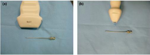
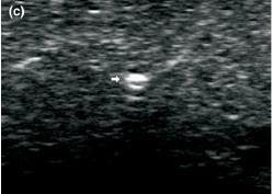
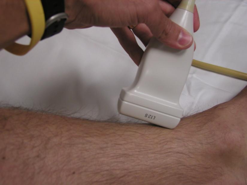
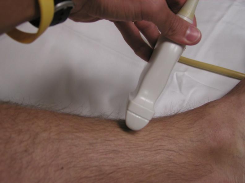
27 Transducers Linear array End of transducer flat Sound waves exit perpendicular Less anisotropy Limited field of view Good for superficial structures Curvilinear array End of transducer curved Sound waves emitted in a fan Increased anisotropy Large field of view Good for deep structures

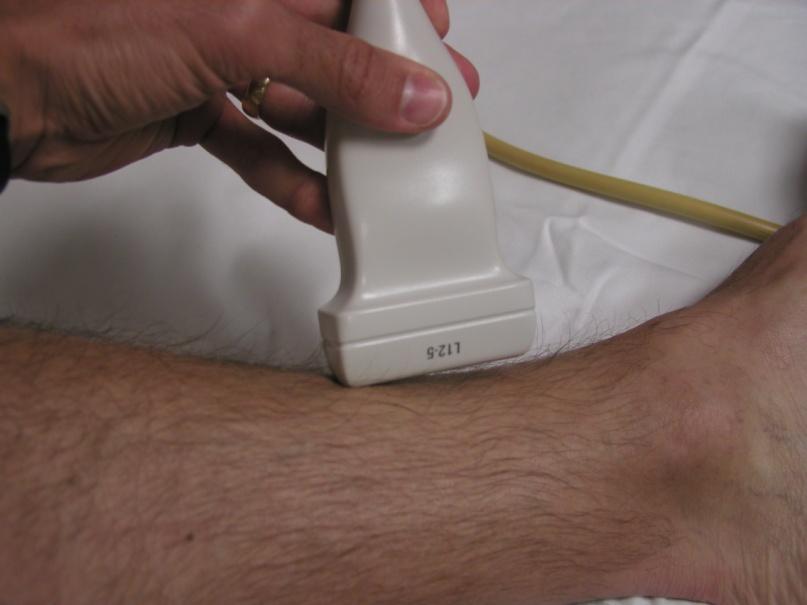
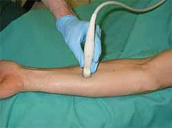
28 Transducer positioning Stabilize transducer with simultaneous contact with the transducer, skin surface, and examiner s hand maintains proper pressure of transducer on the skin avoids involuntary movement of the transducer allows fine adjustments in transducer positioning Full contact sport Jay Smith
29 Normal US Characteristics


30 Normal Appearance - Muscle Longitudinal Feather or veins on a leaf pattern Transverse Starry night pattern


31 Normal Appearance - Tendon Longitudinal Fibrillar appearance Transverse Broom end pattern


32 Normal Appearance - Ligament MCL Longitudinal Fibrillar MCL Transverse Broom end

33 Normal Appearance - Bone

34 Normal Appearance Hyaline Cartilage
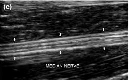
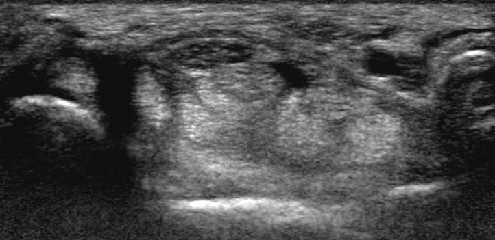

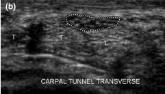
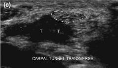
35 Normal Appearance - Nerve Longitudinal Fascicular Transverse Honeycomb pattern


36 Normal Appearance Vasculature Transverse - CPA Transverse Longitudinal - CPA

 Always look for pathology in")
37 Artifacts - Anisotropy Anisotropy US beam did not encounter the structure perpendicular to the plane of that structure but rather an oblique angle normal structure will appear artificially hypoechoic (dark) easily mistaken as pathology Can be avoided by continually manipulating the transducer to direct the US beam perpendicular to the structure (tilting, heel-toe) Always look for pathology in multiple planes

38 Artifacts - Anisotropy

39 Artifacts Posterior reverberation

40 Artifacts Posterior Acoustic Shadowing

41 Artifacts Posterior acoustic enhancement (increased through-transmission)
42 Artifacts Beam-width artifact If a structure is smaller than the ultrasound beam, artifact may be eliminated (e.g. small calcification with no posterior acoustic shadowing)
43 Conclusions Ultrasound has many uses for physiatrists There are many benefits and limitations of US Basic understanding of US physics is important Various tissues can be identified due to their anatomic location, echotexture, and echogenicity Always optimize your image Recognize common artifacts


44 References
Terminology Tissue Appearance
 By Marc Nielsen, MD Advantages/Disadvantages Generation of Image Ultrasound Machine/Transducer selection Modes of Ultrasound Terminology Tissue Appearance Scanning Technique Real-time Portable No ionizing
3/20/2017. Disclosures. Ultrasound Fundamentals. Ultrasound Fundamentals. Bone Anatomy. Tissue Characteristics
 Disclosures Images of ultrasound equipment in this presentation are not an endorsement Fundamentals of Musculoskeletal Ultrasound Physics and Knobology Shane A. Shapiro, M.D. Assistant Professor Orthopedic
Preamble (disclaimer)
") Preamble (disclaimer) PHYSICS AND PRINCIPLES OF HEAD/NECK ULTRASOUND Joseph C. Sniezek, MD FACS LTC, MC, USA Otolaryngology/H&N Surgery Tripler Army Medical Center 1. I am not a physicist 2. ACS has recommended
Preamble (disclaimer) PHYSICS AND PRINCIPLES OF HEAD/NECK ULTRASOUND Joseph C. Sniezek, MD FACS LTC, MC, USA Otolaryngology/H&N Surgery Tripler Army Medical Center 1. I am not a physicist 2. ACS has recommended
The Physics of Ultrasound. The Physics of Ultrasound. Claus G. Roehrborn. Professor and Chairman. Ultrasound Physics
 The Physics of Ultrasound Pipe Organ 10-8000 Emission Dog 452-1080 Man 85-1100 Spectrum Bat 10,000-120,000 Porpoise 7000-120,000 Claus G. Roehrborn Professor and Chairman 10 20 Cycles per second Reception
1 Fundamentals. Basic Definitions and Physics Principles. Fundamentals
 1 To become versed in the language of ultrasonography, it is necessary to review some of the basic principles of physics. The wave physics principles of ordinary (i.e., audible) sound apply to ultrasound
The Essentials Tissue Characterization and Knobology
 The Essentials Tissue Characterization and Knobology Randy E. Moore, DC, RDMS RMSK No relevant financial relationships Ultrasound The New Standard of Care Musculoskeletal sonography has become the standard
Ultrasound Physics & Doppler
 Ultrasound Physics & Doppler Endocrine University 2018 Mark Lupo, MD, FACE, ECNU Objectives Review the essential components of ultrasound physics in neck sonography Demonstrate the importance of ultrasound
Basic of Ultrasound Physics E FAST & Renal Examination. Dr Muhammad Umer Ihsan MBBS,MD, DCH CCPU,DDU1,FACEM
 Basic of Ultrasound Physics E FAST & Renal Examination Dr Muhammad Umer Ihsan MBBS,MD, DCH CCPU,DDU1,FACEM What is Sound? Sound is Mechanical pressure waves What is Ultrasound? Ultrasounds are sound waves
Ultrasound Physics and Knobology Alan Macfarlane. Consultant Anaesthetist Glasgow Royal Infirmary
 Ultrasound Physics and Knobology Alan Macfarlane Consultant Anaesthetist Glasgow Royal Infirmary RAPM 2009; 34: 40-46 Ultrasound Proficiency Understanding US image generation and device operation Image
Ultrasound Physics & Terminology
 Ultrasound Physics & Terminology This module includes the following: Basic physics terms Basic principles of ultrasound Ultrasound terminology and terms Common artifacts seen Doppler principles Terms for
Basic Physics of Ultrasound and Knobology
 WELCOME TO UTMB Basic Physics of Ultrasound and Knobology By Daneshvari Solanki, FRCA Laura B. McDaniel Distinguished Professor Anesthesiology and Pain Medicine University of Texas Medical Branch Galveston,
Diagnostic Ultrasound. Sutiporn Khampunnip, M.D.
 Diagnostic Ultrasound Sutiporn Khampunnip, M.D. Definition of Ultrasound Ultrasound is simply sound waves, like audible sound. High-frequency sound and refers to mechanical vibrations above 20 khz. Human
Introduction to Ultrasound Guided Region Anesthesia
 Introduction to Ultrasound Guided Region Anesthesia Brian D. Sites, MD Dept of Anesthesiology Dartmouth-Hitchcock Medical Center INTRODUCTION Welcome to Introduction to Ultrasound Guided Regional Anesthesia.
Ultrasound Principles cycle Frequency Wavelength Period Velocity
 ! Teresa S. Wu, MD, FACEP Director, EM Ultrasound Program & Fellowship Co-Director, Simulation Based Training Program & Fellowship Associate Program Director, EM Residency Program Maricopa Medical Center
Physical Principles of Ultrasound
 Physical Principles of Ultrasound Grateful appreciation to Richard A. Lopchinsky, MD, FACS and Nancy H. Van Name, RDMS, RTR, and MarleneKattaron, RDMS 2000 UIC All Rights Reserved. Course Objectives Identify
Ultrasonic Testing Level I:
 Ultrasonic Testing Level I: 1- Sound Wave - Introduction - ASNT Level I - Sound Wave Propagation - Velocity / Frequency / Wave Length - Acoustic Impedance - Energy / Intensity 2- Ultrasound Wave Modes
What is Ultrasound? What is Ultrasound? B A. Basic Principles of Ultrasound. Basic Principles of Ultrasound. Basic Principles of Ultrasound
 Introduction to Ultrasound Principles Mani Montazemi, RDMS Baylor College of Medicine Division of Maternal-Fetal Medicine Department of Obstetrics and Gynecology Manager, Maternal Fetal Center Imaging
Dr Emma Chung. Safety first - Physical principles for excellent imaging
 Safety first - Physical principles for excellent imaging Dr Emma Chung Lecturer in Medical Physics, University of Leicester Clinical Scientist, University Hospitals of Leicester NHS Trust Thanks to Caroline
Ultrasound Knobology
 Ultrasound Knobology Raj Dasgupta MD, FACP, FCCP, FASSM Assistant Professor of Clinical Medicine Pulmonary / Critical Care / Sleep Medicine University of Southern California (USC) Objectives Physics of
WELCOME! Introduction to Bedside Ultrasound
 WELCOME! Introduction to Bedside Ultrasound TEACHERS University of California-Irvine School of Medicine Nathan Molina nathan.d.molina@gmail.com Trevor Plescia taplescia90@gmail.com Jack Silva jpsilva42@gmail.com
Introduction & Physics of ED Ultrasound. Objectives. What? - Limited Studies. Who? - ED Docs
 Introduction & Physics of ED Ultrasound Martine Sargent, MD Ultrasound Director, Assistant Professor UCSF Department of Emergency Medicine San Francisco General Hospital & Trauma Center Objectives Who?
Lesson 03: Sound Wave Propagation and Reflection. This lesson contains 15 slides plus 14 multiple-choice questions.
 Lesson 03: Sound Wave Propagation and Reflection This lesson contains 15 slides plus 14 multiple-choice questions. Accompanying text for the slides in this lesson can be found on pages 8 through 14 in
DIGITAL IMAGE PROCESSING IN ULTRASOUND IMAGES
 DIGITAL IMAGE PROCESSING IN ULTRASOUND IMAGES Kamaljeet Kaur Computer Science & Engineering Department Guru Nanak Dev Engg. College, Ludhiana. Punjab-India meetk.89@gmail.com ABSTRACT-- Image processing
What is Ultrasound? Resolution Image production Attenuation Imaging modes Ultrasound artifacts... 7
 What is Ultrasound?... 1 Resolution... 3 Image production... 3 Attenuation... 4 Imaging modes... 5 Ultrasound artifacts... 7 0 What is Ultrasound? High frequency sound of frequencies 2-50 MHz is used in
Chapter 2 Pitfalls in Musculoskeletal Ultrasound
 Chapter 2 Pitfalls in Musculoskeletal Ultrasound Violeta Maria Vlad MD, PhD Introduction Taking a good ultrasound (US) picture is an art. Interpreting it is a science. This is in fact everything US is
Ultrasonography of the Neck as an Adjunct to FNA. Nicole Massoll M.D.
 Ultrasonography of the Neck as an Adjunct to FNA Nicole Massoll M.D. Basic Features of Head and Neck Ultrasound and Anatomy Nicole Massoll M.D. University of Arkansas for Medical Sciences, Little Rock
INTRODUCTION. Getting the best scan. Choosing a probe. Choosing the frequency
 Getting the best scan Choosing a probe Select the most appropriate probe for the particular scan required. s vary in their: operating frequency range higher ultrasound frequencies provide better discrimination
Sound in medicine. CH.12. Dr.Rajaa أ.م.د. رجاء سهيل جنم جامعة تكريت كلية طب االسنان. General Properties of Sound
 CH.12. Dr.Rajaa Sound in medicine أ.م.د. رجاء سهيل جنم جامعة تكريت كلية Sound : It is the audible waves of frequency between 20 Hz and 20 khz. Infrasound : refers to the sound of frequency below the normal
Table of contents. Foreword. Preface. 1 Introduction Historical Perspective 00
 Table of contents Foreword Preface 1 Introduction 00 1.1 Historical Perspective 00 2 Fundamentals of musculoskeletal ultrasound 00 2.1 Frequency and wavelength 00 2.2 Generating ultrasound waves 00 2.3
Ultrasound guidance in regional anesthesia has
 Ultrasound and Regional Anesthesia Artifacts and Pitfall Errors Associated With Ultrasound-Guided Regional Anesthesia. Part I: Understanding the Basic Principles of Ultrasound Physics and Machine Operations
Musculoskeletal Ultrasound Fundamentals
 Fundamentals Benjamin D. Levine, M.D. Associate Professor of Radiology Musculoskeletal Imaging Dept. of Radiological Sciences UCLA Health System I. Image Optimization II. Image Interpretation Artifacts
Supplement (videos)
") Supplement (videos) Ruben s tube (sound): http://www.youtube.com/watch?v=gpcquuwqayw Doppler US (diagnostic use): http://www.youtube.com/watch?v=fgxzg-j_hfw http://www.youtube.com/watch?v=upsmenyoju8 High
Supplement (videos) Ruben s tube (sound): http://www.youtube.com/watch?v=gpcquuwqayw Doppler US (diagnostic use): http://www.youtube.com/watch?v=fgxzg-j_hfw http://www.youtube.com/watch?v=upsmenyoju8 High
Ultrasound. Principles of Medical Imaging. Contents. Prof. Dr. Philippe Cattin. MIAC, University of Basel. Oct 17th, 2016
 Ultrasound Principles of Medical Imaging Prof. Dr. Philippe Cattin MIAC, University of Basel Contents Abstract 1 Image Generation Echography A-Mode B-Mode M-Mode 2.5D Ultrasound 3D Ultrasound 4D Ultrasound
Basic Ultrasound Physics Board Review Questions
 Basic Ultrasound Physics Board Review Questions Sidney K. Edelman, PhD ESP Ultrasound The Woodlands, TX Question 1 What is the wavelength of 2 MHz sound in soft tissue? 1. 1.54 mm 2. 0.75 mm 3. 0.75 cm
Point-of-Care Ultrasound: An Introduction
 Point-of-Care Ultrasound: An Introduction Delegation Teaching Package for Registered Respiratory Therapists and Anesthesia Assistants Developed by: Rob Bryan RRT, AA Edited by: Kelly Hassall RRT, FCSRT,
Background & Indications Probe Selection
 Teresa S. Wu, MD, FACEP Director, EM Ultrasound Program & Fellowship Co-Director, Simulation Based Training Program & Fellowship Associate Program Director, EM Residency Program Maricopa Medical Center
Ultrasound Guided Injections
 Ultrasound Guided Injection Technique More accurate injections Better Results! 1 Benefits: Increased Level of Certainty ie : really know how accurate PRP/Prolotherapy Avoid damage to articular cartilage
ULTRASOUND IMAGING EE 472 F2018. Prof. Yasser Mostafa Kadah
 ULTRASOUND IMAGING EE 472 F2018 Prof. Yasser Mostafa Kadah www.k-space.org Recommended Textbook Diagnostic Ultrasound: Physics and Equipment, 2nd ed., by Peter R. Hoskins (Editor), Kevin Martin (Editor),
ULTRASOUND. OB/Gyn (Core) Ultrasound PIEZOELECTRIC EFFECT. Principles of Ultrasound Physics and Instrumentation. Nathan Pinkney, BS, CDOS
 Ultrasound PIEZOELECTRIC EFFECT. Principles of Ultrasound Physics and Instrumentation. Nathan Pinkney, BS, CDOS") 1 OB/Gyn (Core) Ultrasound Principles of Ultrasound Physics and Instrumentation Nathan Pinkney, BS, CDOS Philadelphia College of Osteopathic Medicine 2016 ULTRASOUND CATEGORIES OF SOUND INFRASOUND = below
1 OB/Gyn (Core) Ultrasound Principles of Ultrasound Physics and Instrumentation Nathan Pinkney, BS, CDOS Philadelphia College of Osteopathic Medicine 2016 ULTRASOUND CATEGORIES OF SOUND INFRASOUND = below
ULTRASOUND NOMENCLATURE
 Chapter 1: Ultrasound Nomenclature, Image Orientation, and Basic Instrumentation CYNTHIA SIKOWSKI Ultrasound waves are sound waves that have a frequency exceeding 20,000 Hz. When sound waves are transmitted
Chapter 14. Imaging Artifacts
 Chapter 14 Image Artifacts The complex physical interactions that occur between an ultrasound beam and human anatomy and the intricate and sophisticated technological components of a sonographic imaging
Breast Imaging Essentials
 Breast Imaging Essentials Module 9 Transcript 2016 ASRT. All rights reserved. Breast Imaging Essentials Module 9 Breast Ultrasound 1. ASRT Animation 2. Welcome Welcome to Module 9 of Breast Imaging Essentials
Ultrasound in Anesthesia: Applying Scientific Principles to Clinical Practice
 AANA Journal Course Update for Nurse Anesthetists 3 6 CE Credits* Ultrasound in Anesthesia: Applying Scientific Principles to Clinical Practice Christian R. Falyar, CRNA, DNAP The use of ultrasound as
Musculoskeletal Ultrasound: Basics, Utility, and Clinical Applications
 Musculoskeletal Ultrasound: Basics, Utility, and Clinical Applications Andrew Lavigne, MD, FRCPC Physical Medicine and Rehabilitation CSCN Diplomat (EMG) Dip Sport Medicine Eugene Maida, MD, PGY-4 Resident
CSB 046 Complementary Imaging Techniques
 CSB 046 Complementary Imaging Techniques - Quizzes are only ultrasound, final includes nuc med and ultrasound Week 1 Intro to Ultrasound Physics - Uses 1 to 20 MHz frequencies, which is way above the sound
Application of Phased Array Radar Theory to Ultrasonic Linear Array Medical Imaging System
 Application of Phased Array Radar Theory to Ultrasonic Linear Array Medical Imaging System R. K. Saha, S. Karmakar, S. Saha, M. Roy, S. Sarkar and S.K. Sen Microelectronics Division, Saha Institute of
Introduction to Biomedical Imaging
 Alejandro Frangi, PhD Computational Imaging Lab Department of Information & Communication Technology Pompeu Fabra University www.cilab.upf.edu Basic principles. Comparison to X-rays Ultrasound > 20kHz
Lesson 07: Ultrasound Transducers. This lesson contains 73 slides plus 16 multiple-choice questions.
 Lesson 07: Ultrasound Transducers This lesson contains 73 slides plus 16 multiple-choice questions. This lesson was derived from pages 33 through 42 in the textbook: Ultrasound Transducers Ultrasound Transducers
Diploma of Medical Ultrasonography (DMU) Physical Principles of Ultrasound and Instrumentation Syllabus
 Physical Principles of Ultrasound and Instrumentation Syllabus") Diploma of Medical Ultrasonography (DMU) Physical Principles of Ultrasound and Instrumentation Syllabus Page 1 of 7 11/18 Candidates are expected to cover all of the content of this syllabus when preparing
Diploma of Medical Ultrasonography (DMU) Physical Principles of Ultrasound and Instrumentation Syllabus Page 1 of 7 11/18 Candidates are expected to cover all of the content of this syllabus when preparing
Thyroid Ultrasound Physics and Doppler
 Thyroid Ultrasound Physics and Doppler Advanced AACE-ACE US training course 2017 Dev Abraham MD, MRCP(UK), ECNU, FACE Professor of Medicine, University of Utah No Disclosures Natural Ability to see with
The 2 nd Cambridge Advanced Emergency Ultrasound Course
 The 2 nd Cambridge Advanced Emergency Ultrasound Course Addenbrooke s Hospital Cambridge Sept 2008 1 2 Faculty! UK! USA! Australia! Toshiba! Emergency Medicine! Radiology 3 Programme! Day 1 Introduction
CONTENTS. Test Number cpd Tanya Reynolds (Nat. Dip. Diag. Rad., B. Tech. Diag. Rad., B. Tech. Ultrasound)
") CONTENTS page 1-15 page 16 BASIC 2-DIMENSIONAL ULTRASOUND PRINCIPLES Multiple Choice Test Test Number cpd 41640 Tanya Reynolds (Nat. Dip. Diag. Rad., B. Tech. Diag. Rad., B. Tech. Ultrasound) Tanya is
CONTENTS page 1-15 page 16 BASIC 2-DIMENSIONAL ULTRASOUND PRINCIPLES Multiple Choice Test Test Number cpd 41640 Tanya Reynolds (Nat. Dip. Diag. Rad., B. Tech. Diag. Rad., B. Tech. Ultrasound) Tanya is
Introduction to Musculoskeletal Ultrasound. Disclosures. Evidence Based Medicine Key References 8/30/2017
 Introduction to Musculoskeletal Ultrasound Johannes Roth MD, PhD, FRCPC, RhMSUS Professor of Pediatrics University of Ottawa Gurjit S Kaeley MBBS, MRCP, RhMSUS Professor of Medicine Division Chief Director
An Overview of Ultrasound Testing For Lesion Detection in Human Kidney
 Journal of Tomography System & Sensors Application Vol.1, Issue 1, June 2018 An Overview of Ultrasound Testing For Lesion Detection in Human Kidney Aina Fadhilah Abd Rahim 1, Zawin Najah Abd Halim 1, Jaysuman
Descriptions of NDT Projects Fall 2004 October 31, 2004
 Descriptions of NDT Projects Fall 2004 October 31, 2004 Introduction There are two separate NDT labs in Magister: ULTRA for ultrasound and EDDY for eddy current. Both labs are equipped with mechanical
The Elbow 3/5/2015. The Elbow Scanning Sequence. * Anterior Joint (The anterior Pyramid ) * Lateral Epicondyle * Medial Epicondyle * Posterior Joint
 * Lateral Epicondyle * Medial Epicondyle * Posterior Joint") Scanning Sequence * Anterior Joint (The anterior Pyramid ) * Lateral Epicondyle * Medial Epicondyle * Posterior Joint Anterior Elbow Pyramid Courtesy of Jay Smith, MD. Vice chair PMR Mayo Clinic Rochester,
Scanning Sequence * Anterior Joint (The anterior Pyramid ) * Lateral Epicondyle * Medial Epicondyle * Posterior Joint Anterior Elbow Pyramid Courtesy of Jay Smith, MD. Vice chair PMR Mayo Clinic Rochester,
High resolution ultrasound scanner for skin imaging
 High resolution ultrasound scanner for skin imaging Christine Turlat Sales Director Atys medical 17 Parc d Arbora 69510 SOUCIEU EN JARREST Atys company Principle of ultrasound imaging DERMCUP Normal image
Pulse-Echo Ultrasound Imaging. Resolution in Ultrasound Imaging. Doppler Ultrasound. Resolution vs Penetration. Medical Imaging (EL582/BE620/GA4426)
") Medical Imaging (EL582/BE620/GA4426) Pulse-Echo Ultrasound Imaging Ultrasound Imaging Lecture 2 Daniel (Dan) Turnbull, Ph.D. Skirball Institute and Dept of Radiology NYU School of Medicine (daniel.turnbull@med.nyu.edu)
Medical Imaging (EL582/BE620/GA4426) Pulse-Echo Ultrasound Imaging Ultrasound Imaging Lecture 2 Daniel (Dan) Turnbull, Ph.D. Skirball Institute and Dept of Radiology NYU School of Medicine (daniel.turnbull@med.nyu.edu)
Carotid Abnormalities Coils, Kinks and Tortuosity David Lorelli M.D., RVT, FACS Michigan Vascular Association Conference Saturday, October 20, 2012
 Carotid Abnormalities Coils, Kinks and Tortuosity David Lorelli M.D., RVT, FACS Michigan Vascular Association Conference Saturday, October 20, 2012 Page 1 Table of Contents Carotid Anatomy Carotid Duplex
Basic Physics of Ultrasound in Transesophageal Echocardiography
 SPECIAL ARTICLE IJUTPC Basic Physics of Ultrasound in Transesophageal Echocardiography Basic Physics of Ultrasound in Transesophageal Echocardiography 1 Mary Korula, 2 Ravi Hebballi 1 Senior Consultant,
Underwater Acoustic Measurements in Megahertz Frequency Range.
 Underwater Acoustic Measurements in Megahertz Frequency Range. Current State and Prospects of Development in Russia Alexander M. Enyakov,, Many medical applications of underwater acoustic measurements
Abdominal Ultrasound
 Abdominal Ultrasound Imaging Control Buttons Depth The organ imaged should take up 3/4 of the screen Frequency = Penetration Use high frequencies (harmonics) for fluid filled and superficial structures
Proceedings of the 10th International Congress of World Equine Veterinary Association
 www.ivis.org Proceedings of the 10th International Congress of World Equine Veterinary Association Jan. 28 Feb. 1, 2008 - Moscow, Russia Next Congress: Reprinted in IVIS with the permission of the Conference
Concepts of Imaging and Knobology
 Concepts of Imaging and Knobology Pravin Patil, MD FACC FASE Associate Professor of Medicine Director, Cardiovascular Disease Training Program Lewis Katz School of Medicine at Temple University Disclosures
Ultrasonic Testing. Basic Principles
 Ultrasonic Testing Ultrasonic Testing (UT) uses high frequency sound waves (typically in the range between 0.5 and 15 MHz) to conduct examinations and make measurements. Besides its wide use in engineering
Roaa M.Hussein Professor, Department of Physics, College of Science, Ramadi, Iraq. Abstract:
 Abstract: Ultrasound Waves Employment in the Medical Diagnostic for Lıver and Gallbladder Faik H. Antar Professor, Department of Physics, College of Science, AL- Anbar University, Ramadi, Iraq. Ultrasound
Contents. Basic Ultrasound Principles and Terminology. Ultrasound Nodule Characteristics
 Contents Basic Ultrasound Principles and Terminology Basic Ultrasound Principles... 1 Ultrasound System... 2 Linear Transducer for Superficial Images and Ultrasound-Guided FNA... 3 Scanning Planes... 4
for the Veterinary Technician
 An Overview of Abdominal Ultrasound for the Veterinary Technician Valerie Gates, CVT, VTS (ECC) Learning Objective: The reader should gain a basic understanding of ultrasound, including physics, terminology,
Emergency Medicine Interest Group (EMIG) 2016
 2016") Emergency Medicine Interest Group (EMIG) 2016 Welcome to the flipped classroom (learning objectives summary) for the 2016 Emergency Medicine Interest Group (EMIG) Procedures Workshop. Overview - Tuesday
Emergency Medicine Interest Group (EMIG) 2016 Welcome to the flipped classroom (learning objectives summary) for the 2016 Emergency Medicine Interest Group (EMIG) Procedures Workshop. Overview - Tuesday
Chapter 3. Sonographic Image Interpretation
 Chapter 3 Sonographic Image Interpretation Sonograms are two-dimensional gray-scale images that allow assessment and diagnosis of many anatomic and pathologic changes that can occur in the human body.
MUSCULOSKELETAL ULTRASOUND
 MUSCULOSKELETAL ULTRASOUND A brief overview of diagnostic and therapeutic applications in musculoskeletal medicine. By Elmer G. Pinzon, MD, MPH and Randy E. Moore, DC, RDMS The use of musculoskeletal ultrasound
1. Fig. 1 shows data for the intensity of a parallel beam of X-rays after penetration through varying thicknesses of a material
 1. Fig. 1 shows data for the intensity of a parallel beam of X-rays after penetration through varying thicknesses of a material. intensity / MW m 2 thickness / mm 0.91 0.40 0.69 0.80 0.52 1.20 0.40 1.60
FAST Focused Assessment with Sonography in Trauma
 FAST Focused Assessment with Sonography in Trauma Wilma Rodriguez Mojica,MD,FACR Professor of Radiology UPR School of Medicine Ultrasound Section - Radiological Sciences Department OBJECTIVES Understand
1. SCOPE ELIGIBILITY EXAMINATION CONTENT RENEWAL & RECERTIFICATION PROCEDURE ESSENTIAL READING...
 Certification Services Division Newton Building, St George s Avenue Northampton, NN2 6JB United Kingdom Tel: +44(0)1604-893-811. Fax: +44(0)1604-893-868. E-mail: pcn@bindt.org PCN/GEN ISO 20807 Appendix
Flaw Assessment Using Shear wave Phased array Ultrasonic Transducer
 18th World Conference on Nondestructive Testing, 16-20 April 2012, Durban, South Africa Flaw Assessment Using Shear wave Phased array Ultrasonic Transducer Byungsik YOON AUTHOR 1, Hee-Jong LEE CO-AUTHOR
Ultrasound Applied Physics
 Ultrasound Applied Physics University of Toronto Department of Medical Imaging Applied Physics Mini-Course #3 2016 Ultrasound Laboratory Manual and Examination Booklet 1/21/2016 Ultrasound Applied Physics
Physical Principles of Ultrasound
 Physical Principles of Ultrasound Pat F. Fulgham 2 Introduction The use of ultrasound is fundamental to the practice of urology. In order for urologists to best use this technology on behalf of their patients,
4.17. RESEARCHING MODELS WITH AN ULTRASONIC ECHOSCOPE
 4.17. RESEARCHING MODELS WITH AN ULTRASONIC ECHOSCOPE Purpose of experiment Determine the main characteristics of ultrasound waves, and the distances and positions of models using an ultrasonic echoscope.
Focused Musculoskeletal Ultrasound
 Focused Musculoskeletal Ultrasound David Lewis Consultant Emergency Medicine Ipswich (Club Doctor, Ipswich Town FC) Advanced Emergency Ultrasound Objectives! General principles! Musculoskeletal anatomy!
Ultrasound in Peripheral Nerve Interventions
 Ultrasound in Peripheral Nerve Interventions John L. Lin, M.D. Shepherd Center Assistant Clinical Professor Emory University, School of Medicine Outline Ultrasound basics Nerve blocks in physiatric setting
Chapter 1. Principles of medical ultrasound. Overview. Background history: first steps to the piezo-electric effect.
 Chapter 1 Principles of medical ultrasound GRAHAM ARTHURS, PATRICK HILL AND TREVOR FRANKEL Overview This chapter provides an introduction to the ultrasound process for trainees in anesthesia and other
Pragmatic ultrasound in the diagnosis of soft tissue rheumatic pain. Plamen Todorov
 Pragmatic ultrasound in the diagnosis of soft tissue rheumatic pain Plamen Todorov INTRODUCTION Soft tissue rheumatism: nonsystemic, focal pathological syndromes involving the periarticular structures.
Ultrasound Guidance Needle Techniques
 Ultrasound Guidance Needle Techniques Dr TANG Ho-ming AED/UCH USG Guidance Needle Techniques Commonly used in EM 1. Vessel cannulation-peripheral & central 2. Foreign body removal 3. Peripheral nerve/plexus
Ultrasound in Sports Medicine
 Ultrasound in Sports Medicine CASES AND USES T I F FA N Y T S AY, M D T O W S O N O R T H O PA E D I C A S S O C I AT E S T H E P R I M A R Y C A R E A P P R O A C H T O T R E AT I N G T H E I N J U R
Image optimization for critical care US
 Image optimization for critical care US 1 Although we assume you are already familiar with focused US in the ED, it might not hurt to revise the basics: Machines & transducers US appearance of normal tissues
4.17. RESEARCHING MODELS WITH AN ULTRASONIC ECHOSCOPE
 4.17. RESEARCHING MODELS WITH AN ULTRASONIC ECHOSCOPE Purpose of experiment Determine the main characteristics of ultrasound waves, and the distances and positions of models using an ultrasonic echoscope.
The table below shows the density and velocity of waves in two different substances. Density / kg m 3 Velocity / m s 1
 Q1.(a) When ultrasound is incident at an interface between two different media some energy is transmitted and some is reflected. The ratio of the reflected energy intensity I r to the incident energy intensity
Routine Quality Assurance Cookbook
 This Cookbook is a companion guide to the AIUM Routine Quality Assurance (QA) for Diagnostic Ultrasound Equipment document, which outlines the basic QA requirements for AIUM-accredited practices. The Guide
Ultrasound: Past and Present. Lecturer: Dr. John M Hudson, PhD
 Ultrasound: Past and Present Lecturer: Dr. John M Hudson, PhD Disclosures 2 No conflicts of interest to declare Course Outline 3 1. Survey of ultrasound physics & applications 2. (Sep 21) 3. (Sep 28) 4.
Basics of US Regional Anaesthesia. November 2008
 Basics of US Regional Anaesthesia November 2008 Essential Physics HIGH frequency = great resolution but poor penetration LOW frequency = poor resolution but great penetration Potential Advantages of US
Physics. Norman McDicken Tom Anderson CHAPTER ULTRASOUND. Ultrasound Propagation
 CHPTER 2 Physics Norman McDicken Tom nderson This chapter provides an introduction to the physics of medical ultrasound (US). Several books exist that can be consulted to extend the material presented
Chapter 3 Physical Principles of Ultrasound of the Male Genitalia
 Chapter 3 Physical Principles of Ultrasound of the Male Genitalia Bruce R. Gilbert and Pat Fox Fulgham Introduction The value of ultrasound evaluation of the male genitalia depends, in large part, on the
Learning Objectives. Ultrasound for the Primary Care Provider. Portable Ultrasound: Laptops, Tablets, Plug-in Probes, and Pocket devices
 Learning Objectives Ultrasound for the Primary Care Provider Richard Hoppmann, MD, FACP University of South Carolina School of Medicine Assess the main components and functions of a portable ultrasound
Linear Ultrasonic Wave Propagation in Biological Tissues
 Indian Journal of Biomechanics: Special Issue (NCBM 7-8 March 29) Linear Ultrasonic Wave Propagation in Biological Tissues Narendra D Londhe R. S. Anand 2, 2 Electrical Engineering Department, IIT Roorkee,
Basic Training Programme. 16 Februrary 2018, ROTTERDAM. Pre and Post-Course Test Answers
 Basic Training Programme 16 Februrary 2018, ROTTERDAM Pre and Post-Course Test Answers Your details: Name: Conference registration number/ BT delegate number: Email address: Are you already performing
Category Term Definition Comments 1 Major Categories 1a
 Working Lexicon Categories, Terms & Definitions Category Term Definition Comments 1 Major Categories 1a Physiologic Category (consistent with normal ovarian physiology) Follicle Simple 3 cm in premenopausal
How do you use the AiM Vascular Mapping Kit?
 How do you use the AiM Vascular Mapping Kit? Protocol for Successful Skin Mapping Evaluate vessel anatomy, perform measurements, etc. according to your laboratory s protocol prior to applying the AiM device
ULTRASOUND QA SOLUTIONS. Ensure Accurate Screening, Diagnosis & Monitoring DOPPLER FLOW PHANTOMS MULTI-PURPOSE PHANTOMS TRANSDUCER TEST PHANTOMS
 ULTRASOUND QA SOLUTIONS Ensure Accurate Screening, Diagnosis & Monitoring DOPPLER FLOW PHANTOMS MULTI-PURPOSE PHANTOMS TRANSDUCER TEST PHANTOMS INNOVATORS IN ADVANCED ULTRASOUND TECHNIQUES Gammex is the
Knobology for Dummies
 Knobology for Dummies Power On/Off Preset button Patient Information Entry Choose preset Transducer probes Connect and disconnect transducer Approach to the patient (machine placement, comfort, draping,
ULTRASOUND QA SOLUTIONS. Ensure Accurate Screening, Diagnosis & Monitoring DOPPLER FLOW PHANTOMS MULTI-PURPOSE PHANTOMS TRANSDUCER TEST PHANTOMS
 ULTRASOUND QA SOLUTIONS Ensure Accurate Screening, Diagnosis & Monitoring DOPPLER FLOW PHANTOMS MULTI-PURPOSE PHANTOMS TRANSDUCER TEST PHANTOMS INNOVATORS IN ADVANCED ULTRASOUND TECHNIQUES Gammex is the