Practical Reporting of Musculoskeletal Imaging Studies: MRI Elbow
|
|
|
- Bryce Wilson
- 5 years ago
- Views:
Transcription
1 Practical Reporting of Musculoskeletal Imaging Studies: MRI Elbow James F Griffith

2 History Where is pain located? For how long? Trauma if so, what and when Radiographers can get this info
3 Grade. Don t mention any feature without grading it Qualitative measure : Minimal, mild, moderate, severe Quantitative measure: Small, medium, large (mm long x mm deep x mm wide)
4 This talk : outline Epicondylitis Cubital tunnel syndrome Trauma inc biceps insertion
5 Epicondylitis Tendinosis of common extensor tendon origin (CETO) Tendinosis of common flexor tendon origin (CFTO)


6 Epicondylitis : Pathophysiology Cumulative microtrauma Inadequate repair Tendinosis (collagen disruption, proteoglycan deposition, vascular ingrowth, fibroblast proliferation, hyaline degeneration)

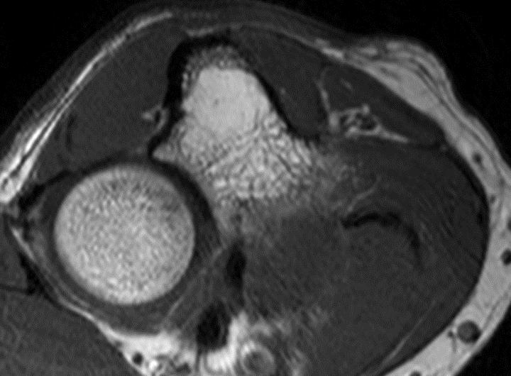
7 Epicondylitis : Why imaging Firm up diagnosis Establish severity Identify tear Check for associated abnormalities Peritendinitis Collateral ligament injury Bone oedema, cortical irregularity
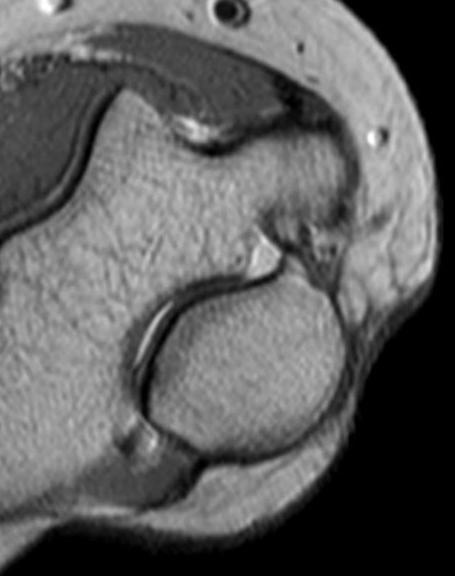
8 Common extensor tendon origin
9 Common extensor tendon origin (CETO) ECRL ECRBr ** ED * ECU ** Possibly due to impingement against capitellum
10 Lateral collateral ligament complex Radial collateral ligament Lateral ulnar collateral ligament
11


12 MR imaging protocol CETO PD axial PD cor CFTO T2-SPAIR axial T2 SPIR obl cor + sagittal images

13 CETO normal Some mild heterogeneity is normal

14 CETO tendinosis: moderate Normal Moderate severity
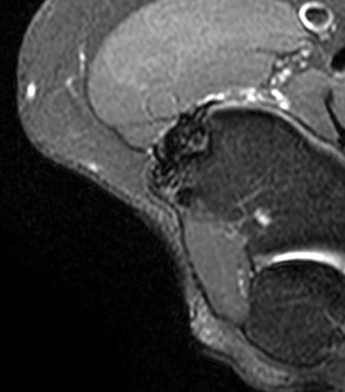
15 CETO tendinosis: moderate Moderate.with tear
16 Lateral collateral ligament complex Radial collateral lig (RCL) Lateral ulnar collateral lig (LUCL)

17 Lateral collateral ligament complex RCL LUCL


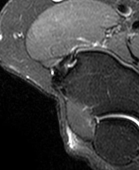
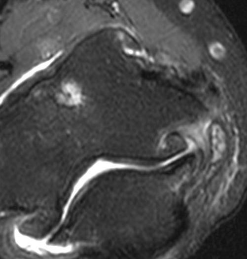
18 CETO tendinosis : moderate Intact RCL Tear CETO Intact LUCL There is moderate CETO tendinosis with a medium-sized (??mm wide x?? mm long) mainly involving the ECRBr Insertional area. The RCL and LUCL are intact


19 Avulsion ECRBr > ECRL >>> ED & ECU ECRBr avulsion ECRL and ECRBr avulsion


20 Avulsion ECRBr > ECRL >>> ED & ECU ECRBr avulsion ECRL and ECRBr avulsion


21 Complete tear CETO and RCL There is moderate CETO tendinosis with a complete avulsivetype tear of the CETO as was as the RC and LUC ligaments


22 Complete tear CETO, RCL and LUCL RCL LUCL

 Place transducer along")
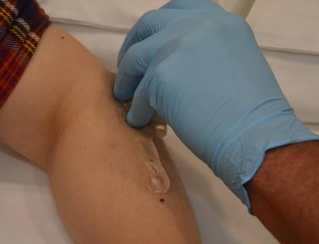
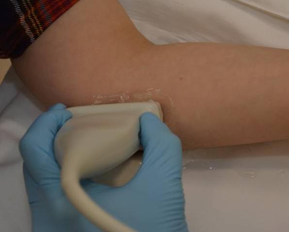
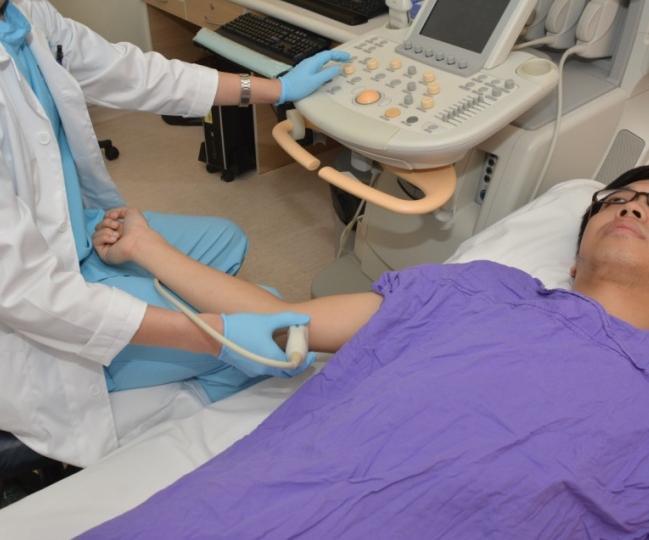
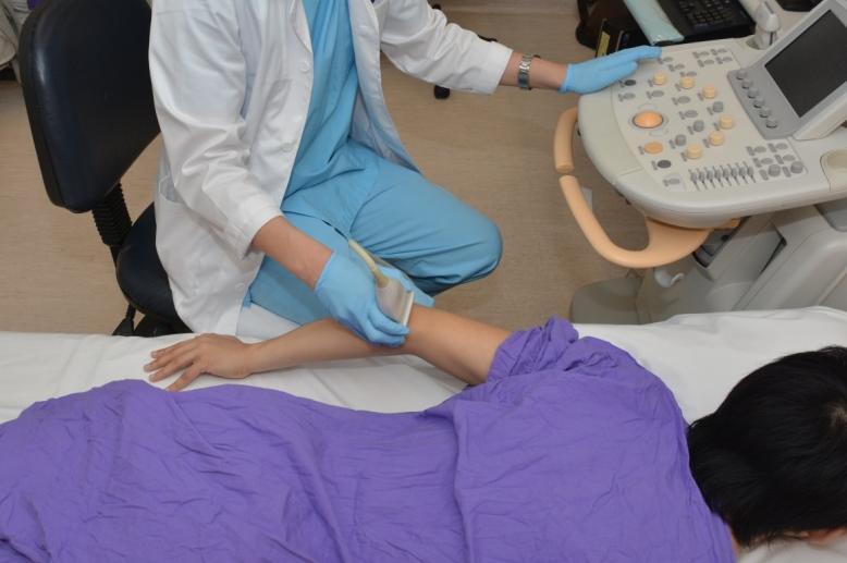
23 Ultrasound protocol : lateral (1) Put finger on lateral epicondyle (2) Imagine line of extensor tendons (3) Place transducer along this line

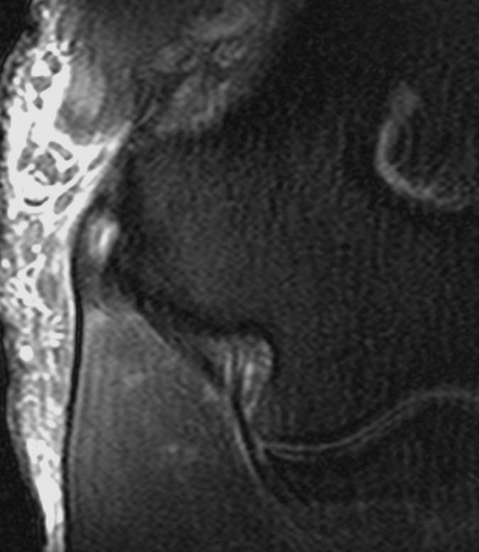
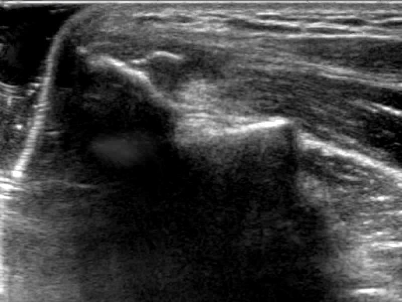
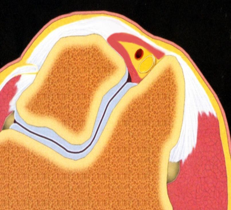
24 Ultrasound protocol: CETO

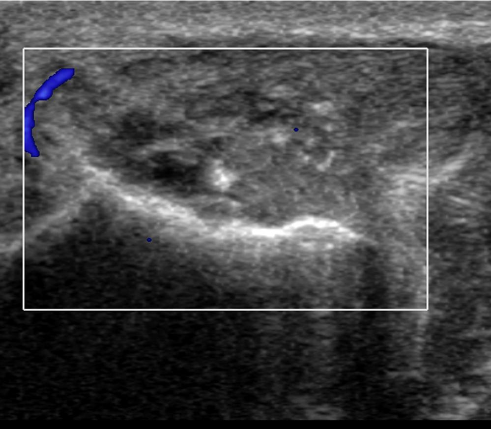
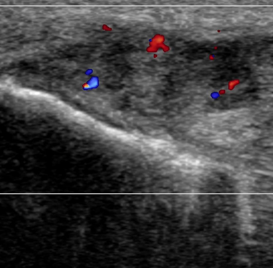
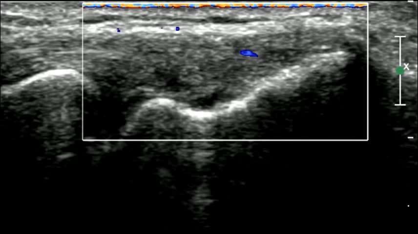
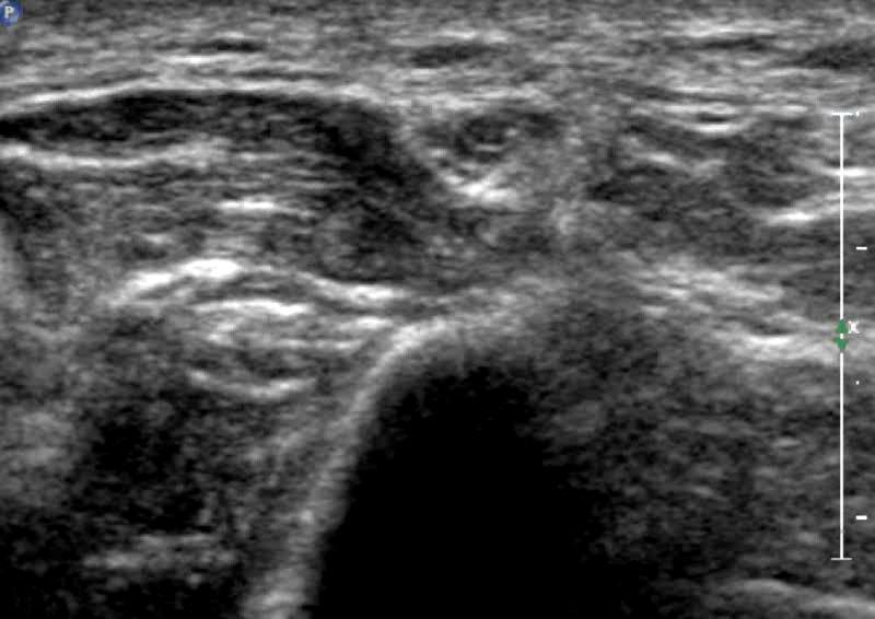
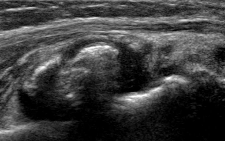
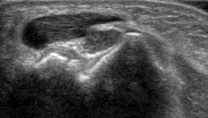
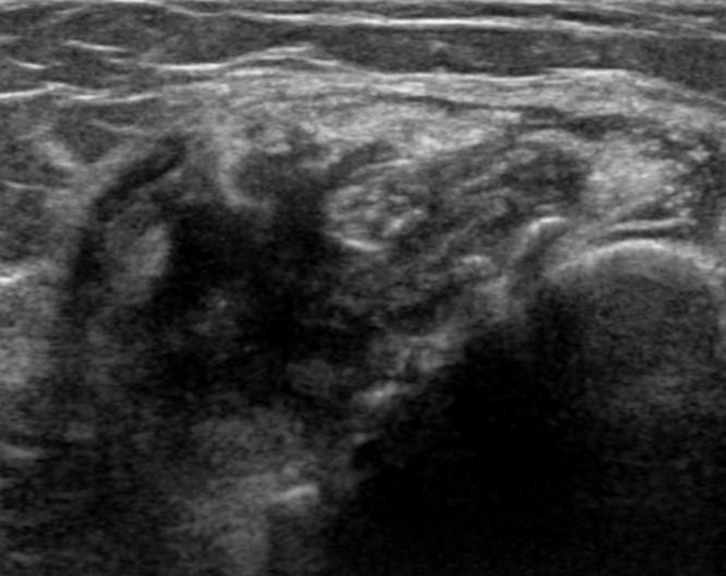
25 Thickening Hypoechogenicity Calcification Tear Cortical irregularity Hyperaemia CETO tendinosis

26 Thickening Hypoechogenicity Calcification Tear Cortical irregularity Hyperaemia CETO tendinosis
27 CETO tendinosis
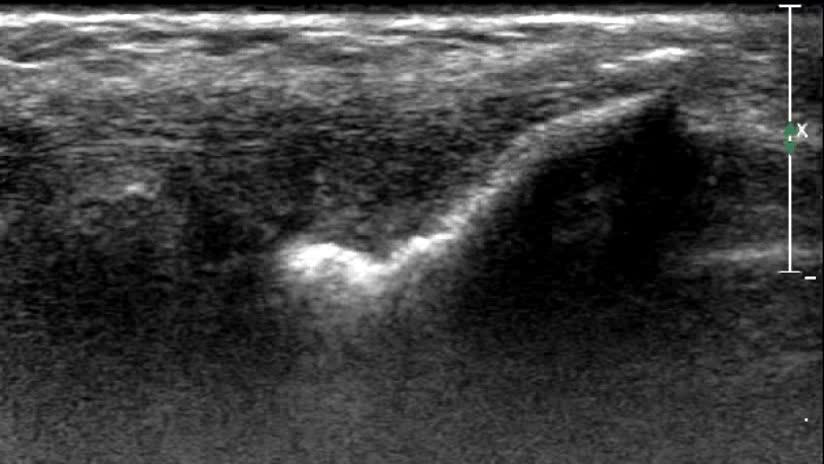
28 CETO tendinosis Lift the transducer off skin


29 CETO tendinosis Normal < 32mm 2 Tendinosis >32mm 2 Baumer P et al 2011


30 Radial synovial fold syndrome No fold Thickened synovial fold but not impacted Impacted synovial fold
31 Common flexor tendon origin (CFTO) PT * FCR * PL FCU FDS
32 Anterior band Transverse band Posterior band Medial collateral ligament

33 Medial collateral ligaments Anterior band Transverse band Posterior band

34 CFTO normal


35 CFTO normal and moderate tendinosis Normal Moderate tendinosis

36 CETO tendinosis and tear

 Place transducer")
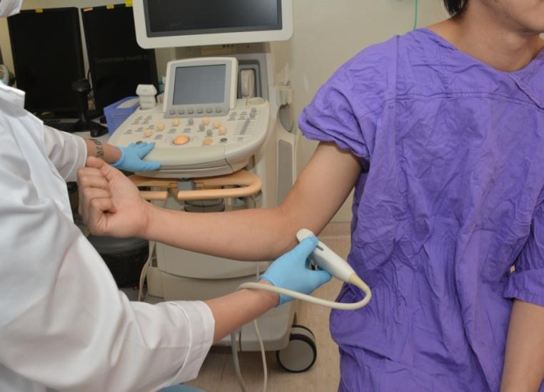
37 Ultrasound protocol : medial (1) Put finger on medial epicondyle (2) Imagine line of flexor tendons (3) Place transducer along this line

38 Ultrasound protocol: CFTO
39 Ultrasound protocol: CFTO
40 US or MR examination? Tendinosis severity Tear depiction Hyperaemia Associated abnormality Operator independent 3rd party review US MRI
41 Epicondylitis : US and MR examination 1/6 have a normal US Represents early non-established disease Proceed to MRI 50% of these will still have normal MRI examination
 Autologous blood injection Botox")
42 Treatment (i) Rest, bracing and physiotherapy Don t use steroids Dry needling Platelet Rich Plasma (PRP) Autologous blood injection Botox
43 Treatment (ii) Extracorporal shock wave therapy Low level laser therapy Surgery Debridement, drilling Release (percutaneous, arthroscopic, open) Suture fixation
44 Epicondylitis : summary Both US and MRI extremely helpful at assessing lateral & medial epicondylitis Report on: Tendinosis severity (mild, moderate, severe) Presence, size and location of tears Vascularity (ultrasound) Collateral ligament integrity
 Paresthesia Hypoesthesia Anaesthesia Muscle weakness Muscle")
45 Clinical Presentation 2 nd most common nerve entrapment, > left side (3:1) Paresthesia Hypoesthesia Anaesthesia Muscle weakness Muscle wasting
46 Cubital Tunnel Anatomy

47 Cubital Tunnel Anatomy FCU heads

48 Aetiology PRIMARY (Younger): Excessive leaning on or flexing of elbow Repeated subluxation/dislocation of ulnar nerve SECONDARY (Older): Osteophytes, loose bodies, synovial proliferation, ganglia, anconeus epitrochlearis m., hypertrophied medial triceps Husarik DB et al. 2009

49 Anconeus epitrochlearis muscle 20% of normal subjects


50 Why image cubital tunnel syndrome? Confirm diagnosis Assess severity Look for secondary cause Look for nerve subluxation Image with.. or


51 MR protocol Axial PD Axial T2 SPAIR ±DWI


52 Ulnar Nerve Calibre 10.1mm mm 2 Normal 11.4mm 2 ± 0.5 Mild UNE : 12.7mm 2 ± 0.5 Severe UNE : 19.4mm 2 ± 2.5 Diagnostic criterion: > 12mm 2 at cubital tunnel Baumer P et al 2011
53 T2-hyperintensity (contrast:noise ratio) CNR = Neural SI Muscle SI Standard deviation Air
")


54 T2-hyperintensity (contrast:noise ratio) = = 15.7 Cubitial tunnel if CNR > 50 at cubital tunnel Baumer P et al 2011


55 DWI (b value 500s/mm2) 10 patients with cubital tunnel syndrome compared to controls All ten showed +ve findings with DWI No controls showed +ve findings No quantitative analysis Iba K et al 2010

56 Diagnostic MR criteria > 12mm 2 CSA ulnar nerve CNR > 50 at cubital tunnel DWI positive signal ulnar nerve Excessive hyperintensity without swelling?? neuritis
57 Ultrasound Cubital Examination Supine Prone Sitting

58 Cubital Tunnel Examination
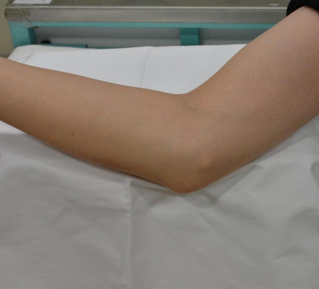
59 Measurements Proximal At Distal
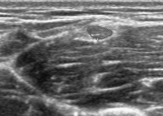
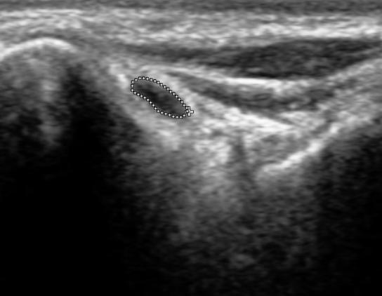
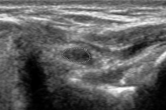
60 Criteria for cubital tunnel syndrome Absolute criteria (CSA ulnar nerve) : Normal < 9mm 2 Symptomatic > 12mm 2 Thoirs K et al 2007 Relative criteria (CSA ulnar nerve) : 2.8: 1 (proximal : at cubital tunnel) Kyoon SJ et al 2008


61 Ulnar nerve swelling 9mm 2 14mm 2 Normal Mild 19mm 2 23mm 2 Moderate Severe
")

62 Ulnar nerve subluxation (20% normal nerves) Olecranon Medial epicondyle Yang SM et al. J Ultrasound Med 2013


63 Ulnar nerve subluxation Due to absence of arcuate ligament


64 2 o cause: Osteophytes


65 2 o cause : Loose bodies

66 2 o cause: Ganglion


67 2 o cause: synovial proliferation Severe rhematoid arthritis


68 2 o cause: Anconeus epitrochlearis m. Medial head triceps slip

69 Diagnostic criteria (ultrasound or MRI) CSA> 12mm 2 CNR > 50 DWI positive or 2.8: 1 (prox : at)
70 Cubital Tunnel Syndrome : summary Readily assessed with ultrasound or MRI Ultrasound more efficient and can assess subluxation MRI possibly more accurate, especially for mild disease if CNR measured ± DWI obtained
71 CONCLUSION CSA > 12mm 2 CNR > 50 DWI positive or 2.8: 1 (prox : at)
72 Biceps tendon FABS view: Flexion Abduction Supination


73 Biceps tendon FABS view: Flexion Abduction Supination view
74 Biceps and brachialis tendon insertions Muscular brachialis insertion Muscular brachialis insertion Tendinous brachialis insertion Biceps tendinous insertion


75 Biceps and brachialis tendon insertions Muscular brachialis insertion Muscular brachialis insertion Tendinous brachialis insertion Biceps tendinous insertion


76 Mild tendinosis biceps tendon Mild tendinosis Mild tendinosis Mild distal biceps tendinosis without tear


77 Severe tendinosis biceps tendons Severe distal biceps tendinosis with moderate-severity tear


78 Severe tendinosis biceps tendons Complete tear distal biceps with retraction by 3cm Operation: short head torn
79 Conclusion Epicondylitis Cubital tunnel syndrome Biceps insertion


80 Thank you
Overuse Injuries of the Upper Extremity. Overuse Injuries 7/23/2018. Peadiatric Overuse Sports Injuries. Al Hess, MD
 Overuse Injuries of the Upper Extremity Al Hess, MD 7/21/2018 1 Overuse Injuries Everything? Not Trauma, infection, tumor, rheumatoid arthritis, osteoarthritis Onset of pain associated with repetitive
Lateral Elbow Pathology
 Lateral Elbow Pathology Jon A. Jacobson, M.D. Professor of adiology Director, Division of Musculoskeletal adiology University of Michigan Disclosures: Consultant: Bioclinica Advisory Board: GE, Philips
Functional Anatomy of the Elbow
 Functional Anatomy of the Elbow Orthopedic Institute Daryl C. Osbahr, M.D. Chief of Sports Medicine, Orlando Health Chief Medical Officer, Orlando City Soccer Club Orthopedic Consultant, Washington Nationals
Elbow Elbow Anatomy. Flexion extension. Pronation Supination. Anatomy. Anatomy. Romina Astifidis, MS., PT., CHT
 Elbow Elbow Anatomy Romina Astifidis, MS., PT., CHT Curtis National Hand Center Baltimore, MD October 6-8, 2017 Link between the arm and forearm to position the hand in space Not just a hinge Elbow = 70%
The Elbow Scanning Protocol
 The Elbow Scanning Protocol Diagnostic Imaging of the Elbow: Introduction The elbow maybe considered as consisting of four quadrants, anterior, medial, lateral and posterior. Ultrasound would normally
Introduction to Ultrasound Examination of the Hand and upper
 Introduction to Ultrasound Examination of the Hand and upper Emil Dionysian, M.D. Ultrasound of upper ext. Upside Convenient Opens another exam dimension Can be like a stethoscope Helps 3-D D visualization
Elbow Pain. Lateral Elbow Pain. Lateral Elbow Pain. tennis elbow lateral epicondylitis extensor tendinopathy
 Elbow Pain Peter Brukner OAM, FACSP Associate Professor in Sports Medicine Centre for Health, Exercise and Sports Medicine University of Melbourne Lateral Elbow Pain tennis elbow lateral epicondylitis
Sports Medicine Unit 16 Elbow
 Sports Medicine Unit 16 Elbow I. Bones a. b. c. II. What movements does the elbow perform? a. Flexion b. c. Pronation d. III. Muscles in motion a. FLEXION (supinated) i Brachialis (pronated) ii (neutral)
OCCUPATIONAL INJURIES OF THE ELBOW
 PLEASE STAND BY WEBINAR WILL BEGIN AT 12:00 PM PST FOR AUDIO: CALL 866-740-1260 / ACCESS CODE: 764-4915# JAMES VAN DEN BOGAERDE, MD OCCUPATIONAL INJURIES OF THE ELBOW Conflict of Interest Disclosure I,
Lecture 9: Forearm bones and muscles
 Lecture 9: Forearm bones and muscles Remember, the region between the shoulder and the elbow = brachium/arm, between elbow and wrist = antebrachium/forearm. Forearm bones : Humerus (distal ends) Radius
Other Elbow Concerns in Overhead Athletes
 Other Elbow Concerns in Overhead Athletes John A. Steubs, M.D. Team Physician, Minnesota Twins TRIA Orthopaedic Center Disclosures None relevant to this presentation. Other Elbow Problems Valgus extension
Slide 1. Slide 2. Slide 3. The Thrower s Elbow: When to Operate. Medial Elbow Pain in the Athlete. Goal of This Talk
 Slide 1 The Thrower s Elbow: When to Operate Luke S. Oh, MD Massachusetts General Hospital Team Physician, Boston Red Sox Team Physician, New England Revolution Consultant, Harvard University Athletics
Medial Collateral Instability of the Elbow. CSES Residents Course Calgary AB February 1-3, 2017 WD Regan MD
 Medial Collateral Instability of the Elbow CSES Residents Course Calgary AB February 1-3, 2017 WD Regan MD Disclosures I have no disclosures to report Anatomy Medial Collateral Ligament Anterior Oblique
I (and/or my co-authors) have something to disclose.
 have something to disclose.") Elbow Anatomy And Biomechanics Nikhil N Verma, MD Director, Division of Sports Medicine Professor, Department of Orthopedics Rush University Medical Center Team Physician, Chicago White Sox and Bulls I
Elbow Anatomy And Biomechanics Nikhil N Verma, MD Director, Division of Sports Medicine Professor, Department of Orthopedics Rush University Medical Center Team Physician, Chicago White Sox and Bulls I
Common Tendon Disorders of the Upper Extremity. Mark Tait MD
 Common Tendon Disorders of the Upper Extremity Mark Tait MD Tendonitis History Pain and swelling (any tendon, any location) Overuse Physical examination findings Localized swelling Pain with resistance
Imaging of the Elbow. Marco Zanetti Radiology Balgrist University Hospital Zurich
 Imaging of the Elbow Marco Zanetti Radiology Balgrist University Hospital Zurich Elbow Case 1 Case 8 Case 2 Case 9 Case 3 Case 10 Case 4 Case 11 Case 5 Case 12 Case 6 Case 13 Case 7 Case 14 Elbow Imaging
This presentation is the intellectual property of the author. Contact them at for permission to reprint and/or distribute.
 B F Morrey, MD Professor of Orthopedic Surgery, UTHSCSA Financial Disclosure Dr. Bernard Morrey has disclosed that he is the Medical director of Tenex Health. OUTLINE Muscles/tendons Ligaments Articulation
MEDIAL EPICONDYLE FRACTURES
 MEDIAL EPICONDYLE FRACTURES Demographic 20% of elbow fractures 60% of which are associated with elbow dislocation. 75% in boys between 6-12 years 20% of elbow dislocation with ME fracture, the ME is incarcerated
Elbow Anatomy, Growth and Physical Exam. Donna M. Pacicca, MD Section of Sports Medicine Division of Orthopaedic Surgery Children s Mercy Hospital
 Elbow Anatomy, Growth and Physical Exam Donna M. Pacicca, MD Section of Sports Medicine Division of Orthopaedic Surgery Children s Mercy Hospital Contributing Factors to Elbow Injury The elbow is affected
Biceps Brachii. Muscles of the Arm and Hand 4/4/2017 MR. S. KELLY
 Muscles of the Arm and Hand PSK 4U MR. S. KELLY NORTH GRENVILLE DHS Biceps Brachii Origin: scapula Insertion: radius, fascia of forearm (bicipital aponeurosis) Action: supination and elbow flexion Innervation:
Grundkurs SGSM-SSMS Sion Sports Elbow. Dr Stéphane Kämpfen
 Grundkurs SGSM-SSMS Sion 2015 Sports Elbow Dr Stéphane Kämpfen Type of pathologies! Acute:! Fractures - dislocation! Sprain! Chronic:! Overload syndrome Fractures - Radial head: Mason classification Mason
Common Elbow Problems
 Common Elbow Problems Duncan Ferguson FRACS Knee and Shoulder Specialist Elbow Instability Common 10-25% of elbow injuries Median age 30 yrs Most simple dislocations are stable after reduction recurrence
ELBOW MRI BASICS BONES/CARTILAGE
 ELBOW MRI BASICS supine vs prone (superman) imaging Coronal for collateral lig and bones Sagittal for biceps/triceps tendons and cartilage Axial for muscles and nerves FABS (flexed elbow, abducted shoulder,
The Elbow 3/5/2015. The Elbow Scanning Sequence. * Anterior Joint (The anterior Pyramid ) * Lateral Epicondyle * Medial Epicondyle * Posterior Joint
 * Lateral Epicondyle * Medial Epicondyle * Posterior Joint") Scanning Sequence * Anterior Joint (The anterior Pyramid ) * Lateral Epicondyle * Medial Epicondyle * Posterior Joint Anterior Elbow Pyramid Courtesy of Jay Smith, MD. Vice chair PMR Mayo Clinic Rochester,
Scanning Sequence * Anterior Joint (The anterior Pyramid ) * Lateral Epicondyle * Medial Epicondyle * Posterior Joint Anterior Elbow Pyramid Courtesy of Jay Smith, MD. Vice chair PMR Mayo Clinic Rochester,
ARM Brachium Musculature
 ARM Brachium Musculature Coracobrachialis coracoid process of the scapula medial shaft of the humerus at about its middle 1. flexes the humerus 2. assists to adduct the humerus Blood: muscular branches
Management of Chronic Elbow Pain
 Mr. Nashat Siddiqui Consultant Upper Limb Orthopaedic Surgeon Management of Chronic Elbow Pain Patients presenting with elbow pain can pose a diagnostic challenge, especially if there is no obvious recent
The Elbow and the cubital fossa. Prof Oluwadiya Kehinde
 The Elbow and the cubital fossa Prof Oluwadiya Kehinde www.oluwadiya.com Elbow and Forearm Anatomy The elbow joint is formed by the humerus, radius, and the ulna Bony anatomy of the elbow Distal Humerus
Ultrasound of the elbow joint - anatomical review of normal structures
 Ultrasound of the elbow joint - anatomical review of normal structures Poster No.: C-2089 Congress: ECR 2015 Type: Educational Exhibit Authors: D. Castelo, E. Matos, F. C. Pires ; Vila Nova de Gaia/PT,
Ultrasound of the elbow, what the radiologist should know.
 Ultrasound of the elbow, what the radiologist should know. Poster No.: C-1679 Congress: ECR 2012 Type: Educational Exhibit Authors: P. Gamo Villegas, J. García Yavar, S. Allodi de la Hoz, J. 1 1 3 2 1
Elbow. Chapter 2 LISTEN. Mechanism of Injury (If Applicable) Pain
 Pain") Chapter 2 Elbow LISTEN Mechanism of Injury (If Applicable) Patient usually remembers their position at the time of injury Certain mechanisms of injury result in characteristic patterns Fall on outstretched
Chapter 2 Elbow LISTEN Mechanism of Injury (If Applicable) Patient usually remembers their position at the time of injury Certain mechanisms of injury result in characteristic patterns Fall on outstretched
Top Elbow Problems: Tennis Elbow, Anyone?
 Disclosure Top Elbow Problems: Tennis Elbow, Anyone? Founder, RunSafe, RaceSafe Founder, SportZPeak Inc. Sanofi, Investigator initiated grant Anthony Luke MD, MPH, CAQ (Sport Med) UCSF Sports Medicine
10/1/2009. October 15, 2009 Christina Kuo MD. Anatomy and pathophysiology of Epicondylitis Diagnosis
 October 15, 2009 Christina Kuo MD Anatomy and pathophysiology of Epicondylitis Diagnosis Treatment options Lawn tennis elbow Morris 1882 - described as an injury occurring from the backhand stroke Age
The Biomechanics of the Human Upper Extremity-The Elbow Joint C. Mirzanli Istanbul Gelisim University
 The Biomechanics of the Human Upper Extremity-The Elbow Joint C. Mirzanli Istanbul Gelisim University Structure of The Elbow Joint A simple hinge joint, actually categorized as a trochoginglymus joint
The Elbow. The Elbow. The Elbow 12/11/2017. Oak Ridge High School Conroe, Texas. Compose of three bones. Ligaments of the Elbow
 Oak Ridge High School Conroe, Texas Compose of three bones The humerus The radius The ulna Ligaments of the Elbow Ulnar collateral ligament Radial collateral ligament Annular ligament 1 The elbow is considered
Elbow Injuries in the Adult Athlete. Tamara A. Scerpella, MD Professor, Orthopedic Surgery University of Wisconsin
 Elbow Injuries in the Adult Athlete Tamara A. Scerpella, MD Professor, Orthopedic Surgery University of Wisconsin Acute Elbow Dislocation Fracture Distal humerus Olecranon Radial head Distal Biceps Rupture
Disclaimer. Evaluation & Treatment of Shoulder and Elbow Pain in the Adult Patient. Objectives. Anatomy
 Evaluation & Treatment of Shoulder and Elbow Pain in the Adult Patient William T. Crowe, RN-C, FNP, MSN, MBA Disclaimer! I, William T Crowe, have relevant financial relationships to be discussed, directly
Management of the Persistently Painful Shoulder and Elbow
 Management of the Persistently Painful Shoulder and Elbow Mr Nashat Siddiqui Consultant Upper Limb Surgeon www.londonupperlimb.com Cannizaro House 2 nd March 2016 How to approach a painful shoulder/elbow
Peripheral Nerve Ultrasound
 Peripheral Nerve Ultrasound Jon A. Jacobson, M.D. Professor of Radiology Director, Division of Musculoskeletal Radiology University of Michigan Normal Peripheral Nerve Ultrasound appearance: Hypoechoic
MUSCLES OF THE ELBOW REGION
 MUSCLES OF THE ELBOW REGION Dr Bronwen Ackermann COMMONWEALTH OF AUSTRALIA Copyright Regulation WARNING This material has been reproduced and communicated to you by or on behalf of the University of Sydney
Joints of the upper limb II
 Joints of the upper limb II Prof. Abdulameer Al-Nuaimi E-mail: a.al-nuaimi@sheffield.ac.uk E. mail: abdulameerh@yahoo.com Elbow joint The elbow joint is connecting the upper arm to the forearm. It is classed
High-resolution ultrasound of the elbow - didactic approach.
 High-resolution ultrasound of the elbow - didactic approach. Poster No.: C-2358 Congress: ECR 2014 Type: Educational Exhibit Authors: C. M. Olchowy, M. Lasecki, U. Zaleska-Dorobisz; Wroclaw/PL Keywords:
The Muscular System. Chapter 10 Part C. PowerPoint Lecture Slides prepared by Karen Dunbar Kareiva Ivy Tech Community College
 Chapter 10 Part C The Muscular System Annie Leibovitz/Contact Press Images PowerPoint Lecture Slides prepared by Karen Dunbar Kareiva Ivy Tech Community College Table 10.9: Muscles Crossing the Shoulder
region of the upper limb between the shoulder and the elbow Superiorly communicates with the axilla.
 1 region of the upper limb between the shoulder and the elbow Superiorly communicates with the axilla. Inferiorly, a number of important structures pass between arm & forearm through cubital fossa. 2 medial
Terrible Triad: Tricks for Dealing with the Unstable Elbow
 Terrible Triad: Tricks for Dealing with the Unstable Elbow Mark A. Mighell, MD Kaitlyn N. Christmas, BS Disclosure Paid Consultation Research Support Speakers Bureau Paid Consultation Speakers Bureau The
Cubital Tunnel Syndrome
 Disclaimer This movie is an educational resource only and should not be used to manage Orthopaedic Health. All decisions about must be made in conjunction with your Physician or a licensed healthcare provider.
ELENI ANDIPA General Hospital of Athens G. Gennimatas
 ELENI ANDIPA General Hospital of Athens G. Gennimatas Technological advances over the last years have caused a dramatic improvement in ultrasound quality and resolution An established imaging modality
Index. radiologic.theclinics.com. Note: Page numbers of article titles are in boldface type.
 Index Note: Page numbers of article titles are in boldface type. A Acromioclavicular joint injuries in football players, 318, 319 ALPSA. See Anterior labroligamentous periosteal sleeve avulsion. Anterior
Golf Injuries in the Upper Extremity
 Golf Injuries in the Upper Extremity David S. Zelouf, MD Philadelphia Hand to Shoulder Center March Meeting 2019 I have nothing to disclose Except that I m an avid, competitive golfer and I ve had golfer
Index. Note: Page numbers of article titles are in boldface type.
 Note: Page numbers of article titles are in boldface type. A ACJ. See Acromioclavicular joint (ACJ) Acromioclavicular joint (ACJ) procedures of, 557 559 Ankle and foot procedures of, 649 671 (See also
Ultrasound of the Knee
 Ultrasound of the Knee Jon A. Jacobson, M.D. Professor of Radiology Director, Division of Musculoskeletal Radiology University of Michigan Disclosures: Consultant: Bioclinica Book Royalties: Elsevier Advisory
Urgent Cases and Foreign Bodies
 Urgent Cases and Foreign Bodies Catherine J. Brandon, MD, MS University of Michigan Ann Arbor, MI, USA Introduction: Patients added on to the schedule from the emergency department or as urgent add-on
Knee, Ankle, and Foot: Normal and Abnormal Features with MRI and Ultrasound Correlation. Disclosures. Outline. Joint Effusion. Suprapatellar recess
 Knee, Ankle, and Foot: Normal and Abnormal Features with MRI and Ultrasound Correlation Jon A. Jacobson, M.D. Professor of Radiology Director, Division of Musculoskeletal Radiology University of Michigan
Elbow joint ultrasonography standard procedure
 Elbow joint ultrasonography standard procedure Poster No.: C-2997 Congress: ECR 2018 Type: Educational Exhibit Authors: A. I. Aguiar, J. A. Torres de Abreu Macedo, M. Barros, P. Gomes, F. Caseiro Alves;
This presentation is the intellectual property of the author. Contact them for permission to reprint and/or distribute.
 COMMON ELBOW INJURIES In The Athlete B F Morrey, MD Professor of Orthopedics UTHSCSA Disclosure Potential conflicts Zimmer royalties, consultant Stryker royalties Tenex Medical director; Interim CEO Professor
Forearm and Wrist Regions Neumann Chapter 7
 Forearm and Wrist Regions Neumann Chapter 7 REVIEW AND HIGHLIGHTS OF OSTEOLOGY & ARTHROLOGY Radius dorsal radial tubercle radial styloid process Ulna ulnar styloid process ulnar head Carpals Proximal Row
Ultrasonography of Peripheral Nerve -upper extremity
 Ultrasonography of Peripheral Nerve -upper extremity Department of Physical Medicine and Rehabilitation Korea University Guro Hospital Korea University College of Medicine Yoon Joon Shik Normal median
CHAPTER 6: THE UPPER EXTREMITY: THE ELBOW, FOREARM, WRIST, AND HAND
 CHAPTER 6: THE UPPER EXTREMITY: THE ELBOW, FOREARM, WRIST, AND HAND KINESIOLOGY Scientific Basis of Human Motion, 12 th edition Hamilton, Weimar & Luttgens Presentation Created by TK Koesterer, Ph.D.,
MSK Imaging Conference. 07/22/2016 Eman Alqahtani, MD, MPH R3/PGY4 UCSD Radiology
 MSK Imaging Conference 07/22/2016 Eman Alqahtani, MD, MPH R3/PGY4 UCSD Radiology A 51 years old female with chronic thumb pain, and inability to actively flex the thumb interphalyngeal joint Possible trigger
#12. Joint نبيل خوري
 #12 30 Anatomy Joint هيام الر جال 9/10/2015 نبيل خوري Salam Awn Some notes before starting : ** Not all slides are included, so I recommend having a look at the slides beside this sheet ** If you find
Elbow Joint Anatomy ELBOW ANATOMY, BIOMECHANICS. Bone Anatomy. Bone Anatomy. Property of VOMPTI, LLC
 ELBOW ANATOMY, BIOMECHANICS AND PATHOLOGY Kristin Kelley, DPT, OCS, FAAOMPT Elbow Joint Anatomy Joint articulations Humeroulnar Radiohumeral Radioulnar (proximal and distal) Orthopaedic Manual Physical
79a Orthopedic Massage: Introduction! Rotator Cuff and Carpal Tunnel!
 79a Orthopedic Massage: Introduction! Rotator Cuff and Carpal Tunnel! 79a Orthopedic Massage: Introduction! Rotator Cuff and Carpal Tunnel! Class Outline" 5 minutes" "Attendance, Breath of Arrival, and
This presentation is the intellectual property of the author. Contact them for permission to reprint and/or distribution.
 COMMON ELBOW INJURIES In The Athlete B F Morrey, MD Professor of Orthopedics UTHSCSA Professor of Orthopedics Mayo Cllnic COMMON SPORTS INJURIES of the ELBOW Disclosure Potential conflicts Zimmer royalities,
Osteology of the Elbow and Forearm Complex. The ability to perform many activities of daily living (ADL) depends upon the elbow.
 depends upon the elbow.") Osteology of the Elbow and Forearm Complex The ability to perform many activities of daily living (ADL) depends upon the elbow. Activities of Daily Living (ADL) Can you think of anything that you do to
Osteology of the Elbow and Forearm Complex The ability to perform many activities of daily living (ADL) depends upon the elbow. Activities of Daily Living (ADL) Can you think of anything that you do to
Ultrasound Evaluation of Posteromedial Ankle Pathology. Andrew C Cordle, M.D., Ph.D. 9/21/2018
 Ultrasound Evaluation of Posteromedial Ankle Pathology Andrew C Cordle, M.D., Ph.D. 9/21/2018 Overview: Pathology of the Posteromedial Ankle Flexor Tendon Pathology Accessory Navicular Bone Pathology Tarsal
Shoulder Elbow Wrist/Hand
 Shoulder Elbow Wrist/Hand Randy E. Moore DC RDMS RMSK General Musculoskeletal Imaging, Inc. 1 Shoulder Tendinosis : 3 key Ultrasound Findings 1. Increased cellularity thickened and ACR inhomogeneous CLV
Inspection. Physical Examination of the Elbow. Anterior Elbow 2/14/2017. Inspection. Carrying angle. Lateral dimple. Physical Exam of the Elbow
 of the Elbow Anthony A. Romeo, MD Professor, Department of Orthopedics Head, Section of Shoulder and Elbow Surgery Rush University President-Elect, American Shoulder Elbow Surgeons Team Physician, Chicago
Ligaments of Elbow hinge: sagittal plane so need lateral and medial ligaments
 Ligaments of Elbow hinge: sagittal plane so need lateral and medial ligaments Ulnar Collateral ligament on medial side; arising from medial epicondyle and stops excess valgus movement (lateral movement)
The Forearm 2. Extensor & lateral Compartments of the Forearm
 The Forearm 2 Extensor & lateral Compartments of the Forearm 1-Lateral Fascial Compartment (at the lateral side of the forearm ) *Some books mention the lateral compartment contain just the Brachioradialis
The use of Ultrasound for the Diagnosis and Treatment of the Musculoskeletal System
 The use of Ultrasound for the Diagnosis and Treatment of the Musculoskeletal System St. Joseph s Refresher Course March 2019 Kenneth Iles, DC John Finkenstadt, MD THIS WORKSHOP WILL FOCUS ON 2 MAIN TOPICS:
The Elbow and Radioulnar Joints Kinesiology. Dr Cüneyt Mirzanli Istanbul Gelisim University
 The Elbow and Radioulnar Joints Kinesiology Dr Cüneyt Mirzanli Istanbul Gelisim University 1 The Elbow & Radioulnar Joints Most upper extremity movements involve the elbow & radioulnar joints. Usually
Elbow. Chapter 2 LISTEN. Mechanism of Injury (If Applicable) Pain
 Pain") Preface The first decade of the twenty-first century has witnessed the continuation of an explosion in our knowledge and understanding of all aspects of disease. Accompanying this has been the increasing
Preface The first decade of the twenty-first century has witnessed the continuation of an explosion in our knowledge and understanding of all aspects of disease. Accompanying this has been the increasing
Long-term sequel of posterolateral rotatory instability of the elbow: a case report
 CASE REPORT Open Access Long-term sequel of posterolateral rotatory instability of the elbow: a case report Chun-Ying Cheng * Abstract The natural course of untreated posterior lateral rotatory instability
Clinical examination of the wrist, thumb and hand
 Clinical examination of the wrist, thumb and hand 20 CHAPTER CONTENTS Referred pain 319 History 319 Inspection 320 Functional examination 320 The distal radioulnar joint.............. 320 The wrist.......................
10/15/2014. Wrist. Clarification of Terms. Clarification of Terms cont
 Wrist Clarification of Terms Palmar is synonymous with anterior aspect of the wrist and hand Ventral is also synonymous with anterior aspect of the wrist and hand Dorsal refers to the posterior aspect
compartments of the forearm
 " forearm posterior compartment " compartments of the forearm Posterior Fascial compartment Muscles: ** The superficial group 1. Extensor carpi radialis brevis 2. Ex. digitorum 3. Ex. digiti minimi 4.
5/8/2017. Finger Injuries in Football. Tendon Injuries of the Hand and Wrist in Football Steve Kronlage, MD Andrews Institute Gulf Breeze, Florida
 Finger Injuries in Football Tendon Injuries of the Hand and Wrist in Football Steve Kronlage, MD Andrews Institute Gulf Breeze, Florida A jammed finger is an injury (at very least a torn ligament) A swollen
79a Orthopedic Massage: Introduction! Rotator Cuff and Carpal Tunnel!
 79a Orthopedic Massage: Introduction! Rotator Cuff and Carpal Tunnel! 79a Orthopedic Massage: Introduction! Rotator Cuff and Carpal Tunnel! Class Outline 5 minutes Attendance, Breath of Arrival, and Reminders
1 Comprehensive Orthopaedic Review Hand and Wrist Tendinopathies Bernard F. Hearon, MD September 2, de Quervain Disorder Fritz de Quervain,
 1 Comprehensive Orthopaedic Review Hand and Wrist Tendinopathies Bernard F. Hearon, MD September 2, 2016 2 de Quervain Disorder Fritz de Quervain, Swiss surgeon, 1895 Stenosing tendinosis, 1st dorsal compartment
Ultrasound of Mid and Hindfoot Pathology
 Ultrasound of Mid and Hindfoot Pathology Levon N. Nazarian, M.D. Professor of Radiology Thomas Jefferson University Hospital Disclosures None relevant to this presentation Educational Objective Following
MR: Finger and Thumb Injuries
 MR: Finger and Thumb Injuries Laura W. Bancroft, M.D. Professor of Radiology University of Central Florida Florida State University Outline Normal anatomy of the fingers and thumb MR imaging protocols
Common Elbow Injuries in the Athlete
 COMMON ELBOW INJURIES In The Athlete B F Morrey, MD Professor of Orthopedics UTHSCSA Common Elbow Injuries in the Athlete Matthew Murray, MD Professor of Orthopedics Mayo Cllnic OUTLINE Muscles/tendons
Dr. Mahir Alhadidi Anatomy Lecture #9 Feb,28 th 2012
 Quick Revision: Upper arm is divided into two compartments: 1. Anterior Compartment: Contains three muscles (Biceps brachii, Coracobrachialis, Brachialis). Innervated by Musculocutaneous nerve. 2. Posterior
Nerves of Upper limb. Dr. Brijendra Singh Professor & Head Department of Anatomy AIIMS Rishikesh
 Nerves of Upper limb Dr. Brijendra Singh Professor & Head Department of Anatomy AIIMS Rishikesh 1 Objectives Origin, course & relation of median & ulnar nerves. Motor & sensory distribution Carpal tunnel
Disclosures. Throwing is NOT Normal MCL RECONSTRUCTION: INDICATIONS, TECHNIQUE, RESULTS. Joshua S. Dines, MD. Sports Medicine and Shoulder Service
 MCL RECONSTRUCTION: INDICATIONS, TECHNIQUE, RESULTS Joshua S. Dines, MD Sports Medicine and Shoulder Service Disclosures Consultant: Arthrex, Conmed Linvatec, Ossur IP/Royalties: Conmed Linvatec Editorial
TOPAZ TM What you should know
 TOPAZ TM What you should know Not for distribution in the United States. Need to know 2 Tendon: is a tough band of fibrous connective tissue that connects muscle to bone and is capable of withstanding
CLINICAL PRESENTATION AND RADIOLOGY QUIZ QUESTION
 Donald L. Renfrew, MD Radiology Associates of the Fox Valley, 333 N. Commercial Street, Suite 100, Neenah, WI 54956 10/6/2012 Radiology Quiz of the Week # 93 Page 1 CLINICAL PRESENTATION AND RADIOLOGY
Fascial Compartments of the Upper Arm
 Fascial Compartments of the Upper Arm The upper arm is enclosed in a sheath of deep fascia and has two fascial septa: 1- Medial fascial septum (medial intermuscular septum): attached to the medial supracondylar
8 Recovering From HAND FRACTURE SURGERY
 8 Recovering From HAND FRACTURE SURGERY Hand fractures are caused by trauma and result in breaking (fracturing) the phalanges or metacarpals. Surgery involves achieving acceptable alignment and providing
Clinical Orthopaedic Rehabilitation Volume 1 and 2
 Clinical Orthopaedic Rehabilitation Volume 1 and 2 COURSE DESCRIPTION This program is a practical, clinical guide that provides guidance on the evaluation, differential diagnosis, treatment, and rehabilitation
The elbow: Anatomy and Pathology of Collateral Ligaments using MRI.
 The elbow: Anatomy and Pathology of Collateral Ligaments using MRI. Award: Magna Cum Laude Poster No.: C-0570 Congress: ECR 2018 Type: Educational Exhibit Authors: J. Acosta Batlle, M. D. Lopez Parra,
Exam of the Injured Hand and Wrist. Christina M. Ward, MD Regions Hospital TRIA Woodbury
 Exam of the Injured Hand and Wrist Christina M. Ward, MD Regions Hospital TRIA Woodbury Disclosures We have no disclosures that are pertinent to this presentation Terminology Ring Long Index Small Thumb
The Rheumatoid Hand Deformities & Management. Dr. Anirudh Sharma Resident Department of Orthopedics
 + The Rheumatoid Hand Deformities & Management Dr. Anirudh Sharma Resident Department of Orthopedics + Why is Rheumatoid Arthritis important? + RA is a very debilitating disease median life expectancy
STRUCTURAL BASIS OF MEDICAL PRACTICE EXAMINATION 5 October 6, 2006
 STRUCTURAL BASIS OF MEDICAL PRACTICE EXAMINATION 5 October 6, 2006 PART l. Answer in the space provided. (8 pts) 1. Identify the structures. (2 pts) B C A. _pisiform B. _ulnar artery A C. _flexor carpi
Technique. Disclosure. Approach to Ultrasound of the Wrist. Objectives. Outline. Technique 14/09/2015
 Approach to Ultrasound of the Wrist Disclosure I have no commercial or financial interests related to the subject matter of this presentation Linda robyn, MD, FRCC MSK Radiologist Objectives At the end
The arm: *For images refer back to the slides
 The arm: *For images refer back to the slides Muscles of the arm: deltoid, triceps (which is located at the back of the arm), biceps and brachialis (it lies under the biceps), brachioradialis (it lies
Lateral elbow tendinopathy
 Lateral elbow tendinopathy Lateral elbow tendinopathy is a common condition with an incidence of 1 2%. The pathology arises from the origin of extensor carpi radialis brevis where changes, consistent with
Connects arm to thorax 3 joints. Glenohumeral joint Acromioclavicular joint Sternoclavicular joint
 Connects arm to thorax 3 joints Glenohumeral joint Acromioclavicular joint Sternoclavicular joint Scapula Elevation Depression Protraction (abduction) Retraction (adduction) Downward Rotation Upward Rotation
*the Arm* -the arm extends from the shoulder joint (proximal), to the elbow joint (distal) - it has one bone ; the humerus which is a long bone
, to the elbow joint (distal) - it has one bone ; the humerus which is a long bone") *the Arm* -the arm extends from the shoulder joint (proximal), to the elbow joint (distal) - it has one bone ; the humerus which is a long bone - muscles in the arm : *brachialis muscle *Biceps brachii
*the Arm* -the arm extends from the shoulder joint (proximal), to the elbow joint (distal) - it has one bone ; the humerus which is a long bone - muscles in the arm : *brachialis muscle *Biceps brachii