European Journal of Neuroscience. Functional specificity of premotor-motor cortical interactions during action selection
|
|
|
- Sophie George
- 5 years ago
- Views:
Transcription
1 Functional specificity of premotor-motor cortical interactions during action selection Journal: Manuscript ID: Manuscript Type: Date Submitted by the Author: EJN R1 Research Report n/a Complete List of Authors: O'Shea, Jacinta; University of Oxford, Dept. of Experimental Psychology Sebastian, Catherine; University College London, Institute of Cognitive Neuroscience Boorman, Erie; University of Oxford, Experimental Psychology Johansen-Berg, Heidi; Oxford University, FMRI Centre Rushworth, Matthew; Oxford University, Experimental Psychology Key Words: action selection, dorsal premotor cortex, TMS, cortico-cortical interactions, primary motor cortex
2 Page 1 of 41 EUROPEAN JOURNAL OF NEUROSCIENCE Receiving Editor: E. A. Murray Functional specificity of premotor-motor cortical interactions during action selection Running title: PMd-M1 interactions during action selection Jacinta O Shea 1, Catherine Sebastian 1, Erie D. Boorman 1, Heidi Johansen-Berg 2 & Matthew F.S. Rushworth 1,2 1. Department of Experimental Psychology University of Oxford South Parks Road Oxford OX1 3UD U.K. 2. FMRIB Centre, John Radcliffe Hospital, University of Oxford Corresponding Author: U.K Telephone: Jacinta O Shea Department of Experimental Psychology University of Oxford South Parks Road Oxford, OX1 3UD Fax: jacinta.oshea@psy.ox.ac.uk Number of pages: 31 Total word count: 8,088 Word count of abstract: 199 words Word count of introduction: 555 words Figures: 7 (plus 1 in supplementary materials) Tables: 1 in supplementary materials Keywords: action selection, cortico-cortical interactions, functional connectivity, motor cortex, dorsal premotor cortex, TMS 1
3 Page 2 of 41 ABSTRACT Functional connections between dorsal premotor cortex (PMd) and primary motor cortex (M1) have been revealed by paired-pulse transcranial magnetic stimulation (TMS). We tested if such connections would be modulated during a cognitive process (response selection) known to rely on those circuits. PMd-M1 TMS applied 75ms after a cue to select a manual response facilitated motor-evoked potentials (MEPs). MEPs were facilitated at 50ms in a control task of response execution, suggesting that PMd-M1 interactions at 75ms are functionally specific to the process of response selection. At 100ms, PMd-M1 TMS delayed choice reaction time (RT). Importantly, the MEP (at 75ms) and the RT (at 100ms) effects were correlated in a way that was hand-specific. When the response was made with the M1-contralateral hand, MEPs correlated with slower RTs. When the response was made with the M1-ipsilateral hand, MEPs correlated with faster RTs. Paired-pulse TMS confined to M1 did not produce these effects, confirming the causal influence of PMd inputs. This study shows that a response selection signal evolves in PMd early during the reaction period (75-100ms), impacts on M1, and affects behaviour. Such interactions are temporally, anatomically, and functionally specific, and have a causal role in choosing which movement to make. 2
4 Page 3 of 41 INTRODUCTION Paired-pulse transcranial magnetic stimulation (TMS), in which one TMS pulse is applied over each of two brain areas, makes it possible to interrogate functional cortical interactions on a sub-second timescale. In the motor system, paired-pulse TMS studies exploit the fact that stimulation of primary motor cortex (M1) elicits a motor-evoked potential (MEP), which can be used as an index of M1 excitability. One can test the causal influence of a connected area on M1 by first stimulating that area, and measuring consequent changes in the amplitude of MEPs (Di Lazzaro et al., 1999; Ferbert et al., 1992; Netz et al., 1995). The technique is thus complementary to combined TMS/neuroimaging approaches (Bestmann et al., 2005; Lee et al., 2003; O'Shea et al., 2007a), but its particular advantage is that it can reveal with sub-second resolution how activity changes in one brain area causally impact on activity in connected areas (O'Shea et al., 2007b). Paired-pulse TMS has revealed resting-state physiological connections to M1 from premotor, parietal and supplementary motor areas of the same hemisphere (Civardi et al., 2001; Koch et al., 2007; Koch et al., 2006). Inter-hemispheric connections between dorsal premotor cortex (PMd) and M1 have also been demonstrated, with paired-pulse TMS facilitating (Baumer et al., 2006) or inhibiting MEPs (Mochizuki et al., 2004) as a function of stimulation intensity. An important extension of the paired-pulse technique is to assess how resting-state connections are modulated by psychological context. There is evidence from the visual system, where phosphene induction is used to index visual cortex excitability, that resting-state V1-V5 connections are modulated by the performance of a visual motion detection task (Pascual- Leone & Walsh, 2001; Silvanto et al., 2005a; Silvanto et al., 2005b). According to this logic, inter-hemispheric PMd-M1 connections should be modulated when task demands recruit those circuits. The PMd plays an important role in the selection of responses for execution, specifically response selection based on learned stimulus-response mapping rules (Passingham, 3
5 Page 4 of ; Petrides, 1985; Wise & Murray, 2000). PMd shows greater activation during tasks that require the selection of a different response on every trial, compared to tasks that instruct the same response on every trial (Cavina-Pratesi et al., 2006; Grol et al., 2006; Johansen-Berg et al., 2002; Rushworth et al., 2003; Schluter et al., 2001; Thoenissen et al., 2002). Although PMd is activated bilaterally during response selection, imaging and TMS studies suggest that left PMd exerts dominance over right PMd: TMS of either PMd disrupts response selection with the contralateral hand, but only left PMd TMS disrupts selection with the ipsilateral hand (Johansen-Berg et al., 2002; Schluter et al., 2001; Schluter et al., 1998). We combined paired-pulse TMS with two tasks: a visuomotor choice reaction time (RT) task that emphasized response selection, and a control simple RT task that deemphasized selection. With this task manipulation we aimed to isolate PMd-M1 interactions that were functionally specific to the process of response selection. We predicted that pairedpulse TMS would modulate MEPs early during the reaction period, because previous singlepulse TMS data suggest that this is when the PMd contributes to response selection (Johansen- Berg et al., 2002; Schluter et al., 1999; Schluter et al., 1998). We further predicted that MEP modulation would be of greater magnitude or duration, and have greater relevance for selection behaviour, after TMS of the (dominant) left PMd than after TMS of the right PMd. MATERIALS & METHODS Subjects Eighteen healthy volunteers participated in this study (age range 23-30). In Experiment 1, ten subjects (six female) were tested in counter-balanced order on both task sessions. Two further subjects participated in only one of the two sessions, yielding a total of eleven subjects per session. In Experiment 2, eight subjects (five females) were tested in counter-balanced order on both task sessions. One further subject participated in only one of the two sessions. In total, seven subjects participated in all sessions of Experiments 1 and 2, so across-experiment 4
6 Page 5 of 41 analyses were performed on the data from those seven subjects. Six subjects (four female) were tested in Experiment 3, one of whom had participated in both sessions of Experiments 1 and 2 two months previously. All subjects were right-handed and reported an absence of psychiatric or neurological disease in their known family history. All subjects gave written informed consent. The study was carried out under permission from the Central Oxford Research Ethics Committee (COREC, 05-Q ) and in accordance with the Declaration of Helsinki. Behavioural Tasks Two tasks were used: an experimental choice RT task (Select) (Experiments 1 and 3) and a control simple RT task (Execute) (Experiment 2). The Select task emphasized response selection: subjects had to select one of two button press responses with the index finger of their right or left hand on each trial, according to the identity of a visual cue. A small circle or large square instructed a right hand response, while a large circle or small square instructed a left hand response (Fig. 1A). These four stimulus-response mappings were counterbalanced across subjects. The fact that neither size nor shape instructed responses in a simple way meant that actions had to be selected with care, even after practice. By contrast, the Execute task de-emphasized selection: subjects made the same response on every trial regardless of which cue was presented. Thus, there was only one stimulus-response mapping, and the responding hand was always contralateral to the stimulated M1 (Fig. 1A). It has been shown that PMd, especially left PMd, is more active during the Select than the Execute task, and that single-pulse PMd TMS disrupts performance on the Select task more than the Execute task (Johansen-Berg et al., 2002; Rushworth et al., 2003; Schluter et al., 1999; Schluter et al., 1998). In both tasks, visual stimuli were presented until the response was detected, and subjects were instructed to respond as quickly and accurately as possible. All experiments were controlled by Turbo Pascal software. 5
7 Page 6 of 41 Fig 1 here Experiment 1: Timing of PMd-M1 physiological interactions during response selection Experiment 1 addressed three issues about inter-hemispheric PMd-M1 interactions during response selection: 1) timing; 2) hemispheric asymmetry; and 3) behavioural relevance. Experiment 1 had two sessions, each conducted on different days. In one session the left PMd-right M1 (lpmd-rm1) pathway was tested; in the other session the right PMd-left M1 (rpmd-lm1) pathway was tested. Session order was counterbalanced across subjects. In both sessions, subjects performed the Select task while TMS was applied to the relevant PMd-M1. There were two types of TMS trials. On single-pulse trials, a single test TMS pulse was applied over the relevant M1, and the resulting motor evoked potentials (MEPs) were recorded from the first dorsal inter-osseous (FDI) muscle of the contralateral hand. The peakto-peak amplitude of the MEP and the reaction time (RT) recorded on each of those singlepulse trials constituted the baseline measurements. On paired-pulse trials, the M1 test pulse was preceded by a conditioning TMS pulse delivered through a second TMS coil positioned over the contralateral PMd (Fig.1A). The interval between the paired pulses (IPI) was 8ms, based on previous resting-state MEP studies of PMd-M1 interactions that showed this IPI to be most effective (Baumer et al., 2006; Mochizuki et al., 2004). Physiological (MEP) and behavioural (RT) measurements from the baseline single-pulse trials were contrasted with those on paired-pulse trials to test the effect of the conditioning TMS applied to PMd. On each trial of the Select task, a visual shape stimulus was presented until a response was detected. MEP and RT data were recorded during the response period, followed by a variable inter-trial interval ( s). To establish the critical time window for PMd-M1 interactions, the M1 TMS pulse (the only one on single-pulse trials) was delivered at each of five stimulus-onset asynchronies (SOAs): 50, 75, 100, 125 or 150ms after the onset of the visual stimulus (Fig.1B). There were twenty single- and twenty paired-pulse TMS trials per 6
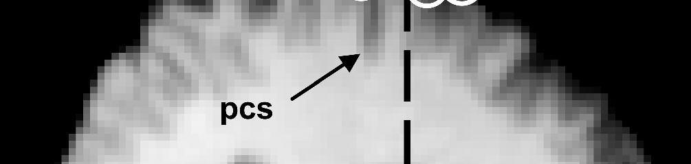
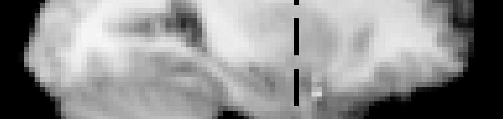
8 Page 7 of 41 SOA. In each condition, half of trials required a left hand response and half required a right hand response. Although MEPs were recorded on every trial, they were always recorded from only one hand (that contralateral to the stimulated M1). This meant that on half of trials MEPs and RTs were recorded from the same hand (contralateral), while on the other half of trials RTs were recorded from the other hand (ipsilateral to the stimulated M1) (Fig.1A). Only data from correct trials were analysed. In total there were ten trials per condition: TMS (single versus paired-pulse) * SOA (50, 75, 100, 125, 150ms) * Hand (contralateral versus ipsilateral). In addition, a further six single-pulse TMS trials were presented at the start of the session. These were designed to allow MEP amplitudes to stabilize and were excluded from the analysis. M1 TMS pulses were applied over the motor cortex hotspot, defined as the optimal scalp position at which the lowest intensity TMS evoked a just-noticeable twitch from the relaxed contralateral FDI muscle. PMd TMS was delivered at scalp coordinates 2cm anterior and 1cm medial from the hotspot. This procedure for targeting PMd has been used in a number of previous studies, which have shown that single-pulse TMS at these coordinates slows RT on the Select task used here (Johansen-Berg et al., 2002; Schluter et al., 1998). We verified the cortical locations of these TMS sites anatomically in nine subjects using Brainsight frameless stereotaxy (Rogue Research, Montreal, Canada)(Fig.2). Individual subjects structural MRI scans were registered to the Montreal Neurological Institute (MNI) 152-mean brain T1 template. This confirmed that PMd TMS was applied just anterior to the dorsal branch of the precentral sulcus [mean MNI coordinates: x= + 28 (SE± 1.75), y= -5 (± 2.94), z= 71 (± 1.42)]. M1 TMS was applied over the motor hand hook in the central sulcus [mean MNI coordinates: x= + 32 (SE± 2.36), y= -21 (± 2.61), z= 69 (± 1.72)]. Both locations correspond well with published probabilistic coordinates and sulcal landmarks for PMd and M1 (Amiez et al., 2006; Chouinard et al., 2003; Fink et al., 1997; Johansen-Berg et al., 2002; Yousry et al., 1997). 7
9 Page 8 of 41 Stimulation intensities were determined separately for each hemisphere. M1 TMS pulses were applied at the minimum intensity required to evoke an MEP of ~1mV peak-topeak amplitude from the relaxed contralateral FDI muscle on ten consecutive trials. Conditioning PMd TMS pulses were applied at 110% of the resting motor threshold (RMT) for M1 of that hemisphere. RMT was defined as the minimum intensity TMS required to evoke a ~50TV MEP from the relaxed contralateral FDI muscle on 5/10 trials. Mean stimulation intensities for M1 were 51.6 (SE +.29, left) and 51.1 (SE +.27, right), and for PMd were 44.4 (SE +.22, left) and 44.6 (SE +.21, right) of maximum stimulator output. TMS pulses were delivered using two monophasic Magstim 200 machines (Magstim Company, Carmarthenshire, Wales, U.K.). Pulses were applied to the PMd through a 50mm figure-ofeight coil and to M1 through a 70mm figure-of-eight coil. Both coils was held tangential to the skull, with the M1 coil handle oriented posteriorly at ~45 and the PMd coil handle oriented laterally at ~90 from the mid-sagittal axis (Fig.1A). MEPs were recorded from the contralateral FDI using Ag-AgCl electrodes and a tendon-belly montage. Electromyographic (EMG) responses were amplified, filtered and sampled using a CED 1902 amplifier, a CED 1401 analogue-to-digital converter and a Pentium 4 computer running Signal (version 2.14) software (Cambridge Electronic Design Ltd., Cambridge, UK) on a PC computer running Windows 98. Signals were sampled at 10,000 Hz and band-pass filtered between 10 and 10,000 Hz. Fig 2 here Experiment 2: Functional specificity of PMd-M1 physiological interactions The aim of Experiment 2 was to establish whether the PMd-M1 physiological interactions (observed in Experiment 1) were functionally specific to the process of response selection, or whether similar effects would be observed during performance of a task with a more limited response selection component. Subjects performed the Execute task, which required them to 8
10 Page 9 of 41 make the same index finger button press response with the same hand on every trial, no matter which shape stimulus was presented. Subjects always responded with the hand contralateral to the stimulated M1, from which MEPs were recorded. So, unlike in Experiment 1, there was no factor of Hand, such that the total trial number was halved. Hence, both sessions (lpmd-rm1 and rpmd-lm1) were conducted on the same day, with order counterbalanced across subjects. Mean stimulation intensities for M1 were 47.6 (SE +.33, left) and 44.7 (SE +.27, right), and for PMd were 41.9 (SE +.23, left) and 43.4 (SE +.26, right). All other procedures were identical to Experiment 1. Experiment 3: Anatomical specificity of PMd-M1 physiological interactions Experiment 3 aimed to establish whether the physiological effects of paired-pulse TMS depended on the anatomical location, PMd, at which the conditioning pulse was applied, or whether the same effects could be obtained by conditioning TMS elsewhere in the motor system. Subjects performed the Select task, and on paired-pulse trials both TMS pulses were delivered over left M1 through a single TMS coil (M1-M1 TMS). The inter-pulse interval and TMS intensities were the same as in Experiment 1, and all other procedures were identical. The mean stimulation intensity was 44.7 (SE +.5) for the conditioning pulse and 50.8 (SE +.45) for the test pulse. Data Analysis All within-experiment analyses compared the effect of paired- versus single-pulse TMS on mean MEP amplitudes (in millivolts, mv) and mean RTs (ms). For all analyses between experiments, the data for each subject and condition were transformed into percentage change values [ie: % MEP = paired-pulse/single-pulse * 100]. This transformation controlled for overall differences in MEP amplitude across experiments caused by differences in task (Select vs. Execute, Experiment 1 vs. 2) or differences in the anatomical site at which 9
11 Page 10 of 41 conditioning TMS was applied (PMd-M1 vs. M1-M1 TMS, Experiment 1 vs. 3). For illustration purposes, all results are displayed in percentage change format. Two outlier datapoints from Experiment 1 (1 MEP, 1 RT) that were more than two standard deviations from the mean were removed from all analyses. Paired-pulse TMS effects were identified using repeated measures ANOVAs with Huyn-Feldt correction and subsequent paired or onesample t-tests corrected for multiple comparisons (Holm 1979). Analyses were carried out using a within-subjects approach whenever possible (see Methods, Subjects). During the simple RT task (Execute, Experiment 2), subjects responded quickly (the earliest quartile of RTs occurred between 150 and 200ms after trial onset). Hence, in the 150ms SOA condition, subjects frequently responded prior to or during the TMS. To identify and eliminate any trials so contaminated by EMG activity, every trial for every subject and condition in every experiment was individually inspected. All trials showing evidence of EMG contamination were eliminated from the dataset prior to analysis. This trial-by-trial inspection procedure revealed that the data for the 150ms SOA condition of the Execute task (Experiment 2) were so frequently contaminated by voluntary muscle activity that they could not be analysed. In all other conditions, however, because MEPs occurred prior to EMG onset, individual trial data were only very rarely contaminated and hence rejected. RESULTS Experiments 1 & Functional Specificity and Timing of PMd-M1 physiological interactions The aim of our study was to determine whether patterns of PMd-M1 functional connectivity differ during action choice ( select task) versus action execution ( execute task). Hence, we carried out a between-experiments analysis on the % MEP data from Experiments 1 and 2 - in order to compare directly the patterns of PMd-M1 functional connectivity observed under the two different task conditions. 10
12 Page 11 of 41 A three-way analysis of variance (ANOVA) was conducted on the % MEP data (for raw data see Supplementary Fig.1) with one between-subjects factor of Task [Select (Experiment 1) versus Execute (Experiment 2)], and two within-subjects factors of Hemisphere (lpmd-rm1 versus rpmd-lm1) and SOA (50, 75, 100, 125ms). In Experiment 2, all responses were made with the hand contralateral to the stimulated M1. Hence, for this across-experiments comparison, the data from Experiment 1 were pooled over the factor of hand before being submitted to analysis. There was a significant Task * SOA interaction (F(3,15) = 5.490, p = 0.001), suggesting that paired-pulse TMS modulated MEP amplitude in both the Select and Execute tasks, but that the relevant SOA differed between the tasks. The Task * Hemisphere interaction approached significance (F(1,5) = 6.110, p = 0.056). There were no other effects, trends or interactions. To further investigate the Task * SOA interaction separate ANOVAs were conducted on the data from each experiment. Mean MEP amplitude data from the Select task (Experiment 1) were analysed using a four-way repeated measures ANOVA with factors of Hemisphere (rpmd-lm1 vs. lpmd-rm1), TMS (single- vs. paired-pulse), SOA (50, 75, 100, 125, 150ms) and Hand (contralateral vs. ipsilateral). The factor of Hand was included because although MEPs were always recorded from the hand contralateral to M1 stimulation, on half of correct trials the response was made with the other (ipsilateral) hand. The TMS * SOA interaction was significant (F(4,32) = 2.854, p = 0.039). There were no other effects, trends or interactions. Paired samples t-tests (pooled over Hemisphere and Hand) showed that paired-pulse TMS facilitated MEP amplitude significantly at 75ms (t(8) = , p = 0.036), and there was a non-significant trend towards facilitation at 50ms (t(9) = , p = 0.084) (Fig 3A). MEP inhibition at 125 and 150ms was not significant (p >.22). Mean MEP amplitude data from the Execute task (Experiment 2) were analysed using a three-way repeated measures ANOVA with the same factors of Hemisphere, TMS and SOA (50, 75, 100, 125ms). There was a main effect of SOA (F(3,24) = 7.092, p = 0.023) and a 11
13 Page 12 of 41 marginally-significant TMS * SOA interaction (F(3,24) = 2.998, p = 0.051). A paired samples t-test revealed significant paired-pulse MEP facilitation at the 50ms SOA (t(8) = 4.202, p = 0.003) (Fig 3B). The tendency towards MEP inhibition at 100ms was not significant (p >.21). These results reveal the timing of task-dependent inter-hemispheric PMd-M1 interactions, and establish their functional specificity by contrasting the patterns of MEP modulation across the Select and the Execute tasks. Paired-pulse TMS significantly facilitated MEP amplitude when applied at an SOA of 50ms in the Execute task (with a non-significant trend in the Select task). In the Select task only, significant MEP facilitation occurred at 75ms only, suggesting that such facilitation is functionally specific to the process of response selection. Fig 3 here 2. No difference in PMd-M1 physiological interactions as a function of Hemisphere or Hand Although the between-experiment ANOVA showed that the Task * Hemisphere interaction approached significance (p = 0.056), separate within-experiment analyses found no main effect of Hemisphere in either task (Experiment 1: Select task p =.133; Experiment 2: Execute task p =.14). More importantly, there was no evidence of hemispheric asymmetry in the pattern of functional connectivity during either task: in both between- and within-experiment analyses, none of the interactions between Hemisphere and TMS approached significance (all p > 0.16). Further exploratory analyses on the MEP data separated by Hemisphere found no evidence to support our a priori hypothesis that conditioning TMS of the left PMd would have a greater effect on MEPs than conditioning TMS of the right PMd. Rather, the results suggest that the timing of inter-hemispheric functional connectivity is the same for both the lpmd-rm1 and the rpmd-lm1 pathway. 12
14 Page 13 of 41 There was no evidence that paired-pulse MEP modulation differed as a function of which hand was selected to make the response (all p > 0.77). In fact, when all data from the critical 75ms SOA were analysed (all sessions and subjects), the magnitude of the % MEP effect showed a significant positive correlation between: a) trials in which the contralateral hand was selected to respond; and b) trials in which the ipsilateral hand was selected to respond (Spearman s r =.23, N = 100, p = 0.021) (Fig. 4). In other words, paired-pulse MEP modulation was similar regardless of which hand was used to respond, suggesting that the MEP facilitation effect by itself is not a correlate of the response selection process. Fig 4 here 3. Behavioural relevance of PMd-M1 physiological interactions We have previously shown that single-pulse PMd TMS applied at an SOA of 100ms during the Select task can delay choice RT (Jenkinson & Smith, 2001; O'Shea et al., 2007a; Schluter et al., 1998). Whereas right PMd TMS slows RTs with the left hand only, left PMd TMS slows RTs with either hand (Johansen-Berg et al., 2002; Schluter et al., 1998). This reflects the established functional dominance of left versus right PMd for response selection (Rushworth et al., 2003). Based on these previous findings, in the present study we had strong a priori directional predictions about the nature of the behavioural effect we expected to be produced by PMd- M1 TMS. First, we predicted that PMd-M1 TMS applied at the 100ms SOA would delay RTs. Second, we predicted a greater effect of left than right PMd TMS. Finally, we expected the RT delay to occur on trials in which the hand contralateral (but not ipsilateral) to the stimulated M1 was selected to make the response. That is, in the present inter-hemispheric paired-pulse design, M1 TMS was applied on every trial, eliciting MEPs from the contralateral hand and itself affecting responses made with that hand. Hence, the present RT analysis aimed to measure the additional impact of the PMd TMS pulse on RTs with the contralateral hand (Fig. 1A). 13
15 Page 14 of 41 Mean RTs from the Select task (Experiment 1)(Supplementary Table 1) were analysed using a four-way repeated measures ANOVA with factors of Hemisphere (rpmd-lm1 vs. lpmd-rm1), TMS (single- vs. paired-pulse), Hand (contralateral vs. ipsilateral) and SOA (50, 75, 100, 125, 150ms). The four-way interaction of Hemisphere * TMS * Hand * SOA was significant (F(4,28) = 5.856, p <.001). To further explore this, separate follow-up ANOVAs were conducted on the data separated by Hemisphere. A three-way ANOVA on the data from the left PMd session (lpmd-rm1) found a main effect of SOA (F(4,36) = 5.122, p =.002) and a three-way interaction of Hand * TMS * SOA (F(4,36) = 4.453, p =.005). We separated the data according to Hand and ran two paired-samples t-tests at the 100ms SOA, since this was the condition for which we had strong a priori predictions. As expected, paired-pulse TMS delayed RTs significantly on trials in which responses were made with the hand contralateral to the stimulated M1 (left hand) (t(9) = , p = 0.05, one-tailed)(fig.5). There was no difference between single- and paired-pulse RTs when responses were made with the hand ipsilateral to the stimulated M1 (right hand) (p >.67). The same ANOVA applied to the RT data from the right PMd session (rpmd-lm1) found no significant effects or interactions (3- way interaction of Hand * TMS * SOA: p =.146). Thus, paired-pulse TMS of lpmd-rm1 at 100ms significantly delayed RTs with the contralateral hand, replicating established findings of left PMd dominance for response selection behaviour. As Figure 5 shows clearly, the RT slowing effect was specific to the 100ms SOA condition. There was no evidence of an RT delay in any other condition. Although faster RTs occurred at the 50ms SOA, behavioural deficits rather than facilitations are the gold standard for claiming that TMS has causally impacted on cognitive function (O'Shea & Walsh, 2007). Hence, while the 100ms RT delay can be clearly ascribed to a functional interference effect of the TMS, the faster RTs at 50ms likely reflect a non-specific alerting effect caused by the acoustic and somatosensory artefacts of the TMS discharge, a phenomenon that is often reported (eg: Schluter et al., 1998). 14
16 Page 15 of 41 Fig 5 here To investigate the potential significance of PMd-M1 physiological interactions for response selection behaviour, we tested for a relationship between the paired-pulse RT and MEP effects. Since conditioning PMd TMS facilitated MEPs (at 75ms) and delayed RTs (at 100ms), we expected a positive correlation between the two effects. To enable statistical correlations to be computed, the data were transformed into % MEP and % RT values (see Data Analysis). Analyses of the MEP data had shown that the pattern of PMd-M1 functional connectivity did not change as a function of which hemisphere was stimulated or which hand was selected to make the response (all p > 0.16)(Fig. 4). By contrast, the RT effect was both hemisphere and hand-specific, significant only after lpmd but not rpmd TMS, and occurring only on trials in which responses were made with the contralateral hand (Fig. 5). Hence, correlation analyses were carried out on the data separated by Hand. We first analysed trials in which responses were made with the contralateral hand (left hand in the lpmd-rm1 session; right hand in the rpmd-lm1 session) (Fig. 1A). As expected, there was a significant positive correlation between the % MEP effect (at 75ms) and % RT effect (at 100ms) (Spearman s r 0.434, N = 19, p = 0.032, one-tailed). That is, when responses were made with the contralateral hand, conditioning TMS of left or right PMd both facilitated MEPs (at 75ms) and slowed RTs (at 100ms) (Fig. 6). This slowing effect on the hand contralateral to the stimulated M1 made it easier to select responses on trials in which the visual stimulus instructed a response with the hand ipsilateral to the stimulated M1. Thus, on those trials, the relationship was reversed: there was a significant negative correlation between % MEP (at 75ms) and % RT (at 100ms) effects (Spearman s r = , N = 19, p = 0.042, one-tailed)(fig. 6). Importantly, similar analyses performed on MEP data from the 50ms SOA condition found no significant correlations (all p >.138). This confirms that only those PMd- 15
17 Page 16 of 41 M1 physiological interactions that were specific to the Select task (at 75ms) were significantly correlated with response selection behaviour. In summary, the physiological and behavioural effects of paired-pulse TMS correlated significantly, suggesting that these two effects shared a common origin. That this relationship was hand-specific strongly suggests that these correlations are a functional marker of the process of response selection. Whereas the MEP effect at 75ms was not hand-specific, the RT effect at 100ms was. The hand-specificity of the RT effect, combined with the inverse correlation patterns between the RT and MEP modulation for the contralateral versus ipsilateral hand suggests that the computational state of PMd evolves during this time window (75-100ms) to generate a response selection decision that causally impacts on M1 and mediates manual response behaviour. Fig 6 here Experiments 1 & 3. Anatomical specificity of PMd-M1 physiological interactions To determine whether the demonstrated patterns of functional connectivity during the Select task depended specifically on inputs from PMd, we compared the % MEP data from Experiments 1 and 3. In Experiment 3, both TMS pulses were applied to left M1 so the analysis compared those data with data from the rpmd-lm1 session of Experiment 1. An ANOVA with one between-subjects factor (Experiment: 1 versus 3) and two within-subjects factors (SOA, Hand) revealed only a main effect of Experiment (F(1,14) = , p < 0.001), indicating that the pattern of paired-pulse modulation during the Select task differed significantly depending on whether conditioning TMS was applied to PMd or M1 (compare Fig. 7 with Fig. 3A). This analysis shows that PMd input is critical to the pattern of functional connectivity identified in Experiment 1: an entirely distinct pattern of modulation was produced when paired-pulse TMS was confined to M1. 16
18 Page 17 of 41 Fig 7 here DISCUSSION We set out to establish whether the physiological connections between PMd and contralateral M1 that have been demonstrated at rest would be modulated when cognitive demands recruit those circuits. Previous studies have shown that MEP amplitude (M1 excitability) can be altered by applying a conditioning TMS pulse to the contralateral PMd 8ms prior to the M1 pulse (Baumer et al., 2006; Mochizuki et al., 2004). Hence, we investigated whether the causal impact of the PMd pulse on M1, at this same inter-pulse interval, would change during the process of response selection, a function for which the PMd is specialized (Amiez et al., 2006; Murray et al., 2000; Passingham, 1993; Passingham & Toni, 2001; Petrides, 2005; Thoenissen et al., 2002; Toni et al., 2001). Timing of PMd-M1 physiological interactions during response selection In Experiment 1 we found that the impact of the conditioning PMd TMS pulse on M1 excitability during the Select task varied over time. Paired-pulse TMS significantly increased the amplitude of MEPs when applied 75ms after the onset of the response instruction cue (Fig. 3A). Conditioning PMd TMS at later times did not cause a significant change in the effect of the M1 test pulse. The changing impact of TMS-induced activity in PMd on M1 suggests that endogenous changes in PMd-M1 functional connectivity occur early during the task, consistent with a process of response selection. The early timing of the physiological modulation identified by the present study (75ms) is consistent with previous behavioural TMS studies that have disrupted response selection performance by applying single-pulse PMd TMS early ( ms) during the reaction time period(johansen-berg et al., 2002; Mochizuki et al., 2005; Schluter et al., 1999; Schluter et al., 1998). Such previous behavioural findings were interpreted as evidence for an early period of response selection, mediated by PMd, followed by a later period of response execution, 17
19 Page 18 of 41 involving M1. The present study stimulated both of these areas in quick succession, whilst measuring the effects on M1 excitability, thus providing physiological evidence about the direction of causality and the timing of PMd-M1 interactions during response selection. The early timing is consistent with single unit recording studies in macaques which have shown that PMd neurons encode the significance of visual cues for response selection, and that PMd neurons are active at approximately similar early time periods from the onset of the cue to move (Boussaoud & Wise, 1993; Cisek et al., 2003; Johnson et al., 1996; Okano, 1992; Wise et al., 1997). In comparison to PMd, M1 neurons begin to encode the movement to be made at slightly later, but overlapping, time periods. Perhaps most notably, the MEP facilitation at 75ms replicates the timing of PMd-M1 interactions reported by Koch, et al. (2006), who combined a similar paired-pulse TMS protocol with an auditory choice reaction time task. The evidence for similar timing of PMd- M1 interactions in these two different studies using two different stimulus modalities (auditory and visual) strengthens the claim that these interactions reflect processes of response selection. However, unlike Koch and colleagues, we conducted an additional experiment to establish empirically whether these state-dependent PMd-M1 interactions were functionally specific to the cognitive process of response selection. Functional specificity of PMd-M1 physiological interactions To establish functional specificity, we manipulated response selection demands by using two tasks. Whereas on a given trial of the Select task (Experiment 1) subjects had to select one of two responses based on four different stimulus-response mappings, in the Execute task (Experiment 2) subjects had to select the same response on every trial whichever stimulus was presented (one stimulus-response mapping). Positron emission tomography (PET) and functional magnetic resonance imaging (fmri) studies that have compared similar pairs of tasks have shown significantly greater activation of PMd in tasks that emphasize 18
20 Page 19 of 41 response selection over execution(amiez et al., 2006; Johansen-Berg et al., 2002; Rushworth et al., 2003; Schluter et al., 2001). Correspondingly, the comparative analysis of Experiments 1 and 2 showed that the effect of paired-pulse TMS differed significantly between the two tasks. Conditioning TMS of PMd facilitated MEPs in both tasks, but at different times. In the Execute task, MEP facilitation occurred at 50ms (Fig.3B). In the Select task, significant MEP facilitation occurred 75ms after the response instruction cue, although there was also a (non-significant) tendency towards facilitation at the 50ms SOA (Fig.3A). Most importantly, the significant dissociation between the tasks establishes the functional specificity of PMd-M1 interactions at the 75ms SOA. Since MEP facilitation at 75ms occurred in the choice RT task, but not in the simple RT task, this argues that the PMd-M1 interactions observed at 75ms reflect processes specific to response choice. Although response selection demands in the Execute task were minimal, they may not have been entirely absent. The blocked nature of the Execute task would have enabled subjects to prepare the same response in advance on every trial, rather than having to select a response only when the visual cue was presented. Nevertheless, subjects still had to select when to respond (at the appearance of the visual cue, which had a variable onset). The early timing of the MEP facilitation (50ms) may reflect the first arrival of visual input to PMd neurons - signalling the onset of the visual cue and the activation of a pre-selected response. Activity changes occur at approximately similar latencies in macaque PMd neurons when responses are selected under the simplest of conditions, but do not occur until several tens of milliseconds later when a more complex learned conditional visuomotor association rule is used to select between competing response options (Cisek & Kalaska, 2005). Response preparation is itself associated with changes in PMd activity (Cavina-Pratesi et al., 2006; Mars et al., 2007; Schluter et al., 1999; Thoenissen et al., 2002; Toni et al., 1999; Wise & Mauritz, 1985). This may account for the small degree of fmri activation typically observed in bilateral PMd 19
21 Page 20 of 41 during simple RT tasks (Jenkinson & Smith, 2001; O'Shea et al., 2007a; Rushworth et al., 2003; Schluter et al., 2001). Behavioural relevance of PMd-M1 physiological interactions More direct evidence that PMd-M1 interactions at 75ms reflect processes of response selection comes from the analysis of the behavioural data. A previous paired-pulse study of PMd-M1 interactions did not report any behavioural consequences of TMS on choice RT (Koch et al., 2006). However, the Select task used in our study featured four stimulus-response mappings, and thus had greater response selection demands than the simple audio-motor response task used by Koch et al. Hence, our task may have had greater sensitivity to detect behavioural effects. In the Select task, a conditioning TMS pulse applied to left PMd at 100ms slowed RTs when responses were made with the hand contralateral to the M1 TMS pulse. Note that this slowing effect of paired-pulse PMd-M1 TMS is measured relative to the effect of single-pulse TMS of M1 alone, identifying the locus of behavioural interference as PMd (Fig. 5). A similar delaying effect of single-pulse PMd TMS at 100ms on choice RT has been reported in a number of previous studies (Johansen-Berg et al., 2002; Schluter et al., 1999; Schluter et al., 1998). In all of those studies, the RT delay was greater in the Select than the Execute task; was more prominent in the hand contralateral to the stimulated PMd; and was greater and more bilateral (affecting responses with either hand) after left than right PMd TMS. Note that in the present study, RT slowing selectively affected the hand contralateral to the stimulated M1 (and thus ipsilateral to the stimulated PMd). That this occurred after left (but not right) PMd TMS thus replicates the known behavioural dominance of left (over right) PMd for the selection of responses to be made with the ipsilateral hand. To investigate the potential significance of PMd-M1 physiological interactions for response selection behaviour, we tested for a relationship between the Select task-specific MEP effect (at 75ms) and the RT slowing effect (at 100ms). On trials in which responses were 20
22 Page 21 of 41 made with the M1-contralateral hand, conditioning TMS of left or right PMd both facilitated MEPs (at 75ms) and slowed RTs (at 100ms) (Fig. 6). This slowing effect on the contralateral hand made it easier to select a response when the visual cue instructed a response with the other hand. Hence, on those ipsilateral hand response trials, the MEP/RT relationship was inverted, reflected in a significant negative correlation (Fig. 6). That the relationship was handspecific strongly suggests that these MEP/RT correlations are a functional marker of the process of manual response choice. The two measures appear to be differentially sensitive: while the MEP effect did not differ as a function of which hand was selected to make the response, the RT effect was hand-specific. Thus, the two measures appear to reflect the computational state of PMd at two distinct phases a state prior to selection (at 75ms), and a later state (at 100ms) by which time the manual response choice is evident and causally affects behaviour. The hand-specific pattern of correlation suggests that both effects share a common origin that is related to a particular cognitive process, response selection. They further suggest that the nature of PMd-M1 interactions evolves during this interval (75-100ms) - from a state in which the response selection decision is not yet evident (at 75ms), to one in which the selection decision causally impacts on M1 and can be read out in behaviour (at 100ms). Cisek and Kalaska (2005) have described a series of neural events in the macaque PMd that unfold between 50 and just over 100ms after cue presentation that are related to different aspects of response selection. The present PMd TMS-induced MEP changes at 50 and 75ms, and the behavioural change at 100ms, suggest a similar evolution of response selection processes in human PMd from the time at which the response instruction cue is initially registered to the time at which the response is selected. No hemispheric asymmetry in PMd-M1 interactions Koch, et al. (2006) reported that conditioning stimulation of left PMd had a greater impact on right M1 than did right PMd TMS on left M1. Given the established behavioural dominance of left PMd over right PMd for response selection (Johansen-Berg et al., 2002; Mochizuki et al., 21
23 Page 22 of ; Schluter et al., 2001; Schluter et al., 1998), we also expected that conditioning TMS of left PMd would have a greater physiological effect than conditioning TMS of right PMd. However, we were unable to find any physiological evidence for hemispheric dominance. Arguably, the inter-hemispheric nature of the TMS protocol means it is not optimized to detect hemispheric asymmetries. More importantly, however, the present replication of similar MEP modulation in both hemispheres underlines the importance of the 75ms time-point, first identified by Koch and colleagues, for PMd-M1 physiological interactions during response selection. Anatomical specificity In Experiment 3 we confirmed that the observed pattern of paired-pulse MEP modulation during the Select task depended critically on PMd inputs. When the identical paired-pulse TMS protocol was applied to M1, it produced a generalized pattern of MEP facilitation that was entirely distinct from the temporally-specific modulation produced by PMd-M1 TMS (Fig. 7). This confirms that, while M1 may be susceptible to prior conditioning pulses applied via a variety of routes (indirectly via PMd, or directly via M1 itself), there is only a brief window during the RT period when manipulations of PMd activity impact on M1 activity. These results complement and extend the demonstration that intra-cortical facilitation (ICF) and short intra-cortical inhibition (SICI) effects produced by paired-pulse TMS of M1 do not resemble the effect on MEPs produced by paired-pulse TMS of PMd-M1 (Koch et al., 2006). Conditioning stimulation of PMd may exert its influence on contralateral M1 via a number of anatomical routes. One possible route is via a direct transcallosal projection linking left and right PMd, followed by an intra-hemispheric connection between PMd and M1. Tracer injection studies in macaques have demonstrated transcallosal projections linking homotopic regions of PMd in each hemisphere (Boussaoud et al., 2005; Marconi et al., 2003), and there are strong intra-hemispheric projections from PMd to M1 (Dum & Strick, 2005; Miyachi et al., 2005). Using implanted intracortical microwires, it has been shown that direct electrical stimulation of the macaque ventral premotor cortex can facilitate the impact of 22
24 Page 23 of 41 ipsilateral M1 stimulation on electromyographic activity (Cerri et al., 2003; Shimazu et al., 2004). However, it has not been possible to examine whether such effects are modulated by cognitive state, because the animals in those preparations were anaesthetized. An alternative route by which conditioning TMS of PMd may exert its effect is via a direct projection from PMd to contralateral M1. Tracing evidence has revealed direct transcallosal projections from PMd to contralateral M1 in the macaque monkey brain (Boussaoud et al., 2005; Marconi et al., 2003). Similar direct and indirect transcallosal pathways are likely to exist in the human brain. Recently, we have used diffusion weighted imaging and tractography to investigate white matter pathways in human subjects who have participated in paired-pulse TMS experiments (Boorman et al., 2007). We have shown that the diffusion anisotropy of white matter adjacent to PMd, and in its transcallosal projection region, is correlated with the size of the PMd conditioning effect on MEPs. Conclusions A couple of recent studies have applied paired-pulse TMS to M1 at very short intervals to identify cortico-cortical functional connectivity changes over a sub-second time-course during the preparation of a movement (Cattaneo et al., 2005; Prabhu et al., 2007). The anatomical origin of the modulatory influence over M1 in these studies is not clear. The present study demonstrates that changes in functional connectivity occur in the pathway linking PMd and contralateral M1 when a response is being selected. These findings confirm important aspects of the timing of PMd-M1 interactions first identified by Koch and colleagues (2006). Importantly, however, the present study further demonstrates that: these state-dependent PMd-M1 interactions are functionally specific to a particular cognitive process, response selection; are anatomically specific to the PMd-M1 pathway; and have a causal impact on response selection behaviour. ACKNOWLEDGEMENTS 23
25 Page 24 of 41 Funded by the MRC, U.K. with additional support from the Stevenson Junior Research Fellowship, University College Oxford (J.O S), the Wellcome Trust (H.J-B. E.D.B.), the MRC (studentship to C.S.) and the Royal Society (M.F.S.R). Corresponding author: Jacinta O Shea, Department of Experimental Psychology, University of Oxford, South Parks Road, Oxford, OX1 3UD, U.K. jacinta.oshea@psy.ox.ac.uk ABBREVIATIONS Ag-AgCl silver-silver chloride ANOVA - analysis of variance EMG electromyographic FDI - first dorsal inter-osseous muscle IPI inter-pulse interval M1 primary motor cortex MEP motor-evoked potential MNI - Montreal Neurological Institute mv - millivolts PMd dorsal premotor cortex SOA - stimulus-onset asynchrony TMS transcranial magnetic stimulation SUPPLEMENTARY MATERIAL See attached FIGURE LEGENDS Figure 1. Experimental procedure. A) Experimental set-up. During the Select task, a single shape stimulus was presented on each trial, and subjects made an index finger buttonpress response with the right or left hand according to a learned rule. There were four 24
26 Page 25 of 41 stimulus-response (S-R) mappings. For half of subjects, a large square or a small circle cued a left hand response, and a large circle or a small square cued a right hand response. For the other half of subjects, the S-R mappings were reversed. One TMS coil was placed over the dorsal premotor cortex (PMd) and the other over the contralateral primary motor cortex (M1). On paired-pulse trials, there was a 8ms interval (IPI) between the first conditioning TMS pulse (to PMd) and the second test pulse (to M1). Motor-evoked potentials (MEPs) were recorded from the first dorsal inter-osseous (FDI) muscle contralateral to the stimulated M1. RTs were recorded from both hands. B) Timecourse of a single trial. A visual shape stimulus was presented until the button press response. Following stimulus presentation, TMS was delivered according to the trial type. On single-pulse trials, a single TMS pulse was applied to M1; on paired-pulse TMS trials a conditioning TMS pulse was applied to PMd 8ms prior to the M1 pulse. TMS onset occurred at one of five SOAs: 50, 75, 100, 125 or 150ms after the onset of the visual stimulus. MEPs and RTs were recorded following TMS. There was a variable inter-trial interval ( seconds). Figure 2. Stimulation sites. Each circle represents the MNI coordinates for an individual subject in Experiment 1 at which TMS was applied over PMd or M1. It is clear that PMd sites cluster above the precentral sulcus (pcs), while M1 sites cluster above the central sulcus (cs). Sections show the left hemisphere group average sagittal plane (PMd: x = -28; M1: x = -32). Dashed line denotes y = 0. Coordinate range for PMd: + 21 <x< 37, -13 <y< 15, 63 <z< 76; and M1: + 21 <x< 43, -5 <y< -32, 63 <z< 69. Figure 3. Timing and functional specificity of PMd-M1 interactions. Graphs show the mean % change in MEP amplitude (% MEP) on paired-pulse compared to single-pulse TMS trials at different SOAs during the Select and Execute tasks (paired-pulse/single-pulse * 100). In combination, the patterns of MEP facilitation across the two tasks suggest that the timing 25
27 Page 26 of 41 of PMd-M1 interactions is early (50, 75ms), and that PMd-M1 interactions at 75ms are functionally specific to the cognitive processes of response choice. The data in both panels have been combined over factors of Hemisphere (lpmd-rm1, rpmd-lm1 sessions), and in A) have also been combined over Hand (contralateral, ipsilateral). A) Select task. Paired-pulse TMS facilitated MEPs at the 75ms SOA, indicating that PMd-M1 interactions occur at that time interval. B) Execute task. Paired-pulse compared to single-pulse TMS facilitated MEPs at 50ms. (* p <0.05, error bars = 1SEM). Figure 4. No difference in PMd-M1 interactions during the Select task as a function of which hand is selected to make the response. Graph shows the mean % change in MEP amplitude (% MEP) caused by paired-pulse TMS at the 75ms SOA of the Select task (Experiment 1). The x axis shows data from trials in which the response was made with the same hand from which MEPs were recorded (ie: the hand contralateral to M1); the y axis shows data from trials in which the response was made with the other hand (ipsilateral to M1). The significant positive correlation between the two types of data recorded in each session shows that conditioning PMd TMS facilitated MEPs at 75ms irrespective of which hand was selected to make the response. Black circles are data from the rpmd-lm1 session; white circles are data from the lpmd-rm1 session. Each circle represents the mean % MEP change for a single subject at a single SOA. Data are from all eleven subjects and all five SOAs. Figure 5. Conditioning TMS of left PMd at 100ms delayed choice RT with the contralateral hand. The graph shows the percentage change in reaction time (%RT) after paired-pulse PMd-M1 TMS compared to single-pulse M1 TMS in the Select task of Experiment 1. Paired-pulse TMS at 100ms caused a significant delay in RTs on trials in which the response was made with the hand contralateral to the stimulated M1. (* p <0.05, error bars = 1SEM). 26
28 Page 27 of 41 Figure 6. Physiological and behavioural effects of PMd-M1 TMS during the Select task correlate in a hand-specific manner. The graph plots the percentage change in MEPs at 75ms (x axis) against the percentage change in reaction times at 100ms (y axis) caused by paired-pulse compared to single-pulse TMS. MEP and RT changes correlated significantly in a hand-specific manner. On trials in which subjects selected a response with the hand contralateral to the stimulated M1, the degree of MEP facilitation (at 75ms) correlated positively with the RT change (at 100ms). On trials in which subjects selected a response with the hand ipsilateral to the stimulated M1, the degree of MEP facilitation (at 75ms) correlated negatively with the RT change (at 100ms). Black circles are data from the rpmd-lm1 session; white circles are data from the lpmd-rm1 session. Each circle represents the mean % MEP and % RT changes for a single subject. Figure 7. Anatomical Specificity of PMd-M1 interactions during the Select task. Graphs show the mean % change in MEP amplitude (% MEP) at different SOAs on paired-pulse compared to single-pulse TMS trials of Experiment 3. Single or paired pulses of TMS were applied to left M1 while subjects performed the Select task. A) % MEP facilitation on trials in which responses were made with the same hand from which MEPs were being recorded. B) % MEP facilitation on trials in which responses were made with the other hand. Paired-pulse TMS of M1 significantly facilitated MEPs, but this generalized pattern of facilitation differed significantly from that the temporally-specific modulation caused by paired-pulse TMS of PMd-M1 (compare with Fig. 4A). (* p <0.05, error bars = 1SEM). 27
29 Page 28 of 41 REFERENCES Amiez, C., Kostopoulos, P., Champod, A.S. & Petrides, M. (2006) Local morphology predicts functional organization of the dorsal premotor region in the human brain. J Neurosci, 26, Baumer, T., Bock, F., Koch, G., Lange, R., Rothwell, J.C., Siebner, H.R. & Munchau, A. (2006) Magnetic stimulation of human premotor or motor cortex produces interhemispheric facilitation through distinct pathways. Journal of Physiology (London), 572, Bestmann, S., Baudewig, J., Siebner, H.R., Rothwell, J.C. & Frahm, J. (2005) BOLD MRI responses to repetitive TMS over human dorsal premotor cortex. Neuroimage, 28, Boorman, E.D., O'Shea, J., Sebastian, C., Rushworth, M.F.S. & Johansen-Berg, H. (2007) Individual differences in white matter microstructure reflect variation in functional connectivity during choice. Current Biology, in press. Boussaoud, D., Tanne-Gariepy, J., Wannier, T. & Rouiller, E.M. (2005) Callosal connections of dorsal versus ventral premotor areas in the macaque monkey: a multiple retrograde tracing study. BMC Neurosci, 6, 67. Boussaoud, D. & Wise, S.P. (1993) Primate frontal cortex: neuronal activity following attentional versus intentional cues. Exp. Brain. Res., 95, Cattaneo, L., Voss, M., Brochier, T., Prabhu, G., Wolpert, D.M. & Lemon, R.N. (2005) A cortico-cortical mechanism mediating object-driven grasp in humans. Proc Natl Acad Sci U S A, 102, Cavina-Pratesi, C., Valyear, K.F., Culham, J.C., Kohler, S., Obhi, S.S., Marzi, C.A. & Goodale, M.A. (2006) Dissociating arbitrary stimulus-response mapping from movement planning during preparatory period: evidence from event-related functional magnetic resonance imaging. J Neurosci, 26, Cerri, G., Shimazu, H., Maier, M.A. & Lemon, R.N. (2003) Facilitation from ventral premotor cortex of primary motor cortex outputs to macaque hand muscles. J Neurophysiol, 90, Chouinard, P.A., Van Der Werf, Y.D., Leonard, G. & Paus, T. (2003) Modulating neural networks with transcranial magnetic stimulation applied over the dorsal premotor and primary motor cortices. J Neurophysiol, 90, Cisek, P., Crammond, D.J. & Kalaska, J.F. (2003) Neural Activity in Primary Motor and Dorsal Premotor Cortex In Reaching Tasks With the Contralateral Versus Ipsilateral Arm. J Neurophysiol, 89, Cisek, P. & Kalaska, J.F. (2005) Neural correlates of reaching decisions in dorsal premotor cortex: specification of multiple direction choices and final selection of action. Neuron, 45, Civardi, C., Cantello, R., Asselman, P. & Rothwell, J.C. (2001) Transcranial magnetic stimulation can be used to test connections to primary motor areas from frontal and medial cortex in humans. Neuroimage, 14, Di Lazzaro, V., Oliviero, A., Profice, P., Insola, A., Mazzone, P., Tonali, P. & Rothwell, J.C. (1999) Direct demonstration of interhemispheric inhibition of the human motor cortex produced by transcranial magnetic stimulation. Exp Brain Res, 124, Dum, R.P. & Strick, P.L. (2005) Frontal lobe inputs to the digit representations of the motor areas on the lateral surface of the hemisphere. J Neurosci, 25,
30 Page 29 of 41 Ferbert, A., Priori, A., Rothwell, J.C., Day, B.L., Colebatch, J.G. & Marsden, C.D. (1992) Interhemispheric inhibition of the human motor cortex. J Physiol, 453, Fink, G.R., Frackowiak, R.S., Pietrzyk, U. & Passingham, R.E. (1997) Multiple nonprimary motor areas in the human cortex. J Neurophysiol, 77, Grol, M.J., de Lange, F.P., Verstraten, F.A., Passingham, R.E. & Toni, I. (2006) Cerebral changes during performance of overlearned arbitrary visuomotor associations. J Neurosci, 26, Jenkinson, M. & Smith, S. (2001) A global optimisation method for robust affine registration of brain images. Med Image Anal, 5, Johansen-Berg, H., Rushworth, M.F.S., Bogdanovic, M.D., Kischka, U., Wimalaratna, S. & Matthews, P.M. (2002) The role of ipsilateral premotor cortex in hand movement after stroke. Proc Natl Acad Sci U S A, 99, Johnson, P.B., Ferraina, S., Bianchi, L. & Caminiti, R. (1996) Cortical networks for visual reaching: physiological and anatomical organization of frontal and parietal lobe arm regions. Cerebral Cortex, 6, Koch, G., Fernandez Del Olmo, M., Cheeran, B., Ruge, D., Schippling, S., Caltagirone, C. & Rothwell, J.C. (2007) Focal stimulation of the posterior parietal cortex increases the excitability of the ipsilateral motor cortex. J Neurosci, 27, Koch, G., Franca, M., Del Olmo, M.F., Cheeran, B., Milton, R., Alvarez Sauco, M. & Rothwell, J.C. (2006) Time course of functional connectivity between dorsal premotor and contralateral motor cortex during movement selection. J Neurosci, 26, Lee, L., Siebner, H.R., Rowe, J.B., Rizzo, V., Rothwell, J.C., Frackowiak, R.S. & Friston, K.J. (2003) Acute remapping within the motor system induced by low-frequency repetitive transcranial magnetic stimulation. J Neurosci, 23, Marconi, B., Genovesio, A., Giannetti, S., Molinari, M. & Caminiti, R. (2003) Callosal connections of dorso-lateral premotor cortex. Eur J Neurosci, 18, Mars, R.B., Coles, M.G.H., Hulstijn, W. & Toni, I. (2007) Cerebral dynamics and topography of preparatory activity. Cortex, in press. Miyachi, S., Lu, X., Inoue, S., Iwasaki, T., Koike, S., Nambu, A. & Takada, M. (2005) Organization of multisynaptic inputs from prefrontal cortex to primary motor cortex as revealed by retrograde transneuronal transport of rabies virus. J Neurosci, 25, Mochizuki, H., Franca, M., Huang, Y.Z. & Rothwell, J.C. (2005) The role of dorsal premotor area in reaction task: comparing the "virtual lesion" effect of paired pulse or theta burst transcranial magnetic stimulation. Exp Brain Res, 167, Mochizuki, H., Huang, Y.Z. & Rothwell, J.C. (2004) Interhemispheric interaction between human dorsal premotor and contralateral primary motor cortex. J Physiol, 561, Murray, E.A., Bussey, T.J. & Wise, S.P. (2000) Role of prefrontal cortex in a network for arbitrary visuomotor mapping. Exp. Brain. Res., 133, Netz, J., Ziemann, U. & Homberg, V. (1995) Hemispheric asymmetry of transcallosal inhibition in man. Exp Brain Res, 104,
31 Page 30 of 41 O'Shea, J., Johansen-Berg, H., Trief, D., Gobel, S.M. & Rushworth, M.F.S. (2007a) Functionally-Specific Reorganisation in Human Premotor Cortex. Neuron, 54, O'Shea, J., Taylor, P.C.J. & Rushworth, M.F.S. (2007b) Imaging causal interactions during sensorimotor processing. Cortex, in press. O'Shea, J. & Walsh, V. (2007) Transcranial magnetic stimulation. Curr Biol, 17, R Okano, K. (1992) Temporal priority of premotor cortex over nearby areas in receiving visual cues in primates. NeuroReport, 3, Pascual-Leone, A. & Walsh, V. (2001) Fast backprojections from the motion to the primary visual area necessary for visual awareness. Science, 292, Passingham, R.E. (1993) The Frontal Lobes and Voluntary Action. Oxford University Press, Oxford. Passingham, R.E. & Toni, I. (2001) Contrasting the dorsal and ventral visual systems: guidance of movement versus decision making. Neuroimage, 14, S Petrides, M. (1985) Deficits in nonspatial conditional associative learning after periarcuate lesions in the monkey. Behav. Brain Res., 16, Petrides, M. (2005) Lateral prefrontal cortex: architectonic and functional organization. Philos Trans R Soc Lond B Biol Sci, 360, Prabhu, G., Voss, M., Brochier, T., Cattaneo, L., Haggard, P. & Lemon, R. (2007) Excitability of human motor cortex inputs prior to grasp. Journal of Physiology (London). Rushworth, M.F.S., Johansen-Berg, H., Gobel, S.M. & Devlin, J.T. (2003) The left parietal and premotor cortices: motor attention and selection. Neuroimage, 20 Suppl 1, S Schluter, N.D., Krams, M., Rushworth, M.F.S. & Passingham, R.E. (2001) Cerebral dominance for action in the human brain: the selection of actions. Neuropsychologia, 39, Schluter, N.D., Rushworth, M.F.S., Mills, K.R. & Passingham, R.E. (1999) Signal-, set-, and movement-related activity in the human premotor cortex. Neuropsychologia, 37, Schluter, N.D., Rushworth, M.F.S., Passingham, R.E. & Mills, K.R. (1998) Temporary interference in human lateral premotor cortex suggests dominance for the selection of movements. A study using transcranial magnetic stimulation. Brain, 121 ( Pt 5), Shimazu, H., Maier, M.A., Cerri, G., Kirkwood, P.A. & Lemon, R.N. (2004) Macaque Ventral Premotor Cortex Exerts Powerful Facilitation of Motor Cortex Outputs to Upper Limb Motoneurons. J Neurosci, 24, Silvanto, J., Cowey, A., Lavie, N. & Walsh, V. (2005a) Striate cortex (V1) activity gates awareness of motion. Nat Neurosci, 8, Silvanto, J., Lavie, N. & Walsh, V. (2005b) Double dissociation of V1 and V5/MT activity in visual awareness. Cereb Cortex, 15, Thoenissen, D., Zilles, K. & Toni, I. (2002) Differential involvement of parietal and precentral regions in movement preparation and motor intention. J Neurosci, 22, Toni, I., Ramnani, N., Josephs, O., Ashburner, J. & Passingham, R.E. (2001) Learning arbitrary visuomotor associations: temporal dynamic of brain activity. Neuroimage, 14,
32 Page 31 of 41 Toni, I., Schluter, N.D., Josephs, O., Friston, K. & Passingham, R.E. (1999) Signal-, set- and movement-related activity in the human brain: an event-related fmri study. Cereb Cortex, 9, Wise, S.P., Boussaoud, D., Johnson, P.B. & Caminiti, R. (1997) Premotor and parietal cortex: corticortical connectivity and combinatorial computations. Annu. Rev. Neurosci., 20, Wise, S.P. & Mauritz, K.H. (1985) Set-related neuronal activity in the premotor cortex of rhesus monkeys: effects of changes in motor set. Proc R Soc Lond B Biol Sci, 223, Wise, S.P. & Murray, E.A. (2000) Arbitrary associations between antecedents and actions. TINS, 23, Yousry, T.A., Schmid, U.D., Alkadhi, H., Schmidt, D., Peraud, A., Buettner, A. & Winkler, P. (1997) Localization of the motor hand area to a knob on the precentral gyrus. A new landmark. Brain, 120 ( Pt 1),
33 Page 32 of x415mm (600 x 600 DPI)


34 Page 33 of 41
Neurophysiological Basis of TMS Workshop
 Neurophysiological Basis of TMS Workshop Programme 31st March - 3rd April 2017 Sobell Department Institute of Neurology University College London 33 Queen Square London WC1N 3BG Brought to you by 31 March
Transcranial Magnetic Stimulation
 Transcranial Magnetic Stimulation Session 4 Virtual Lesion Approach I Alexandra Reichenbach MPI for Biological Cybernetics Tübingen, Germany Today s Schedule Virtual Lesion Approach : Study Design Rationale
Introduction to TMS Transcranial Magnetic Stimulation
 Introduction to TMS Transcranial Magnetic Stimulation Lisa Koski, PhD, Clin Psy TMS Neurorehabilitation Lab Royal Victoria Hospital 2009-12-14 BIC Seminar, MNI Overview History, basic principles, instrumentation
Practical. Paired-pulse on two brain regions
 Practical Paired-pulse on two brain regions Paula Davila Pérez, MD Berenson-Allen Center for Noninvasive Brain Stimulation Beth Israel Deaconess Medical Center Harvard Medical School Plans for the afternoon
Dissociating the Role of Prefrontal and Premotor Cortices in Controlling Inhibitory Mechanisms during Motor. preparation.
 806 The Journal of Neuroscience, January 18, 2012 32(3):806 816 Behavioral/Systems/Cognitive Dissociating the Role of Prefrontal and Premotor Cortices in Controlling Inhibitory Mechanisms during Motor
TMS Disruption of Time Encoding in Human Primary Visual Cortex Molly Bryan Beauchamp Lab
 TMS Disruption of Time Encoding in Human Primary Visual Cortex Molly Bryan Beauchamp Lab This report details my summer research project for the REU Theoretical and Computational Neuroscience program as
Behavioral/Systems/Cognitive
 5944 The Journal of Neuroscience, June 4, 2008 28(23):5944 5953 Behavioral/Systems/Cognitive Functional Interplay between Posterior Parietal and Ipsilateral Motor Cortex Revealed by Twin-Coil Transcranial
Role of the Human Medial Frontal Cortex in Task Switching: A Combined fmri and TMS Study
 J Neurophysiol 87: 2577 2592, 2002; 10.1152/jn.00812.2001. Role of the Human Medial Frontal Cortex in Task Switching: A Combined fmri and TMS Study M.F.S. RUSHWORTH, 1,2 K. A. HADLAND, 1, T. PAUS, 3 AND
Neurosoft TMS. Transcranial Magnetic Stimulator DIAGNOSTICS REHABILITATION TREATMENT STIMULATION. of motor disorders after the stroke
 Neurosoft TMS Transcranial Magnetic Stimulator DIAGNOSTICS REHABILITATION TREATMENT of corticospinal pathways pathology of motor disorders after the stroke of depression and Parkinson s disease STIMULATION
Neuro-MS/D DIAGNOSTICS REHABILITATION TREATMENT STIMULATION. Transcranial Magnetic Stimulator. of motor disorders after the stroke
 Neuro-MS/D Transcranial Magnetic Stimulator DIAGNOSTICS of corticospinal pathway pathology REHABILITATION of motor disorders after the stroke TREATMENT of depression and Parkinson s disease STIMULATION
Resistant Against De-depression: LTD-Like Plasticity in the Human Motor Cortex Induced by Spaced ctbs
 Cerebral Cortex July 2015;25:1724 1734 doi:10.1093/cercor/bht353 Advance Access publication January 31, 2014 Resistant Against De-depression: LTD-Like Plasticity in the Human Motor Cortex Induced by Spaced
Neural Correlates of Human Cognitive Function:
 Neural Correlates of Human Cognitive Function: A Comparison of Electrophysiological and Other Neuroimaging Approaches Leun J. Otten Institute of Cognitive Neuroscience & Department of Psychology University
TREATMENT-SPECIFIC ABNORMAL SYNAPTIC PLASTICITY IN EARLY PARKINSON S DISEASE
 TREATMENT-SPECIFIC ABNORMAL SYNAPTIC PLASTICITY IN EARLY PARKINSON S DISEASE Angel Lago-Rodriguez 1, Binith Cheeran 2 and Miguel Fernández-Del-Olmo 3 1. Prism Lab, Behavioural Brain Sciences, School of
The Contribution of TMS-EEG Coregistration in the Exploration of the Human Connectome
 IRCCS San Giovanni di Dio Fatebenefratelli The Contribution of TMS-EEG Coregistration in the Exploration of the Human Connectome Marta Bortoletto Carlo Miniussi Cognitive Neuroscience Section, The Saint
Resistance to forgetting associated with hippocampus-mediated. reactivation during new learning
 Resistance to Forgetting 1 Resistance to forgetting associated with hippocampus-mediated reactivation during new learning Brice A. Kuhl, Arpeet T. Shah, Sarah DuBrow, & Anthony D. Wagner Resistance to
Action Complexity Modulates Corticospinal Excitability During Action Observation. Michelle Kaye Hutchison. Bachelor of Psychology (Honours)
") Running head: CORTICAL EXCITABILITY DURING ACTION OBSERVATION 1 Action Complexity Modulates Corticospinal Excitability During Action Observation Michelle Kaye Hutchison Bachelor of Psychology (Honours)
Running head: CORTICAL EXCITABILITY DURING ACTION OBSERVATION 1 Action Complexity Modulates Corticospinal Excitability During Action Observation Michelle Kaye Hutchison Bachelor of Psychology (Honours)
Modulation of interhemispheric inhibition by volitional motor activity: an ipsilateral silent period study
 J Physiol 587.22 (2009) pp 5393 5410 5393 Modulation of interhemispheric inhibition by volitional motor activity: an ipsilateral silent period study Fabio Giovannelli 1,2, Alessandra Borgheresi 1, Fabrizio
Non-therapeutic and investigational uses of non-invasive brain stimulation
 Non-therapeutic and investigational uses of non-invasive brain stimulation Robert Chen, MA, MBBChir, MSc, FRCPC Catherine Manson Chair in Movement Disorders Professor of Medicine (Neurology), University
Motor Systems I Cortex. Reading: BCP Chapter 14
 Motor Systems I Cortex Reading: BCP Chapter 14 Principles of Sensorimotor Function Hierarchical Organization association cortex at the highest level, muscles at the lowest signals flow between levels over
Primary motor cortical metaplasticity induced by priming over the supplementary motor area
 J Physiol 587.20 (2009) pp 4845 4862 4845 Primary motor cortical metaplasticity induced by priming over the supplementary motor area Masashi Hamada 1, Ritsuko Hanajima 1, Yasuo Terao 1,ShingoOkabe 1, Setsu
Neuro-MS/D Transcranial Magnetic Stimulator
 Neuro-MS/D Transcranial Magnetic Stimulator 20 Hz stimulation with 100% intensity Peak magnetic field - up to 4 T High-performance cooling: up to 10 000 pulses during one session Neuro-MS.NET software
The Central Nervous System
 The Central Nervous System Cellular Basis. Neural Communication. Major Structures. Principles & Methods. Principles of Neural Organization Big Question #1: Representation. How is the external world coded
Supplementary Information on TMS/hd-EEG recordings: acquisition and preprocessing
 Supplementary Information on TMS/hd-EEG recordings: acquisition and preprocessing Stability of the coil position was assured by using a software aiming device allowing the stimulation only when the deviation
Neurophysiology of systems
 Neurophysiology of systems Motor cortex (voluntary movements) Dana Cohen, Room 410, tel: 7138 danacoh@gmail.com Voluntary movements vs. reflexes Same stimulus yields a different movement depending on context
Planning Face, Hand, and Leg Movements: Anatomical Constraints on Preparatory Inhibition.
 1 1 2 Title Planning Face, Hand, and Leg Movements: Anatomical Constraints on Preparatory Inhibition. 3 4 5 6 7 8 9 10 Ludovica Labruna 1,2, Claudia Tischler 1, Cristian Cazares 3, Ian Greenhouse 4, Julie
Water immersion modulates sensory and motor cortical excitability
 Water immersion modulates sensory and motor cortical excitability Daisuke Sato, PhD Department of Health and Sports Niigata University of Health and Welfare Topics Neurophysiological changes during water
Manuscript under review for Psychological Science. Direct Electrophysiological Measurement of Attentional Templates in Visual Working Memory
 Direct Electrophysiological Measurement of Attentional Templates in Visual Working Memory Journal: Psychological Science Manuscript ID: PSCI-0-0.R Manuscript Type: Short report Date Submitted by the Author:
NeuroImage. Brain behavior correlates of optimizing learning through interleaved practice
 NeuroImage 56 (2011) 1758 1772 Contents lists available at ScienceDirect NeuroImage journal homepage: www.elsevier.com/locate/ynimg Brain behavior correlates of optimizing learning through interleaved
The neurolinguistic toolbox Jonathan R. Brennan. Introduction to Neurolinguistics, LSA2017 1
 The neurolinguistic toolbox Jonathan R. Brennan Introduction to Neurolinguistics, LSA2017 1 Psycholinguistics / Neurolinguistics Happy Hour!!! Tuesdays 7/11, 7/18, 7/25 5:30-6:30 PM @ the Boone Center
Circuits & Behavior. Daniel Huber
 Circuits & Behavior Daniel Huber How to study circuits? Anatomy (boundaries, tracers, viral tools) Inactivations (lesions, optogenetic, pharma, accidents) Activations (electrodes, magnets, optogenetic)
EEG Analysis on Brain.fm (Focus)
") EEG Analysis on Brain.fm (Focus) Introduction 17 subjects were tested to measure effects of a Brain.fm focus session on cognition. With 4 additional subjects, we recorded EEG data during baseline and while
EEG Analysis on Brain.fm (Focus) Introduction 17 subjects were tested to measure effects of a Brain.fm focus session on cognition. With 4 additional subjects, we recorded EEG data during baseline and while
L'application de la TMS dans l'étude des fonctions cognitives
 L'application de la TMS dans l'étude des fonctions cognitives Chotiga Pattamadilok Laboratoire Parole et Langage Aix-en-Provence RMN- Oct. 2016 Magnetic stimulation Silvanus P. Thompson: on inserting the
Ch 5. Perception and Encoding
 Ch 5. Perception and Encoding Cognitive Neuroscience: The Biology of the Mind, 2 nd Ed., M. S. Gazzaniga, R. B. Ivry, and G. R. Mangun, Norton, 2002. Summarized by Y.-J. Park, M.-H. Kim, and B.-T. Zhang
Effect of Transcranial Magnetic Stimulation(TMS) on Visual Search Task
 on Visual Search Task") INTERNATIONAL JOURNAL OF APPLIED BIOMEDICAL ENGINEERING VOL.2, NO.2 2009 1 Effect of Transcranial Magnetic Stimulation(TMS) on Visual Search Task K. Iramina, Guest member ABSTRACT Transcranial magnetic
INTERNATIONAL JOURNAL OF APPLIED BIOMEDICAL ENGINEERING VOL.2, NO.2 2009 1 Effect of Transcranial Magnetic Stimulation(TMS) on Visual Search Task K. Iramina, Guest member ABSTRACT Transcranial magnetic
Twelve right-handed subjects between the ages of 22 and 30 were recruited from the
 Supplementary Methods Materials & Methods Subjects Twelve right-handed subjects between the ages of 22 and 30 were recruited from the Dartmouth community. All subjects were native speakers of English,
NeuroImage 85 (2014) Contents lists available at ScienceDirect. NeuroImage. journal homepage:
 Contents lists available at ScienceDirect. NeuroImage. journal homepage:") NeuroImage 85 (2014) 924 933 Contents lists available at ScienceDirect NeuroImage journal homepage: www.elsevier.com/locate/ynimg Predicting behavioural response to TDCS in chronic motor stroke Jacinta
NeuroImage 85 (2014) 924 933 Contents lists available at ScienceDirect NeuroImage journal homepage: www.elsevier.com/locate/ynimg Predicting behavioural response to TDCS in chronic motor stroke Jacinta
Neuroimaging. BIE601 Advanced Biological Engineering Dr. Boonserm Kaewkamnerdpong Biological Engineering Program, KMUTT. Human Brain Mapping
 11/8/2013 Neuroimaging N i i BIE601 Advanced Biological Engineering Dr. Boonserm Kaewkamnerdpong Biological Engineering Program, KMUTT 2 Human Brain Mapping H Human m n brain br in m mapping ppin can nb
Ch 5. Perception and Encoding
 Ch 5. Perception and Encoding Cognitive Neuroscience: The Biology of the Mind, 2 nd Ed., M. S. Gazzaniga,, R. B. Ivry,, and G. R. Mangun,, Norton, 2002. Summarized by Y.-J. Park, M.-H. Kim, and B.-T. Zhang
Cerebral Cortex 1. Sarah Heilbronner
 Cerebral Cortex 1 Sarah Heilbronner heilb028@umn.edu Want to meet? Coffee hour 10-11am Tuesday 11/27 Surdyk s Overview and organization of the cerebral cortex What is the cerebral cortex? Where is each
Daina S. E. Dickins, 1 Marc R. Kamke, 1 and Martin V. Sale 1,2. 1. Introduction
 Hindawi Volume 217, Article ID 831949, 14 pages https://doi.org/1.1155/217/831949 Research Article Corticospinal Plasticity in Bilateral Primary Motor Cortices Induced by Paired Associative Stimulation
SUPPLEMENTARY MATERIAL. Table. Neuroimaging studies on the premonitory urge and sensory function in patients with Tourette syndrome.
 SUPPLEMENTARY MATERIAL Table. Neuroimaging studies on the premonitory urge and sensory function in patients with Tourette syndrome. Authors Year Patients Male gender (%) Mean age (range) Adults/ Children
NEURO-MS TMS. Diagnostic Monophasic Magnetic Stimulator
 NEURO-MS Diagnostic Monophasic Magnetic Stimulator Diagnostics of neurological disorders Powerful monophasic stimulus Ergonomic and lightweight coils of different shapes and sizes Configurations for single
STRUCTURAL ORGANIZATION OF THE NERVOUS SYSTEM
 STRUCTURAL ORGANIZATION OF THE NERVOUS SYSTEM STRUCTURAL ORGANIZATION OF THE BRAIN The central nervous system (CNS), consisting of the brain and spinal cord, receives input from sensory neurons and directs
Paired-Pulse TMS to one Brain Region. Joyce Gomes-Osman Research Fellow Berenson-Allen Center for Non-Invasive Stimulation LEASE DO NOT COPY
 Paired-Pulse TMS to one Brain Region Joyce Gomes-Osman Research Fellow Berenson-Allen Center for Non-Invasive Stimulation Paired-Pulse Paradigms Sequential pulses applied to the same cortical region Variable
Selective bias in temporal bisection task by number exposition
 Selective bias in temporal bisection task by number exposition Carmelo M. Vicario¹ ¹ Dipartimento di Psicologia, Università Roma la Sapienza, via dei Marsi 78, Roma, Italy Key words: number- time- spatial
Disruption of State Estimation in the Human Lateral Cerebellum
 Disruption of State Estimation in the Human Lateral Cerebellum R. Chris Miall 1*, Lars O. D. Christensen 2, Owen Cain 1, James Stanley 1 PLoS BIOLOGY 1 School of Psychology, University of Birmingham, Birmingham,
Decoding a Perceptual Decision Process across Cortex
 Article Decoding a Perceptual Decision Process across Cortex Adrián Hernández, 1 Verónica Nácher, 1 Rogelio Luna, 1 Antonio Zainos, 1 Luis Lemus, 1 Manuel Alvarez, 1 Yuriria Vázquez, 1 Liliana Camarillo,
Supplementary materials for: Executive control processes underlying multi- item working memory
 Supplementary materials for: Executive control processes underlying multi- item working memory Antonio H. Lara & Jonathan D. Wallis Supplementary Figure 1 Supplementary Figure 1. Behavioral measures of
The Nervous System. Neuron 01/12/2011. The Synapse: The Processor
 The Nervous System Neuron Nucleus Cell body Dendrites they are part of the cell body of a neuron that collect chemical and electrical signals from other neurons at synapses and convert them into electrical
Making the blindsighted see
 Neuropsychologia 45 (2007) 3346 3350 Note Making the blindsighted see Juha Silvanto a,, Alan Cowey b, Nilli Lavie a, Vincent Walsh a a Institute of Cognitive Neuroscience and Department of Psychology,
SUPPLEMENTARY INFORMATION
 doi:10.1038/nature11239 Introduction The first Supplementary Figure shows additional regions of fmri activation evoked by the task. The second, sixth, and eighth shows an alternative way of analyzing reaction
Brief communication Contralateral visual search deficits following TMS
 501 Journal of Neuropsychology (2008), 2, 501 508 q 2008 The British Psychological Society The British Psychological Society www.bpsjournals.co.uk Brief communication Contralateral visual search deficits
Differential modulation of intracortical inhibition in human motor cortex during selective activation of an intrinsic hand muscle
 J Physiol (2003), 550.3, pp. 933 946 DOI: 10.1113/jphysiol.2003.042606 The Physiological Society 2003 www.jphysiol.org Differential modulation of intracortical inhibition in human motor cortex during selective
Final Report. Title of Project: Quantifying and measuring cortical reorganisation and excitability with post-stroke Wii-based Movement Therapy
 Final Report Author: Dr Penelope McNulty Qualification: PhD Institution: Neuroscience Research Australia Date: 26 th August, 2015 Title of Project: Quantifying and measuring cortical reorganisation and
Modulation of single motor unit discharges using magnetic stimulation of the motor cortex in incomplete spinal cord injury
 1 SHORT REPORT Division of Neuroscience and Psychological Medicine, Imperial College School of Medicine, Charing Cross Hospital, London W 8RF, UK H C Smith NJDavey D W Maskill P H Ellaway National Spinal
DATA MANAGEMENT & TYPES OF ANALYSES OFTEN USED. Dennis L. Molfese University of Nebraska - Lincoln
 DATA MANAGEMENT & TYPES OF ANALYSES OFTEN USED Dennis L. Molfese University of Nebraska - Lincoln 1 DATA MANAGEMENT Backups Storage Identification Analyses 2 Data Analysis Pre-processing Statistical Analysis
INFLUENCE OF SI ON INTERHEMISPHERIC INHIBITION
 INFLUENCE OF SI ON INTERHEMISPHERIC INHIBITION INFLUENCE OF PRIMARY SOMATOSENSORY CORTEX ON INTERHEMISPHERIC INHIBITION BY CHRISTOPHER M. ZAPALLOW, B.SC. A Thesis Submitted to the School of Graduate Studies
Interpreting Instructional Cues in Task Switching Procedures: The Role of Mediator Retrieval
 Journal of Experimental Psychology: Learning, Memory, and Cognition 2006, Vol. 32, No. 3, 347 363 Copyright 2006 by the American Psychological Association 0278-7393/06/$12.00 DOI: 10.1037/0278-7393.32.3.347
We are IntechOpen, the world s leading publisher of Open Access books Built by scientists, for scientists. International authors and editors
 We are IntechOpen, the world s leading publisher of Open Access books Built by scientists, for scientists 3,900 116,000 120M Open access books available International authors and editors Downloads Our
Using Transcranial magnetic stimulation to improve our understanding of Transverse Myelitis
 Using Transcranial magnetic stimulation to improve our understanding of Transverse Myelitis Kathy Zackowski, PhD, OTR Kennedy Krieger Institute Johns Hopkins University School of Medicine TMS (transcranial
Human Paleoneurology and the Evolution of the Parietal Cortex
 PARIETAL LOBE The Parietal Lobes develop at about the age of 5 years. They function to give the individual perspective and to help them understand space, touch, and volume. The location of the parietal
Premotor transcranial direct current stimulation (tdcs) affects primary motor excitability in humans
 affects primary motor excitability in humans") European Journal of Neuroscience, Vol. 27, pp. 1292 1300, 2008 doi:10.1111/j.1460-9568.2008.06090.x Premotor transcranial direct current stimulation (tdcs) affects primary motor excitability in humans
European Journal of Neuroscience, Vol. 27, pp. 1292 1300, 2008 doi:10.1111/j.1460-9568.2008.06090.x Premotor transcranial direct current stimulation (tdcs) affects primary motor excitability in humans
 Peripheral facial paralysis (right side). The patient is asked to close her eyes and to retract their mouth (From Heimer) Hemiplegia of the left side. Note the characteristic position of the arm with
Reduced dynamic range to tune the sensory-motor coupling on the left, at least in males who stutter
 Updated Perspectives on the Neural Bases of Stuttering: Sensory & Motor Mechanisms Underlying Dysfluent Speech Reduced dynamic range to tune the sensory-motor coupling on the left, at least in males who
Biomedical Research 2013; 24 (3): ISSN X
: ISSN X") Biomedical Research 2013; 24 (3): 359-364 ISSN 0970-938X http://www.biomedres.info Investigating relative strengths and positions of electrical activity in the left and right hemispheres of the human brain
Biomedical Research 2013; 24 (3): 359-364 ISSN 0970-938X http://www.biomedres.info Investigating relative strengths and positions of electrical activity in the left and right hemispheres of the human brain
Control of visuo-spatial attention. Emiliano Macaluso
 Control of visuo-spatial attention Emiliano Macaluso CB demo Attention Limited processing resources Overwhelming sensory input cannot be fully processed => SELECTIVE PROCESSING Selection via spatial orienting
Behavioral Task Performance
 Zacks 1 Supplementary content for: Functional Reorganization of Spatial Transformations After a Parietal Lesion Jeffrey M. Zacks, PhD *, Pascale Michelon, PhD *, Jean M. Vettel, BA *, and Jeffrey G. Ojemann,
Brain and Cognition. Cognitive Neuroscience. If the brain were simple enough to understand, we would be too stupid to understand it
 Brain and Cognition Cognitive Neuroscience If the brain were simple enough to understand, we would be too stupid to understand it 1 The Chemical Synapse 2 Chemical Neurotransmission At rest, the synapse
Supplementary Information Methods Subjects The study was comprised of 84 chronic pain patients with either chronic back pain (CBP) or osteoarthritis
 or osteoarthritis") Supplementary Information Methods Subjects The study was comprised of 84 chronic pain patients with either chronic back pain (CBP) or osteoarthritis (OA). All subjects provided informed consent to procedures
Supplementary Information Methods Subjects The study was comprised of 84 chronic pain patients with either chronic back pain (CBP) or osteoarthritis (OA). All subjects provided informed consent to procedures
Do women with fragile X syndrome have problems in switching attention: Preliminary findings from ERP and fmri
 Brain and Cognition 54 (2004) 235 239 www.elsevier.com/locate/b&c Do women with fragile X syndrome have problems in switching attention: Preliminary findings from ERP and fmri Kim Cornish, a,b, * Rachel
Limits to the Use of Iconic Memory
 Limits to Iconic Memory 0 Limits to the Use of Iconic Memory Ronald A. Rensink Departments of Psychology and Computer Science University of British Columbia Vancouver, BC V6T 1Z4 Canada Running Head: Limits
Copyright 2002 American Academy of Neurology. Volume 58(8) 23 April 2002 pp
 23 April 2002 pp") Copyright 2002 American Academy of Neurology Volume 58(8) 23 April 2002 pp 1288-1290 Improved executive functioning following repetitive transcranial magnetic stimulation [Brief Communications] Moser,
Copyright 2002 American Academy of Neurology Volume 58(8) 23 April 2002 pp 1288-1290 Improved executive functioning following repetitive transcranial magnetic stimulation [Brief Communications] Moser,
Transcranial direct current stimulation modulates shifts in global/local attention
 University of New Mexico UNM Digital Repository Psychology ETDs Electronic Theses and Dissertations 2-9-2010 Transcranial direct current stimulation modulates shifts in global/local attention David B.
Diagnosing Complicated Epilepsy: Mapping of the Epileptic Circuitry. Michael R. Sperling, M.D. Thomas Jefferson University Philadelphia, PA
 Diagnosing Complicated Epilepsy: Mapping of the Epileptic Circuitry Michael R. Sperling, M.D. Thomas Jefferson University Philadelphia, PA Overview Definition of epileptic circuitry Methods of mapping
FINAL PROGRESS REPORT
 (1) Foreword (optional) (2) Table of Contents (if report is more than 10 pages) (3) List of Appendixes, Illustrations and Tables (if applicable) (4) Statement of the problem studied FINAL PROGRESS REPORT
Seizure onset can be difficult to asses in scalp EEG. However, some tools can be used to increase the seizure onset activity over the EEG background:
 This presentation was given during the Dianalund Summer School on EEG and Epilepsy, July 24, 2012. The main purpose of this introductory talk is to show the possibilities of improved seizure onset analysis
Naoyuki Takeuchi, MD, PhD 1, Takeo Tada, MD, PhD 2, Masahiko Toshima, MD 3, Yuichiro Matsuo, MD 1 and Katsunori Ikoma, MD, PhD 1 ORIGINAL REPORT
 J Rehabil Med 2009; 41: 1049 1054 ORIGINAL REPORT REPETITIVE TRANSCRANIAL MAGNETIC STIMULATION OVER BILATERAL HEMISPHERES ENHANCES MOTOR FUNCTION AND TRAINING EFFECT OF PARETIC HAND IN PATIENTS AFTER STROKE
Can brain stimulation help with relearning movement after stroke?
 stroke.org.uk Final report summary Can brain stimulation help with relearning movement after stroke? The effect of transcranial direct current stimulation on motor learning after stroke PROJECT CODE: TSA
Sum of Neurally Distinct Stimulus- and Task-Related Components.
 SUPPLEMENTARY MATERIAL for Cardoso et al. 22 The Neuroimaging Signal is a Linear Sum of Neurally Distinct Stimulus- and Task-Related Components. : Appendix: Homogeneous Linear ( Null ) and Modified Linear
Imaging and stimulating human brain plasticity
 Imaging and stimulating human brain plasticity Heidi Johansen-Berg! Nuffield Dept of Clinical Neurosciences, University of Oxford Overview Structural plasticity: from micro to macro scale! Plasticity of
Ube3a is required for experience-dependent maturation of the neocortex
 Ube3a is required for experience-dependent maturation of the neocortex Koji Yashiro, Thorfinn T. Riday, Kathryn H. Condon, Adam C. Roberts, Danilo R. Bernardo, Rohit Prakash, Richard J. Weinberg, Michael
The Journal of Physiology Neuroscience
 J Physiol 592.1 (2014) pp 153 169 153 The Journal of Physiology Neuroscience Corticospinal modulation induced by sounds depends on action preparedness Welber Marinovic 1,2, James R. Tresilian 3, Aymar
A model of the interaction between mood and memory
 INSTITUTE OF PHYSICS PUBLISHING NETWORK: COMPUTATION IN NEURAL SYSTEMS Network: Comput. Neural Syst. 12 (2001) 89 109 www.iop.org/journals/ne PII: S0954-898X(01)22487-7 A model of the interaction between
Gross Organization I The Brain. Reading: BCP Chapter 7
 Gross Organization I The Brain Reading: BCP Chapter 7 Layout of the Nervous System Central Nervous System (CNS) Located inside of bone Includes the brain (in the skull) and the spinal cord (in the backbone)
The Integration of Features in Visual Awareness : The Binding Problem. By Andrew Laguna, S.J.
 The Integration of Features in Visual Awareness : The Binding Problem By Andrew Laguna, S.J. Outline I. Introduction II. The Visual System III. What is the Binding Problem? IV. Possible Theoretical Solutions
Seeing through the tongue: cross-modal plasticity in the congenitally blind
 International Congress Series 1270 (2004) 79 84 Seeing through the tongue: cross-modal plasticity in the congenitally blind Ron Kupers a, *, Maurice Ptito b www.ics-elsevier.com a Center for Functionally
Exclusion criteria and outlier detection
 1 Exclusion criteria and outlier detection 1 2 Supplementary Fig. 1 31 subjects complied with the inclusion criteria as tested during the familiarization session. The upper part of the figure (ovals) indicates
Oscillations: From Neuron to MEG
 Oscillations: From Neuron to MEG Educational Symposium, MEG UK 2014, Nottingham, Jan 8th 2014 Krish Singh CUBRIC, School of Psychology Cardiff University What are we trying to achieve? Bridge the gap from
Cortical Control of Movement
 Strick Lecture 2 March 24, 2006 Page 1 Cortical Control of Movement Four parts of this lecture: I) Anatomical Framework, II) Physiological Framework, III) Primary Motor Cortex Function and IV) Premotor
Neuro-MEP-Micro EMG EP. 2-Channel Portable EMG and NCS System with a Built-in Miniature Dedicated Keyboard. EMG according to international standards
 Neuro-MEP-Micro 2-Channel Portable EMG and NCS System with a Built-in Miniature Dedicated Keyboard EMG according to international standards Instant analysis of high-quality responses Over 50 EMG and EP
Timing and Sequence of Brain Activity in Top-Down Control of Visual-Spatial Attention
 Timing and Sequence of Brain Activity in Top-Down Control of Visual-Spatial Attention Tineke Grent- t-jong 1,2, Marty G. Woldorff 1,3* PLoS BIOLOGY 1 Center for Cognitive Neuroscience, Duke University,
Recovery mechanisms from aphasia
 Recovery mechanisms from aphasia Dr. Michal Ben-Shachar 977 Acquired language and reading impairments 1 Research questions Which brain systems can support recovery from aphasia? Which compensatory route
Retinotopy & Phase Mapping
 Retinotopy & Phase Mapping Fani Deligianni B. A. Wandell, et al. Visual Field Maps in Human Cortex, Neuron, 56(2):366-383, 2007 Retinotopy Visual Cortex organised in visual field maps: Nearby neurons have
5th Mini-Symposium on Cognition, Decision-making and Social Function: In Memory of Kang Cheng
 5th Mini-Symposium on Cognition, Decision-making and Social Function: In Memory of Kang Cheng 13:30-13:35 Opening 13:30 17:30 13:35-14:00 Metacognition in Value-based Decision-making Dr. Xiaohong Wan (Beijing
Final Summary Project Title: Cognitive Workload During Prosthetic Use: A quantitative EEG outcome measure
 American Orthotic and Prosthetic Association (AOPA) Center for Orthotics and Prosthetics Leraning and Outcomes/Evidence-Based Practice (COPL) Final Summary 2-28-14 Project Title: Cognitive Workload During
The Tools: Imaging the Living Brain
 The Tools: Imaging the Living Brain I believe the study of neuroimaging has supported the localization of mental operations within the human brain. -Michael I. Posner, 2003 Neuroimaging methods Since Descarte
Manipulating the Experienced Onset of Intention after Action Execution
 Manipulating the Experienced Onset of Intention after Action Execution Hakwan C. Lau 1,2, Robert D. Rogers 2, and Richard E. Passingham 1,2 Abstract & Using transcranial magnetic stimulation (TMS), we
Stimulation over the human supplementary motor area interferes with the organization of future elements in complex motor sequences
 Brain (1997), 120, 1587 1602 Stimulation over the human supplementary motor area interferes with the organization of future elements in complex motor sequences Christian Gerloff, Brian Corwell, Robert
Movimento volontario dell'arto superiore analisi, perturbazione, ottimizzazione
 Movimento volontario dell'arto superiore analisi, perturbazione, ottimizzazione Antonio Currà UOS Neurologia Universitaria, Osp. A. Fiorini, Terracina UOC Neuroriabilitazione ICOT, Latina, Dir. Prof. F.
Representation of Goal and Movements without Overt Motor Behavior in the Human Motor Cortex: A Transcranial Magnetic Stimulation Study
 11134 The Journal of Neuroscience, September 9, 2009 29(36):11134 11138 Brief Communications Representation of Goal and Movements without Overt Motor Behavior in the Human Motor Cortex: A Transcranial