Neuroimaging. BIE601 Advanced Biological Engineering Dr. Boonserm Kaewkamnerdpong Biological Engineering Program, KMUTT. Human Brain Mapping
|
|
|
- Kristin Pitts
- 5 years ago
- Views:
Transcription


1 11/8/2013 Neuroimaging N i i BIE601 Advanced Biological Engineering Dr. Boonserm Kaewkamnerdpong Biological Engineering Program, KMUTT 2 Human Brain Mapping H Human m n brain br in m mapping ppin can nb be ddefined fin d as to visualize is alize brain areas and their interconnection engaged in a certain function by using non-invasive techniques. Source: H. Shibasaki. Human brain mapping: Hemodynamic response and electrophysiology. Clinical Neurophysiology, vol. 119, pp , Apr The main objective is to understand how brain works. works Activation studies depend on imaging changes in brain state within the same scanning session. 1
2 11/8/ Brief History of Human Brain Mapping In 1983, half life 1983 Herscovitch et al. al introduced the use of short short-half-life radiotracers and positron emission tomography (PET) in nuclear medicine. The first activation maps appeared in Lauter et al., 1985 and Fox et al., Up until this time, regional differences among brain scans had been characterized using hand-drawn regions of interest (ROI), reducing h d d off thousands hundreds h d off voxels l to a h handful df l off ROI measurements. The idea of making voxel-specific statistical inferences, through the use of statistical parametric maps, emerged in response to the clear need to make inferences about brain responses without knowing where those responses were going to be expressed. 4 Neuroimaging Modalities The non-invasive techniques currently available for brain mapping are largely divided into two groups based on their principles: Electrophysiological principle Hemodynamic principle Electroencephalography (EEG) Positron Emission Tomography (PET) Magnetoencephalography (MEG) Single-photon Emission Computed Tomography (SPECT) Transcranial Magnetic Stimulation (TMS) Functional MRI (fmri) Near-infrared Spectroscopy (NIRS) 2
3 5 Statistical Parametric Mapping Statistical ti ti parametric mapping is used to identify regionally specific effects in neuroimaging data. Statistical parametric mapping is a prevalent approach to characterizing functional anatomy, specialization and diseaserelated changes. Statistical i parametric mapping is a voxel-based approach, employing topological inference, to make some comment about regionally specific responses to experimental factors. 6 Objective of Study Experimental Design Experiment Inferences in neuroimaging could be about differences expressed when comparing one group of subjects to another or, within subjects, changes over a sequence of observations according to the structural differences and, in turn, neurophysiological measures of brain functions. Image/Signal Data Image Transformation Modeling Inference In order to assign an observed response to a particular brain structure, or cortical area, the data are usually realigned, normalized and mapped into an anatomical space. Output Brain Activity Model 3

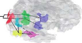
4 7 Experimental Design Regional physiology will vary systematically with the degree of cognitive or sensorimotor processing or deficits thereof. The difference between two tasks can be formulated as a separable cognitive or sensorimotor component and that regionally specific differences in haemodynamic responses, evoked by the two tasks, identify the corresponding functionally selective area. Cognitive Subtraction One tests a single hypothesis pertaining to the activation in one task relative to another. Cognitive conjunctions combine a series of subtractions, so several hypotheses are tested, asking whether the activations, in a series of task pairs, are collectively significant. 8 Consider the problem of identifying regionally specific activations due to a particular cognitive component. If one can identify a series of task pairs whose differences have only that component in common, then the region which activates, in all the corresponding subtractions, can be associated with the common component. An fmri study of visual motion processing using radially moving dots To identify areas involved in visual motion, a stationary dots condition was subtracted from the moving dots conditions. 4


5 11/8/ Image Transformation Objectives: j performing the inversion of forward or generative models of how data are caused decomposing the inversion of forward spatiotemporal models into spatial and temporal parts characterizing and removing anatomical differences A series of spatial transformations is performed to reduce unwanted variance components in the voxel time-series induced by movement or shape differences among a series of scans. Processes: realign the data undo the effects of subject movement during the scanning session spatially smooth before inverting the temporal part of the model transform into a standard anatomical space (e.g. Talairach and Tournoux, 1988) using linear or non-linear warps 5
 of the brain is defined largely by its connections.")
6 11 12 Modeling Neuroscience depends on conceptual, anatomical, statistical ti ti and causal models that link ideas about how the brain works to observed neuronal responses. The functional role of any component (e.g. cortical area, subarea or neuronal population) of the brain is defined largely by its connections. General Linear Model (GLM): expresses an observed response y in terms of a linear combination of explanatory variables in the design matrix X plus a well-behaved error term. 6
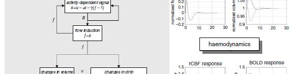
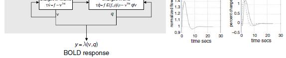
7 13 Biophysical Models 14 Dynamic Causal Modeling 7

8 15 Inference The critical issue is whether we want to make an inference about the effect in relation to the within-subject variability or with respect to the between-subject variability. This distinction relates directly to the difference between fixed and random-effect analyses. Ui Using statistics i that compare interesting i effects and the error with classical inference, Bayesian inference and other alternatives 16 8

9 17 Neurovascular Coupling Although functional neuroimaging is now widely used for noninvasively investigating human brain functions in the field of basic and clinical neuroscience, how accurately those images based on the hemodynamic principles reflect neuronal electrical activity is still not clearly understood. Experimentally, in both animal and human, the hemodynamic response has been shown to be a function of electrophysiological activity at least within a certain range, but it is seen over a relatively larger area in space than the electrophysiological activity and lasts longer in time beyond the saturation of local neuronal activity. Detailed mechanisms underlying the coupling of neuronal signals to local vasodilation have not been clarified. 18 Multimodal Neuroimaging Each technique in fntinln functional neuroimaging in based on hemodynamic principle and electrophysiology has unique features in terms of temporal and spatial resolution. The combined use of two or more techniques is expected to complement each other and thus provide more information than the use of a single technique. 1 st Modality 2 nd Modality Neuroimaging Process Information 9

10 19 Multimodal Neuroimaging In practical, the multi-modalmodal approach can be divided into two categories: separate sessions having the underlying disadvantage of uncontrolled background due to uncontrollable experimental conditions between the two sessions simultaneously sessions drawing special attention of many investigators in recent years requiring artifact elimination of interferences from one another 20 EEG-MEG EEG provides electrophysiological data with high temporal resolution but poor spatial resolution. MEG can detect only current flow in the tangential orientation. Nevertheless, MEG is not affected by shunting effect that causes distortion in EEG. Sharon et al. conducted an empirical study of brain activity under focal visual stimuli and found that the combined EEG- MEG yielded more accurate localization or cortical source estimation than either unimodality measurement. 10


11 21 EEG-PET PET scanner detects the emitted radioisotopes at the hemodynamically or metabolically activated region and constructs signal data into images. Combining two modalities from different principles in order to reveal the underlying mechanism in neurovascular coupling between hemodynamic response and electrophysiological activity 22 EEG-fMRI fmri provides images with good spatial resolution but inadequate temporal resolution. fmri is not recommended to operate on infants, toddlers and patients with inserted metallic implants. Th i l f EEG d The simultaneous use of EEG and fmri become attractive and popular in multimodal neuroimaging as the integration of high temporal resolution from EEG and high spatial resolution from fmri. 11


12 11/8/ EEG-NIRS NIRS optically measuring the changes of concentrations of oxyhemoglobin to support h l bi and d deoxyhemoglobin d h l bi in i the h tissue i neuronal activity Both are infant- and toddler-friendly technologies. Compared to fmri, NIRS has better temporal resolution and adequate spatial resolution. NIRS can be recorded concurrentlyy with EEG in a more natural manner for a longer time than fmri. Compared to EEG-fMRI, the integrative analysis of EEG-NIRS for understanding the underlying mechanisms in neurovascular coupling is still in its infancy; it has, however, great potential to improved overall spatiotemporal resolution for the purpose of human brain mapping, cognitive neuroscience studies, and development of neuroimaging diagnostic system
13 25 Research Directions of Human Brain Mapping Choosing the most appropriate pri technique available for solving each specific question Using diffusion fmri which has higher temporal resolution than the conventional blood oxygenation level-dependent (BOLD) analysis of hemodynamic response Ui Using rtms in causing plastic changes or virtual lesions to enable us to more precisely activate a small cortical area in studies Source: H. Shibasaki. Human brain mapping: Hemodynamic response and electrophysiology. Clinical Neurophysiology, vol. 119, pp , Apr References H. Shibasaki. Human brain mapping: Hemodynamic response and electrophysiology. Clinical Neurophysiology, vol. 119, pp , Apr Statistical parametric mapping: The analysis of functional brain images, Eds. Karl J. Friston, John T. Ashburner, Stefan J. Kiebel, Thomas E. Nichols, and William D. Penny, Academic Press, D. Sharon, M.H. Hämäläinen, R.B.H. Tootell, E. Halgren, and J.W. Belliveau. The advantage of combining MEG and EEG: Comparison to fmri in focally stimulated visual cortex. NeuroImage, vol. 36, pp , Jul
Beyond fmri. Joe Kable Summer Workshop on Decision Neuroscience August 21, 2009
 Beyond fmri Joe Kable Summer Workshop on Decision Neuroscience August 21, 2009 What are the strengths of fmri?! Noninvasive, safe! Can be done in humans! Verified correlate of neural activity! Great spatio-temporal
Introduction to Brain Imaging
 Introduction to Brain Imaging Human Brain Imaging NEUR 570 & BIC lecture series September 9, 2013 Petra Schweinhardt, MD PhD Montreal Neurological Institute McGill University Montreal, Canada Various techniques
Neural Correlates of Human Cognitive Function:
 Neural Correlates of Human Cognitive Function: A Comparison of Electrophysiological and Other Neuroimaging Approaches Leun J. Otten Institute of Cognitive Neuroscience & Department of Psychology University
Outline. Biological Psychology: Research Methods. Dr. Katherine Mickley Steinmetz
 Biological Psychology: Research Methods Dr. Katherine Mickley Steinmetz Outline Neuroscience Methods Histology Electrophysiological Recordings Lesion Neuroimaging Neuroanatomy Histology: Brain structure
The Central Nervous System
 The Central Nervous System Cellular Basis. Neural Communication. Major Structures. Principles & Methods. Principles of Neural Organization Big Question #1: Representation. How is the external world coded
The neurolinguistic toolbox Jonathan R. Brennan. Introduction to Neurolinguistics, LSA2017 1
 The neurolinguistic toolbox Jonathan R. Brennan Introduction to Neurolinguistics, LSA2017 1 Psycholinguistics / Neurolinguistics Happy Hour!!! Tuesdays 7/11, 7/18, 7/25 5:30-6:30 PM @ the Boone Center
MSc Neuroimaging for Clinical & Cognitive Neuroscience
 MSc Neuroimaging for Clinical & Cognitive Neuroscience School of Psychological Sciences Faculty of Medical & Human Sciences Module Information *Please note that this is a sample guide to modules. The exact
Laurent Itti: CS564 Brain Theory and Artificial Intelligence. Lecture 4: Experimental techniques in visual neuroscience. Reading Assignments: None!
 CS 564 Brain Theory and Artificial Intelligence Lecture 4: Experimental techniques in visual neuroscience Reading Assignments: None! 1 Today we will briefly review - electrophysiological recording and
COGNITIVE SCIENCE 17. Peeking Inside The Head. Part 1. Jaime A. Pineda, Ph.D.
 COGNITIVE SCIENCE 17 Peeking Inside The Head Part 1 Jaime A. Pineda, Ph.D. Imaging The Living Brain! Computed Tomography (CT)! Magnetic Resonance Imaging (MRI)! Positron Emission Tomography (PET)! Functional
Edinburgh Imaging Academy online distance learning courses. Functional Imaging
 Functional Imaging Semester 2 / Commences January 10 Credits Each Course is composed of Modules & Activities. Modules: BOLD Signal IMSc NI4R Experimental Design IMSc NI4R Pre-processing IMSc NI4R GLM IMSc
AdvAnced TMS. Research with PowerMAG Products and Application Booklet
 AdvAnced TMS Research with PowerMAG Products and Application Booklet Table of ConTenTs Introduction p. 04 Legend p. 06 Applications» navigated TMS p. 08» clinical Research p. 10» Multi-Modal TMS p. 12»
COGNITIVE NEUROSCIENCE
 HOW TO STUDY MORE EFFECTIVELY (P 187-189) Elaborate Think about the meaning of the information that you are learning Relate to what you already know Associate: link information together Generate and test
Introduction to Computational Neuroscience
 Introduction to Computational Neuroscience Lecture 10: Brain-Computer Interfaces Ilya Kuzovkin So Far Stimulus So Far So Far Stimulus What are the neuroimaging techniques you know about? Stimulus So Far
Bayesian Inference. Thomas Nichols. With thanks Lee Harrison
 Bayesian Inference Thomas Nichols With thanks Lee Harrison Attention to Motion Paradigm Results Attention No attention Büchel & Friston 1997, Cereb. Cortex Büchel et al. 1998, Brain - fixation only -
Non-Invasive Techniques
 Non-Invasive Techniques Key: Does not hurt the organism Psychology 372 Physiological Psychology Steven E. Meier, Ph.D. Listen to the audio lecture while viewing these slides or view the video presentation
Non-Invasive Techniques
 Many Procedures Non-Invasive Techniques Key: Does not hurt the organism Psychology 372 Physiological Psychology Steven E. Meier, Ph.D. Listen to the audio lecture while viewing these slides or view the
Course proposal. Neuroengineering
 Course proposal Neuroengineering Scientific Area: Bioengineering Biosignals and biomedical systems Weight: Credits: 6 ECTS Contact time: 56h Autonomous time: 112h Total time: 168h Rationale Neuroengineering
Functional MRI Mapping Cognition
 Outline Functional MRI Mapping Cognition Michael A. Yassa, B.A. Division of Psychiatric Neuro-imaging Psychiatry and Behavioral Sciences Johns Hopkins School of Medicine Why fmri? fmri - How it works Research
Nuclear imaging of the human brain
 Nuclear imaging of the human brain Steven Laureys Coma Science Group Cyclotron Research Centre & Neurology Dept. University of Liège, Belgium Neuroimaging structure function Neuroimaging: Modalities Structural
Prof. Greg Francis 1/2/19
 Brain scans PSY 200 Greg Francis Lecture 03 How to study the brain without killing someone. Scanning Technology provides insight into brain processes w EEG recordings w MRI w Non-invasive Maps of brain
An Overview of BMIs. Luca Rossini. Workshop on Brain Machine Interfaces for Space Applications
 An Overview of BMIs Luca Rossini Workshop on Brain Machine Interfaces for Space Applications European Space Research and Technology Centre, European Space Agency Noordvijk, 30 th November 2009 Definition
Development of a New Rehabilitation System Based on a Brain-Computer Interface Using Near-Infrared Spectroscopy
 Development of a New Rehabilitation System Based on a Brain-Computer Interface Using Near-Infrared Spectroscopy Takafumi Nagaoka, Kaoru Sakatani, Takayuki Awano, Noriaki Yokose, Tatsuya Hoshino, Yoshihiro
Subject: Magnetoencephalography/Magnetic Source Imaging
 01-95805-16 Original Effective Date: 09/01/01 Reviewed: 07/26/18 Revised: 08/15/18 Subject: Magnetoencephalography/Magnetic Source Imaging THIS MEDICAL COVERAGE GUIDELINE IS NOT AN AUTHORIZATION, CERTIFICATION,
Neuroimaging and Neurostimulation: Going inside the black box
 Neuroimaging and Neurostimulation: Going inside the black box Benzi M. Kluger M.D., M.S. Director, Movement Disorders Center Associate Professor of Neurology & Psychiatry University of Colorado OUTLINE
Research Perspectives in Clinical Neurophysiology
 Research Perspectives in Clinical Neurophysiology A position paper of the EC-IFCN (European Chapter of the International Federation of Clinical Neurophysiology) representing ~ 8000 Clinical Neurophysiologists
Experimental Design. Outline. Outline. A very simple experiment. Activation for movement versus rest
 Experimental Design Kate Watkins Department of Experimental Psychology University of Oxford With thanks to: Heidi Johansen-Berg Joe Devlin Outline Choices for experimental paradigm Subtraction / hierarchical
PHYSICS OF MRI ACQUISITION. Alternatives to BOLD for fmri
 PHYSICS OF MRI ACQUISITION Quick Review for fmri HST-583, Fall 2002 HST.583: Functional Magnetic Resonance Imaging: Data Acquisition and Analysis Harvard-MIT Division of Health Sciences and Technology
Computational Cognitive Neuroscience (CCN)
") introduction people!s background? motivation for taking this course? Computational Cognitive Neuroscience (CCN) Peggy Seriès, Institute for Adaptive and Neural Computation, University of Edinburgh, UK
introduction people!s background? motivation for taking this course? Computational Cognitive Neuroscience (CCN) Peggy Seriès, Institute for Adaptive and Neural Computation, University of Edinburgh, UK
Chapter 2 Knowledge Production in Cognitive Neuroscience: Tests of Association, Necessity, and Sufficiency
 Chapter 2 Knowledge Production in Cognitive Neuroscience: Tests of Association, Necessity, and Sufficiency While all domains in neuroscience might be relevant for NeuroIS research to some degree, the field
HST 583 fmri DATA ANALYSIS AND ACQUISITION
 HST 583 fmri DATA ANALYSIS AND ACQUISITION Neural Signal Processing for Functional Neuroimaging Neuroscience Statistics Research Laboratory Massachusetts General Hospital Harvard Medical School/MIT Division
Stuttering Research. Vincent Gracco, PhD Haskins Laboratories
 Stuttering Research Vincent Gracco, PhD Haskins Laboratories Stuttering Developmental disorder occurs in 5% of children Spontaneous remission in approximately 70% of cases Approximately 1% of adults with
P2 Visual - Perception
 P2 Visual - Perception 2014 SOSE Neuroimaging of high-level visual functions gyula.kovacs@uni-jena.de 11/09/06 Functional magnetic resonance imaging (fmri) The very basics What is fmri? What is MRI? The
Competing Streams at the Cocktail Party
 Competing Streams at the Cocktail Party A Neural and Behavioral Study of Auditory Attention Jonathan Z. Simon Neuroscience and Cognitive Sciences / Biology / Electrical & Computer Engineering University
Statistical parametric mapping
 350 PRACTICAL NEUROLOGY HOW TO UNDERSTAND IT Statistical parametric mapping Geraint Rees Wellcome Senior Clinical Fellow,Institute of Cognitive Neuroscience & Institute of Neurology, University College
INTRO TO BOLD FMRI FRANZ JOSEPH GALL ( ) OUTLINE. MRI & Fast MRI Observations Models Statistical Detection
 OUTLINE. MRI & Fast MRI Observations Models Statistical Detection") INTRO TO BOLD FMRI 2014 M.S. Cohen all rights reserved mscohen@g.ucla.edu OUTLINE FRANZ JOSEPH GALL (1758-1828) MRI & Fast MRI Observations Models Statistical Detection PAUL BROCA (1824-1880) WILLIAM JAMES
INTRO TO BOLD FMRI 2014 M.S. Cohen all rights reserved mscohen@g.ucla.edu OUTLINE FRANZ JOSEPH GALL (1758-1828) MRI & Fast MRI Observations Models Statistical Detection PAUL BROCA (1824-1880) WILLIAM JAMES
Biomedical Imaging: Course syllabus
 Biomedical Imaging: Course syllabus Dr. Felipe Orihuela Espina Term: Spring 2015 Table of Contents Description... 1 Objectives... 1 Skills and Abilities... 2 Notes... 2 Prerequisites... 2 Evaluation and
Chapter 5 The Research Methods of Biopsychology
 Chapter 5 The Research Methods of Biopsychology Understanding What Biopsychologists Do This multimedia product and its contents are protected under copyright law. The following are prohibited by law: any
Experimental design for Cognitive fmri
 Experimental design for Cognitive fmri Alexa Morcom Edinburgh SPM course 2017 Thanks to Rik Henson, Thomas Wolbers, Jody Culham, and the SPM authors for slides Overview Categorical designs Factorial designs
3/1/18. Overview of the Talk. Important Aspects of Neuroimaging Technology
 3/1/18 Considerations for the Use of Neuroimaging for Predicting Recovery of Speech and Language in Aphasia Linda I. Shuster, Ph.D., CCC-SLP Overview of the Talk Important aspects of neuroimaging technology
fmri: What Does It Measure?
 fmri: What Does It Measure? Psychology 355: Cognitive Psychology Instructor: John Miyamoto 04/02/2018: Lecture 02-1 Note: This Powerpoint presentation may contain macros that I wrote to help me create
Statistical Analysis of Sensor Data
 Statistical Analysis of Sensor Data Stefan Kiebel Max Planck Institute for Human Cognitive and Brain Sciences Leipzig, Germany Overview 1 Introduction 2 Within-subject analysis 3 Between-subject analysis
Physiological and Physical Basis of Functional Brain Imaging 6. EEG/MEG. Kâmil Uludağ, 20. November 2007
 Physiological and Physical Basis of Functional Brain Imaging 6. EEG/MEG Kâmil Uludağ, 20. November 2007 Course schedule 1. Overview 2. fmri (Spin dynamics, Image formation) 3. fmri (physiology) 4. fmri
Announcements. Final Exam will be a take-home exam. Format similar to the short assignment (no multiple choice, etc.)
") Announcements Final Exam will be a take-home exam Format similar to the short assignment (no multiple choice, etc.) Will be handed out at end of last class period (Thursday June 5 th ) Due by 6 pm June
Announcements Final Exam will be a take-home exam Format similar to the short assignment (no multiple choice, etc.) Will be handed out at end of last class period (Thursday June 5 th ) Due by 6 pm June
Methods for assessing the brain basis of developmental disorders
 Announcements LIGN171: Child Language Acquisition http://ling.ucsd.edu/courses/lign171 Final Exam will be a take-home exam Format similar to the short assignment (no multiple choice, etc.) Will be handed
Experimental Design. Thomas Wolbers Space and Aging Laboratory Centre for Cognitive and Neural Systems
 Experimental Design Thomas Wolbers Space and Aging Laboratory Centre for Cognitive and Neural Systems Overview Design of functional neuroimaging studies Categorical designs Factorial designs Parametric
Comparing event-related and epoch analysis in blocked design fmri
 Available online at www.sciencedirect.com R NeuroImage 18 (2003) 806 810 www.elsevier.com/locate/ynimg Technical Note Comparing event-related and epoch analysis in blocked design fmri Andrea Mechelli,
Neurophysiological Basis of TMS Workshop
 Neurophysiological Basis of TMS Workshop Programme 31st March - 3rd April 2017 Sobell Department Institute of Neurology University College London 33 Queen Square London WC1N 3BG Brought to you by 31 March
Daniel Bulte. Centre for Functional Magnetic Resonance Imaging of the Brain. University of Oxford
 Daniel Bulte Centre for Functional Magnetic Resonance Imaging of the Brain University of Oxford Overview Signal Sources BOLD Contrast Mechanism of MR signal change FMRI Modelling Scan design details Factors
WHAT DOES THE BRAIN TELL US ABOUT TRUST AND DISTRUST? EVIDENCE FROM A FUNCTIONAL NEUROIMAGING STUDY 1
 SPECIAL ISSUE WHAT DOES THE BRAIN TE US ABOUT AND DIS? EVIDENCE FROM A FUNCTIONAL NEUROIMAGING STUDY 1 By: Angelika Dimoka Fox School of Business Temple University 1801 Liacouras Walk Philadelphia, PA
Methods for Seeing the Brain (http://www.pbs.org/wnet/brain/scanning/)
") Mind-Brain There are 2 broad approaches to connecting brain and cognitive activity: 1. (modern) Imaging techniques 2. (classic) Patients with brain trauma H. Jackson, A.R. Luria, O. Sacks Methods for Seeing
Mind-Brain There are 2 broad approaches to connecting brain and cognitive activity: 1. (modern) Imaging techniques 2. (classic) Patients with brain trauma H. Jackson, A.R. Luria, O. Sacks Methods for Seeing
Neuroimaging and Assessment Methods
 Psych 2200, Lecture 5 Experimental Design and Brain Imaging Methods Tues Sept 15, 2015 Revised TA office hours (Sam), today 4-5p, and wed 11:30-1:30. I will not have office hours this thurs but you should
Concurrent near-infrared spectroscopy (NIRS) and functional magnetic resonance imaging (fmri) of the brain
 and functional magnetic resonance imaging (fmri) of the brain") Motor cortex activation fmri Near-infrared imaging Concurrent near-infrared spectroscopy (NIRS) and functional magnetic resonance imaging (fmri) of the brain Sergio Fantini s group, Department of Biomedical
Motor cortex activation fmri Near-infrared imaging Concurrent near-infrared spectroscopy (NIRS) and functional magnetic resonance imaging (fmri) of the brain Sergio Fantini s group, Department of Biomedical
Cerebral Cortex 1. Sarah Heilbronner
 Cerebral Cortex 1 Sarah Heilbronner heilb028@umn.edu Want to meet? Coffee hour 10-11am Tuesday 11/27 Surdyk s Overview and organization of the cerebral cortex What is the cerebral cortex? Where is each
Supporting Information
 Supporting Information Moriguchi and Hiraki 10.1073/pnas.0809747106 SI Text Differences in Brain Activation Between Preswitch and Postswitch Phases. The paired t test was used to compare the brain activation
Large, High-Dimensional Data Sets in Functional Neuroimaging
 Goals of Functional Neuroimaging Identify Regional Specializations of the Brain Large, High-Dimensional Data Sets in Functional Neuroimaging 1 Goals of Functional Neuroimaging Goals of Functional Neuroimaging
Causality from fmri?
 Causality from fmri? Olivier David, PhD Brain Function and Neuromodulation, Joseph Fourier University Olivier.David@inserm.fr Grenoble Brain Connectivity Course Yes! Experiments (from 2003 on) Friston
Brain and Cognition. Cognitive Neuroscience. If the brain were simple enough to understand, we would be too stupid to understand it
 Brain and Cognition Cognitive Neuroscience If the brain were simple enough to understand, we would be too stupid to understand it 1 The Chemical Synapse 2 Chemical Neurotransmission At rest, the synapse
Inverse problems in functional brain imaging Identification of the hemodynamic response in fmri
 Inverse problems in functional brain imaging Identification of the hemodynamic response in fmri Ph. Ciuciu1,2 philippe.ciuciu@cea.fr 1: CEA/NeuroSpin/LNAO May 7, 2010 www.lnao.fr 2: IFR49 GDR -ISIS Spring
Biomedical Research 2013; 24 (3): ISSN X
: ISSN X") Biomedical Research 2013; 24 (3): 359-364 ISSN 0970-938X http://www.biomedres.info Investigating relative strengths and positions of electrical activity in the left and right hemispheres of the human brain
Biomedical Research 2013; 24 (3): 359-364 ISSN 0970-938X http://www.biomedres.info Investigating relative strengths and positions of electrical activity in the left and right hemispheres of the human brain
Computational Cognitive Neuroscience (CCN)
") How are we ever going to understand this? Computational Cognitive Neuroscience (CCN) Peggy Seriès, Institute for Adaptive and Neural Computation, University of Edinburgh, UK Spring Term 2010 Practical
How are we ever going to understand this? Computational Cognitive Neuroscience (CCN) Peggy Seriès, Institute for Adaptive and Neural Computation, University of Edinburgh, UK Spring Term 2010 Practical
Neural Modeling and Functional Brain Imaging: An Overview. Barry Horwitz Brain Imaging & Modeling Section NIDCD, NIH
 Neural Modeling and Functional Brain Imaging: An Overview Barry Horwitz Brain Imaging & Modeling Section NIDCD, NIH Methods to Understand Neural Basis of Human Cognition 1. Brain lesions & cognitive neuropsychology
FALSE POSITIVES IN FUNCTIONAL NEAR- INFRARED TOPOGRAPHY
 FALSE POSITIVES IN FUNCTIONAL NEAR- INFRARED TOPOGRAPHY Ilias Tachtsidis 1, Terence S. Leung 1, Anchal Chopra 1, Peck H. Koh 1, Caroline B. Reid 1, and Clare E. Elwell 1 Abstract: Functional cranial near-infrared
The Cognitive Neuroscientist s Toolkit
 The Cognitive Neuroscientist s Toolkit Jesse Rissman CS 182 Guest Lecture, 2/1/07 QuickTimeª and a TIFF (Uncompressed) decompressor are needed to see this picture. LESION PET fmri EEG/MEG TMS A little
LESSON 1.3 WORKBOOK. How can we study the behaving brain?
 LESSON 1.3 WORKBOOK How can we study the behaving brain? We are in the middle of a technological revolution when it comes to how closely we can look at the behaving brain. Scientists and doctors now have
Methods of Visualizing the Living Human Brain
 Methods of Visualizing the Living Human Brain! Contrast X-rays! Computerized Tomography (CT)! Magnetic Resonance Imaging (MRI)! Positron Emission Tomography (PET)! Functional MRI! Magnetoencephalography
NEUROSCIENCE. W1 Divisions of the nervous system PSYC1002 NOTES
 PSYC1002 NOTES NEUROSCIENCE W1 Divisions of the nervous system Nervous system: - CNS o Brain and spinal cord - Peripheral Nervous System o Sensory nerves o Motor nerves o Autonomic nervous system o Enteric
Magnetic resonance imaging (MRI) is a noninvasive
 is a noninvasive") Incidental Findings in Magnetic Resonance Imaging (MRI) Brain Research Charles A. Nelson Magnetic resonance imaging (MRI) is a noninvasive imaging tool that utilizes a strong magnetic field and radio frequency
Incidental Findings in Magnetic Resonance Imaging (MRI) Brain Research Charles A. Nelson Magnetic resonance imaging (MRI) is a noninvasive imaging tool that utilizes a strong magnetic field and radio frequency
EEG Wave of the Future: The Video-EEG and fmri Suite?
 Current Literature In Clinical Science EEG Wave of the Future: The Video-EEG and fmri Suite? Mapping Preictal and Ictal Haemodynamic Networks Using Video-Electroencephalography and Functional Imaging.
Computational Cognitive Neuroscience (CCN)
") How are we ever going to understand this? Computational Cognitive Neuroscience (CCN) Peggy Seriès, Institute for Adaptive and Neural Computation, University of Edinburgh, UK Spring Term 2013 Practical
How are we ever going to understand this? Computational Cognitive Neuroscience (CCN) Peggy Seriès, Institute for Adaptive and Neural Computation, University of Edinburgh, UK Spring Term 2013 Practical
Brain Activity Measurement during Program Comprehension with NIRS
 Brain Activity Measurement during Program Comprehension with NIRS Yoshiharu Ikutani Department of Information Engineering Nara National College of Technology Japan, Nara, Yamatokoriyama Email: ikutani@info.nara-k.ac.jp
Experimental design. Alexa Morcom Edinburgh SPM course Thanks to Rik Henson, Thomas Wolbers, Jody Culham, and the SPM authors for slides
 Experimental design Alexa Morcom Edinburgh SPM course 2013 Thanks to Rik Henson, Thomas Wolbers, Jody Culham, and the SPM authors for slides Overview of SPM Image Image timeseries timeseries Design Design
The Tools: Imaging the Living Brain
 The Tools: Imaging the Living Brain I believe the study of neuroimaging has supported the localization of mental operations within the human brain. -Michael I. Posner, 2003 Neuroimaging methods Since Descarte
Population Inference post Model Selection in Neuroscience
 Population Inference post Model Selection in Neuroscience Genevera I. Allen Dobelman Family Junior Chair, Department of Statistics and Electrical and Computer Engineering, Rice University, Department of
Neurovascular Physiology and Pathophysiology
 Neurovascular Physiology and Pathophysiology The physiological questions aim at understanding the molecular and biochemical mechanisms, by which the brain adapts local blood flow to neuronal activity and
Interpretation of clinical functional neuroimaging studies
 Interpretation of clinical functional neuroimaging studies 2 Geoffrey K Aguirre INTRODUCTION The first functional magnetic resonance imaging (fmri) scans of a human being were obtained on a clinical MRI
Classification and Statistical Analysis of Auditory FMRI Data Using Linear Discriminative Analysis and Quadratic Discriminative Analysis
 International Journal of Innovative Research in Computer Science & Technology (IJIRCST) ISSN: 2347-5552, Volume-2, Issue-6, November-2014 Classification and Statistical Analysis of Auditory FMRI Data Using
Molecular Imaging and the Brain
 Molecular imaging technologies are playing an important role in neuroimaging, a branch of medical imaging, by providing a window into the living brain. Where CT and conventional MR imaging provide important
MEDICAL REVIEW Functional Magnetic Resonance Imaging: From Acquisition to Application
 Functional Magnetic Resonance Imaging: From Acquisition to Application Gail Yarmish * and Michael L. Lipton *, Departments of Radiology *, and Neuroscience Albert Einstein College of Medicine Bronx, New
Combining tdcs and fmri. OHMB Teaching Course, Hamburg June 8, Andrea Antal
 Andrea Antal Department of Clinical Neurophysiology Georg-August University Goettingen Combining tdcs and fmri OHMB Teaching Course, Hamburg June 8, 2014 Classical Biomarkers for measuring human neuroplasticity
A U. Methods of Cognitive Neuroscience 2/8/2016. Neat Stuff! Cognitive Psychology
 Methods of Cognitive Neuroscience Neat Stuff! Optogenetics http://spie.org/newsroom/technical-articles-archive/videos/0411-boyden Stimulating the brain with light Cognitive Psychology Mental Representation
Experimental Design I
 Experimental Design I Topics What questions can we ask (intelligently) in fmri Basic assumptions in isolating cognitive processes and comparing conditions General design strategies A few really cool experiments
Oximeters. Hsiao-Lung Chan, Ph.D. Dept Electrical Engineering Chang Gung University, Taiwan
 Oximeters Hsiao-Lung Chan, Ph.D. Dept Electrical Engineering Chang Gung University, Taiwan chanhl@mail.cgu.edu.tw Oxygen transport in blood Hemoglobin Oxygen O Deoxygen +O O Oximeters Arterial saturation
Define functional MRI. Briefly describe fmri image acquisition. Discuss relative functional neuroanatomy. Review clinical applications.
 Dr. Peter J. Fiester November 14, 2012 Define functional MRI. Briefly describe fmri image acquisition. Discuss relative functional neuroanatomy. Review clinical applications. Briefly discuss a few examples
Optical Illusions and Human Visual System: Can we reveal more? CIS Micro-Grant Executive Report and Summary of Results
 Optical Illusions and Human Visual System: Can we reveal more? CIS Micro-Grant 2011 Executive Report and Summary of Results Prepared By: Principal Investigator: Siddharth Khullar 1, Ph.D. Candidate (sxk4792@rit.edu)
PRESS NOTES YORK INSTRUMENTS Last updated 2018
 PRESS NOTES YORK INSTRUMENTS Last updated 2018 York Instruments: Company Profile York Instruments is a medical technology company set to radically transform the field of functional brain imaging. The company
Statistical Analysis Methods for the fmri Data
 Basic and Clinical Summer 2011, Volume 2, Number 4 Statistical Analysis Methods for the fmri Data Mehdi Behroozi 1, Mohammad Reza Daliri 1, Huseyin Boyaci 2 1. Biomedical Engineering Department, Faculty
Incorporation of Imaging-Based Functional Assessment Procedures into the DICOM Standard Draft version 0.1 7/27/2011
 Incorporation of Imaging-Based Functional Assessment Procedures into the DICOM Standard Draft version 0.1 7/27/2011 I. Purpose Drawing from the profile development of the QIBA-fMRI Technical Committee,
Supplementary Online Content
 Supplementary Online Content Gregg NM, Kim AE, Gurol ME, et al. Incidental cerebral microbleeds and cerebral blood flow in elderly individuals. JAMA Neurol. Published online July 13, 2015. doi:10.1001/jamaneurol.2015.1359.
Sum of Neurally Distinct Stimulus- and Task-Related Components.
 SUPPLEMENTARY MATERIAL for Cardoso et al. 22 The Neuroimaging Signal is a Linear Sum of Neurally Distinct Stimulus- and Task-Related Components. : Appendix: Homogeneous Linear ( Null ) and Modified Linear
Neuroimaging methods vs. lesion studies FOCUSING ON LANGUAGE
 Neuroimaging methods vs. lesion studies FOCUSING ON LANGUAGE Pioneers in lesion studies Their postmortem examination provided the basis for the linkage of the left hemisphere with language C. Wernicke
Experimental design. Experimental design. Experimental design. Guido van Wingen Department of Psychiatry Academic Medical Center
 Experimental design Guido van Wingen Department of Psychiatry Academic Medical Center guidovanwingen@gmail.com Experimental design Experimental design Image time-series Spatial filter Design matrix Statistical
BOLD signal dependence on blood flow and metabolism. Outline
 BOLD signal dependence on blood flow and metabolism R. Hoge, MGH NMR Center Outline physiological events accompanying neuronal activation factors affecting BOLD signal sensitivity BOLD response dynamics
Basic concepts and overview
 1 Basic concepts and overview Karl J. Friston Contents I. Introduction II. Principles of functional organization and implications for imaging II.A. Specialisation or integration? II.B. Functional integration
ASSUMPTION OF COGNITIVE UNIFORMITY
 The Human Brain cerebral hemispheres: two most important divisions of the brain, separated by the longitudinal fissure corpus callosum: a large bundle of axons that constitutes the major connection between
Comparison of Neuronal and Hemodynamic Measures of the Brain Response to Visual Stimulation: An Optical Imaging Study
 Human Brain Mapping 13:13 25(2001) Comparison of Neuronal and Hemodynamic Measures of the Brain Response to Visual Stimulation: An Optical Imaging Study Gabriele Gratton,* Marsha Ruth Goodman-Wood, and
Est-ce que l'eeg a toujours sa place en 2019?
 Est-ce que l'eeg a toujours sa place en 2019? Thomas Bast Epilepsy Center Kork, Germany Does EEG still play a role in 2019? What a question 7T-MRI, fmri, DTI, MEG, SISCOM, Of ieeg course! /HFO, Genetics
Brain Imaging Techniques
 2 Brain Imaging Techniques A D I N A L. R O S K I E S 1. I N T R O D U C T I O N Several recently developed techniques allow researchers to investigate the neural basis of cognitive function in the living
Intrinsic Signal Optical Imaging
 Intrinsic Signal Optical Imaging Introduction Intrinsic signal optical imaging (ISOI) is a technique used to map dynamics in single cells, brain slices and even and most importantly entire mammalian brains.
Experimental design of fmri studies
 Experimental design of fmri studies Kerstin Preuschoff Computational Neuroscience Lab, EPFL LREN SPM Course Lausanne April 10, 2013 With many thanks for slides & images to: Rik Henson Christian Ruff Sandra