Advanced Imaging Technology. Chamaree Chuapetcharasopon, MD TIMAC-11, 13 March 2013
|
|
|
- Bruce Cobb
- 5 years ago
- Views:
Transcription
1 Advanced Imaging Technology Chamaree Chuapetcharasopon, MD TIMAC-11, 13 March 2013
2 Advanced Imaging Technology Radiology Cardiology Gastroenterology OB


3 NEW 4D Ultrasound OB

4

5
6 Bronchoscopy Endoscopic imaging of the lung Flexible fiberoptic scope Patient is conscious and aware Exam tend to be repeated at follow up Multiple light source, fluorescence

7
8 Otolaryngology Endoscopic imaging of the ear nose and throat Numerous types and styles of endoscopes Patient is conscious and aware Exams tend to be repeated at follow up Video frame rates can reach 200 fps Requires synchronized audio

9

10

11

12
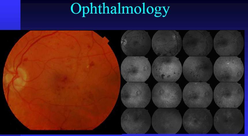
13 Ophthalmology Many types of image devices Non dilated, Non touch examination 30
14
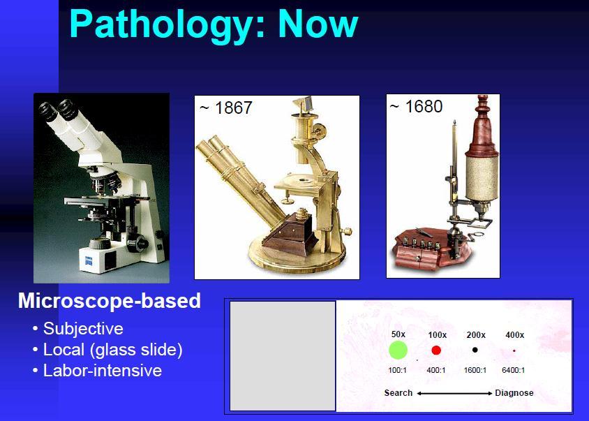
15 Pathology Anatomical Pathology Require high definition details Better than 0.1 micron resolution Rapid (subminute) image gathering True color >20 GB images
16


17


18 Dr Keith Foord East Sussex Hospitals, UK


19 Dr Keith Foord East Sussex Hospitals, UK


20 40 Dr Keith Foord East Sussex Hospitals, UK


21 Image users real time guided therapy and surgery Sinus surgery. The cross lines show The position of the probe on the corresponding CT in x,y and z axes midturbvirtendosc.jpg GE Medical Systems


22 Digital OR Images BrainLAB AG Smith and Nephew website :UPMC Southside Hospital
23 Advanced Imaging Technology Gastroenterology Orthopedics Otolaryngology Pathology Cardiology OB-Gyn Radiology
24 Advanced Imaging Technology Terminology General Radiology ร งส ว ทยาท วไป Diagnostic Radiology ร งส ว น จฉ ย Interventional Radiology ร งส ร วมร กษา Radiation Oncology (Therapy) ร งส ร กษา Nuclear Medicine เวชศาสตร น วเคล ยร
25 Advanced Imaging Technologies IT World ( Information Technology ) Digital Data Data Transfer
26 Advanced Imaging Technology Analog to Digital Analog: Camera + Film Post Processing Picture Digital: Digital Camera + Data storage Data Transfer Computer Display Display Monitor Printer Internets Save file CD ROM Hard Drive Picture Scanner Digital Data
27 Advanced Imaging Technology Analog: Plain film Mammogram Special Procedure Upper GI, Barium Enema, IVP, Angiogram Ultrasound ( US ) NM ( Nuclear Medicine ) PET Scan ( Positron Emission Tomography ) Digital CR, DR FFDM Full field digital mammogram CT Tomosynthesis MRI
28 Advanced Imaging Technology Digital Data: CT ( Computed Tomography ) MRI ( Magnetic resonance imaging ) NM ( Nuclear Medicine ) PET (Positron Emission Tomography) CR, DR (computed radiograph, digital radiograph) Mammogram Ultrasound DSI (digital subtraction imaging) DSA (digital subtraction angiogram)
29 Advanced Imaging Technology CR/DR CT scan MRI PET/CT PET/MRI HIFU


30 Old days

31 Introduction to CR

32 Introduction to CR


33 Introduction to CR and display monitors


34 DR Wired Wireless


35
36 Advanced Imaging Technology CT scan (Computerized tomography) Conventional CT Spiral, helical CT 64 slices 256 slices 640 slices Dual sources


37 CT Scan Third generation Conventional Spiral

38 MSCT New Concepts to Understand

39


40 Many vendors


41 Example of CT work place


42 CAC Scoring

43 CT Contrast study


44 LAD disease

45 Volume Rendering Whole heart images

46 CT Chest Whole chest images


47


48 Other clinical applications
49 Advanced Imaging Technology MRI Low field, mid filed, high field 1.5 Tesla 3 Tesla Open MRI Standing MRI


50 MRI High field MR machine

51 Phase array body coil


52 Different images of the same territory T1 T2 Proton density


53 MRI CT MR and CT of HCC cirrhotic liver
54 Hemangioma


55 MRCP


56 MRCP


57 Neurovascular application


58 Nerve fiber tracking


59 Peripheral vascular coil

60 Peripheral vascular images


61 Contrast Enhanced MRA
62 Advanced Imaging Technology PET/CT PET/MRI


63 PET/CT (Positron Emission Tomography/ Computerized Tomography)
 Anato-metabolic Information Anatomy,")


64 Anatomy vs Function Anatomical (CT) Functional (PET) Anato-metabolic Information Anatomy, morphology Function, metabolism Anatomy+Function Detection Size, morphology Altered metabolism Size, Location, Metabolism Image details No or indirect Little, or no anatomy Function+Anatomy function
65 Principle of PET e + Neutron deficient isotope: unstable Positron emission Positron meets electron:annihilation 511 kev -ray e kev -ray Emission is on straight line!

66
67 Uptake of FDG Kapoor, V. et al. Radiographics 2004;24:


68 PET/CT Fused PET/CT FORE AWOSEM CT PET Upper limit Topogram Lower limit CT acquisition attenuation correction scatter correction
69


70 Why PET/CT? - Difference from other Modality Anatomy vs Function X-ray CT MR PET

71 BI PET CT SIEMENS PET-CT (64 SLICES)


72 Evaluation Radionuclides given Having PET/CT Scan
73 Indications for PET imaging with FDG Breast cancer Cervical cancer Colorectal cancer Esophageal cancer Head and neck cancer (non-thyroid, non-cns) Lymphoma Melanoma Non small cell lung cancer Solitary pulmonary nodules Follicular cell thyroid cancer
74 Radionuclides Radionuclides used in PET scanning are typically isotopes with short half-lives carbon-11 (~20 min), nitrogen-13 (~10 min), oxygen-15 (~2 min), fluorine-18 (~110 min) rubidum-82(~1.27 min).


75 PET/MRI
76 Advanced Imaging Technology CR/DR CT scan MRI PET/CT PET/MRI HIFU
77 What is HIFU? (High Intensity Focused Ultrasound) High energy focused ultrasound beam. Brought to a tight focus in tissue at a distance from the source. Absorption of the energy leads to tissue heating. Causes localized temperature rise at the focal point. Sharply demarcated area of coagulative necrosis. No damage to overlying and surrounding tissue.
 Skin Tumour")

78 Lesion of coagulation necrosis at focus (12x3mm) Skin Tumour Target organ (e.g. liver) Transducer Undamaged tissue in front of focus
79 HIFU 3-Dimensional Therapy Plan One pulse of HIFU exposure Multiple-pulse Scanning beam Line Volume Slice

80 Mechanisms of HIFU Ablation Thermal Effect --Heat (56-100⁰ C)

81
82 Clinical Advantages of HIFU HIFU does not suffer from the following problems associated with Radiotherapy & Chemotherapy: Lethargy Sickness Lowered immunity to infection Hair loss Haematological disorders Multiple hospital visits
83 Clinical Advantages of HIFU One time Treatment Real-time US-guided Procedure Low cost Non Toxic Not limited by tumour size Short hospital stay (one day) Minor complications
 3. Frequency Generator and amplifier 4.")
![ψ-motion Device (rotates transducer about y-axis [transverse]) 5.](/docs-images/94/122313180/images/84-1.jpg "Integrated Treatment Transducer (diagnostic US probe and HIFU transducer) in degassed water reservoir 6.")
84 Operator Console 2. γ-motion Device (rotates transducer about x-axis [long axis of bed]) 3. Frequency Generator and amplifier 4. ψ-motion Device (rotates transducer about y-axis [transverse]) 5. Integrated Treatment Transducer (diagnostic US probe and HIFU transducer) in degassed water reservoir 6. 4-dimension Motion Device (translates transducer in x, y and z, rotates plane of diagnostic image) 7. Treatment Bed

85 Model-JC Focused Ultrasound Tumour Therapy System Regulatory Approvals: Europe: CE, MHRA China: SFDA Korea: KFDA


86


87 New Haifu Products for Clinical Trial An MRI-guided Extracorporeal HIFU Device (Haifu-Siemens)
88 Minimal invasive treatment of malignant hepatic tumors 1. Radio-frequency ablation 2. Microwave ablation 3. Laser ablation 4. Cryoablation 5. Ethanol ablation 6. Chemoembolization


89 Minimal invasive treatment of malignant hepatic tumors Rita 15 G, 4-8 Prong, Radiotherapeutics 14 G10 Prong Radionics17 G/3- needle


90 Minimal invasive treatment of malignant hepatic tumors CC/03


91 Minimal invasive treatment of malignant hepatic tumors


92 Minimal invasive treatment of malignant hepatic tumors


93 Minimal invasive treatment of malignant hepatic tumors


94 Minimal invasive treatment of malignant hepatic tumors

95 Minimal invasive treatment of malignant hepatic tumors TACE


96 Minimal invasive treatment of malignant hepatic tumors CC/03


97 Minimal invasive treatment of malignant hepatic tumors CC/03

98
99 Advanced Imaging Technology Take home messages New technologies occur every day. How to use them appropriately? We (who?) need to consider Appropriateness criteria Cost effectiveness Patient benefit

100 Thank you for your attention
Index. Surg Oncol Clin N Am 16 (2007) Note: Page numbers of article titles are in boldface type.
 Note: Page numbers of article titles are in boldface type.") Surg Oncol Clin N Am 16 (2007) 465 469 Index Note: Page numbers of article titles are in boldface type. A Adjuvant therapy, preoperative for gastric cancer, staging and, 339 B Breast cancer, metabolic
Surg Oncol Clin N Am 16 (2007) 465 469 Index Note: Page numbers of article titles are in boldface type. A Adjuvant therapy, preoperative for gastric cancer, staging and, 339 B Breast cancer, metabolic
Molecular Imaging and Cancer
 Molecular Imaging and Cancer Cancer causes one in every four deaths in the United States, second only to heart disease. According to the U.S. Department of Health and Human Services, more than 512,000
Nuclear Medicine and PET. D. J. McMahon rev cewood
 Nuclear Medicine and PET D. J. McMahon 150504 rev cewood 2018-02-15 Key Points Nuclear Medicine and PET: Imaging: Understand how Nuc Med & PET differ from Radiography & CT by the source of radiation. Be
Certification Review. Module 28. Medical Coding. Radiology
 Module 28 is the study of x-rays, using radiant energy and other imaging techniques, such as resonance imaging or ultrasound, to diagnose illnesses and diseases. Vocabulary Barium enema (BE): lower gastrointestinal
PET IMAGING (POSITRON EMISSION TOMOGRAPY) FACT SHEET
 FACT SHEET") Positron Emission Tomography (PET) When calling Anthem (1-800-533-1120) or using the Point of Care authorization system for a Health Service Review, the following clinical information may be needed to
Positron Emission Tomography (PET) When calling Anthem (1-800-533-1120) or using the Point of Care authorization system for a Health Service Review, the following clinical information may be needed to
Head and Neck Cancer. What is head and neck cancer?
 Scan for mobile link. Head and Neck Cancer Head and neck cancer is a group of cancers that usually originate in the squamous cells that line the mouth, nose and throat. Typical symptoms include a persistent
created by high-voltage devices Examples include medical and dental x-rays, light, microwaves and nuclear energy
 What is radiation? Radiation is energy emitted from a source, that travels through space and can penetrate matter. Listed below are two types that we are exposed to and contribute to our overall radiation
Improving Methods for Breast Cancer Detection and Diagnosis. The National Cancer Institute (NCI) is funding numerous research projects to improve
 is funding numerous research projects to improve") CANCER FACTS N a t i o n a l C a n c e r I n s t i t u t e N a t i o n a l I n s t i t u t e s o f H e a l t h D e p a r t m e n t o f H e a l t h a n d H u m a n S e r v i c e s Improving Methods for
CANCER FACTS N a t i o n a l C a n c e r I n s t i t u t e N a t i o n a l I n s t i t u t e s o f H e a l t h D e p a r t m e n t o f H e a l t h a n d H u m a n S e r v i c e s Improving Methods for
Breast Cancer PET/CT Imaging Protocol
 Breast Cancer PET/CT Imaging Protocol Scanning Protocol: Patients are scanned from the top of the neck through the pelvis. Arms-up position is used to avoid beam-hardening artifact in the chest and abdomen.
RADIOLOGY (MEDICAL IMAGING)
") RADIOLOGY (MEDICAL IMAGING) Radiology is the study of the diagnosis of disease by the use of radiant energy (radiation). In the past this meant the use of X-rays to make an image. Today many other forms
RADIOLOGY (MEDICAL IMAGING) Radiology is the study of the diagnosis of disease by the use of radiant energy (radiation). In the past this meant the use of X-rays to make an image. Today many other forms
High Tech Imaging Quick Reference Guide
 High Tech Imaging Quick Reference Guide 1 High Tech Imaging Authorizations may now be requested through our secure provider portal, BlueAccess. Getting Started Step 1: Log into BlueAccess from www.bcbst.com
What is head and neck cancer? How is head and neck cancer diagnosed and evaluated? How is head and neck cancer treated?
 Scan for mobile link. Head and Neck Cancer Head and neck cancer is a group of cancers that start in the oral cavity, larynx, pharynx, salivary glands, nasal cavity or paranasal sinuses. They usually begin
HSC Physics. Module 9.6. Medical Physics
 HSC Physics Module 9.6 Medical Physics Contextual Outline 9.6 Medical Physics (28 indicative hours) The use of other advances in technology, developed from our understanding of the electromagnetic spectrum,
Anthem Blue Cross and Blue Shield Virginia Advanced Imaging Procedures Requiring Precertification Revised 02/13/2013
 Anthem Blue Cross and Blue Shield Virginia Advanced Imaging Procedures Requiring Precertification Revised 02/13/2013 Modality and CT Head CTA Head: Cerebrovascular MRI Head MRA Head: Cerebrovascular Functional
COMENIUS-Project: SM&CLIL Radiation & Medicine
 Medical imaging refers to the techniques and processes used to create images of the human body (or parts thereof) for clinical purposes. Thanks to modern mathematics and computer technology, medical imaging
Appendix A: Introduction to Imaging Modalities for Which Data Were Collected in the 2017 Imaging Inventory
 Appendix A: Introduction to Imaging Modalities for Which Data Were Collected in the 207 Imaging Inventory Computed Tomography Computed tomography (CT) employs X-rays as a source of ionizing radiation,
Medical Diagnostic Imaging
 Medical Diagnostic Imaging Laboratories Medical Diagnostic Imaging Lab Name Location Person in Charge Programs Served Courses Served Patient Care and Management (2) Introduction to MDI Radiographic Technique
Cardiac Imaging Tests
 Cardiac Imaging Tests http://www.medpagetoday.com/upload/2010/11/15/23347.jpg Standard imaging tests include echocardiography, chest x-ray, CT, MRI, and various radionuclide techniques. Standard CT and
Chapter 16 Worksheet Code It
 Name: Class: Date: ID: A Chapter 16 Worksheet 3 2 1 Code It True/False Indicate whether the statement is true or false. 1. CT scans generate three-dimensional images. 2. An ultrasound produces images of
Austin Radiological Association Ga-68 NETSPOT (Ga-68 dotatate)
") Austin Radiological Association Ga-68 NETSPOT (Ga-68 dotatate) Overview Ga-68 dotatate binds to somatostatin receptors, with highest affinity for subtype 2 receptors (sstr2). It binds to cells that express
Austin Radiological Association Ga-68 NETSPOT (Ga-68 dotatate) Overview Ga-68 dotatate binds to somatostatin receptors, with highest affinity for subtype 2 receptors (sstr2). It binds to cells that express
Therapy - Minimize harm to patients
 Therapy - Minimize harm to patients CHONGQING HAIFU MEDICAL TECHNOLOGY CO., LTD Tel: 86-23-6788 6799/6788 6195/6788 6199 Fax: 86-23-6788 6168 Email: sales@hifu.cn Website: www.haifumedical.com Address:
HEALTHFIRST 2011 RADIOLOGY PROGRAM CODE LIST
 HEALTHFIRST 2011 RADIOLOGY PROGRAM CODE LIST Outpatient Radiology utilization call Carecore at 1-877-773-6964 Modality CPT CODE Description CT SCANS 70450 CT HEAD/BRAIN W/O CONTRAST CT SCANS 70460 CT HEAD/BRAIN
Radiologic Imaging Magnetic Resonance Imaging (MRI)
") Radiologic Imaging X-ray has always been the golden rule in diagnosing and treating podiatric patients. Unfortunately, for some patients the diagnosis is not as evident. That is when we need to utilize
Radiologic Imaging X-ray has always been the golden rule in diagnosing and treating podiatric patients. Unfortunately, for some patients the diagnosis is not as evident. That is when we need to utilize
Life saver 9. Life saver 9. Life saver 9. Life saver 8. Mobility of device 3. Mobility of device 1. PET scan. Ultra Violet Lamp. MRI scanner.
 Ultra Violet Lamp MRI scanner Used to stop jaundice in babies helping the tiny liver clean the blood. Also used to treat people with TB, Lupus or even treat acne. Mobility of device 3 Uses magnets to change
ADI Procedure Codes. August 2016 Revised April 2017 Page 1 of 7 ADI Procedure Codes
 Code Description 70450 CT Head without contrast 70460 CT Head with contrast 70470 CT Head with & without contrast 70480 CT Orbit, et al without contrast 70481 CT Orbit, et al with contrast 70482 CT Orbit,
POSITRON EMISSION TOMOGRAPHY (PET)
") Status Active Medical and Behavioral Health Policy Section: Radiology Policy Number: V-27 Effective Date: 08/27/2014 Blue Cross and Blue Shield of Minnesota medical policies do not imply that members should
Status Active Medical and Behavioral Health Policy Section: Radiology Policy Number: V-27 Effective Date: 08/27/2014 Blue Cross and Blue Shield of Minnesota medical policies do not imply that members should
Arteriogram An X-ray of an artery after the injection of dye.
 A Abscess A localized collection of pus in any part of the body, usually surrounded by inflamed tissue. Anesthetic An agent that causes loss of sensation with or without the loss of consciousness. Angiography,
Typical PET Image. Elevated uptake of FDG (related to metabolism) Lung cancer example: But where exactly is it located?
 Lung cancer example: But where exactly is it located?") Typical PET Image Elevated uptake of FDG (related to metabolism) Lung cancer example: But where exactly is it located? PET/CT Oncology Imaging Anatometabolic fusion images are useful in the management
Typical PET Image Elevated uptake of FDG (related to metabolism) Lung cancer example: But where exactly is it located? PET/CT Oncology Imaging Anatometabolic fusion images are useful in the management
Thoracic Diagnostic Assessment Program. Patient information for. Last revised: November
 Thoracic Diagnostic Assessment Program Patient information for Last revised: November 2016 1 A list of your tests and appointments Diagnostic tests 2 3 4 Specialist appointments Doctor: Specialty: Notes:
Los Angeles Radiological Society 62 nd Annual Midwinter Radiology Conference January 31, 2010
 Los Angeles Radiological Society 62 nd Annual Midwinter Radiology Conference January 31, 2010 Self Assessment Module on Nuclear Medicine and PET/CT Case Review FDG PET/CT IN LYMPHOMA AND MELANOMA Submitted
Description MRI, TMJ C T Head Without Contrast C T Head With Contrast C T Head Without & With Contrast
 s Requiring Prior Authorization for the Advanced Imaging 70336 MRI, TMJ 70450 C T Head Without Contrast 70460 C T Head With Contrast 70470 C T Head Without & With Contrast 70480 C T Orbit Without Contrast
A. DeWerd. Michael Kissick. Larry. Editors. The Phantoms of Medical. and Health Physics. Devices for Research and Development.
 Larry Editors A. DeWerd Michael Kissick The Phantoms of Medical and Health Physics Devices for Research and Development ^ Springer Contents 1 Introduction to Phantoms of Medical and Health Physics 1 1.1
Medical Use of Radioisotopes
 Medical Use of Radioisotopes Therapy Radioisotopes prove to be useful in the application of brachytherapy, the procedure for using temporary irradiation close to the area of disease (i.e. cancer) 10% Medical
45 Hr PET Registry Review Course
 45 HR PET/CT REGISTRY REVIEW COURSE Course Control Document Timothy K. Marshel, MBA, R.T. (R), (N)(CT)(MR)(NCT)(PET)(CNMT) The PET/CT Training Institute, Inc. SNMMI-TS 028600-028632 45hr CEH s Voice Credits
Last Updated: 2/10/2017 Implementation date: 4/3/2017 Radiology & Cardiology Prior Authorization / Utilization Management Procedure List
 Last Updated: 2/10/2017 Implementation date: 4/3/2017 Radiology & Cardiology Prior Authorization / Utilization Management Procedure List Deal Sheet Group Product Category CPT CPT Description 3D Imaging
Cigna - Prior Authorization Procedure List: Radiology & Cardiology
 Cigna - Prior Authorization Procedure List: Radiology & Cardiology Category CPT Code CPT Code Description 93451 Right heart catheterization 93452 Left heart catheterization 93453 Combined right and left
ADVANCES IN RADIATION TECHNOLOGIES IN THE TREATMENT OF CANCER
 ADVANCES IN RADIATION TECHNOLOGIES IN THE TREATMENT OF CANCER Bro. Dr. Collie Miller IARC/WHO Based on trends in the incidence of cancer, the International Agency for Research on Cancer (IARC) and WHO
PET/CT Frequently Asked Questions
 PET/CT Frequently Asked Questions General Q: Is FDG PET specific for cancer? A: No, it is a marker of metabolism. In general, any disease that causes increased metabolism can result in increased FDG uptake
Breast Imaging Update: Old Dog New Tricks
 Breast Imaging Update: Old Dog New Tricks Claire McKay, DO M&S Imaging Assoc. San Antonio, TX cmckayhart@juno.com Goals Describe modalities available, old and new Provide understanding of pros and cons
China Medical Technologies, Inc.
 China Medical Technologies, Inc. China Medical Technologies, Inc. (CMT) is a high-tech enterprise, trading on Nasdaq with the ticker CMED. We currently conduct our operations principally through our wholly-owned
Esophageal Cancer. What is esophageal cancer?
 Scan for mobile link. Esophageal Cancer Esophageal cancer occurs when cancer cells develop in the esophagus. The two main types are squamous cell carcinoma and adenocarcinoma. Esophageal cancer may not
ANNOUNCING THE NEW STONY BROOK UNIVERSITY OUTPATIENT IMAGING CENTER
 ANNOUNCING THE NEW STONY BROOK UNIVERSITY OUTPATIENT IMAGING CENTER PROVIDING THE MOST ADVANCED DIAGNOSTICS FOR THE HIGHEST QUALITY OF CARE Call Our Dedicated Line: (631) 638-2121 When you need the benefit
Brain Tumors. What is a brain tumor?
 Scan for mobile link. Brain Tumors A brain tumor is a collection of abnormal cells that grows in or around the brain. It poses a risk to the healthy brain by either invading or destroying normal brain
Introduction Pediatric malignancies Changing trends & Radiation burden Radiation exposure from PET/CT Image gently PET & CT modification - PET/CT
 Introduction Pediatric malignancies Changing trends & Radiation burden Radiation exposure from PET/CT Image gently PET & CT modification - PET/CT protocols Tips Leukaemia / lymphoma: ~ 35% acute lymphoblastic
Medical Imaging. Alex Elliott Western Infirmary Glasgow
 Medical Imaging Alex Elliott Western Infirmary Glasgow History of medical imaging X-rays - Roentgen, 1895 Nuclear medicine - Cassen, 1951 Ultrasound Donald, 1962 SPECT - Kuhl, Edwards, 1963 PET Ter-Pogossian,
Esophageal cancer. What is esophageal cancer? Esophageal cancer is a disease in which malignant (cancer) cells form in the tissues of the esophagus.
 cells form in the tissues of the esophagus.") Esophageal Cancer Esophageal cancer What is esophageal cancer? What are risk factors? Signs and symptoms Tests for esophageal cancer Stages of esophageal cancer Treatment options What is esophageal cancer?
Esophageal Cancer Esophageal cancer What is esophageal cancer? What are risk factors? Signs and symptoms Tests for esophageal cancer Stages of esophageal cancer Treatment options What is esophageal cancer?
Medical imaging X-ray, CT, MRI, scintigraphy, SPECT, PET Györgyi Műzes
 Medical imaging X-ray, CT, MRI, scintigraphy, SPECT, PET Györgyi Műzes Semmelweis University, 2nd Dept. of Medicine Medical imaging: definition technical process of creating visual representations about
CT Imaging at the Point-of-Care
 ENGLISH True Dedication The new Planmed Verity Extremity CT Scanner revolutionizes extremity CT imaging. The compact unit brings 3D imaging at emergency departments, orthopedic clinics or trauma centers
MR IMAGING OF THE LUMBAR SPINE A TEACHING ATLAS 1ST EDITION
 page 1 / 5 page 2 / 5 mr imaging of the pdf MR Imaging of Parotid Tumors: Typical Lesion Characteristics in MR Imaging Improve Discrimination between Benign and Malignant Disease MR Imaging of Parotid
Radiology Codes Requiring Authorization*
 70336 Magnetic resonance (eg, proton) imaging, temporomandibular joint(s) 70450 Computed tomography, head or brain; without contrast material 70460 Computed tomography, head or brain; with contrast material(s)
General Nuclear Medicine
 General Nuclear Medicine What is General Nuclear Medicine? What are some common uses of the procedure? How should I prepare? What does the equipment look like? How does the procedure work? How is the procedure
Expanding therapy options for
 MR systems Sonalleve MR-HIFU Expanding therapy options for women s health and oncology 2 Discover the freedom of patient-friendly and non-invasive therapy options Sonalleve MR-HIFU is an innovative therapy
Contrast-Enhanced Digital Mammography
 2015 ARRS Breast Symposium Contrast-Enhanced Digital Mammography John Lewin, M.D. Diversified Radiology of Colorado CEDM - Outline History Technique Literature Review / Cases Clinical Status Inexpensive,
CLINICAL RADIATION SCIENCES (CLRS)
") Clinical Radiation Sciences (CLRS) 1 CLINICAL RADIATION SCIENCES (CLRS) CLRS 101. Introduction to Clinical Radiologic Sciences. 1 Hour. Semester course; 1 lecture hour. 1 credit. Presentation and discussion
Clinical Radiation Sciences (CLRS) 1 CLINICAL RADIATION SCIENCES (CLRS) CLRS 101. Introduction to Clinical Radiologic Sciences. 1 Hour. Semester course; 1 lecture hour. 1 credit. Presentation and discussion
New Visions in PET: Surgical Decision Making and PET/CT
 New Visions in PET: Surgical Decision Making and PET/CT Stanley J. Goldsmith, MD Director, Nuclear Medicine Professor, Radiology & Medicine New York Presbyterian Hospital- Weill Cornell Medical Center
HEALTHCARE AI DEVELOPMENT CYCLE
 Dr. Keith Dreyer Chief Science Officer, ACR Data Science Institute ACR Board of Chancellors, Chairman Informatics Commission Chief Data Science Officer, MGH, BWH, Partners Healthcare Associate Professor
Vaginal cancer: Know what to expect
 Vaginal cancer: Know what to expect For women with vaginal cancer What is the vagina? The vagina is a hollow canal that connects the cervix and the uterus to the outside. of the body. When a woman gives
X-Ray & CT Physics / Clinical CT
 Computed Tomography-Basic Principles and Good Practice X-Ray & CT Physics / Clinical CT INSTRUCTORS: Dane Franklin, MBA, RT (R) (CT) Office hours will be Tuesdays from 5pm to 6pm CLASSROOM: TIME: REQUIRED
Molecular Imaging and Breast Cancer
 Molecular Imaging and Breast Cancer Breast cancer forms in tissues of the breast usually in the ducts, tubes that carry milk to the nipple, and lobules, the glands that make milk. It occurs in both men
Radionuclides in Medical Imaging. Danielle Wilson
 Radionuclides in Medical Imaging Danielle Wilson Outline Definitions History and development Radionuclide applications & techniques in imaging Conclusion Definition #1 : Radionuclide An unstable nucleus
DOWNLOAD OR READ : MRI CLINICAL MAGNETIC RESONANCE IMAGING PDF EBOOK EPUB MOBI
 DOWNLOAD OR READ : MRI CLINICAL MAGNETIC RESONANCE IMAGING PDF EBOOK EPUB MOBI Page 1 Page 2 mri clinical magnetic resonance imaging mri clinical magnetic resonance pdf mri clinical magnetic resonance
HEPATIC METASTASES. We can state 3 types of metastases depending on their treatment options:
 HEPATIC METASTASES 1. Definition Metastasis means the spread of cancer. Cancerous cells can separate from the primary tumor and enter the bloodstream or the lymphatic system (the one that produces, stores,
Introduction. Cardiac Imaging Modalities MRI. Overview. MRI (Continued) MRI (Continued) Arnaud Bistoquet 12/19/03
 MRI (Continued) Arnaud Bistoquet 12/19/03") Introduction Cardiac Imaging Modalities Arnaud Bistoquet 12/19/03 Coronary heart disease: the vessels that supply oxygen-carrying blood to the heart, become narrowed and unable to carry a normal amount
Introduction Cardiac Imaging Modalities Arnaud Bistoquet 12/19/03 Coronary heart disease: the vessels that supply oxygen-carrying blood to the heart, become narrowed and unable to carry a normal amount
Introduction to the Course and the Techniques. Jeffry R. Alger, PhD Ahmanson-Lovelace Brain Mapping Center Department of Neurology
 Introduction to the Course and the Techniques Jeffry R. Alger, PhD Ahmanson-Lovelace Brain Mapping Center Department of Neurology (jralger@ucla.edu) CTSI Neuroimaging April 2014 Rationale for the Course
Cigna - Prior Authorization Procedure List: Radiology & Cardiology
 Cigna - Prior Authorization Procedure List: Radiology & Cardiology Product Category CPT Code CPT Code Description Radiology MR 70336 MRI Temporomandibular Joint(s), (TMJ) Radiology CT 70450 CT Head or
Austin Radiological Association BRAIN AMYLOID STUDY (F-18-Florbetapir)
") Austin Radiological Association BRAIN AMYLOID STUDY (F-18-Florbetapir) Overview The Brain Amyloid Study with F-18-florbetapir depicts the extracellular deposition of B- amyloid (Aβ) peptides (or plaques
Austin Radiological Association BRAIN AMYLOID STUDY (F-18-Florbetapir) Overview The Brain Amyloid Study with F-18-florbetapir depicts the extracellular deposition of B- amyloid (Aβ) peptides (or plaques
BlueAdvantage SM. & BlueChoice SM Radiology Prior Authorization Program Code List CPT /HCPS
 BlueAdvantage SM & BlueChoice SM Radiology Prior Authorization Program Code List CPT /HCPS 70336 MRI TMJ 70450 CT Head Without Contrast 70460 CT Head With Contrast 70470 CT Head Without & With Contrast
MEASUREMENT OF EFFECT SOLID TUMOR EXAMPLES
 MEASUREMENT OF EFFECT SOLID TUMOR EXAMPLES Although response is not the primary endpoint of this trial, subjects with measurable disease will be assessed by standard criteria. For the purposes of this
Diagnostic Imaging Providers Privileging Guidelines
 Diagnostic Imaging Providers Privileging Guidelines The following guidelines are intended to promote reasonable and consistent quality and safety standards for the provision of imaging services. These
Radiology. General radiology department. X-ray
 The radiology directorate provides a diagnostic, interventional and therapeutic service for its local population, and a tertiary service for the region. It also provides support to some national work such
Molecular Imaging and the Brain
 Molecular imaging technologies are playing an important role in neuroimaging, a branch of medical imaging, by providing a window into the living brain. Where CT and conventional MR imaging provide important
Breast Cancer. What is breast cancer?
 Scan for mobile link. Breast Cancer Breast cancer is a malignant tumor in or around breast tissue. It usually begins as a lump or calcium deposit that develops from abnormal cell growth. Most breast lumps
WN MEDICAL IMAGING. RADIOTHERAPY. MEDICAL PHYSICS. NUCLEAR MEDICINE. RADIOACTIVITY
 WN MEDICAL IMAGING. RADIOTHERAPY. MEDICAL PHYSICS. NUCLEAR MEDICINE. RADIOACTIVITY Definitions : (from various online encyclopaedias/dictionaries) Medical Imaging/Radiology/ Diagnostic imaging: the use
Outline. Biological Psychology: Research Methods. Dr. Katherine Mickley Steinmetz
 Biological Psychology: Research Methods Dr. Katherine Mickley Steinmetz Outline Neuroscience Methods Histology Electrophysiological Recordings Lesion Neuroimaging Neuroanatomy Histology: Brain structure
Relative Survival Rate
 5X Su r vi val Th r ou gh Ear l y Det ect i on Cancer Stage I II III IV Relative Survival Rate 55,2% 28% 8% 2% 90% TIME AND COST IS W AITING IN LINE OBJ ECTIVES Reduce Screening Time By Reduce Screening
Appendix 1: Regional Lymph Node Stations for Staging Esophageal Cancer
 Appendix 1: Regional Lymph Node Stations for Staging Esophageal Cancer Locoregional (N stage) disease was redefined in the seventh edition of the AJCC Cancer Staging Manual as any periesophageal lymph
Computed tomography. Department of Radiology, University Medical School, Szeged
 Computed tomography Department of Radiology, University Medical School, Szeged voxel +1-4 +2 +5 +3 +1 0-2 pixel -2 0 +1-4 -6 +5 +2 +1 Department of Radiology, University Medical School, Szeged
Monitoring Patients Undergoing Cancer Therapy. By Timothy K. Egan
 F E A T U R E By Timothy K. Egan Before placement in a computed tomography scanner, a patient is fitted with an immobilization device. Immobilization ensures that the same area of the patient is scanned
Principles of nuclear metabolic imaging. Prof. Dr. Alex Maes AZ Groeninge Kortrijk and KULeuven Belgium
 Principles of nuclear metabolic imaging Prof. Dr. Alex Maes AZ Groeninge Kortrijk and KULeuven Belgium I. Molecular imaging probes A. Introduction - Chemical disturbances will precede anatomical abnormalities
Prof. Dr. NAGUI M. ABDELWAHAB,M.D.; MARYSE Y. AWADALLAH, M.D. AYA M. BASSAM, Ms.C.
 Role of Whole-body Diffusion MR in Detection of Metastatic lesions Prof. Dr. NAGUI M. ABDELWAHAB,M.D.; MARYSE Y. AWADALLAH, M.D. AYA M. BASSAM, Ms.C. Cancer is a potentially life-threatening disease,
Breast Cancer. What is breast cancer?
 Scan for mobile link. Breast Cancer Breast cancer is a malignant tumor in or around breast tissue. It usually begins as a lump or calcium deposit that develops from abnormal cell growth. Most breast lumps
Physical Bases : Which Isotopes?
 Physical Bases : Which Isotopes? S. Gnesin Institute of Radiation Physics, Lausanne University Hospital, Lausanne, Switzerland 1/53 Theranostic Bruxelles, 2 Octobrer 2017 Theranostic : use of diagnostic
PET-CT for radiotherapy planning in lung cancer: current recommendations and future directions
 PET-CT for radiotherapy planning in lung cancer: current recommendations and future directions Gerry Hanna Centre for Cancer Research and Cell Biology Queen s University of Belfast @gerryhanna Talk Outline
Here are examples of bilateral analog mammograms from the same patient including CC and MLO projections.
 Good afternoon. It s my pleasure to be discussing Diagnostic Breast Imaging over the next half hour. I m Wei Yang, Professor of Diagnostic Radiology and Chief, the Section of Breast Imaging as well as
performed to help sway the clinician in what the appropriate diagnosis is, which can substantially alter the treatment of management.
 Hello, I am Maura Polansky at the University of Texas MD Anderson Cancer Center. I am a Physician Assistant in the Department of Gastrointestinal Medical Oncology and the Program Director for Physician
RADIOLOGIC TECHNOLOGY (526)
") RADIOLOGIC TECHNOLOGY (526) 526-133 DMS General Procedures 2 Radiologic Technology (526) 1 526-130 Introduction to Diagnostic Medical Sonography This course introduces the student to the history of ultrasound
RADIOLOGIC TECHNOLOGY (526) 526-133 DMS General Procedures 2 Radiologic Technology (526) 1 526-130 Introduction to Diagnostic Medical Sonography This course introduces the student to the history of ultrasound
Diagnostic Imaging & Interventional Radiology. Unsurpassed Quality, Safety, Comfort and Convenience
 Diagnostic Imaging & Interventional Radiology Unsurpassed Quality, Safety, Comfort and Convenience A crucial first step in addressing any medical concern is accurate diagnosis. Imaging allows doctors to
TIN1.1 3/10/06 10:31 AM Page 1. Section 1 The Basics
 TIN1.1 3/10/06 10:31 AM Page 1 Section 1 The Basics TIN1.1 3/10/06 10:31 AM Page 2 TIN1.1 3/10/06 10:31 AM Page 3 1.1 What is Medical Imaging? Like any other specialty, medical imaging and imaging nursing
Optical Imaging: Technology and Applications for Radiology
 September 2004 Optical Imaging: Technology and Applications for Radiology Rima Arnaout Harvard Medical School Year III Our Patient: 61 yo female, yearly mammogram Suspicious right breast mass: spiculated,,
Radiofrequency ablation combined with conventional radiotherapy: a treatment option for patients with medically inoperable lung cancer
 Radiofrequency ablation combined with conventional radiotherapy: a treatment option for patients with medically inoperable lung cancer Poster No.: C-0654 Congress: ECR 2011 Type: Scientific Paper Authors:
Index. C Cancer, Carcinoid syndrome, 413. D DCIS. See Ductal carcinoma in situ (DCIS) DEB-TACE. See Drug eluting beads-tace (DEB-TACE)
 DEB-TACE. See Drug eluting beads-tace (DEB-TACE)") Index A Ablation, 179 199, 843 853 devices, 179, 199 liver tumors, 527 techniques, 516 Ablative radiation therapy, 456 Accelerated partial breast, 894, 896 898 Adaptive radiation therapy, 586, 598, 602
Index A Ablation, 179 199, 843 853 devices, 179, 199 liver tumors, 527 techniques, 516 Ablative radiation therapy, 456 Accelerated partial breast, 894, 896 898 Adaptive radiation therapy, 586, 598, 602
Australian and New Zealand College of Veterinary Scientists. Fellowship Examination. Veterinary Radiology Paper 1
 Australian and New Zealand College of Veterinary Scientists Fellowship Examination June 2014 Veterinary Radiology Paper 1 Perusal time: Twenty (20) minutes Time allowed: Three (3) hours after perusal Section
 Page 1 of 5 Patient Safety: Radiation Dose in X-Ray and CT Exams What are x-rays and what do they do? X-rays are forms of radiant energy, like light or radio waves. Unlike light, x-rays can penetrate the
Radiology Update 2017
 Radiology Update 2017 John K. Phillips, MD Affiliated Assistant Professor of Radiology University of Tennessee Health Sciences Center Chief, Radiology and Nuclear Medicine VA Memphis Disclosures Financial:
COMPUTED TOMOGRAPHY COURSE
 CT Radiography RAD 421 4 th year semester 2 Course Lecture Tutorial Practical Credit hours CT Radiography 2-1 2 Course Description The course explores the basic physical and technical principles of CT
A Patient s Guide to SRS
 A Patient s Guide to SRS Stereotactic Radiosurgery 230 Nebraska St. Sioux City, IA 51101 NOTES 230 Nebraska St. Sioux City, IA 51101 Contents page Introduction 1 SRS and how it works 2 The technology involved
Damian Dupuy, MD. Image Guided Intervention (IGI) Studies 10:25 11:05 AM
 Studies 10:25 11:05 AM") Damian Dupuy, MD Image Guided Intervention (IGI) Studies 10:25 11:05 AM Image Guided Intervention (IGI) Studies Damian E. Dupuy, M.D., FACR Professor of Diagnostic Imaging The Warren Alpert Medical School
Damian Dupuy, MD Image Guided Intervention (IGI) Studies 10:25 11:05 AM Image Guided Intervention (IGI) Studies Damian E. Dupuy, M.D., FACR Professor of Diagnostic Imaging The Warren Alpert Medical School
HST.582J / 6.555J / J Biomedical Signal and Image Processing Spring 2007
 MIT OpenCourseWare http://ocw.mit.edu HST.582J / 6.555J / 16.456J Biomedical Signal and Image Processing Spring 2007 For information about citing these materials or our Terms of Use, visit: http://ocw.mit.edu/terms.
CNS Imaging. Dr Amir Monir, MD. Lecturer of radiodiagnosis.
 CNS Imaging Dr Amir Monir, MD Lecturer of radiodiagnosis www.dramir.net Types of radiological examinations you know Plain X ray X ray with contrast GIT : barium (swallow, meal, follow through, enema) ERCP