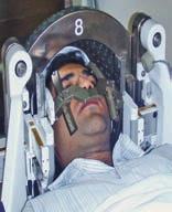
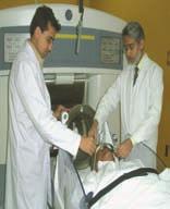
We all welcome you to visit the facility at any time, please feel free to ask any question from any member of our team.
|
|
|
- Barry Holt
- 6 years ago
- Views:
Transcription
1

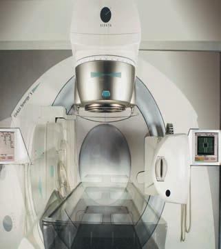
2 Imenant Faculty of Neuclear Medicine 1. Dr. Shahid Kamal M.D. Ph.D (Specialist in Neuclear Medicine) Former Director KIRIN & JPMC (N,M). Director, Nuclear Medicine & Molecular Imaging Neurospinal & Medicine 2. Dr. Asif Mateen FRCR Oncological Radiologist 20 years experience in England. Chief Radiologist Neurospinal & Medical Institute. 3. Dr. Sattar M. Hashim M.D. Ph.d Chief Consultant Neurosurgeon & Managing Director Neurospinal & Medical Institute We all welcome you to visit the facility at any time, please feel free to ask any question from any member of our team. n n n n n n n State of art molecular Imaging. Characterise and monitor cancer lesians in sub m.m.s from head to toe, making cancer cure possible and effective at early stage. Best option for understanding of disease, its progression and response to treatment, Surgery, radiotherapy, chemotherapy. Complete screaning of cancer and other diseases. Smart auto cardiac registration, non-invasive quantitative assessment of cardiac blood flow and coronary flow reserve. Smart neuro account registration clear and quick assessment of hyper and hypo metabolic brain regions. Friendly for children, minimum dose and maximum speed. n PET CT provide accurate high precision radiation therapy planning.


3 PET/CT Scan INTRODUCTION Neurospinal & Medical Institute (NMI), by the grace of God, has been serving the nation since its establishment in Our spirit of serving the nation with most advanced medical technology for diagnosis and therapeutics, has led us offering the best facilities for In and Out patient care, ICU, OT, X-ray, Ultrasound, EEG, EMG, laboratory, Multi-slice CT Scanner, Open MRI, 1.5 T high strength MRI and now a state of art PET-CT with Cyclotron for simultaneous radioisotope preparation under one roof to avid decay of radio isotope,use of less isotope & less radiation to body. General and specialized, neurosurgery like microsurgery, stereotactic surgery (biopsy & movement disorders), skull base neuroendoscopy, pediatric neurosurgery other disciplines of medicine, e.g., Neurology, orthopedics, general medicine and general surgery is practiced by eminent clinicians. Abnormal PET-CT body Scan 3


4 PET/CT Scan Positron Emission Tomography PET/CT combines the strengths of two well-established imaging modalities, CT for anatomy and PET for function, into a single imaging device. By imaging with the two modalities in a single scan, disease can be both identified and localized, potentially resulting in an earlier diagnosis and more accurate staging. scanners provide accurately aligned anatomical and functional images of a patient, allowing functional abnormalities to be localized and distinguished from normal uptake of the PET tracer, which increases physician confidence in arriving at a correct diagnosis. Patient Management Fusing PET and CT images more accurately localizes metabolic activity to the appropriate anatomic structure or location, such as a lymph node, a bowel loop, or a vocal cord. For differentiation of tumor versus necrosis or scar tissue, knowing which part of a mass has viable tumor instead of necrosis or scar tissue assists in targeting the right areas to sample or remove. o Example: Patient with suspected recurrent rectal carcinoma where there is typically scar tissue at the site of a previous AP resection. CT alone is often unable to determine if a tumor is present, and if present, CT may not distinguish the actual viable tumor focus from surrounding scar tissue. PET/CT can clearly show hypermetabolic tumor focus within an otherwise hypometabolic region of scar tissue, enabling a high-yield directed needle biopsy or open biopsy. (Left) Axial T1 C+ MR shows minimal abnormal enhancement corresponding to a posterior left parietal lesion g in this patient with a newly diagnosed WHO grade II glioma. (Right) Axial fused PET/MR shows relative hypometabolism that corresponds to the posterior left parietal lesion g characteristic of a WHO grade II lesion 4 Radiation oncologists need maximum information for radiation therapy planning, and PET/CT data files can be imported into many radiation therapy planning computers. For example, IMRT can deliver precise amounts of radiation to complex 3- D volumes, fully utilizing PET/CT datasets. IV contrast may or may not be used with PET/CT, depending on the studies being completed. PET/CT scanners have advanced imaging algorithms that allow for the use of IV contrast when needed.


5 Advantages of PET/CT Cancer diagnosis - head to toe (whole body) PET/CT provides physicians with superior information for determining tissue characterizations and classifications, staging of cancers, restaging of cancers, patient prognosis and monitoring the effectiveness of cancer therapies. Advantages include: Superior lesion localization from near-perfect anatomical/functional registration with fewer motion artifacts. Better distinction between physiological uptake and pathological uptake. Consolidation of patient's imaging studies. Shorter scan time (avg. 30 minutes to complete) by using CT for attenuation correction. This aids in patient comfort and minimizes claustrophobia problems. RT Planning Radiation Therapy Treatment Planning is a promising area for PET/CT. The PET/CT, with a continuous 70cm patient bore, will accommodate many therapy treatment pallets. The image data set created by the PET/CT can be sent to nearly every treatment planning system on the market today. The Radiation Therapist can use the data set to define treatment contour maps without having to subject the patient to an additional CT scan. Cardiology Applications With the recent inclusion of PET studies for evaluating Myocardial Perfusion, and with the introduction of the 16 row, isotropic CT, we now have the potential to evaluate cardiac disease with increased information, and with exquisite detail. Neurological Applications Axial CECT (with well-timed portal venous phase) of a patient with a history of breast cancer and recent rise in CA shows no evidence of hepatic lesions. In conjunction with PET imaging, CT shows great potential with its ability to quickly scan and demonstrate perfusion characteristics within specific areas of the brain. More research needs to be done in this area, but the potential usefulness of the fused PET scan and CT perfusion scan show great potential for early diagnosis and staging of these disease types. Axial fused PET/CT shows at least 6 FDGavid bilodar hepatic metastatic lesions g. One of the major added benefits of PET/CT is detection of lesions not identifiable on CT, even with good contrst enhancement. 5


6 Oncology In oncology, PET permits a physician to accurately image many organs of the body with a single scan in order to detect malignancy. PET has demonstrated usefulness in cost-effective wholebody metastatic surveys, avoiding biopsies. The benefits of PET include non-invasive differentiation of tumors from radiation necrosis, the possibility to change the course of ineffective chemotherapy and avoidance of unnecessary diagnostic and therapeutic procedures. Applications for PET in oncology include: Colorectal Cancer t Detect locally recurrent or distant metastatic disease in patients with elevated or rising CEA who may be candidates for surgical re-excision. PET/Scan also rule out distant metastases for preoperative evaluation. Brain Tumor t Differentiate a recurrent tumor from radiation necrosis. t Differentiate primary CNS lymphoma from toxoplasmosis. t Exclude brain metastatic disease. Graphic shows a circumferential mass in the sigmoid colon g that causes marked narrowing of the colonic lemen. This appearance is often termed apple core lesion on barium enema. Coronal PET (A), axial CT (B) and fused PET/CT (C) show a focal area of intense FDG activity g, corresponding to primary colon carcinoma in the proximal ascending colon. Head & Neck Cancer t Determine extent of local, regional and distant disease. t Detect recurrent/residual tumor following definitive therapy. (Left) Axial T1WL MR shows a WHO grade I tectal lesion g that had been stable for several years and showed no increased metabolic activity on the original PET scan. A follow-up PET scan was ordered due to new worsening clinical symptoms. (Right) Axial fused PET/MR shows a new focus of moderately increased FDG activity correlating to the center of the tectal mass g, suggestive of high grade transformation of this previously low grade glioma. Breast Cancer t Identify involved axillary nodes or distant metastatic disease. t Exclude local recurrence of disease. t Evaluate response to treatment. Axial CT (left) and fused PET/CT (right) show a right parapharyngeal space mass g and a mucosal lesion only identified on the fused PET/CT g. Follow-up axial CT (left) and fused PET/CT (right) after surgery demonstrate no residual or recurrent tumor. 6 (Left) Axial CECT shows a heterogeneously enhancing mass in the left breast g that is compatible with breast cancer. (Right) Axial fused PET/CT shows focal intense FDG activity g that correlates with the enhancing mass in the left breast, compatible with primary breast carcinoma.


 Axial CECT shows a peripherally based right lower lobe nodule g.")


 revealed no definite evidence for disease above the diaphragm.")
 Axial fused PET/CT shows focal intense FDG activity correlating with a small lymph node g,")
7 Lung Cancer t Distinguish malignant pulmonary nodules from benign ones. t Stage mediastinal or distant metastatic disease. t Use as part of radiotherapy treatment planning. t Detect recurrent/residual tumor following definitive therapy. Musculoskeletal Tumors Evaluate local extent of disease and exclude distant metastases. Measure treatment response and exclude recurrent/residual tumor following definitive therapy. (Left) Axial CECT shows a peripherally based right lower lobe nodule g. (Right) Axial fused PET/CT shows the same lesion with intense FDG uptake g, compatible with a primary lung cancer. (Left) Axial CECT shows no obvious metastatic lesions, although there is questionable abnormal enhancement of the left gluteal muscle g that was not called prospectively on CT. (Right) Axial fused PET/CT shows focal intense FDG activity correlating with the lesion in the left gluteal muscle g, compatible with metastatic disese. Lymphoma Determine extent of disease and measure treatment response. Ovarian Cancer Detect recurrent/residual tumor prior to surgical exploration or additional chemotherapy. (Left) Axial CECT shows confluent mesenteric and retroperitoneal soft tissue involvement g in this patient recently diagnosed with diffuse large B-cell lymphoma. (Right) Axial fused PET/CT shows intense increased FDG activity throughout the confluent adenopathy g. CT (not shown) revealed no definite evidence for disease above the diaphragm. (Left) Axial CECT shows a normal-sized left common iliac lymph node g, not identified prospectively on CT. (Right) Axial fused PET/CT shows focal intense FDG activity correlating with a small lymph node g, compatible with recurrent malignancy. Melanoma Identify extent of local and regional disease spread in patients with high risk melanoma (e.g., primary tumor less than 4mm) or in suspected recurrence. (Left) Coronal PET shows focal uptake in the rib g, corresponding to a metastasis in this patient with known melanoma. (Right) Axial fused PET/CT shows that the same lesion demonstrates increased FDG uptake g. 7


. Dementia Differentiate Alzheimer's disease from multi-infarct dementia.")


8 Pancreatic Cancer Differentiation of benign processes such as pancreatitis, mucinous cyst adenoma and pseudocyst from malignant disease. Rule out distant metastases during preoperative evaluation. Neurology In neurology, PET/Scan provides information for assessing various neurological diseases such as brain tumor, Alzheimer's disease and other dementias, Parkinson's disease, and Huntington's disease. Additionally, it localizes epileptic foci for qualifying and identifying the site requiring surgical intervention. It also allows the characterization, grading and assessment of possible brain tumor recurrence (see previous section). Dementia Differentiate Alzheimer's disease from multi-infarct dementia. (Left) Axial CECT shows a slightly hypodense lesion in the pancreatic body g with distal ductal dilation and atrophy g. (Right) Axial fused PET/CT of the same patient demonstrates increased metabolic activity g, consistent with pancreatic adenocarcinoma. Thyroid Cancer Detect metastatic or locally recurrent disease in patient with elevated thyroglobulin after definitive initial treatment and negative I-131 examination. Epilepsy Localize the seizure focus in patients with intractable complex seizure disorders. Stroke Evaluate extent of disease and recovery following therapy. (Left) Axial NECT shows an enlarged thyroid anterior to the trachea g. (Right) Axial Fused PET/CT shows that the enlarged thyroid has intense, diffuse FDG uptake g. Subsequent bipsy revealed anaplastic thyroid carcinoma. epileptic foci Stroke 8


9 Cardiology In cardiology, PET/Scan enables physicians to screen for coronary artery disease, to assess flow rates and flow reserve and to distinguish viable from nonviable myocardium for bypass and transplant candidates. Myocardial Viability distinguish viable myocardium from infarcted tissue in patients with suspected hibernating or stunned myocardium. Coronary Artery Disease Identify ischemic disease. Preoperative Prognostic Assessment Evaluate extent of disease in patients being considered for interventional revascularization or transplantation procedures. Pyrexia of unknown origin (PUO) Useful for identifying disease foci and excluding malignancy in highly selected patients where conventional imaging is negative or equivocal. Paraneoplastic Indicated in selected patients with non-metastatic manifestations of syndrome neoplastic disease e.g. neurological signs or raise antibodies to exclude or confirm an occult primary tumor when conventional imaging is negative or equivocal. Vasculitis Useful in the assessment of patterns of vasculitis and disease response. May have value in the assessment of aortic aneurysm follow up post stenting/surgical intervention, if graft infection is suspected.

10 How does PETCT work? During a PET scan, the patient is first injected with a radiopharmaceutical compound, usually FDG, a radioactive glucose compound. The compound distributes throughout the body and accumulates in various organs depending on the metabolic activity within the organ or tissue. Because cancer cells usually have a higher metabolic rate than surrounding cells, they absorb more of the tracer and will show up more prominently on the image. The PET CT scanner detects the FDG accumulated in glucose-avid organs or tissues and creates images that are displayed as colour-coded images. PET CT and Cancer a. Diagnose cancer, from head to toe. b. Staging of cancer: PET is extremely sensitive in detemining the full extent of disease, especially in cancers. Confirmation of metastatic disease allow the physician and patient to more accurately decide on how to proceed with the patients s management. c. Cheking for recurrences: PET/CT is currently considered to be the most accurate diagnostic procedure to differentiate tumor recurrences from radiation necrosis or post-surgical changes. Such an approach allows for the development of a more rational treatment plan for the patient. d. Assessing the Effectiveness of radiotherapy, Chemotherapy: The level of tumor metabolism is compared on PPET scans befor and after chemotherapy radiotherapy cycle. A successful response seen on PET scan frequently precedes alterations in anatomy and is considered to be an earlier indicator of tumor shrinkage than might be seen with other diagnostic modalities. d. Provide key diagnostic information for Alzheimer s disease in select patients. e. Differentiate among dementia disorder, including Alzheimer s disease, Parkinson t disese, and epileptic seizure disorders f. Stroke PET CT and Neurology a. Diagnosis of Brain Tumors b. PET/CT can reveal abnormal functional and structural patterns in the brain, helping to assess patients with various disorders, including epilepsy and dementia c. Localize areas of the brain causing epileptic seizures determine whether surgery is an option. 10
 surgery, or heart transplantation.")


11 PET CT and Cardiology PET CT provides unparalleled insight into myocardial viability. This often helps to determine the optimal treatment path identifying whether a patient is a candidate for coronary angioplasty, coronary artery bypass graft (CABG) surgery, or heart transplantation. At our centre we have the unique capability of fusing PET/CT images with CT coronary angiography for the first time in south asia. PET CT and infection There is growing body of evidence that PET CT is very valuable in diagnosis of infection and for follow-up to determine eradiction of disease. It can be very useful in patients with fever of unknown origin (PUO). PET CT may provide objective evidence of when to stop treatment in selected patients with tubercular infections. It may also be useful in determining infection in joint implants. Exam Preparation There is very little preparation needed for a PET CT exam. Typically you will be asked not to eat 6-8 hours prior to the exam but you should drink a lot of water before the scan. If you are taking medication please consult with your physician before the exam. Most medications can be taken on the day of the exam. Please avoid alcohol and strenuous exercise 24 hours before your appointment. If you are diabetic it is important that your blood sugar be in control. If your blood sugar is high on the day of the scan the doctors at the PET CT centre may have to correct it by injecting a short-acting insulin. Alternatively your scan may be deferred till such time that your sugar comes under control. Please do not wear any jewellery. Please bring any related test reports including CT or MRI films. PET CT Scans are not done on pregnant patients. So please consult with your doctor if you think you are pregnant. Plan to spend about 3 hours at the PET CT centre. The Exam Prior to the exam you will receive a small injection of radioactive sugar (FDG). You will be asked to sit or lie down on a comfortable chair or bed for minutes while the FDG travels throughout your body. It is important that you do not talk, read, walk around or chew gum during this period. In some 11 cases you may be asked to drink medicine (oral contrast). This will be at regular intervals before the actual scan is performed. After this short time, the technologist will assist you to the scanner. An intravenous non-ionic iodinated contrast may be injected for the ct scan if required. It is important that you don t move for the duration of the scan. The length of the exam is determined by your height and area of interest. Most PET CT scans at our centre are typically completed within minutes. After the Exam Once the scan has been performed you may resume normal daily activity. Even though the FDG and contrast will quickly leave your body, you can expedited the process by drinking plenty of water after your scan is complete. Your PET CT result will not be immediately available. You can collect your scan report the next day. Important Since the radioactive glucose prepared for you is expensive and has a very short life it is imperative that you keep your appointment on time. In case you need to cancel your appointment please inform us at least 24 hours ahead.
 scanning has brought about a revolution in the imaging of many common cancers and, as it becomes widely available in developed countries, is increasingly being")
 as the radiopharmaceutical, in combination with structural imaging, such as X ray computed tomography (CT) scanning,")

12 PET-CT Guided Radiation Planning Accurate imaging is central to the treatment planning process for most malignancies managed with curative intent using radiation therapy. Positron emission tomography (PET) scanning has brought about a revolution in the imaging of many common cancers and, as it becomes widely available in developed countries, is increasingly being incorporated into routine radiation therapy planning. PET scanning, usually employing Fluorine-18-fluorodeoxyglucose (18F- FDG) as the radiopharmaceutical, in combination with structural imaging, such as X ray computed tomography (CT) scanning, currently provides the most accurate available information on tumours extent and distribution for many common cancers,including lymphomas and epithelial malignancies of the lung, esophagus, cervix and head and neck. As PET becomes more widely used for radiation therapy planning, it is important that it is introduced rationally and used appropriately for malignancies in which it provides significant incremental information aside from that obtained from structural imaging. Functional imaging with PET can provide information that can influence RT planning in a number of ways. Some of the most important include the following: n PET can reveal targets that are not well visualized by CT/magnetic resonance (MR) structural imaging. These targets may be remote from the primary tumours, such as unsuspected lymph node or distant metastases, or they may be additional neoplastic regions adjacent to the tumours volume defined by CT/MR imaging. n PET makes it less likely that treatment will be given to equivocal regions on CT/MR which do not actually contain tumours. These regions may also be remote, such as benign reactive lymphadenopathy, or adjacent to the tumour volume defined by CT/MRI, such as atelectatic regions of lung. n The imaging of biologic inhomogeneities within sub volumes of the tumour may offer the possibility to adapt doses to local differences in radiosensitivity (known as dose painting, not yet been shown to be of value in any tumour site). n PET can be useful for the evaluation of residual masses after chemotherapy in conditions like lymphoma, helping to determine which regions, if any, require radiation therapy and helping to choose between a lower dose for presumed microscopic residual disease or a higher dose for gross residual disease. Feasibility studies have found that the use of 18F-FDG-PET/CT for planning three dimensional conformal radiation therapy improves the standardization of volume delineation compared with CT alone for a number of cancers that are well-imaged on PET. It is simply easier for physicians to visualize the tumours clearly. In addition, there is the attractive possibility that sub-regions within the tumours can be targeted selectively, either with higher radiation doses on a gross level or more feasibly with specific pharmaceutical agents at a molecular level. x-knife 12 Gamma-Knife

 Axial fused PET/MR shows a new focus of moderately increased FDG activity correlating to the center of the tectal mass g,")


13 Further Applications of PET PRIMARY BRAIN NEOPLASMS (Left) Axial T1WL MR shows a WHO grade I tectal lesion g that had been stable for several years and showed no increased metabolic activity on the original PET scan. A follow-up PET scan was ordered due to new worsening clinical symptoms. (Right) Axial fused PET/MR shows a new focus of moderately increased FDG activity correlating to the center of the tectal mass g, suggestive of high grade transformation of this previously low grade glioma. PRIMARY BRAIN NEOPLASMS Axil FLAIR MR shows abnormal signal g in a patient with a glioblastoma multiforema treated with gamma knife. Differential is radiation necrosis vs. recurrent tumor. Axil PET shows correlative intense FDG activity g compatible with residual/recurrent tumor rather than radiation necrosis. PRIMARY BRAIN NEOPLASMS (Left) Axial T1 C+ MR in this patient with a history of prior glioblastoma status post resection and radiation shows extensive rim enhancement g, worrisome for possible recurrent tumor. (Right) Axial fused fused PET/MR shows no increased metabolic activity in the area of abnormal enhancement g, compatible with radiation with radiation necrosis. Both radiation necrosis and residual/recurrent tumor may have abnormal enhancement on MR, and PET can often to be used to distinguish between the two diagnoses. 13
 and fused PET/CT (right) show a right parapharyngeal space mass")
 and fused PET/CT (right) after surgery")
, axial CT (B) and fused PET/CT (C) show")


14 Further Applications of PET HEAD AND NECK CANCER, SQUAMOUS Axial CT (left) and fused PET/CT (right) show a right parapharyngeal space mass g and a mucosal lesion only identified on the fused PET/CT g. Follow-up axial CT (left) and fused PET/CT (right) after surgery demonstrate no residual or recurrent tumor. THYROID CANCER Coronal PET (A), axial CT (B) and fused PET/CT (C) show a subtle recurrent thyroid carcinoma g in a patient with rising thyroglobulin levels and a negative iodine study. Axial CT (top) and fused PET/CT (bottom) show focal thyroid cancer recurrence in the thyroidectomy bed g. SOLITARY PULMONARY NODULES Infection: Tuberculosis Malignancy: Metastasis Inflammation: Pseudotumor 14


15 Further Applications of PET LUNG CANCER (Left) Axial CECT shows a peripherally based right lower lobe nodule g. (Right) Axial fused PET/CT shows the same lesion with intense FDG uptake g, compatible with a primary lung cancer. BREAST CANCER (Left) Axial CECT shows a heterogeneously enhancing mass in the left breast g that is compatible with breast cancer. (Right) Axial fused PET/CT shows focal intense FDG, activity g that correlates with the enhancing mass in the left breast, compatible with primary breast carcinoma. ESOPHAGEAL CANCER (Left) Coronal PET (A), axial CT (B) and fused PET/CT (C) images demonstrate intense FDG activity along the gastroesophageal junction g in this patient with newly diagnosed esophageal carcinoma. (Right) Specimen shows an infiltrative and nodular lesion at the gastroesophageal junction g, representing the patient s primary esophageal carcinoma. 15
 PET/CT study")

 Axial CT (top) and fused PET/CT (bottom) demonstrate a")


 Axial CECT shows confluent mesenteric")
 Axial fused")
16 Further Applications of PET COLORECTAL CANCER Physiologic Anorectal Junction Polyp Villous Adenoma HODGKIN LYMPHOMA (Left) PET/CT study demonstrates intense FDG activity g on the coronal PET (A) corresponding to a large anterior mediastinal mass g on axial CT (B) and fused PET/CT (C). (Right) Axial CT (top) and fused PET/CT (bottom) demonstrate a large anterior mediastinal mass with intense FDG activity g, compatible with the patient s history of recently diagnosed Hodgkin lymphoma. The majority of odgkin lymphomas are FDG avid, and this is a typical representation. NON-HODGKIN LYMPHOMA (Left) Axial CECT shows confluent mesenteric and retroperitoneal soft tissue involvement g in this patient recently diagnosed with diffuse large B-cell lymphoma. (Right) Axial fused PET/CT shows intense increased FDG activity throughout the confluent adenopathy g. CT (not shown) revealed no definite evidence for disease above the diaphragm. 16


 Axial fused PET/CT shows intense FDG activity within")


 Axial used PET/CT of the same patient as the previous")
17 Further Applications of PET CERVICAL CARCINOMA (Left) Axial CECT shows a heterogeneously enhancing cervical mass ---, compatible with the patient s primary cervica! carcinoma. In addition, there are cystic metastases involving the left adnexa (Right) Axial fused PET/CT shows intense FDG activity within both the primary cervical mass --- and the left adnexa In general, cervical carcinoma tends to be FDG avid, as are most squamous cell carcinomas throughout the body. NEUROENDOCRINE TUMORS Cholangiocarcinoma Hepatocellular Carcinoma Metastasis PRIMARY BONE NEOPLASMS 17 (Left) Axial CECT shows a small, almost imperceptible region of increased soft tissue g superficial to the body of the left mandible. (Right) Axial used PET/CT of the same patient as the previous image reveals intense FDG uptake in the same region g, compatible with malignancy.


18 Further Applications of PET METASTATIC LESIONS OF THE BONES (Left) Axial CECT shows subtle mixed lytic metastases g throughout this vertebral body in a patient with breast cancer. (Right) Axial fused PET/CT of the same patient shows diffuse FDG activity g from the numerous small lytic metastases. MULTIPLE MYELOMA (Left) Axial CECT shows an expansile, lyctic lesion g in a rib with cortical disruption. (Right) Axial fused PET/CT of the same patient shows moderately increased FDG activity g within the lesion. THYMIC PROCESSES (Left) Axial CECT shows a heterogeneously enhancing soft tissue lesion that has convex margins g with the left mediastinal surface and may invade the pleura. This was pathologically proven to be invasive thymoma. (Right) axial fused PET/CT shows asymmetric increased FDG activity in the right aspect g of the invasive thymoma, which corresponds to the more solid-appearing component of the lesion. 18

 Coronal PET (A), Axial CT (B) and from the same patient, demonstrating a hepatic lesion.")


 Axial CECT shows a focal area of thickening along the right posterior lateral aspect of the")
19 Further Applications of PET HEPATOCELLULAR CARCINOMA (Left) Axial CECT shows almost complete replacement of the left hepatic lobe g with tumor in this patient with hepatocellular carcinoma. Approximately 50% of hepatocellular carclnoma primary lesions may not be FDG avid. (Right) Coronal PET (A), Axial CT (B) and from the same patient, demonstrating a hepatic lesion. RENAL CELL CARCINOMA (Left) Axial contrast enema shows a low attenuation mass g arising from the medial aspect of the left kidney. (Right) Axial fused PET/CT shows mild to moderate increased FDG activity within the left renal mass g with moderately hypermetabolic retroperitoneal adenopathy g. The overall appearance is compatible with renal cell carcinoma and regional metastases. BLADDER CARCINOMA (Left) Axial CECT shows a focal area of thickening along the right posterior lateral aspect of the bladder g, compatible with this patient s known history of primary transitional cell carcinoma. (Right) Axial fused PET/CT shows an asymmetrical focus of FDG activity correlating with the patient s primary transitional cell carcinoma g. Note slight misregistration of the images caused by filling of the urinary bladder during the PET portion of the exam. 19

20
PET IMAGING (POSITRON EMISSION TOMOGRAPY) FACT SHEET
 FACT SHEET") Positron Emission Tomography (PET) When calling Anthem (1-800-533-1120) or using the Point of Care authorization system for a Health Service Review, the following clinical information may be needed to
Positron Emission Tomography (PET) When calling Anthem (1-800-533-1120) or using the Point of Care authorization system for a Health Service Review, the following clinical information may be needed to
PET/CT Frequently Asked Questions
 PET/CT Frequently Asked Questions General Q: Is FDG PET specific for cancer? A: No, it is a marker of metabolism. In general, any disease that causes increased metabolism can result in increased FDG uptake
Clinical indications for positron emission tomography
 Clinical indications for positron emission tomography Oncology applications Brain and spinal cord Parotid Suspected tumour recurrence when anatomical imaging is difficult or equivocal and management will
Molecular Imaging and Cancer
 Molecular Imaging and Cancer Cancer causes one in every four deaths in the United States, second only to heart disease. According to the U.S. Department of Health and Human Services, more than 512,000
performed to help sway the clinician in what the appropriate diagnosis is, which can substantially alter the treatment of management.
 Hello, I am Maura Polansky at the University of Texas MD Anderson Cancer Center. I am a Physician Assistant in the Department of Gastrointestinal Medical Oncology and the Program Director for Physician
POSITRON EMISSION TOMOGRAPHY (PET)
") Status Active Medical and Behavioral Health Policy Section: Radiology Policy Number: V-27 Effective Date: 08/27/2014 Blue Cross and Blue Shield of Minnesota medical policies do not imply that members should
Status Active Medical and Behavioral Health Policy Section: Radiology Policy Number: V-27 Effective Date: 08/27/2014 Blue Cross and Blue Shield of Minnesota medical policies do not imply that members should
Optimized. clinical pathway. propels high utilization of PET/MR at Pitié-Salpêtrière Hospital
 Optimized propels high utilization of PET/MR at Pitié-Salpêtrière Hospital clinical pathway As one of Europe s largest teaching hospitals, Pitié-Salpêtrière Hospital is renowned for its innovative research
Radiology Pathology Conference
 Radiology Pathology Conference Sharlin Johnykutty,, MD, Cytopathology Fellow Sara Majewski, MD, Radiology Resident Friday, August 28, 2009 Presentation material is for education purposes only. All rights
Index. Surg Oncol Clin N Am 16 (2007) Note: Page numbers of article titles are in boldface type.
 Note: Page numbers of article titles are in boldface type.") Surg Oncol Clin N Am 16 (2007) 465 469 Index Note: Page numbers of article titles are in boldface type. A Adjuvant therapy, preoperative for gastric cancer, staging and, 339 B Breast cancer, metabolic
Surg Oncol Clin N Am 16 (2007) 465 469 Index Note: Page numbers of article titles are in boldface type. A Adjuvant therapy, preoperative for gastric cancer, staging and, 339 B Breast cancer, metabolic
objectives Pitfalls and Pearls in PET/CT imaging Kevin Robinson, DO Assistant Professor Department of Radiology Michigan State University
 objectives Pitfalls and Pearls in PET/CT imaging Kevin Robinson, DO Assistant Professor Department of Radiology Michigan State University To determine the regions of physiologic activity To understand
Dr Sneha Shah Tata Memorial Hospital, Mumbai.
 Dr Sneha Shah Tata Memorial Hospital, Mumbai. Topics covered Lymphomas including Burkitts Pediatric solid tumors (non CNS) Musculoskeletal Ewings & osteosarcoma. Neuroblastomas Nasopharyngeal carcinomas
Positron Emission Tomography Computed Tomography (PET/CT)
") Positron Emission Tomography Computed Tomography (PET/CT) What is Positron Emission Tomography Computed Tomography (PET/CT) Scanning? What are some common uses of the procedure? How should I prepare for
Positron Emission Tomography Computed Tomography (PET/CT) What is Positron Emission Tomography Computed Tomography (PET/CT) Scanning? What are some common uses of the procedure? How should I prepare for
Imaging Patient Education What you should know about your PET/CT Bone Scan.
 Imaging Patient Education What you should know about your PET/CT Bone Scan. Purpose: PET/CT uses a small amount of a radioactive sodium fluoride product to detect cancer within the bones. A low dose CT
An Introduction to PET Imaging in Oncology
 January 2002 An Introduction to PET Imaging in Oncology Janet McLaren, Harvard Medical School Year III Basics of PET Principle of Physiologic Imaging: Allows in vivo visualization of structures by their
Brain Tumors. What is a brain tumor?
 Scan for mobile link. Brain Tumors A brain tumor is a collection of abnormal cells that grows in or around the brain. It poses a risk to the healthy brain by either invading or destroying normal brain
Appendix 1: Regional Lymph Node Stations for Staging Esophageal Cancer
 Appendix 1: Regional Lymph Node Stations for Staging Esophageal Cancer Locoregional (N stage) disease was redefined in the seventh edition of the AJCC Cancer Staging Manual as any periesophageal lymph
Imaging in gastric cancer
 Imaging in gastric cancer Gastric cancer remains a deadly disease because of late diagnosis. Adenocarcinoma represents 90% of malignant tumors. Diagnosis is based on endoscopic examination with biopsies.
Dr Claire Smith, Consultant Radiologist St James University Hospital Leeds
 Dr Claire Smith, Consultant Radiologist St James University Hospital Leeds Imaging in jaundice and 2ww pathway Image protocol Staging Limitations Pancreatic cancer 1.2.4 Refer people using a suspected
42 yr old male with h/o Graves disease and prior I 131 treatment presents with hyperthyroidism and undetectable TSH. 2 hr uptake 20%, 24 hr uptake 50%
 Pinhole images of the neck are acquired in multiple projections, 24hrs after the oral administration of approximately 200 µci of I123. Usually, 24hr uptake value if also calculated (normal 24 hr uptake
Los Angeles Radiological Society 62 nd Annual Midwinter Radiology Conference January 31, 2010
 Los Angeles Radiological Society 62 nd Annual Midwinter Radiology Conference January 31, 2010 Self Assessment Module on Nuclear Medicine and PET/CT Case Review FDG PET/CT IN LYMPHOMA AND MELANOMA Submitted
Prof. Dr. NAGUI M. ABDELWAHAB,M.D.; MARYSE Y. AWADALLAH, M.D. AYA M. BASSAM, Ms.C.
 Role of Whole-body Diffusion MR in Detection of Metastatic lesions Prof. Dr. NAGUI M. ABDELWAHAB,M.D.; MARYSE Y. AWADALLAH, M.D. AYA M. BASSAM, Ms.C. Cancer is a potentially life-threatening disease,
Take Home Quiz 1 Please complete the quiz below prior to the session. Use the Multiple Primary and Histology Rules
 Take Home Quiz 1 Please complete the quiz below prior to the session. Use the Multiple Primary and Histology Rules Case 1 72 year old white female presents with a nodular thyroid. This was biopsied in
FDG PET/CT STAGING OF LUNG CANCER. Dr Shakher Ramdave
 FDG PET/CT STAGING OF LUNG CANCER Dr Shakher Ramdave FDG PET/CT STAGING OF LUNG CANCER FDG PET/CT is used in all patients with lung cancer who are considered for curative treatment to exclude occult disease.
Medical Policy An independent licensee of the Blue Cross Blue Shield Association
 PET Scanning: Oncologic Applications Page 1 of 88 Medical Policy An independent licensee of the Blue Cross Blue Shield Association Title: Positron Emission Tomography (PET) Scanning: Oncologic Applications
Cancer of Unknown Primary (CUP)
") Cancer of Unknown Primary (CUP) Pathways and Guidelines V1.0 London Cancer September 2013 The following pathways and guidelines document has been compiled by the London Cancer CUP technical subgroup and
Cancer of Unknown Primary (CUP) Pathways and Guidelines V1.0 London Cancer September 2013 The following pathways and guidelines document has been compiled by the London Cancer CUP technical subgroup and
Molecular Imaging and the Brain
 Molecular imaging technologies are playing an important role in neuroimaging, a branch of medical imaging, by providing a window into the living brain. Where CT and conventional MR imaging provide important
Medical imaging X-ray, CT, MRI, scintigraphy, SPECT, PET Györgyi Műzes
 Medical imaging X-ray, CT, MRI, scintigraphy, SPECT, PET Györgyi Műzes Semmelweis University, 2nd Dept. of Medicine Medical imaging: definition technical process of creating visual representations about
Positron Emission Tomography - Computed Tomography (PET/CT)
") Scan for mobile link. Positron Emission Tomography - Computed Tomography (PET/CT) Positron emission tomography (PET) uses small amounts of radioactive materials called radiotracers, a special camera and
Scan for mobile link. Positron Emission Tomography - Computed Tomography (PET/CT) Positron emission tomography (PET) uses small amounts of radioactive materials called radiotracers, a special camera and
Lymphoma Read with the experts
 Lymphoma Read with the experts Marc Seltzer, MD Associate Professor of Radiology Geisel School of Medicine at Dartmouth Director, PET-CT Course American College of Radiology Learning Objectives Recognize
Molecular Imaging and Breast Cancer
 Molecular Imaging and Breast Cancer Breast cancer forms in tissues of the breast usually in the ducts, tubes that carry milk to the nipple, and lobules, the glands that make milk. It occurs in both men
Nuclear Medicine in Oncology
 Radiopharmaceuticals Nuclear Medicine in Oncology Practice Pharmaceutical Radionuc lide Function Tumor type Diphosphonates Tc-99m Osteoblast Bone tumor & metast. Ga-citrate Ga-67 Fe-analogue Bronchogenous
CLINICAL PRESENTATION AND RADIOLOGY QUIZ QUESTION
 Donald L. Renfrew, MD Radiology Associates of the Fox Valley, 333 N. Commercial Street, Suite 100, Neenah, WI 54956 4/30/2011 Radiology Quiz of the Week # 18 Page 1 CLINICAL PRESENTATION AND RADIOLOGY
Evaluation and Management of Thyroid Nodules. Nick Vernetti, MD, FACE Palm Medical Group Las Vegas, Nevada
 Evaluation and Management of Thyroid Nodules Nick Vernetti, MD, FACE Palm Medical Group Las Vegas, Nevada Disclosure Consulting Amgen Speaking Amgen Objectives Understand the significance of incidental
Imaging in Pediatric Thyroid disorders: US and Radionuclide imaging. Deepa R Biyyam, MD Attending Pediatric Radiologist
 Imaging in Pediatric Thyroid disorders: US and Radionuclide imaging Deepa R Biyyam, MD Attending Pediatric Radiologist Imaging in Pediatric Thyroid disorders: Imaging modalities Outline ACR-SNM-SPR guidelines
Oncologic Applications of PET Scanning
 6.01.26 Oncologic Applications of PET Scanning Section 6.0 Radiology Subsection Effective Date February 15, 2015 Original Policy Date January 26, 2009 Next Review Date December 2015 Description Positron
Radiology-Pathology Conference
 July 31, 2009 Radiology-Pathology Conference Daniel T Ginat, M.D., M.S. Sharlin Johnykutty,, M.D. Presentation material is for education purposes only. All rights reserved. 2009 URMC Radiology Page 1 of
Nuclear Sciences and Medicine
 Nuclear Sciences and Medicine Rethy Chhem, MD, PhD (Edu), PhD (His), FRCPC Division of Human Health Guest Professor, Medical University of Vienna International Atomic Energy Agency Medical Imaging X-rays
Nuclear Medicine and PET. D. J. McMahon rev cewood
 Nuclear Medicine and PET D. J. McMahon 150504 rev cewood 2018-02-15 Key Points Nuclear Medicine and PET: Imaging: Understand how Nuc Med & PET differ from Radiography & CT by the source of radiation. Be
Shadow because the air
 Thyroid Ultrasound Thyroid US examination needs: 1. high frequency transducer 2. extended patient's neck 3. check all the neck area because the swelling could be in areas other than the thyroid such as
SEER Summary Stage Still Here!
 SEER Summary Stage Still Here! CCRA NORTHERN REGION STAGING SYMPOSIUM SEPTEMBER 20, 2017 SEER Summary Stage Timeframe: includes all information available through completion of surgery(ies) in the first
Staging Colorectal Cancer
 Staging Colorectal Cancer CT is recommended as the initial staging scan for colorectal cancer to assess local extent of the disease and to look for metastases to the liver and/or lung Further imaging for
Disclosure. Acknowledgement. What is the Best Workup for Rectal Cancer Staging: US/MRI/PET? Rectal cancer imaging. None
 What is the Best Workup for Rectal Cancer Staging: US/MRI/PET? Zhen Jane Wang, MD Assistant Professor in Residence UC SF Department of Radiology Disclosure None Acknowledgement Hueylan Chern, MD, Department
FDG-PET/CT for cancer management
 195 REVIEW FDG-PET/CT for cancer management Hideki Otsuka, Naomi Morita, Kyo Yamashita, and Hiromu Nishitani Department of Radiology, Institute of Health Biosciences, The University of Tokushima, Graduate
Breast Cancer PET/CT Imaging Protocol
 Breast Cancer PET/CT Imaging Protocol Scanning Protocol: Patients are scanned from the top of the neck through the pelvis. Arms-up position is used to avoid beam-hardening artifact in the chest and abdomen.
Page: 1 of 29. For this policy, PET scanning is discussed for the following 4 applications in oncology:
 Emission Tomography Scanning Page: 1 of 29 Last Review Status/Date: June 2015 Description Positron emission tomography (PET) scans are based on the use of positron-emitting radionuclide tracers coupled
HEPATIC METASTASES. We can state 3 types of metastases depending on their treatment options:
 HEPATIC METASTASES 1. Definition Metastasis means the spread of cancer. Cancerous cells can separate from the primary tumor and enter the bloodstream or the lymphatic system (the one that produces, stores,
Brain Tumor Treatment
 Scan for mobile link. Brain Tumor Treatment Brain Tumors Overview A brain tumor is a group of abnormal cells that grows in or around the brain. Tumors can directly destroy healthy brain cells. They can
FieldStrength. Leuven research is finetuning. whole body staging
 FieldStrength Publication for the Philips MRI Community Issue 40 May 2010 Leuven research is finetuning 3.0T DWIBS for whole body staging The University Hospital of Leuven is researching 3.0T whole body
Exercise 15: CSv2 Data Item Coding Instructions ANSWERS
 Exercise 15: CSv2 Data Item Coding Instructions ANSWERS CS Tumor Size Tumor size is the diameter of the tumor, not the depth or thickness of the tumor. Chest x-ray shows 3.5 cm mass; the pathology report
New Visions in PET: Surgical Decision Making and PET/CT
 New Visions in PET: Surgical Decision Making and PET/CT Stanley J. Goldsmith, MD Director, Nuclear Medicine Professor, Radiology & Medicine New York Presbyterian Hospital- Weill Cornell Medical Center
Functional aspects of anatomical imaging techniques
 Functional aspects of anatomical imaging techniques Nilendu Purandare Associate Professor & Consultant Radiologist Tata Memorial Centre Functional/metabolic/molecular imaging (radioisotope scanning) PET
Liver Tumors. Prof. Dr. Ahmed El - Samongy
 Liver Tumors Prof. Dr. Ahmed El - Samongy Objective 1. Identify the most important features of common benign liver tumors 2. Know the risk factors, diagnosis, and management of hepatocellular carcinoma
PET CT for Staging Lung Cancer
 PET CT for Staging Lung Cancer Rohit Kochhar Consultant Radiologist Disclosures Neither I nor my immediate family members have financial relationships with commercial organizations that may have a direct
Imaging Work-Up of a Neck Mass - Adults & Children
 Disclosures Imaging Work-Up of a Neck Mass - Adults & Children I have nothing to disclose Christine M Glastonbury MBBS Professor of Radiology & Biomedical Imaging Otolaryngology-Head & Neck Surgery and
Thyroid Nodule. Disclosure. Learning Objectives P A P A P A 3/18/2014. Nothing to disclose.
 Thyroid Nodule Evaluating the patient with a thyroid nodule and some management options. Miguel V. Valdez PA C Disclosure Nothing to disclose. Learning Objectives Examination of thyroid gland Options for
Breast Cancer. What is breast cancer?
 Scan for mobile link. Breast Cancer Breast cancer is a malignant tumor in or around breast tissue. It usually begins as a lump or calcium deposit that develops from abnormal cell growth. Most breast lumps
Breast Cancer Diagnosis, Treatment and Follow-up
 Breast Cancer Diagnosis, Treatment and Follow-up What is breast cancer? Each of the body s organs, including the breast, is made up of many types of cells. Normally, healthy cells grow and divide to produce
What Is Nuclear Medicine?
 What Is Nuclear Medicine? An Introductory Guide For Patients And Their Families What is nuclear medicine? Nuclear medicine is a type of medical imaging that uses small amounts of radioactive material (called
Management of Neck Metastasis from Unknown Primary
 Management of Neck Metastasis from Unknown Primary.. Definition Histologic evidence of malignancy in the cervical lymph node (s) with no apparent primary site of original tumour Diagnosis after a thorough
Esophageal Cancer. Source: National Cancer Institute
 Esophageal Cancer Esophageal cancer forms in the tissues that line the esophagus, or the long, hollow tube that connects the mouth and stomach. Food and drink pass through the esophagus to be digested.
Medical Policy An independent licensee of the Blue Cross Blue Shield Association
 PET Scanning: Oncologic Applications Page 1 of 42 Medical Policy An independent licensee of the Blue Cross Blue Shield Association Title: See also: Positron Emission Tomography (PET) Scanning: Oncologic
General Nuclear Medicine
 General Nuclear Medicine What is General Nuclear Medicine? What are some common uses of the procedure? How should I prepare? What does the equipment look like? How does the procedure work? How is the procedure
IMAGING GUIDELINES - COLORECTAL CANCER
 IMAGING GUIDELINES - COLORECTAL CANCER DIAGNOSIS The majority of colorectal cancers are diagnosed on colonoscopy, with some being diagnosed on Ba enema, ultrasound or CT. STAGING CT chest, abdomen and
Boot Camp Case Scenarios
 Boot Camp Case Scenarios Case Scenario 1 Patient is a 69-year-old white female. She presents with dyspnea on exertion, cough, and right rib pain. Patient is a smoker. 9/21/12 CT Chest FINDINGS: There is
PET imaging of cancer metabolism is commonly performed with F18
 PCRI Insights, August 2012, Vol. 15: No. 3 Carbon-11-Acetate PET/CT Imaging in Prostate Cancer Fabio Almeida, M.D. Medical Director, Arizona Molecular Imaging Center - Phoenix PET imaging of cancer metabolism
FOR CMS (MEDICARE) MEMBERS ONLY NATIONAL COVERAGE DETERMINATION (NCD) FOR MAGNETIC RESONANCE IMAGING:
 MEMBERS ONLY NATIONAL COVERAGE DETERMINATION (NCD) FOR MAGNETIC RESONANCE IMAGING:") National Imaging Associates, Inc. Clinical guidelines BONE MARROW MRI Original Date: July 2008 Page 1 of 5 CPT Codes: 77084 Last Review Date: September 2014 NCD 220.2 MRI Last Effective Date: July 2011
National Imaging Associates, Inc. Clinical guidelines BONE MARROW MRI Original Date: July 2008 Page 1 of 5 CPT Codes: 77084 Last Review Date: September 2014 NCD 220.2 MRI Last Effective Date: July 2011
CT PET SCANNING for GIT Malignancies A clinician s perspective
 CT PET SCANNING for GIT Malignancies A clinician s perspective Damon Bizos Head, Surgical Gastroenterology Charlotte Maxeke Johannesburg Academic Hospital Case presentation 54 year old with recent onset
Cardiac Nuclear Medicine
 Cardiac Nuclear Medicine What is Cardiac Nuclear Medicine? What are some common uses of the procedure? How should I prepare? What does the equipment look like? How does the procedure work? How is the procedure
Breast Cancer. What is breast cancer?
 Scan for mobile link. Breast Cancer Breast cancer is a malignant tumor in or around breast tissue. It usually begins as a lump or calcium deposit that develops from abnormal cell growth. Most breast lumps
F NaF PET/CT in the Evaluation of Skeletal Malignancy
 F NaF PET/CT in the Evaluation of Skeletal Malignancy Andrei Iagaru, MD September 26, 2013 School of of Medicine Ø Introduction Ø F NaF PET/CT in Primary Bone Cancers Ø F NaF PET/CT in Bone Metastases
CT Contrast Protocols for Different Organ Imaging
 CT Contrast Protocols for Different Organ Imaging g Paul Shreve, M.D. Advanced Radiology Services, P.C. & Spectrum Health Grand Rapids, MI, USA Correlative Imaging Council Society of Nuclear Medicine 56
Lung Cancer Imaging. Terence Z. Wong, MD,PhD. Department of Radiology Duke University Medical Center Durham, NC 9/9/09
 Lung Cancer Imaging Terence Z. Wong, MD,PhD Department of Radiology Duke University Medical Center Durham, NC 9/9/09 Acknowledgements Edward F. Patz, Jr., MD Jenny Hoang, MD Ellen L. Jones, MD, PhD Lung
Positron emission tomography/computer tomography in the evaluation of head and neck cancer treatment
 Positron emission tomography/computer tomography in the evaluation of head and neck cancer treatment Severina Šedienė 1, Ilona Kulakienė 1, Viktoras Rudžianskas 2 1 Lithuanian University of Health Sciences,
Contents. Basic Ultrasound Principles and Terminology. Ultrasound Nodule Characteristics
 Contents Basic Ultrasound Principles and Terminology Basic Ultrasound Principles... 1 Ultrasound System... 2 Linear Transducer for Superficial Images and Ultrasound-Guided FNA... 3 Scanning Planes... 4
The Use of PET Scanning in Urologic Oncology
 The Use of PET Scanning in Urologic Oncology Dr Nicholas C. Buchan Uro-oncology Fellow 1 2 Aims To understand the basic concepts underlying PET scanning. Understand the emerging role of PET Scanning for
Jeffrey C. Weinreb, MD, FACR Yale School of Medicine Yale-New Haven Hospital
 Jeffrey C. Weinreb, MD, FACR Yale School of Medicine Yale-New Haven Hospital jeffrey.weinreb@yale.edu 1991 1997 Whole body MRI: multistation approach x z Isocenter: Table Move: Multiple Steps Whole body
Principles of nuclear metabolic imaging. Prof. Dr. Alex Maes AZ Groeninge Kortrijk and KULeuven Belgium
 Principles of nuclear metabolic imaging Prof. Dr. Alex Maes AZ Groeninge Kortrijk and KULeuven Belgium I. Molecular imaging probes A. Introduction - Chemical disturbances will precede anatomical abnormalities
Case Scenario 1 History and Physical 3/15/13 Imaging Pathology
 Case Scenario 1 History and Physical 3/15/13 The patient is an 84 year old white female who presented with an abnormal mammogram. The patient has a five year history of refractory anemia with ringed sideroblasts
Karoline Nowillo, MD. February 1, 2008
 Case Presentation Karoline Nowillo, MD SUNY Downstate t February 1, 2008 Case Presentation Chief complaint enlarging goiter x 8 months History of present illness shortness of breath, heaviness in chest
Pancreas Case Scenario #1
 Pancreas Case Scenario #1 An 85 year old white female presented to her primary care physician with increasing abdominal pain. On 8/19 she had a CT scan of the abdomen and pelvis. This showed a 4.6 cm mass
Cancer of Unknown Primary (CUP) Protocol
 Protocol") 1 Department of Oncology. Cancer of Unknown Primary (CUP) Protocol Version: Document type: Document sponsor Designation Document author [ s] Designation[s] Approving committee / Group Ratified by: Date
1 Department of Oncology. Cancer of Unknown Primary (CUP) Protocol Version: Document type: Document sponsor Designation Document author [ s] Designation[s] Approving committee / Group Ratified by: Date
11/21/13 CEA: 1.7 WNL
 Case Scenario 1 A 70 year-old white male presented to his primary care physician with a recent history of rectal bleeding. He was referred for imaging and a colonoscopy and was found to have adenocarcinoma.
Nuclear medicine in oncology. 1. Diagnosis 2. Therapy
 Nuclear medicine in oncology 1. Diagnosis 2. Therapy Diagnosis - Conventional methods - Nonspecific radiopharmaceuticals cumulating in tumours - Specific radiopharmaceuticals (receptor- and immunoscintigraphy)
Sectional Anatomy Quiz II
 Sectional Anatomy II Rashid Hashmi Rural Clinical School, University of New South Wales, Wagga Wagga, New South Wales, Australia A R T I C L E I N F O Article type: Article history: Received: 3 Aug 2017
Radiology Pathology Conference
 Radiology Pathology Conference Nadia F. Yusaf, M.D. PGY-3 1/29/2010 Presentation material is for education purposes only. All rights reserved. 2010 URMC Radiology Page 1 of 90 Case 1 60 year- old man presents
GUNDERSEN HEATLH SYSTEM NUCLEAR MEDICINE DEPARTMENT PROTOCOL MANUAL
 GUNDERSEN HEATLH SYSTEM NUCLEAR MEDICINE DEPARTMENT PROTOCOL MANUAL PROCEDURE: POSITRON EMISSION TOMOGRAPHY (PET) SECTION: PET 12.1 ORIGINAL DATE: 2 13 02 DATE REVISED: 6 14-17 REVIEWED: ANNUAL 1 Indications
Intraoperative staging of GIT cancer using Intraoperative Ultrasound
 Intraoperative staging of GIT cancer using Intraoperative Ultrasound Thesis For Fulfillment of MSc Degree In Surgical Oncology By Abdelhalim Salah Abdelhalim Moursi M.B.B.Ch (Cairo University ) Supervisors
Slide 1. Slide 2. Slide 3. Investigation and management of lung cancer Robert Rintoul. Epidemiology. Risk factors/aetiology
 Slide 1 Investigation and management of lung cancer Robert Rintoul Department of Thoracic Oncology Papworth Hospital Slide 2 Epidemiology Second most common cancer in the UK (after breast). 38 000 new
8. The polyp in the illustration can be described as (circle all that apply) a. Exophytic b. Pedunculated c. Sessile d. Frank
 a. Exophytic b. Pedunculated c. Sessile d. Frank") Quiz 1 Overview 1. Beginning with the cecum, which is the correct sequence of colon subsites? a. Cecum, ascending, splenic flexure, transverse, hepatic flexure, descending, sigmoid. b. Cecum, ascending,
Quiz 1 Overview 1. Beginning with the cecum, which is the correct sequence of colon subsites? a. Cecum, ascending, splenic flexure, transverse, hepatic flexure, descending, sigmoid. b. Cecum, ascending,
Breast Cancer Imaging
 Breast Cancer Imaging I. Policy University Health Alliance (UHA) will cover breast imaging when such services meet the medical criteria guidelines (subject to limitations and exclusions) indicated below.
Case Scenario 1 Worksheet. Primary Site C44.4 Morphology 8743/3 Laterality 0 Stage/ Prognostic Factors
 CASE SCENARIO 1 9/10/13 HISTORY: Patient is a 67-year-old white male and presents with lesion located 4-5cm above his right ear. The lesion has been present for years. No lymphadenopathy. 9/10/13 anterior
PET/CT F-18 FDG. Objectives. Basics of PET/CT Imaging. Objectives. Basic PET imaging
 Basics of PET/CT Imaging Kevin Robinson, DO Department of Radiology Michigan State University Objectives Basic PET imaging Evaluating the therapeutic response Evaluating the big 5 Lymphoma Breast Lung
Alison Douglass Gillian Lieberman, MD. November. Colon Cancer. Alison Douglass, Harvard Medical School Year III Gillian Lieberman, MD
 November Colon Cancer Alison Douglass, Harvard Medical School Year III Our Patient Mr. K. is a 67 year old man with no prior medical problems other than hemorrhoids which have caused occasional rectal
COMENIUS-Project: SM&CLIL Radiation & Medicine
 Medical imaging refers to the techniques and processes used to create images of the human body (or parts thereof) for clinical purposes. Thanks to modern mathematics and computer technology, medical imaging
Prostate Case Scenario 1
 Prostate Case Scenario 1 H&P 5/12/16: A 57-year-old Hispanic male presents with frequency of micturition, urinary urgency, and hesitancy associated with a weak stream. Over the past several weeks, he has
Liver Cancer (Hepatocellular Carcinoma or HCC) Overview
 Overview") Liver Cancer (Hepatocellular Carcinoma or HCC) Overview Recent advances in liver cancer care seek to address the rising incidence of liver cancer, which has steadily increased over the past three decades.
Liver Cancer (Hepatocellular Carcinoma or HCC) Overview Recent advances in liver cancer care seek to address the rising incidence of liver cancer, which has steadily increased over the past three decades.
Pancreatic Cancer. What is pancreatic cancer?
 Scan for mobile link. Pancreatic Cancer Pancreatic cancer is a tumor of the pancreas, an organ that is located behind the stomach in the abdomen. Pancreatic cancer does not always cause symptoms until
High Tech Imaging Quick Reference Guide
 High Tech Imaging Quick Reference Guide 1 High Tech Imaging Authorizations may now be requested through our secure provider portal, BlueAccess. Getting Started Step 1: Log into BlueAccess from www.bcbst.com
PET/MR:Techniques, Indications and Applications
 PET/MR:Techniques, Indications and Applications Franz Wolfgang Hirsch Professor and Head of the Department of Pediatric Radiology University Hospital Leipzig / Germany Children s Hospital University Leipzig
Cancer , The Patient Education Institute, Inc. ocf80101 Last reviewed: 06/08/2016 1
 Cancer Introduction Cancer begins in your cells, which are the building blocks of your body. Extra cells can form a mass called a tumor. Some tumors aren t cancerous, while other ones are. Cells from cancerous