Pathology of the Hematopoietic System GROSS/HISTO LAB
|
|
|
- Roderick Hudson
- 5 years ago
- Views:
Transcription
1 Pathology of the Hematopoietic System GROSS/HISTO LAB Paul Hanna (thanks to Dr s Aburto, Martinson & Fenton) Fall 2014


2 Slide 1 Spleen from a Beaver Give a morphologic diagnosis and possible etiology & name of disease
3 Slide 1 Spleen from a Beaver 1) Splenitis, suppurative (necrosuppurative), multifocal, acute, severe 2) Fracisella tularensis / Tularemia Yersina pseudotuberculosis / Yersiniosis
4 Slide 2 Canine parathyroid glands (note thyroids are normal size). Give a morphologic diagnosis and give two possible causes.
5 Slide 2 Canine parathyroid glands (note thyroids are normal size). Note, all 4 parathyroid glands are symmetrically enlarged 1) Diffuse parathyroid gland hyperplasia 2A) Renal secondary hyperparathyroidism (hyperphosphatemia and impaired activation of Vit D 3 ) 2B) Nutritional secondary hyperparathyroidism (excess dietary phosphorous & low Ca/Vit D 3 )
6 Case #1 History: 8 year old beagle Neck pain for the past couple of weeks Paresis, followed by paralysis developed over the past few days

7 Gross Description courtesy of Dr L Pack, AVC
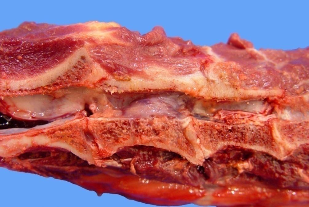
8 Gross Description A nodular mass is present within the second cervical vertebrae. It protrudes into the spinal canal (compressing the spinal cord at this site) courtesy of Dr L Pack, AVC

9 Gross Description
10 Gross Description On section, a soft tan to pink gelatinous mass is present within the marrow cavity of the vertebral body with slight dorsal protrusion into the spinal canal.
11 Differential Diagnoses:
12 Differential Diagnoses: Multiple myeloma, osteosarcoma, osteomyelitis
13 Histology
14 Histologic Description:

15 Histologic Description: There is a marked increase in the cellularity of the bone marrow (100% cells / 0% adipose tissue)


16 Histologic Description: Sheets of tightly packed neoplastic round cells infiltrate the marrow cavity of the vertebral body

17 Histologic Description: The cells are round with distinct cell margins and a moderate amount of pale basophilic cytoplasm occasionally with perinuclear clearing. The nuclei are eccentric and round with coarse clumped hyperchromatic chromatin
18 Case #1 Histologic Description (summary): Sheets of tightly packed neoplastic round cells infiltrate the marrow cavity of the vertebral body. The cells are round with distinct cell margins and a moderate amount of pale basophilic cytoplasm with occasional perinuclear clearing. The nuclei are round with clumped chromatin Morphologic Diagnosis:
19 Case #1 Histologic Description (summary): Sheets of tightly packed neoplastic round cells infiltrate the marrow cavity of the vertebral body. The cells are round with distinct cell margins and a moderate amount of pale basophilic cytoplasm with occasional perinuclear clearing. The nuclei are round with clumped chromatin Morphologic Diagnosis: Myeloma, vertebral body Possible clinical pathology findings:
20 Case #1 Histologic Description (summary): Sheets of tightly packed neoplastic round cells infiltrate the marrow cavity of the vertebral body. The cells are round with distinct cell margins and a moderate amount of pale basophilic cytoplasm with occasional perinuclear clearing. The nuclei are round with clumped chromatin Morphologic Diagnosis: Myeloma, vertebral body Possible clinical pathology findings: Hypergammaglobulinemia, hypercalcemia, Bence-Jones proteinuria


21 courtesy of Dr A Matthews AVC Ancillary / helpful diagnostic findings Hypercalcemia increased osteoclastic activity within bone lesions courtesy of Dr. A Matthews, AVC Hypergammaglobulinemia clonal neoplastic plasma cells secrete Ig s (or Ig fragments) of one class, ie monoclonal gammopathy Bence-Jones proteinuria Immunoglobulin light chains pass through glomeruli into urine Radiographs showed punched out lesions in the vertebrae

22 Case #2 Signalment: 1.5 year old, laying hen History: The bird has been down for a day and has not been eating All images for this case were provided by Dr Heather Fenton, AVC

23 Spleen Liver Bone


24 Spleen Normal chicken spleen
25 Spleen Gross Description
26 Spleen Gross Description The spleen is markedly enlarged and misshapen with coalescent yellow-tan nodules scattered over the surface

27 Spleen Gross Description These extend into the parenchyma on cut surface
28 Spleen Histologic Description

29 Spleen Histologic Description Discrete eosinophilic nodules are present within the spleen.
30 Spleen Histologic Description
31 Spleen Histologic Description The nodules are composed of central regions of necrosis surrounded by a thick cellular rim. What are the cells?
32 Spleen Histologic Description The nodules are composed of central regions of necrosis surrounded by a thick cellular rim. What are the cells?

33 Spleen Histologic Description The cells consist primarily of epithelioid macrophages and rare heterophils. Admixed with these cells are occasional lymphocytes, plasma cells and fibroblasts and what other cell type?
34 Spleen Histologic Description Scattered multinucleated giant cells
35 Spleen Morphologic Diagnosis
36 Spleen Morphologic Diagnosis Splenitis, granulomatous, multifocal to coalescing, chronic, marked
37 Spleen Acid-fast stain Etiology and Disease Name


38 Spleen Acid-fast stain Etiology and Disease Name Etiology: Mycobacterium avium Disease Name: Avian tuberculosis
39 Case #2 Histologic Description (summary): Scattered throughout the spleen are discrete foci of necrosis surrounded by infiltrates of epithelioid macrophages and multinucleated giant cells admixed with fewer lymphocytes and plasma cells Acid-fast bacilli are present within the lesion Morphologic Diagnosis: Splenitis, granulomatous, multifocal to coalescing, chronic, marked, with intralesional acid-fast bacilli Etiology and Disease Name: Mycobacterium avium Avian tuberculosis

 Affects")
40 Case #2 Avian Tuberculosis Mycobacterium avium complex (serotypes 1, 2 and 3) Can infect an extensive range of species In birds usually chickens, captive wild birds and turkeys Free range farming systems and keeping breeders for several years are conducive to the spread of this disease. Main sources of infection are infected individuals and the contaminated environment (water and soil) Affects multiple organs Granulomatous osteomyelitis and hepatitis

41 Case #3 Signalment: 9 yr-old MC cat History: Cat had been anorexic and was beginning to lose weight. On physical exam the cat was dehydrated. Abdominal palpation revealed enlarged irregular kidneys. Blood chemistry revealed elevated urea and creatinine.
42

43 Gross Description Marked enlargement of the kidneys Nodular/irregular capsular surface
44 Gross Description
45 Gross Description On cut surface, there are multifocal to coalescing, white, round to irregular masses scattered throughout both of the kidneys
46 Morphologic Diagnosis

47 Morphologic Diagnosis Multiple renal masses, probable malignant neoplasia


48 Need Histology! Feline Infectious Peritonitis (Pyogranulomatous nephritis) Compare previous slides with this case of multifocal granulomatous nephritis due to FIP. These lesions can easily be mistaken for renal lymphoma! Two main differentials for this lesion in cats: Lymphoma and FIP Renal lymphoma


49 LYMPHOMA Renal lymphoma vs. Pyogranulomatous nephritis (FIP, dry form) Renal lesions, cat FIP

50 Histologic Description
51 Histologic Description At low power, we can appreciate a nodule within the tissue (kidney) that looks more blue than normal suggests an increase in the cellularity of the tissue

52 Histologic Description Description: Densely cellular sheet-like infiltrates efface the normal renal architecture leaving few remaining glomeruli

53 At 40x
.")
54 Histologic Description These cells have distinct cell margins, scant eosinophilic cytoplasm, and round nuclei with stippled chromatin and prominent nucleoli. Mitoses are present (arrow).
55 Case #3 Histologic Description (summary): In this section of kidney, most of the normal architecture has been effaced by dense sheet-like cellular infiltrates consisting of large numbers of neoplastic round cells. These neoplastic round cells have small amounts of well-delineated pale acidophilic cytoplasm. The nuclei are round to slightly ovoid with stippled chromatin and distinct nucleoli. Morphologic Diagnosis: Renal lymphoma Comments: Lymphoma is the most common renal tumor in the cat. It may cause kidney deficiency/failure by destroying parenchyma or by obstructing urine outflow


56 Gross Description The inguinal and mesenteric lymph nodes are moist, pale and enlarged 2-3 times normal size. Morphologic diagnosis: Inguinal/ mesenteric lymphadenomegaly Case #4 Images from Noah s arkive


57 Gross Description The pericardial and bone marrow fat tissue are replaced by clear yellow gelatinous material. Morphologic diagnosis: Pericardial and bone marrow fat: Serous atrophy, diffuse, marked. From Noah s arkive

 with large botryoid intracytoplasmic viral")
58 Case #4 Etiology / Disease: Porcine circovirus type 2 / Postweaning multisystemic wasting syndrome Histology: Granulomatous infiltration of the node (diffuse granulomatous lymphadenitis) with large botryoid intracytoplasmic viral inclusions Images from Noah s arkive
 compressed by a subdural (intradural)")

59 7 year-old cow with posterior paralysis Description: Cross section of spinal cord (lumbar segment) compressed by a subdural (intradural) white, solid, ovoid mass. Morphologic diagnosis: 1. Subdural lymphoma 2. Spinal cord compression; lumbar segment. From Noah s arkive Case #5


60 Tonsil: Squamous cell carcinoma Tonsil: Lymphoma From Noah s arkive Pathologic Basis of Veterinary Disease, 4 th ed.


61 Splenic hematoma, dog. Locally, there is marked expansion and replacement of the splenic parenchyma by a mass of blood. The capsule is markedly distended; cut surface reveals extensive areas of necrosis and fibrin.
62 Splenic Nodules important differentials Splenic nodules with a bloody consistency Hematoma Hemangioma Hemangiosarcoma Splenic infarcts Incompletely contracted areas of the spleen Splenic nodules with a firm consistency Nodular hyperplasia Primary Neoplasia (eg lymphoma, histiocytic sarcoma) Metastatic (secondary) Neoplasia Abscess Granuloma
63 Diffuse Splenomegaly important differentials Diffuse splenomegaly with a bloody consistency = Bloody Spleens Acute septicemia Acute hemolytic anemia Splenic torsion Barbiturate anesthesia/euthanasia Vascular pooling Diffuse splenomegaly with a firm consistency = Meaty Spleens Septicemia Salmonella Hemolytic anemia Neoplasia eg lymphoma, mast cell tumor, histiocytic sarcoma Granulomatous disease Amyloidosis


64 Siderofibrosis = Gamna-Gandy bodies, siderocalcified plaques Histo: Hematoidin, hemosiderin and mineral deposits Pathologic Basis of Veterinary Disease, 4 th ed.
 nodular masses.")
65 Splenic hemangiosarcoma, dog. Multiple, variable sized, dark red, soft (bloody) nodular masses. These lesions often develop infarction and/or rupture, followed by bleeding.

66 Splenic lacerations and hematoma in horse that was hit-by-car

67 Splenic lacerations and hematoma in horse that was hit-by-car
HISTOPATHOLOGY. Shannon Martinson
 HISTOPATHOLOGY Shannon Martinson March 2013 Case #1 History: 8 year old beagle Neck pain for the past couple of weeks Paresis, followed by paralysis developed over the past few days Gross Description courtesy
Pathology of the Hematopoietic System. Case studies
 Pathology of the Hematopoietic System Case studies Shannon Martinson, September 2015 Signalment: 9 yr-old MC cat Case Study 1 History: Cat had been anorexic and developed bleeding in the eyes Physical
Pathology of the Hematopoietic System - Lab.
 Pathology of the Hematopoietic System - Lab http://people.upei.ca/smartinson/ Shannon Martinson, September 2015 Case #1 Signalment: 96 kg gilt History: Pig from minimal disease herd. Sudden death Case
Inflammation Laboratory 3 Emphasis: Chronic inflammation and healing. Shannon Martinson: VPM 152: April 2013
 Inflammation Laboratory 3 Emphasis: Chronic inflammation and healing Shannon Martinson: http://people.upei.ca/smartinson VPM 152: April 2013 Example A Reproductive tract and colon/rectum from a sheep Previous
Pathology of the Hematopoietic System. Lecture 3: Thymus and Spleen
 Pathology of the Hematopoietic System Lecture 3: Thymus and Spleen Enrique Aburto, March 2014 Thymus Structure and function White to pink, lobulated organ within the anterior mediastinum Ruminants and
Pathology of the Hematopoietic System. Lecture 3: Spleen and thymus
 Pathology of the Hematopoietic System Lecture 3: Spleen and thymus Shannon Martinson, March 2012 Spleen Structure and Function Present in the left cranial part of the abdomen within the greater omentum
Avian Pathology. Bacterial diseases: histo slides. ECVP-ESVP Summer School 2012 Frédérique NGUYEN
 Avian Pathology Bacterial diseases: histo slides ECVP-ESVP Summer School 2012 Frédérique NGUYEN Bacterial diseases: histo slides B1. Turkey. Organs? Morphologic diagnosis? Special procedure? B2. Hen. Organ?
Pathology of the Hematopoietic System. Lecture 3: Spleen and thymus
 Pathology of the Hematopoietic System Lecture 3: Spleen and thymus Shannon Martinson, March 2011 Spleen Structure and Function Present in the left cranial part of the abdomen within the greater omentum
Pathology of the Alimentary Tract
 Pathology of the Alimentary Tract Lab 2: Lower alimentary tract SI, LI, cecum, and peritoneum GIST in the cecum of a dog Shannon Martinson: http://people.upei.ca/smartinson VPM 221: November, 2011 3 year
Cellular Pathology. Histopathology Lab #2 (web) Paul Hanna Jan 2018
 Paul Hanna Jan 2018") Cellular Pathology Histopathology Lab #2 (web) Paul Hanna Jan 2018 Slide #91 Clinical History: a necropsy was performed on an aged cat the gross pathological changes included: widespread subcutaneous edema
Cellular Pathology Histopathology Lab #2 (web) Paul Hanna Jan 2018 Slide #91 Clinical History: a necropsy was performed on an aged cat the gross pathological changes included: widespread subcutaneous edema
Diagnostic Cytology of Cancer Cases
 Diagnostic Cytology of Cancer Cases Somporn Techangamsuwan Companion Animal Cancer Research Unit (CAC-RU) Department of Pathology, Faculty of Veterinary Science, Chulalongkorn University 1 Tumor or Non-tumor
Pathology of the Hematopoietic System. Lecture 1: Introduction, Bone Marrow, and Blood Cells
 Pathology of the Hematopoietic System Lecture 1: Introduction, Bone Marrow, and Blood Cells Shannon Martinson, March 2010 Hematopoietic system Tizzard, Veterinary Immunology,, 9 th Ed, Saunders Myeloid
Pathology of the Hematopoietic System
 Pathology of the Hematopoietic System Lecture 1: Introduction, Bone Marrow, and Blood Cells http://people.upei.ca/smartinson/ Shannon Martinson, April 2018 Hematopoietic system - Introduction Myeloid Tissue
WSC , Conference 9, Case 1. Tissue from a nyala.
 WSC 2009-2010, Conference 9, Case 1. Tissue from a nyala. MICROSCOPIC DESCRIPTION: Heart, atrium (1 pt.): Approximately 40% of the atrial myocardium is replaced by areas of fibrous connective tissue (1
Disorders of Cell Growth & Neoplasia. Histopathology Lab
 Disorders of Cell Growth & Neoplasia Histopathology Lab Paul Hanna April 2010 Case #84 Clinical History: 5 yr-old, West Highland White terrier. skin mass from axillary region. has been present for the
Respiratory Pathology Lab 2: Lung. Shannon Martinson,
 Respiratory Pathology Lab 2: Lung Shannon Martinson, 2017 http://people.upei.ca/smartinson/ Case 1 Signalment: 9 month old DSH cat History: Poor doer with stunted growth One month of lethargy one day the
Liver Lab #2. Bacterial Hepatitis
 Liver Lab #2 Bacterial Hepatitis Case: O12561-04. Adult ewe. Describe the lesion: Multifocal large nodules ranging in size from 1-3.5cm in greatest diameter are present within the liver and are filled
Plasma cell myeloma (multiple myeloma)
") Plasma cell myeloma (multiple myeloma) Common lymphoid neoplasm, present at old age (70 years average) Remember: plasma cells are terminally differentiated B-lymphocytes that produces antibodies. B-cells
Plasma cell myeloma (multiple myeloma) Common lymphoid neoplasm, present at old age (70 years average) Remember: plasma cells are terminally differentiated B-lymphocytes that produces antibodies. B-cells
Burkitt lymphoma. Sporadic Endemic in Africa associated with EBV Translocations involving MYC gene on chromosome 8
 Heme 8 Burkitt lymphoma Sporadic Endemic in Africa associated with EBV Translocations involving MYC gene on chromosome 8 Most common is t(8;14) Believed to be the fastest growing tumor in humans!!!! Morphology
Table of Contents. Preface xi. Acknowledgments xiii. Part I Overview of the Diagnostic Process 1. 1 Overview of Grading and Staging 3
 Table of Contents Preface xi Acknowledgments xiii Part I Overview of the Diagnostic Process 1 1 Overview of Grading and Staging 3 Identification of the process 3 Identification of tumor types 5 Grading
This is the second learning component (Learning Component 2) in our first learning module (Learning Module 1). In this component we review a very
 in our first learning module (Learning Module 1). In this component we review a very") This is the second learning component (Learning Component 2) in our first learning module (Learning Module 1). In this component we review a very basic response to injury inflammation. We ll look at examples
This is the second learning component (Learning Component 2) in our first learning module (Learning Module 1). In this component we review a very basic response to injury inflammation. We ll look at examples
VPM Pigment and other tissue deposits. Shannon Martinson
 VPM 152 - Pigment and other tissue deposits Shannon Martinson http://people.upei.ca/smartinson/ Case 1 Signalment: 2 month old heifer beef calf Clinical History: Lateral recumbency for 4 days Tachycardia,
Endocrine Lab. Heather Fenton VPM 222 November
 Endocrine Lab Heather Fenton VPM 222 November 27 2012 Case 1: Nursery pig Case 1: Nursery pig Description: There are multifocal round (approximately 1cm diameter) firm lesions within the adrenal gland
Cellular Pathology Gross Pathology Laboratory 2 Cell Injury. VPM 152: General Pathology Instructor: Chelsea Martin Winter 2016
 Cellular Pathology Gross Pathology Laboratory 2 Cell Injury VPM 152: General Pathology Instructor: Chelsea Martin Winter 2016 Gross Specimens The following slides consist of images from the specimens presented
VETERINARY HEMATOLOGY ATLAS OF COMMON DOMESTIC AND NON-DOMESTIC SPECIES COPYRIGHTED MATERIAL SECOND EDITION
 VETERINARY HEMATOLOGY ATLAS OF COMMON DOMESTIC AND NON-DOMESTIC SPECIES SECOND EDITION COPYRIGHTED MATERIAL CHAPTER ONE HEMATOPOIESIS GENERAL FEATURES All blood cells have a finite life span, but in normal
Lách
 Lách Lách Lách Lách Splenogonadal fusion. Splenic tissue is attached to testicular tissue. Pseudocyst (false or secondary cyst). A, Outer aspect. Pseudocyst (false or secondary cyst). B, Inner surface.
What s your diagnosis? Malori Marotz. Squirt, an 8month old mix breed puppy. History:
 What s your diagnosis? Malori Marotz Squirt, an 8month old mix breed puppy History: The owner obtained squirt at 12 weeks of age. The owner reported that Squirt was passing soft stools lately and he is
Pathology of Hematopoietic and Lymphoid tissue
 CONTENTS Pathology of Hematopoietic and Lymphoid tissue White blood cells and lymph nodes Quantitative disorder of white blood cells Reactive lymphadenopathies Infectious lymphadenitis Tumor metastasis
A Look at the Lymphoid System Rose Raskin, DVM, PhD, DACVP Purdue University West Lafayette, IN
 A Look at the Lymphoid System Rose Raskin, DVM, PhD, DACVP Purdue University West Lafayette, IN The lymphoid organs commonly biopsied include the peripheral and internal lymph nodes, spleen, and occasionally
Hematopathology Lab. Third year medical students
 Hematopathology Lab Third year medical students Objectives Identify the lesion Know the specific name of the lesion Know associated disease Know relevant pathologic background Spherocytes: appear small,
Inflammation Laboratory 1
 Inflammation Laboratory 1 Lab1 Emphasis: The exudates of acute inflammation Descriptions Morphologic Diagnoses Shannon Martinson: http://people.upei.ca/smartinson VPM 152: February 2012 Describing Lesions
Pathology of the Liver and Biliary Tract 5 Diseases of the Biliary Tract. Shannon Martinson, March 2017
 Pathology of the Liver and Biliary Tract 5 Diseases of the Biliary Tract Shannon Martinson, March 2017 http://people.upei.ca/smartinson/ OUTLINE Normal anatomy & function Hepatobiliary injury and responses
Inflammation Laboratory 1
 Inflammation Laboratory 1 Lab1 Emphasis: The exudates of acute inflammation Descriptions Morphologic Diagnoses Shannon Martinson: http://people.upei.ca/smartinson VPM 152: March 2013 Describing Lesions
Case Scenario 1: Thyroid
 Case Scenario 1: Thyroid History and Physical Patient is an otherwise healthy 80 year old female with the complaint of a neck mass first noticed two weeks ago. The mass has increased in size and is palpable.
Tumors of the Spleen
 Tumors of the Spleen 803-808-7387 www.gracepets.com These notes are provided to help you understand the diagnosis or possible diagnosis of cancer in your pet. For general information on cancer in pets
Almost any suspected tumor can be aspirated easily and safely. Some masses are more risky to aspirate including:
 DOES THIS PATIENT HAVE CANCER? USING IN-HOUSE CYTOLOGY TO HELP YOU MAKE THIS DIAGNOSIS. Joyce Obradovich, DVM, Diplomate, ACVIM (Oncology) Animal Cancer & Imaging Center, Canton, Michigan Almost every
Figure 2: Lymph node Cortical follicular (F) and paracortical (PC) atrophy, with narrowing of the cortex relative to the medulla (M).
 and paracortical (PC) atrophy, with narrowing of the cortex relative to the medulla (M).") Figure 1: Lymph node Follicular hyperplasia, with expansion of the follicular germinal centres (F) by large blast cells. Paracortical hyperplasia, with expansion of the paracortex (PC) by small lymphocytes.
Figure 1: Lymph node Follicular hyperplasia, with expansion of the follicular germinal centres (F) by large blast cells. Paracortical hyperplasia, with expansion of the paracortex (PC) by small lymphocytes.
MECHANISMS OF HUMAN DISEASE: LABORATORY SESSIONS LYMPHOMA. April 16, 2008
 MECHANISMS OF HUMAN DISEASE: LABORATORY SESSIONS LYMPHOMA April 16, 2008 FACULTY COPY GOAL: Learn the appearance of normal peripheral blood elements and lymph nodes. Recognize abnormal peripheral blood
Neoplasia 2018 Lecture 2. Dr Heyam Awad MD, FRCPath
 Neoplasia 2018 Lecture 2 Dr Heyam Awad MD, FRCPath ILOS 1. List the differences between benign and malignant tumors. 2. Recognize the histological features of malignancy. 3. Define dysplasia and understand
VPM Pigment and other tissue deposits. Shannon Martinson
 VPM 152 - Pigment and other tissue deposits Shannon Martinson http://people.upei.ca/smartinson/ Case 1: Signalment: 2 month old heifer beef calf Clinical History: Lateral recumbency for 4 days. Tachycardia,
Disturbances of Circulation. Histopathology Lab #2 (Web)
") Disturbances of Circulation Histopathology Lab #2 (Web) Paul Hanna Winter 2015 Slide #96 History: pig was fine in the morning & found dead in the afternoon there was ~100 mls of clear fluid in the pericardial
Disturbances of Circulation Histopathology Lab #2 (Web) Paul Hanna Winter 2015 Slide #96 History: pig was fine in the morning & found dead in the afternoon there was ~100 mls of clear fluid in the pericardial
PROBABLE HODGKIN'S DISEASE IN A DOG: REPORT OF A CASE 1
 PROBABLE HODGKIN'S DISEASE IN A DOG: REPORT OF A CASE 1 LEONARD K. STALKER, M.D. Fellow in Surgery, The Mayo Foundation CARL F. SCHLOTTHAUER, D.V.M. AND WILLIAM H. FELDMAN, D.V.M., M.S. Division of Experimental
Pathology of Hematopoietic and Lymphoid tissue
 Pathology of Hematopoietic and Lymphoid tissue Peerayut Sitthichaiyakul, M.D. Department of Pathology and Forensic Medicine Faculty of Medicine, Naresuan University CONTENTS White blood cells and lymph
PATHOLOGY Intracellular Degeneration LAB 1
 PATHOLOGY Intracellular Degeneration LAB 1 Cellular swelling Liver Organ :- Liver Lesion :- 1. Narrowing of hepatic sinusoids due to the swelling of hepatocyte. 2. The cytoplasm of affected hepatocyte
SESSION 1: GENERAL (BASIC) PATHOLOGY CONCEPTS Thursday, October 16, :30am - 11:30am FACULTY COPY
 PATHOLOGY CONCEPTS Thursday, October 16, :30am - 11:30am FACULTY COPY") SESSION 1: GENERAL (BASIC) PATHOLOGY CONCEPTS Thursday, October 16, 2008 9:30am - 11:30am FACULTY COPY GOAL: Describe the basic morphologic (structural) changes which occur in various pathologic conditions.
SESSION 1: GENERAL (BASIC) PATHOLOGY CONCEPTS Thursday, October 16, 2008 9:30am - 11:30am FACULTY COPY GOAL: Describe the basic morphologic (structural) changes which occur in various pathologic conditions.
WSC , Conference 9. Case 1. Tissue from a rhesus macaque.
 Case 1. Tissue from a rhesus macaque. MICROSCOPIC DESCRIPTION: Esophagus: There is multifocal loss of the mucosal lining (1 pt). In these areas, the denuded subepithelial fibrous connective tissue is infiltrated
What s Your Diagnosis?
 What s Your Diagnosis? Signalment: 5 year old MC Belgian Malinois Presenting Complaint: Perineal hernia as well as not eating or defecating History: The patient presented to the KSU VHC on 7/28/2018 for
Inflammation Laboratory 2. Shannon Martinson: VPM 152: March 2012
 Inflammation Laboratory 2 Shannon Martinson: http://people.upei.ca/smartinson VPM 152: March 2012 Reminder - Creating a Morphologic Diagnosis for Inflammatory Lesions Organ and Process Exudate Distribution
Cytology of NeoPlasia
 PEEr reviewed Cytology of NeoPlasia An Essential Component of Diagnosis Anne Barger, DVM, MS, Diplomate ACVP Cytology is a quick, easy, and inexpensive diagnostic tool. It is commonly used for the diagnosis
Case: The patient is a 73 year old woman with vague complaints of dyspepsia and abdominal pain. Upper endoscopy showed features of gastritis and a
 Case: The patient is a 73 year old woman with vague complaints of dyspepsia and abdominal pain. Upper endoscopy showed features of gastritis and a nodular lesion in the body of the stomach. The patient
Avian Pathology. QUIZ HISTOPATHOLOGY: Answers
 ECVP/ESVP Summer School 202 Avian Pathology QUIZ HISTOPATHOLOGY: Answers Jérôme ABADIE Frédérique NGUYEN Q Tissue from a Hen Q. Hen Lung. Infiltrating and effacing nearly 60% of the pulmonary parenchyma,
LYMPH GLAND. By : Group 1
 LYMPH GLAND By : Group 1 ANATOMY LYMPH NODE Lymphatic Organs Red bone marrow Thymus gland Lymph nodes Lymph nodules Spleen Primary organs Secondary organs Lymph Nodes Firm, smooth-surfaced, bean-shaped
Disorders of Cell Growth & Neoplasia
 General Pathology VPM 152 Disorders of Cell Growth & Neoplasia Lecture 3 Rate of growth, local invasion, and metastasis. Molecular basis of cancer (normal cell-cycle and cellular proliferation). Enrique
, , 2011 HODGKIN LYMPHOMA
 European Federation of Cytology Societies 4tu Annual Tutorial in Cytopathology Trieste, June 6-10, 2011 HODGKIN LYMPHOMA Classification The World Health Organization Classification of Lymphomas (2001)
Kidney Case 1 SURGICAL PATHOLOGY REPORT
 Kidney Case 1 Surgical Pathology Report February 9, 2007 Clinical History: This 45 year old woman was found to have a left renal mass. CT urography with reconstruction revealed a 2 cm medial mass which
Cytology of Neoplasms that Occur on the Limbs Rick Alleman, DVM, PhD, DABVP, DACVP
 Cytology of Neoplasms that Occur on the Limbs Rick Alleman, DVM, PhD, DABVP, DACVP I. Introduction The purpose of this material is to provide information that may be useful in the identification of tumors
Pathology lab 4 DONE BY : MORAD ABU QAMAR
 Pathology lab 4 DONE BY : MORAD ABU QAMAR Chronic interstitial inflammation, lung Certain etiologic agents such as viruses are more likely to lead to chronic inflammation, as seen here in the lung of a
Cell injury, adaptation and death. Unite one Second Lab.
 Cell injury, adaptation and death Unite one Second Lab. The two lung abscesses seen here are examples of liquefactive necrosis in which there is a liquid center in an area of tissue injury. One abscess
What s Your Diagnosis??? Renée Fahrenholz, Class of 2012
 Renée Fahrenholz, Class of 2012 What s Your Diagnosis??? Signalment Emma, a 9 year old, Female, Spayed, Domestic Short Haired Feline Presenting Complaint Weight loss, vomited the morning of her visit,
Osteosclerotic Myeloma (POEMS Syndrome)
") Osteosclerotic Myeloma (POEMS Syndrome) Osteosclerotic Myeloma (POEMS Syndrome) Synonyms Crow-Fukase syndrome Multicentric Castleman disease Takatsuki syndrome Acronym coined by Bardwick POEMS Scheinker,
Osteosclerotic Myeloma (POEMS Syndrome) Osteosclerotic Myeloma (POEMS Syndrome) Synonyms Crow-Fukase syndrome Multicentric Castleman disease Takatsuki syndrome Acronym coined by Bardwick POEMS Scheinker,
2015 Descriptive Vet Path Course. Histo Exam #3 KEY
 2015 Descriptive Vet Path Course Histo Exam #3 KEY Test 3, Slide 1 Tissue from a guinea pig. MORPHOLOGIC DIAGNOSIS: Heart: Multifocally and randomly (1 pt), within the left and right ventricular myocardium
GENERAL ABDOMINAL IMAGING PERITONEAL SPACE, PANCREAS, & SPLEEN. VMB 960 March 25, 2013
 GENERAL ABDOMINAL IMAGING PERITONEAL SPACE, PANCREAS, & SPLEEN VMB 960 March 25, 2013 REFERENCE Chapters 35-36 Pages 650-678 Chapter 37 Pages 694-701 Chapter 3 Pages 38-49 OBJECTIVES Radiography and Ultrasound
What s Your Diagnosis?
 What s Your Diagnosis? Courtney S. Wait Signalment: 11 year old FS Labrador Retriever Presenting Complaint/History: The patient presented to the referring DVM for inappetance, vomiting, lethargy, and anorexia.
Neoplasms of the Canine, Feline and Lemur Liver:
 Neoplasms of the Canine, Feline and Lemur Liver: Classification and Prognosis Annual Seminar of the French Society of Veterinary Pathology John M. Cullen VMD PhD DACVP North Carolina State University Primary
Proceeding of the SEVC Southern European Veterinary Conference
 www.ivis.org Proceeding of the SEVC Southern European Veterinary Conference Oct. 17-19, 2008 Barcelona, Spain http://www.sevc.info Reprinted in the IVIS website with the permission of the SEVC www.ivis.org
Disclosures. Parathyroid Pathology. Objectives. The normal parathyroid 11/10/2012
 Disclosures Parathyroid Pathology I have nothing to disclose Annemieke van Zante MD/PhD Assistant Professor of Clinical Pathology Associate Chief of Cytopathology Objectives 1. Review the pathologic features
Note: The cause of testicular neoplasms remains unknown
 - In the 15- to 34-year-old age group, they are the most common tumors of men. - Tumors of the testis are a heterogeneous group of neoplasms that include: I. Germ cell tumors : 95%; all are malignant.
GENERAL ABDOMINAL IMAGING PERITONEAL SPACE, PANCREAS, & SPLEEN
 GENERAL ABDOMINAL IMAGING PERITONEAL SPACE, PANCREAS, & SPLEEN VMB 960 March 25, 2013 REFERENCE Chapters 35-36 Pages 650-678 Chapter 37 Pages 694-701 Chapter 3 Pages 38-49 OBJECTIVES Radiography and Ultrasound
MORPHOLOGIC DIAGNOSIS: Liver: Hepatitis, necrotizing, multifocal to coalescing, severe, with numerous trichomonads. (3 pt)
") Case 1. Tissue from a pelican. MICROSCOPIC DESCRIPTION: Liver: Approximately 80% (1 pt) of the liver is replaced by multifocal to coalescing areas of coagulative and lytic necrosis. Centrally, within these
Case 1. Tissue from a pelican. MICROSCOPIC DESCRIPTION: Liver: Approximately 80% (1 pt) of the liver is replaced by multifocal to coalescing areas of coagulative and lytic necrosis. Centrally, within these
VPM 152 GENERAL PATHOLOGY Gross Pathology Lab 1
 VPM 152 GENERAL PATHOLOGY Gross Pathology Lab 1 CELLULAR ADAPTATIONS AND POST MORTEM CHANGES Dr Enrique Aburto 421N Path / Micro 15 Jan 2016 Bone Marrow Atrophy Atrophy Serous atrophy of fat Gelatinous
TUMOR,NEOPLASM. Pathology Department, Zhejiang University School of Medicine,
 TUMOR,NEOPLASM Pathology Department, Zhejiang University School of Medicine, 马丽琴,maliqin198@zju.edu.cn The points in this chapter What is a neoplasm (conception) Morphology of neoplasm Macroscopy of Neoplasm
ataxia, head tremors and mild inappentence, was given palliative care
 2013-5-2 Cerebellum, Spleen-Raccoon Ahmed M. Abubakar BOVINE PATHOLOGY CONTRIBUTING INSTITUTION :College of Veterinary Medicine UC Davies Signalment: Wild-caught juvenile male raccoon, ( Procyon lotor)
CONNECTIVE TISSUE (C.T.)
") CONNECTIVE TISSUE (C.T.) Objectives: By the end of this lecture, the student should be able to: 1. Enumerate the general characteristics of C.T. 2. Classify C.T into C.T. proper and special types of C.T.
CONNECTIVE TISSUE (C.T.) Objectives: By the end of this lecture, the student should be able to: 1. Enumerate the general characteristics of C.T. 2. Classify C.T into C.T. proper and special types of C.T.
CINtec p16 INK4a Staining Atlas
 CINtec p16 INK4a Staining Atlas Rating Rating Positive The rating positive will be assigned if the p16 INK4a -stained slide shows a continuous staining of cells of the basal and parabasal cell layers of
Pathology of the Liver and Biliary Tract 5 Diseases of the Biliary Tract. Shannon Martinson, April 2016
 Pathology of the Liver and Biliary Tract 5 Diseases of the Biliary Tract Shannon Martinson, April 2016 http://people.upei.ca/smartinson/ OUTLINE Normal anatomy & function Hepatobiliary Injury and responses
Describing and interpreting gross lesions. Prepared for VPM 4600, May 2018; Shannon Martinson
 Describing and interpreting gross lesions Prepared for VPM 4600, May 2018; Shannon Martinson How to Describe (and Interpret) Lesions Step 1 Step 2 Step 3 Step 4 Look at the specimen: Is it normal or abnormal
PATHOLOGY OF LIVER & BILIARY TRACT. Lecture 5. Idiopathic & proliferative conditions; diseases of the biliary tract
 PATHOLOGY OF LIVER & BILIARY TRACT Lecture 5 Idiopathic & proliferative conditions; diseases of the biliary tract Enrique Aburto Winter 2015 IX. Diseases of uncertain origin Equine serum hepatitis Idiopathic
(Iteceived for publication December 3, 1915)
") TRANSPLANTABLE SARCOMATA OF THE RAT LIVER ARISING IN THE WALLS OF PARASITIC CYSTS G. L. ROHDENBURG, M.D., AND F. D. BULLOCK, M.D. From Colurnbia University, George Crocker Special Re-search Fund, F. C.
TRANSPLANTABLE SARCOMATA OF THE RAT LIVER ARISING IN THE WALLS OF PARASITIC CYSTS G. L. ROHDENBURG, M.D., AND F. D. BULLOCK, M.D. From Colurnbia University, George Crocker Special Re-search Fund, F. C.
FNA of Thyroid. Toward a Uniform Terminology With Management Guidelines. NCI NCI Thyroid FNA State of the Science Conference
 FNA of Thyroid NCI NCI Thyroid FNA State of the Science Conference Toward a Uniform Terminology With Management Guidelines Thyroid Thyroid FNA Cytomorphology NCI Thyroid FNA State of the Science Conference
Figure Schematic illustration of the hematopoietic hierarchy in the bone marrow.
 Slide 1 Figure 13-01. Schematic illustration of the hematopoietic hierarchy in the bone marrow. The bone marrow consists of: (1) stem cells, pluripotent cells capable of self-renewal; (2) committed progenitor
Thyroiditis in the differential diagnosis of lymphoma
 Thyroiditis in the differential diagnosis of lymphoma 2nd Pannonia Congress of Pathology Siofok, Hungary, May 17-19, 2012 Božo Krušlin, M.D., Ph.D., Dpt of Pathology, School of Medicine, University of
CYTOLOGY OF THE LIVER
 CYTOLOGY OF THE LIVER Maxey L. Wellman, DVM, PhD, DACVP (Clinical Pathology) Professor, Department of Veterinary Biosciences, College of Veterinary Medicine, The Ohio State University, Columbus, OH, USA
Principles of Surgical Oncology. Winnie Achilles Tierklinik Hollabrunn Lastenstrasse Hollabrunn
 Principles of Surgical Oncology Winnie Achilles Tierklinik Hollabrunn Lastenstrasse 2 2020 Hollabrunn boexi@gmx.de The first surgery provides the best chance for a cure in an animal with a tumor Clinical
Submission of samples. Cytology of Lumps and Bumps. Evaluation of samples. Use caution interpreting. Criteria of malignancy.
 Submission of samples Cytology of Lumps and Bumps Paul Avery VMD, PhD, DACVP paul.avery@colostate.edu Air dry only No wet fixation using formalin or ethanol Stain 1-2 on-site to evaluate quality Send all
Post Mortem Approach to the Haematopoietic System
 Post Mortem Approach to the Haematopoietic System Examination of the hematopoeitic system involves evaluation of various organ systems including the bone marrow, spleen, lymph nodes and thymus. System
Case year female. Routine Pap smear
 Case 1 57 year female Routine Pap smear Diagnosis? 1. Atypical glandular cells of unknown significance (AGUS) 2. Endocervical AIS 3. Endocervical adenocarcinoma 4. Endometrial adenocarcinoma 5. Adenocarcinoma
Extracellular degeneration
 Extracellular degeneration By Dr. Hemn Hassan Othman PhD, Pathology Fall 2016 1/17/2017 1 Extracellular Degenerations I / Hyaline Degeneration (Hyalinization): The ward hyaline is derived from the Latin
DETERMINATION OF A LYMPHOID PROCESS
 Chapter 2 Applications of Touch Preparation Cytology to Intraoperative Consultations: Lymph Nodes and Extranodal Tissues for Evaluation of Hematolymphoid Disorders INTRODUCTION As discussed in Chap. 1,
COMPANY OR UNIVERSITY
 CONTRIBUTOR NAME Daniel Heinrich, DVM CONTRIBUTOR EMAIL dheinric@umn.edu COAUTHORS Jed Overmann, DVM, DACVP; Davis Seelig DVM, PhD, DACVP & Matthew Sturos, DVM COMPANY OR UNIVERSITY University of Minnesota
Infections and nonmicrobial inflammatory stimuli can cause leukocytosis (as seen in Lab 1) as well as lymph node enlargement (lymphadenopathy).
 as well as lymph node enlargement (lymphadenopathy).") LAB 5: LYMPHOID TISSUE AND SKIN The focus of this week s lab will be pathology of the lymphoid tissue and skin. The lymphoid organs include the thymus, spleen, and lymph nodes. Abnormalities in the lymph
LAB 5: LYMPHOID TISSUE AND SKIN The focus of this week s lab will be pathology of the lymphoid tissue and skin. The lymphoid organs include the thymus, spleen, and lymph nodes. Abnormalities in the lymph
Anaplastic Large Cell Lymphoma (of T cell lineage)
") Anaplastic Large Cell Lymphoma (of T cell lineage) Definition T-cell lymphoma comprised of large cells with abundant cytoplasm and pleomorphic, often horseshoe-shaped nuclei CD30+ Most express cytotoxic
Anaplastic Large Cell Lymphoma (of T cell lineage) Definition T-cell lymphoma comprised of large cells with abundant cytoplasm and pleomorphic, often horseshoe-shaped nuclei CD30+ Most express cytotoxic
Non-Invasive Follicular Thyroid Neoplasm with Papillary-like Nuclei (NIFTP)
") Papillary Thyroid Carcinoma: Follicular Variant Encapsulated Type Replaced by: Non-Invasive Follicular Thyroid Neoplasm with Papillary-like Nuclei (NIFTP) Lester D. R. Thompson www.lester-thompson.com
Papillary Thyroid Carcinoma: Follicular Variant Encapsulated Type Replaced by: Non-Invasive Follicular Thyroid Neoplasm with Papillary-like Nuclei (NIFTP) Lester D. R. Thompson www.lester-thompson.com
Practical Histology. Lab 3: Connective tissue
 Practical Histology Lab 3: Connective tissue Connective tissues Connective tissue provides structural support for the body by binding cells and tissues together to form organs. It also provides metabolic
Neoplasia part I. Dr. Mohsen Dashti. Clinical Medicine & Pathology nd Lecture
 Neoplasia part I By Dr. Mohsen Dashti Clinical Medicine & Pathology 316 2 nd Lecture Lecture outline Review of structure & function. Basic definitions. Classification of neoplasms. Morphologic features.
A case of giant cell tumour of soft parts in a horse Francesco Cian 1, Sarah Whiteoak 2, Jennifer Stewart 1
 A case of giant cell tumour of soft parts in a horse Francesco Cian 1, Sarah Whiteoak 2, Jennifer Stewart 1 1 Animal Health Trust, Newmarket, UK 2 608 Equine and Farm Vets, Rowington, UK Signalment: Horse,
Normal thyroid tissue
 Thyroid Pathology Overview Normal thyroid tissue Normal thyroid tissue with follicles filled with colloid. Thyroid cells form follicles, spheres of epithelial cells (always single layered in health, usually
Canine Liver Eneku Wilfred Bovine Pathology
 2012-1-3 Canine Liver Eneku Wilfred Bovine Pathology Contributor: New Mexico Department of Agriculture Veterinary Diagnostic Services Signalment: 5 month old male Weimaraner dog (Canis familiaris) History:
Systemic Pathology. Practical slides
 Systemic Pathology Practical slides Cardiovascular System CARDIOVASCULAR SYSTEM Slide 1: Coronary artery fresh thrombus Slide 2: Coronary artery organised thrombus Slide 3: Heart Acute myocardial infarction
Schistosome life cycle.
 Schistosomiasis infects approximately 200 million persons and kills approximately 280,000 annually. Most of the mortality comes from hepatic granulomas and fibrosis Schistosoma japonicum and Schistosoma
Blood Cell Identification Graded
 BCP-21 Blood Cell Identification Graded Case History The patient is a 37-year-old female with a history of multiple sickle cell crises. She now presents with avascular necrosis of the left hip. Laboratory