PRESENTATION PLAN. Aim: Bethesda System 2001
|
|
|
- Gillian Daniel
- 6 years ago
- Views:
Transcription


1 REACTIVE CELLULAR CHANGES AND INFECTIONS OF FEMALE GENITAL TRACT Aysun Uğuz, Prof, MD, FIAC Çukurova Üniv. Tıp Fak. Pathology Department-Cytology Division 18.Nisan.2015 Aim: The aim of the presentation is clarifying the cause of infectious disease of female genital tract and the reactive cellular changes to interpretate/ recognize by cytology. PRESENTATION PLAN What Bethesda includes under this title? Causes of infectious disease Reactive cellular changes Repair process Cellular changes due to the RT Atrophy Others... REACTIVE CELLULAR CHANGES AND INFECTIONS OF FEMALE GENITAL TRACT Negative for intraepithelial lesion and malignancy was represented quite heterogenous cytomorphologic features in Bethesda Wide spectrum of benign lesions Bethesda System 2001 Negative for intraepithelial lesions and malignancy Organisms Trichomonas vaginalis Fungal infectious Bacterial vaginosis Actinomyces Cellular changes depend on HPV infection Other neoplastic features (not obligatory to mention in report) Reactive celluler changes İnflammation Radiaton IUD Glandular cells following hysterectomy Atrophy 1
 and further... Case; Age?")
2 CELLULAR CHANGES AND INFECTIONS OF FEMALE GENITAL TRACT When we diagnose; Common point of the heterogenous benign lesions of this category is not to be included the cytomorphologic features of (ASC-US) and further... Case; Age?, Last mens date Hormonal effects, The presence of infectious disease, Atypia history in old smears And if she take any therapy All these that mentioned above have serious effects on cell morphology and clinical information should be included to the whole evaluation process. REACTIVE CELLULAR CHANGES AND INFECTIONS OF FEMALE GENITAL TRACT Cervical cytology is a screening test The aim of the test is to diagnose squamous cell carcinoma and its precursors by cytology. To know what is normal is the base note of the interpretation of the sample regardless conventional or liquid base methods. Squamous and endocervical cells are seen in same morphology in LBP as well as conventional method. REACTIVE CELLULAR CHANGES has... Well described criteria Lack of reproducibility Should be noted the presence of microbial agents in reports Other changes might be mentioned depends of the different requirements of the different clinical departments. TRICHOMONAS VAGINALIS Usually small (especially in liquid base), 15-30µm, oval-round Eccantrically placed faint-vesicular nucleus Cytoplasmic debris can be distinguished from organism itself by analyzing the inner structure difficult to see flagella but can be seen in detailed analysis 2


3 TRİCHOMONAS VAGİNALİS Flagella and pear shaped forms as seen in living organism can be recognazible Eosinophilic cytoplasmic granules Leptotrix can be observed in the background Kite-shape cells can be seen in LPB TRİCHOMONAS VAGİNALİS Presence of inflammatory cells, perinuclear halo in squamous cells and bichromatic staining Organism also can be seen without accompanying of inflammatory cells 3


4 CANDIDA SPECIES Grouping of cells is typical squamous cells can phagocyte neutrophills also is another clue of this infection. Herringbone pattern or shish-kebap pattern with polydust also are useful diagnostically. CANDİDA SPECIES Vacuolated copy paper or moth hole appearance of cytoplasm or reactive nucleus can commonly be observed. CANDIDA SPECIES It is important to differentiate mucus flaments from pseudohyphae 4


5 CANDIDA SPECIES Mucus flaments/strands extend out of the cell groups however it is easy to notice the width of flaments are different and irregular in high magnification No hyphae and spors can be seen Budding yeast forms may occur in Candida infection 5


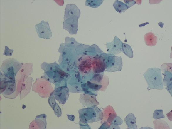
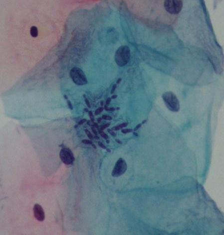
6 BACTERİEL VAGİNOSİS (Cocobasil predominance in the background) BACTERIAL VAGINOSIS The replacement of normal lactobacilli with by shorter bacilli (coccobacilli), curved bacilli and mixed bacteria that gives a filmy appreance in the background is a hallmark of this infection (in conventional smears) Every each squamous cells are covered by the organisms, known as clue cells No lactobacilli BACTERIAL VAGINOSIS BACTERIAL VAGINOSIS On the contrary of conventional smears, LBP s represents claen background associated with clue cells ACTINOMYCES ACTINOMICES Numerous Gram (+), filamentous organisms with acute angle branching patterns can be observed in clumps forms. Numeraous filamentous structures radiating from the center (wolly clumps) Acute inflammatory response associated with polymorphonuclear leucocytes are usually seen In close proximity to IUD in most of the patients 6


7 ACTINOMICES HERPES SYMPLEX VIRUS Ground glass nuclei, margination of chromatine and distinct membrane are diagnostic clues. Multinucleation and nuclear molding are easily identifiable. 7


 It has critical importance")


8 HERPES SYMPLEX VIRUS Eosinophilic nuclear inclusions are prominent which is even more distinctive in LBPs, also without any drying and/or mechanic artifacts SECONDARY REACTIVE CHANGES TO INFLAMMATION REACTIVE CHANGES Slight nuclear enlargement (2 folds of the nuclei of intermedier cell) It has critical importance to distinguish nonspecific halos from the ones associated with HPV infections Cytoplasmic vacuolation and bi-chromatic staining pattern may also be seen REACTIVE CHANGES Clumping neutrophils are demonstrable in the background Cellular details are increased due to extended fixation These cells are seen with slighly enlargement of the nuclei associated with mild hyperchromasia and nucleolei REACTIVE CHANGES Nuclear changes might bring to mind ASC-US diagnosis however, important to require all nuclear changes of ASC- US to refrain any Slight irregularity of nuclear membrane can be noticed However uniformity can be noticed due to the cytoplasmic degeneration 8


9 SECONDARY REACTIVE CHANGES TO REPAIR A B Epihelial cell layers phagocyted the neutrophils is remarkable Cell groups are more spheric and deep focus are necessary due to LBP. No mechanic artefacts FISH SCHOOL REPAIR Eyeliner effect Nuclei with thin chromatin, irregular membranes and presence of nucleolei are noticable with the uniform cell sheets. 9


 RADIATION CHANGES")

10 REPAIR Nuclear polarity (streaming effect) can be observed Differences between repair and SIL Cels are arranged as sheets in repair and this gives a peculiar tissue-culture appearance to the cytoplasms of the cells görünümü vardır. Despite of the enlargement of nucleus, chromatin pattern is seen evenly distributed and homoganous with the regular cell borders sınırlar düzgündür. Nucleoli can be prominent but not in all cell population Differences between repair and SIL To detect SIL cells in epithelial sheets requires further concentration and focusing Size of the nuclei is different and polarity represents prominent Nucleoli can be multiple and prominent RADIATION CHANGES Cell size is increased without any changing in N/C ratio (cytomegali) RADIATION CHANGES Bizarre cell morphology can be observed Large nucleus with a pale chromatin and folding are common in degenerative changes Binucleation and multinucleation also are common. 10


11 RADIATION CHANGES RADIATION CHANGES Nuclei with marked size variation can be observed. enlarged nuclei and normal size nuclei can be seen in the same cell groups. Slight nuclear hyperchromasia. If there is any association of repairment, nuclei can be observed in multiple form as well as the single form Cytoplasmic vacuolation and cytoplasmic polychromatic staining pattern can be encountered. ATROPHY WITH AND WITHOUT INFLAMMATION Flat sheets of parabasallike cells with honeycomb arragement. Relatively high nucleo/cytoplasmic ratio with the particular size of the nuclei Genel bir nükleus büyüklüğü vardır (3-5 folds of intermedier cell nuclei) Nuclear enlargement is relatively descreased compared to the conventiaonal smears ATROPHY Intermedier cells tend to be normochromatic, however, parabasal cells represent slight hyperchromasia with the nuclear elongation. Chromatin is evenly distributed Naked nuclei can be observed due to autolyse, which is much rare in LBPs. 20 x 11


12 ATROPHY Abundant inflammatory exuda and basophilic granular background can mimic tumor diatesis ATROPHY Variable in size and shape histiocytes with multiple round, epithelioid nuclei and foamy-dense cytoplasm OTHER NON-NEOPLASTIC CONDITIONS THAT DO NOT REPLACED IN 2001 BETHESDA SYSTEM 18.Nisan


.")

13 Tubal metaplasia Endocervical columnar cells that arranged in small to pseudostratified crowded groups. Nuclei is oval, round and enlarged, pleomorphism and hypochromasia can be encountered. Evenly distributed chromatin without nucleoli Nucleus-to-cytoplasmic ratio increased. Tubal metaplasia Vacuolation and goblet cell changes might be observed in cytoplasm of the cells. Presence of cilia/ terminal bars is typical for these cells, however isolated cells with cilia is not enough to described as metaplasia. Parakeratosis Various numbers of small, superficial squamous cells with dense-orangiophilic cytoplasm Nuclei is small and picnotic If there is any accompanying morphologic features of atypia, then must be called as atypical cellular chnages (ASC). Isolated cell layers or whorls arrangement is common. Cells can be seen oval, round, polygonal or spindle in shape. Hyperkeratosis Anucelated mature, polygonal squamous cells Keratohyaline granules arecommonly observed as well as ghost cells. Lymphocytic (Follicular) cervicitis Clusters of polymorphic lymphoid cells, macrophages are encountered in mucus deposits. Lymphoid cells can mostly be seen as single cells as well as clusters in LBPs 13

14 Dikkatiniz için 14
LGM International, Inc.
 Liqui-PREP TM Cytology Atlas Preface The following pictures are examples with descriptions of cytology slides processed with the Liqui-PREP TM System.. The descriptions are reviewed by Pathologists. It
Cytoplasmic changes Nuclear changes
 The presence of infection in the female genital tract may procure certain cellular changes in the epithelium. Such changes are seen in nucleus and cytoplasm surrounding the nucleus. Cytoplasmic changes
CINtec p16 INK4a Staining Atlas
 CINtec p16 INK4a Staining Atlas Rating Rating Positive The rating positive will be assigned if the p16 INK4a -stained slide shows a continuous staining of cells of the basal and parabasal cell layers of
Morphology I Slide: 1
 Morphology I Slide: 1 Morphology I Slide: 2 ThinPrep Morphology Normal Cytology Morphology I Slide: 3 CT & Pathologist Training Training program begins with ThinPrep morphology presentation Microscopic
BOSNIAN-TURKISH CYTOPATHOLOGY SCHOOL June 18-19, 2016 Sarajevo. Case Discussions. 60 year old woman Routine gynecologic control LBC
 BOSNIAN-TURKISH CYTOPATHOLOGY SCHOOL June 18-19, 2016 Sarajevo Case Discussions Prof Dr Sıtkı Tuzlalı Tuzlalı Pathology Laboratory 60 year old woman Routine gynecologic control LBC 1 2 Endometrial thickening
SQUAMOUS CELLS: Atypical squamous cells (ASC) - of undetermined significance (ASC-US) - cannot exclude HSIL (ASC-H)
 - of undetermined significance (ASC-US) - cannot exclude HSIL (ASC-H)") SQUAMOUS CELLS: Atypical squamous cells (ASC) - of undetermined significance (ASC-US) - cannot exclude HSIL (ASC-H) ASC refers to cytologic changes suggestive of SIL, which are qualitativley or quantitatively
SQUAMOUS CELLS: Atypical squamous cells (ASC) - of undetermined significance (ASC-US) - cannot exclude HSIL (ASC-H) ASC refers to cytologic changes suggestive of SIL, which are qualitativley or quantitatively
Prepared By Jocelyn Palao and Layla Faqih
 Prepared By Jocelyn Palao and Layla Faqih The structure of the suspected atypical cell should always be compared to the structure of other similar, benign, cells which are present in the smears. The diagnosis
Maturation Index 3/29/2017. Disclosure of Relevant Financial Relationships. Gynecologic Cytology. Normal Maturation of Squamous Epithelium : :
 Gynecologic Cytology Fadi W. Abdul Karim, MD MEd Department of Anatomic Pathology Vice Chair Education RT PLMI Professor of Pathology Cleveland Clinic. Cleveland Ohio Disclosure of Relevant Financial Relationships
Gynecologic Cytopathology: Glandular lesions
 Gynecologic Cytopathology: Glandular lesions Lin Wai Fung (MSc, MPH, CMIAC) 17/4/2014 Glandular lesions of the uterus Endocervix Endometrium Normal endocervical cells Sheets, strips well-preserved architecture:
New Diagnoses Need New Approaches: A Glimpse into the Near Future of Gynecologic Pathology
 New Diagnoses Need New Approaches: A Glimpse into the Near Future of Gynecologic Pathology United States and Canadian Academy of Pathology 102 nd Annual Meeting Baltimore, Maryland Christina S. Kong, M.D.
Non-Neoplastic Findings
 Chapter 2 Non-Neoplastic Findings Nancy A. Young, Marluce Bibbo, Sally-Beth Buckner, Terence J. Colgan, and Marianne U. Prey Negative for Intraepithelial Lesion or Malignancy (when there is no cellular
Hyperchromatic Crowded Groups: What is Your Diagnosis? Session 3000
 Hyperchromatic Crowded Groups: What is Your Diagnosis? Session 3000 Thomas A. Bonfiglio, M.D. Professor Emeritus, Pathology and Laboratory Medicine University of Rochester Disclosures In the past 12 months,
Workshop for O& G trainees and paramedics 17 Dec 2011 Cytological Interpretation
 Workshop for O& G trainees and paramedics 17 Dec 2011 Cytological Interpretation May Yu Director of Cytology Laboratory Service Department of Anatomical & Cellular Pathology Prince of Wales Hospital Cervical
CYTOMORPHOLOGY MODULE 28.1 INTRODUCTION OBJECTIVES 28.2 GENERAL GUIDELINES. Notes
 28 CYTOMORPHOLOGY 28.1 INTRODUCTION Light microscopic examination of stained cells in smears is the method of choice of diagnostic cytology. It allows classification of most normal cells as to type and
Objectives. Atypical Glandular Cells. Atypical Endocervical Cells. Reactive Endocervical Cells
 2013 California Society of Pathologists 66 th Annual Meeting San Francisco, CA Atypical Glandular Cells to Early Invasive Adenocarcinoma: Cervical Cytology and Histology Christina S. Kong, MD Associate
Cytology Report Format
 Squamous Precursor Lesions and Malignancies In Pap Test Dina R. Mody, MD, FCAP Director of Cytology The Methodist Hospital, Houston, TX Professor of Pathology and Laboratory Medicine Weill Medical College
EDUCATIONAL COMMENTARY DISTINGUISHING MORPHOLOGIC LOOK-ALIKES
 EDUCATIONAL COMMENTARY DISTINGUISHING MORPHOLOGIC LOOK-ALIKES Educational commentary is provided through our affiliation with the American Society for Clinical Pathology (ASCP). To obtain FREE CME/CMLE
FNA of Thyroid. Toward a Uniform Terminology With Management Guidelines. NCI NCI Thyroid FNA State of the Science Conference
 FNA of Thyroid NCI NCI Thyroid FNA State of the Science Conference Toward a Uniform Terminology With Management Guidelines Thyroid Thyroid FNA Cytomorphology NCI Thyroid FNA State of the Science Conference
Histopathology: Cervical HPV and neoplasia
 Histopathology: Cervical HPV and neoplasia These presentations are to help you identify basic histopathological features. They do not contain the additional factual information that you need to learn about
Introduction. 23 rd Annual Seminar in Pathology. FLUIDS, Part 1. Pittsburgh, PA Gladwyn Leiman UVMMC, VT
 23 rd Annual Seminar in Pathology Pittsburgh, PA Gladwyn Leiman UVMMC, VT FLUIDS, Part 1 "Blue walls", Claudia Hansen, 2009 Introduction o Challenging to everyone o Almost any benign or malignant process
Table of Contents. 1. Overview. 2. Interpretation Guide. 3. Staining Gallery Cases Negative for CINtec PLUS
 Staining Atlas Table of Contents 1. Overview 1.1 Introduction 1.2 Role of p16 INK4a 1.3 Role of Ki-67 1.4 Molecular Pathogenesis 1.5 p16 INK4a Expression in Cervical Dysplasia 1.6 The Concept of CINtec
QUALITY ASSURANCE PROGRAM CYTOLOGY CYCLE 01/2018 (TRIAL)
") [Pick the Date] FINAL REPORT QUALITY ASSURANCE PROGRAM CYTOLOGY CYCLE 01/2018 (TRIAL) NOTES FROM THE COORDINATOR 1. For this cycle 01/2018, a total of 32 pen drives had been circulated. Twenty-eight institutions
[Pick the Date] FINAL REPORT QUALITY ASSURANCE PROGRAM CYTOLOGY CYCLE 01/2018 (TRIAL) NOTES FROM THE COORDINATOR 1. For this cycle 01/2018, a total of 32 pen drives had been circulated. Twenty-eight institutions
Clinical Practice Guidelines June 2013
 Clinical Practice Guidelines June 2013 General Principles: The Papanicolaou (Pap) smear is widely credited with reducing mortality from cervical cancer, and remains the single best method for the early
Morphologic Clues and Pitfalls for High Grade Lesions in Cervical Cytology
 Morphologic Clues and Pitfalls for High Grade Lesions in Cervical Cytology Ritu Nayar, MD Northwestern University, Feinberg School of Medicine Chicago, IL, USA Disclosures Editor, Cervical Cytology Bethesda
Salivary Glands 3/7/2017
 Salivary Glands 3/7/2017 Goals and objectives Focus on the entities unique to H&N Common board type facts Information for your future practice Salivary Glands Salivary Glands Major gland. Paratid. Submandibular.
Normal Morphology. Anatomic Considerations. Normal Urothelial Histology and Cytology
 1 Normal Morphology Anatomic Considerations The urinary tract can be divided into three regions: the kidney; the calyces, pelves and ureters (upper collecting system or upper tract); and the bladder and
Case 3 - GYN. History: 66 year old, routine Pap test. Dr. Stelow
 Case 3 - GYN History: 66 year old, routine Pap test Dr. Stelow Case 3 66 year year old woman Routine Pap Test Cytologic Features 3 dimensional clusters of cells with small to moderate amount of
Outline 11/2/2017. Pancreatic EUS-FNA general aspects. Cytomorphologic features of solid neoplasms/lesions of the pancreas
 ENDOSCOPIC ULTRASOUND GUIDED-FINE NEEDLE ASPIRATION CYTOLOGY OF PANCREAS Khalid Amin M.D. Assistant Professor Department of Laboratory Medicine and Pathology University of Minnesota Outline Pancreatic
Cervical Cytology Preparations
 GYN Cytology Cervical Cytology Preparations CS TP SP Fadi W. Abdul-Karim, MD MSMedu Department of Anatomic Pathology Vice Chair Education Professor of Pathology Cleveland Clinic Cleveland Ohio Conventional
Salivary Gland Cytology
 Salivary Gland Cytology Diagnostic challenges and potential pitfalls Tarik M. Elsheikh, MD Professor and Medical Director Anatomic Pathology Cleveland Clinic FNA Salivary Gland Lesions Indications Distinguish
Diagnostic Cytology of Cancer Cases
 Diagnostic Cytology of Cancer Cases Somporn Techangamsuwan Companion Animal Cancer Research Unit (CAC-RU) Department of Pathology, Faculty of Veterinary Science, Chulalongkorn University 1 Tumor or Non-tumor
Thyroid master class. Thyroid Fine needle aspiration cytology and liquid-based techniques: Hologic and Becton Dickinson
 Thyroid master class Thyroid Fine needle aspiration cytology and liquid-based techniques: Hologic and Becton Dickinson Principle of LBC Collection of cells in liquid medium Immediate fixation Processor-prepared
Proceeding of the SEVC Southern European Veterinary Conference
 www.ivis.org Proceeding of the SEVC Southern European Veterinary Conference Oct. 17-19, 2008 Barcelona, Spain http://www.sevc.info Reprinted in the IVIS website with the permission of the SEVC www.ivis.org
ACGME Competency / Milestone Assessment. The Pap Test. Ricardo R. Lastra, MD Zubair W. Baloch, MD, PhD
 1 ACGME Competency / Milestone Assessment The Pap Test Ricardo R. Lastra, MD Zubair W. Baloch, MD, PhD Department of Pathology & Laboratory Medicine University of Pennsylvania, Perelman School of Medicine
PAP SMEAR by Dr.Shantha Krishnamurthy MD Senior Consultant Pathology Fortis Hospitals
 PAP SMEAR by Dr.Shantha Krishnamurthy MD Senior Consultant Pathology Fortis Hospitals Historical Named after George Papanicolaou, a Greek American Studied cervical epithelium in menstrual cycle of guinea
Pancreatitis: A Potential Pitfall in Endoscopic Ultrasound Guided Pancreatic FNA
 Pancreatitis: A Potential Pitfall in Endoscopic Ultrasound Guided Pancreatic FNA Jack Yang, MD Department of Pathology, Medical University of South Carolina Objectives Understand the indication of EUS
DOWNLOAD ENTIRE DOCUMENT FROM
 PREVIEW ONLY 1 Atlas on Bethesda system for reporting Thyroid Cytology PREVIEW ONLY 2 OVERVIEW 1. Indications and goal of thyroid FNA 2. Contraindications 3. Procurement of cell sample 4. Staining methods
EU guidelines for reporting gynaecological cytology
 EU guidelines for reporting gynaecological cytology Amanda Herbert Guy s & St Thomas Foundation NHS Trust 5th EFCS Annual Tutorial, Trondheim, Norway 28 th May 1 st June 2012 EU guidelines aim to harmonize
DETERMINATION OF A LYMPHOID PROCESS
 Chapter 2 Applications of Touch Preparation Cytology to Intraoperative Consultations: Lymph Nodes and Extranodal Tissues for Evaluation of Hematolymphoid Disorders INTRODUCTION As discussed in Chap. 1,
, , 2011 HODGKIN LYMPHOMA
 European Federation of Cytology Societies 4tu Annual Tutorial in Cytopathology Trieste, June 6-10, 2011 HODGKIN LYMPHOMA Classification The World Health Organization Classification of Lymphomas (2001)
Index 179. Genital tract contaminants, 17, 20, 22, 150 papilloma virus-infected cells, 47 squamous cells, sources of, 7
 Index Accuracy of urinary cytology, 166 Acute inflammatory cells, 38 catheter sample, 39 herpes simplex infections, 44 carcinomas, 104, 105 non-viral inclusions, 52, 53 voided urine, 17 Adenocarcinoma
EDUCATIONAL COMMENTARY DIFFERENTIATING IMMATURE PERIPHERAL BLOOD CELLS
 Educational commentary is provided through our affiliation with the American Society for Clinical Pathology (ASCP). To obtain FREE CME/CMLE credits click on Continuing Education on the left side of the
By Dr. Mohamed Saad Daoud
 By Dr. Mohamed Saad Daoud Part I Introduction Types of White Blood Cells Genesis of the White Blood Cells Life Span of the White Blood Cells Dr. Mohamed Saad Daoud 2 Leucocytes Introduction: Infectious
Cervical Cancer : Pap smear
 Taking a PAP SMEAR Cervical Cancer : Pap smear George N Papanicolaou introduced cervical cytology in clinical practice in 1940 In 1945, PAP smear was endorsed by American cancer society as an effective
A Study on Diagnostic Accuracy of Cervical Pap Smear by Correlating with Histopathology in a Tertiary Care Centre
 Original Article DOI: 10.21276/APALM.1878 A Study on Diagnostic Accuracy of Cervical Pap Smear by Correlating with Histopathology in a Tertiary Care Centre Rachana L Y, S.S. Hiremath*, Prabhu M H, S.S
SCOPE OF PRACTICE PGY-5
 Recognize normal cytomorphology of cells derived from the respiratory, gastrointestinal, and genitourinary tracts, and body fluid (Cerebrospinal fluid, pleural and peritoneal fluid) Recognize normal cytomorphology
The ABCs of TBS. A Novice's Guide to the Bethesda System
 CE U P D A T E W O M E N ' S HEALTH III Julia Woodruff Wildes, MD The ABCs of TBS A Novice's Guide to the Bethesda System This is the third and final article in a three-part series on women's health. The
Thyroid follicular neoplasms in cytology. Ulrika Klopčič Institute of Oncology, Department of Cytopathology, Ljubljana, Slovenia
 Thyroid follicular neoplasms in cytology Ulrika Klopčič Institute of Oncology, Department of Cytopathology, Ljubljana, Slovenia Lecture overview importance of FNAB in assessing thyroid lesions follicular
Mody. AIS vs. Invasive Adenocarcinoma of the Cervix
 Common Problems in Gynecologic Pathology Michael T. Deavers, M.D. Houston Methodist Hospital, Houston, Texas Common Problems in Gynecologic Pathology Adenocarcinoma in-situ (AIS) of the Cervix vs. Invasive
Conflict of Interest 9/7/2018. Dr. Mody 1. None with vendors of cytology equipment/testing/vaccines Amirsys (now Elsevier)
") Glandular Lesions in Cervicovaginal Cytology: Patterns, Pitfalls and Bethesda Updates Dina R Mody, MD Director of Cytology Laboratories Houston s Methodist Hospital and Bioreference Laboratory The Ibrahim
Glandular Lesions in Cervicovaginal Cytology: Patterns, Pitfalls and Bethesda Updates Dina R Mody, MD Director of Cytology Laboratories Houston s Methodist Hospital and Bioreference Laboratory The Ibrahim
Almost any suspected tumor can be aspirated easily and safely. Some masses are more risky to aspirate including:
 DOES THIS PATIENT HAVE CANCER? USING IN-HOUSE CYTOLOGY TO HELP YOU MAKE THIS DIAGNOSIS. Joyce Obradovich, DVM, Diplomate, ACVIM (Oncology) Animal Cancer & Imaging Center, Canton, Michigan Almost every
Urinary Cytology. Spasenija Savic Prince, MD Pathology, University Hospital Basel, Switzerland
 Urinary Cytology Spasenija Savic Prince, MD Pathology, University Hospital Basel, Switzerland Outline Pre-analytics The Paris System (TPS): Background Diagnostic categories Morphologic criteria for each
A cyto-histopathological correlation study of lesions of uterine cervix
 Original Research Article Mandakini B. Tengli 1,*, Mohammed Mateen Ahmed 2 1 Associate Professor, 2 Assistant Professor, Dept. of Pathology, KBNIMS, Gulbarga *Corresponding Author: Email: mandakinibt@gmail.com
Urine Sediment Photographs
 Urine Sediment Photographs Case History CMP-04 This urine sample is from a 35-year-old female as part of a routine exam. Laboratory data include: Specific Gravity = 1.015; ph = 7.0; ketones, glucose, protein,
EDUCATIONAL COMMENTARY MORPHOLOGIC ABNORMALITIES IN LEUKOCYTES
 EDUCATIONAL COMMENTARY MORPHOLOGIC ABNORMALITIES IN LEUKOCYTES Educational commentary is provided through our affiliation with the American Society for Clinical Pathology (ASCP). To obtain FREE CME/CMLE
Interpretation guide. Abnormal cytology can t hide anymore
 Interpretation guide Abnormal cytology can t hide anymore Unique dual-biomarker technology makes you certain about the presence of transforming HPV infection. The science that creates certainty. Table
Introduction 10/27/2011. Follicular Lesion/Atypia of Undetermined Significance
 Follicular Lesion/Atypia of Undetermined Significance Tarik M. Elsheikh, MD Cleveland Clinic Cleveland, Ohio Diagnostic Categories Proposed by Bethesda System/NCI Thyroid Conference 1. Benign 2. Follicular
Benign and malignant epithelial lesions: Seborrheic keratosis: A common benign pigmented epidermal tumor occur in middle-aged or older persons more
 Benign and malignant epithelial lesions: Seborrheic keratosis: A common benign pigmented epidermal tumor occur in middle-aged or older persons more common on the trunk; but extremities, head and neck are
Cytyc Corporation - Case Presentation Archive - July 2002
 ThinPrep Pap Test History: 34 Year Old Female LMP: Day 20 Specimen Type: Cervical/Vaginal Case provided by Mark Tulecke, M.D. and Gabrielle Trawinski CT (ASCP), Mount Auburn Hospital, Cambridge, Massachusetts.
Participants Identification No. % Evaluation. Mitotic figure Educational Erythrocyte precursor, abnormal 1 0.
 Cell Identification Mitotic figure 212 99.5 Educational Erythrocyte precursor, abnormal BMD-02 The arrowed cell is a mitotic figure. It was correctly identified by 99.5% of the participants. A cell containing
Cervical Cancer Screening for the Primary Care Physician for Average Risk Individuals Clinical Practice Guidelines. June 2013
 Cervical Cancer Screening for the Primary Care Physician for Average Risk Individuals Clinical Practice Guidelines General Principles: Since its introduction in 1943, Papanicolaou (Pap) smear is widely
Case year female. Routine Pap smear
 Case 1 57 year female Routine Pap smear Diagnosis? 1. Atypical glandular cells of unknown significance (AGUS) 2. Endocervical AIS 3. Endocervical adenocarcinoma 4. Endometrial adenocarcinoma 5. Adenocarcinoma
From Morphology to Molecular Pathology: A Practical Approach for Cytopathologists Part 1-Cytomorphology. Songlin Zhang, MD, PhD LSUHSC-Shreveport
 From Morphology to Molecular Pathology: A Practical Approach for Cytopathologists Part 1-Cytomorphology Songlin Zhang, MD, PhD LSUHSC-Shreveport I have no Conflict of Interest. FNA on Lymphoproliferative
Demystifying Endometrial Hyperplasia
 Demystifying Endometrial Hyperplasia A review from Diagnostic Histopathology 19:7 Dr R Hadden ST5 Histopathology Derriford Hospital Plymouth Endometrium Target for sex-steroid hormones Glands Stroma Proliferate
Medullary Thyroid Carcinoma. This case was provided by Treant Hospital, Bethesda, Hoogeveen, The Netherlands
 Medullary Thyroid Carcinoma This case was provided by Treant Hospital, Bethesda, Hoogeveen, The Netherlands ADS-01504 Rev. 001 2016 Hologic, Inc. All rights reserved. Overview Medullary Thyroid Carcinoma
1.Acute and Chronic Cervicitis - At the onset of menarche, the production of estrogens by the ovary stimulates maturation of the cervical and vaginal
 Diseases of cervix I. Inflammations 1.Acute and Chronic Cervicitis - At the onset of menarche, the production of estrogens by the ovary stimulates maturation of the cervical and vaginal squamous mucosa
DIAGNOSTIC CHALLENGES Pancreas FNAB. Dr. M. Weir Oct 2017
 DIAGNOSTIC CHALLENGES Pancreas FNAB Dr. M. Weir Oct 2017 CONFLICT OF INTEREST DISCLOSURE I have not had in the past 3 years, a financial interest, arrangement or affiliation with one or more organizations
Cytology of Inflammatory Cutaneous lesions in the Dog and Cat
 Cytology of Inflammatory Cutaneous lesions in the Dog and Cat Rick L. Cowell, DVM, MS, MRCVS, Diplomate ACVP Clinical Pathologist IDEXX Laboratories Inc A. Cytologic Patterns of Inflammation: 1. Neutrophilic
GYN (Glandulars) Still Difficult After All These Years! Dina R Mody, MD Director of Cytology Laboratories and fellowship Program Methodist Hospital
 Still Difficult After All These Years! Dina R Mody, MD Director of Cytology Laboratories and fellowship Program Methodist Hospital") GYN (Glandulars) Still Difficult After All These Years! Dina R Mody, MD Director of Cytology Laboratories and fellowship Program Methodist Hospital and Bioreference Labs (Houston) Department of Pathology
GYN (Glandulars) Still Difficult After All These Years! Dina R Mody, MD Director of Cytology Laboratories and fellowship Program Methodist Hospital and Bioreference Labs (Houston) Department of Pathology
VETERINARY HEMATOLOGY ATLAS OF COMMON DOMESTIC AND NON-DOMESTIC SPECIES COPYRIGHTED MATERIAL SECOND EDITION
 VETERINARY HEMATOLOGY ATLAS OF COMMON DOMESTIC AND NON-DOMESTIC SPECIES SECOND EDITION COPYRIGHTED MATERIAL CHAPTER ONE HEMATOPOIESIS GENERAL FEATURES All blood cells have a finite life span, but in normal
Clinically Microscopically Pathogenesis: autoimmune not lifetime
 Vulvar Diseases: Can be divided to non-neoplastic and neoplastic diseases. The neoplastic diseases are much less common. Of those, squamous cell carcinoma is the most common. most common in postmenopausal
A pitfall in the cytodiagnosis of sputum of asthmatics
 J. clin. Path. (1964), 17, 84 A pitfall in the cytodiagnosis of sputum of asthmatics BERNARD NAYLOR AND CONSTANTINE RAILEY' From the Department ofpathology, the University of Michigan, Ann Arbor, Michigan,
Cervicovaginal Flora Comparison of Conventional Pap Smears and a Liquid-Based Thin-Layer Preparation
 Anatomic Pathology / CERVICOVAGINAL FLORA Cervicovaginal Flora Comparison of Conventional Pap Smears and a Liquid-Based Thin-Layer Preparation Hidehiro Takei, MD, 1,2 Bernardo Ruiz, MD, PhD, 2 and John
A adipose cells. B capillary. C epithelium
 EPITHELIA Objective The objective of this class is to observe how different epithelia vary in terms of cell shape, size and number of cell layers enabling them to be well adapted for functions in different
FNA OF SALIVARY GLANDS: A PRACTICAL APPROACH
 FNA OF SALIVARY GLANDS: A PRACTICAL APPROACH FNA of Salivary Glands: Challenges Wide range of neoplastic and non-neoplastic lesions Cytological overlap between the different benign and malignant tumors
Blood Cell Identification Graded
 BCP-21 Blood Cell Identification Graded Case History The patient is a 37-year-old female with a history of multiple sickle cell crises. She now presents with avascular necrosis of the left hip. Laboratory
Case 1. Slide 1 History: 65 year old male presents with bilateral pleural effusions, a 40 pack year smoking history and peripheral and hilar lung
 Case 1. Slide 1 History: 65 year old male presents with bilateral pleural effusions, a 40 pack year smoking history and peripheral and hilar lung masses. Specimen shown is from a tap of the pleural effusion.
EDUCATIONAL COMMENTARY BLOOD CELL IDENTIFICATION
 EDUCATIONAL COMMENTARY BLOOD CELL IDENTIFICATION Educational commentary is provided through our affiliation with the American Society for Clinical Pathology (ASCP). To obtain FREE CME/CMLE credits click
Cytyc Corporation - Case Presentation Archive - March 2002
 FirstCyte Ductal Lavage History: 68 Year Old Female Gail Index: Unknown Clinical History: Negative Mammogram in 1995 6 yrs. later presents with bloody nipple discharge Subsequent suspicious mammogram Suspicious
Mody. Atypical Glandular Cells(TBS 2001) Adenocarcinoma In Situ(TBS 2001)
 Adenocarcinoma In Situ(TBS 2001)") Glandular Lesions in Cervicovaginal Cytology Dina R. Mody, MD, FCAP Director of Cytology The Methodist Hospital, Houston, TX Professor of Pathology and Laboratory Medicine Weill Medical College of Cornell
Glandular Lesions in Cervicovaginal Cytology Dina R. Mody, MD, FCAP Director of Cytology The Methodist Hospital, Houston, TX Professor of Pathology and Laboratory Medicine Weill Medical College of Cornell
Hematopathology Lab. Third year medical students
 Hematopathology Lab Third year medical students Objectives Identify the lesion Know the specific name of the lesion Know associated disease Know relevant pathologic background Spherocytes: appear small,
Differentiation of Renal Tubular Epithelium in Renal Transplantation Cytology
 Differentiation of Renal Tubular Epithelium in Renal Transplantation Cytology G. BERRY SCHUMANN, M.D., LAWRENCE J. PALMIERI, B.S., C.T.(ASCP), AND DAVID B. JONES, M.D. Schumann, G. Berry, Palmieri, Lawrence
International Journal of Pharma and Bio Sciences CHROMOPHOBE VARIANT OF RENAL CELL CARCINOMA MASQUARDING AS RENAL ONCOCYTOMA ON CYTOLOGY.
 Case Report Pathology International Journal of Pharma and Bio Sciences ISSN 0975-6299 CHROMOPHOBE VARIANT OF RENAL CELL CARCINOMA MASQUARDING AS RENAL ONCOCYTOMA ON CYTOLOGY. DR.MAMATHA K*, DR. ARAKERI
Lessons From Cases of Screened Women Who Developed Cervical Carcinoma
 Lessons From Cases of Screened Women Who Developed Cervical Carcinoma R. Marshall Austin MD,PhD Magee-Womens Hospital of University of Pittsburgh Medical Center raustin@magee.edu Why Focus Study On Cases
Pathology Slides. [Pathology]
![Pathology Slides. [Pathology]](/thumbs/94/120604575.jpg "Pathology Slides. [Pathology]") Pathology Slides MedicoNotes provides real laboratory pathological slides to aid you to differentiate between different pathological structures under microscope. www.mediconotes.com Histology slides example
Pathology Slides MedicoNotes provides real laboratory pathological slides to aid you to differentiate between different pathological structures under microscope. www.mediconotes.com Histology slides example
This is the second learning component (Learning Component 2) in our first learning module (Learning Module 1). In this component we review a very
 in our first learning module (Learning Module 1). In this component we review a very") This is the second learning component (Learning Component 2) in our first learning module (Learning Module 1). In this component we review a very basic response to injury inflammation. We ll look at examples
This is the second learning component (Learning Component 2) in our first learning module (Learning Module 1). In this component we review a very basic response to injury inflammation. We ll look at examples
Biliary Tract Neoplasia: A Cyto-histologic Review. Michelle Reid, MD, MSc Professor of Pathology Director of Cytopathology Emory University Hospital
 Biliary Tract Neoplasia: A Cyto-histologic Review Michelle Reid, MD, MSc Professor of Pathology Director of Cytopathology Emory University Hospital Bile Duct Brushings (BDB) BDBs are the initial diagnostic
أملس عضلي غرن = Leiomyosarcoma. Leiomyosarcoma 1 / 5
 Leiomyosarcoma 1 / 5 EPIDEMIOLOGY Exact incidence is unknown, but older studies suggest that leiomyosarcomas comprise approximately 3 percent of soft-tissue sarcomas. Superficial leiomyosarcoma occurs
Chapter 2 Normal Components
 Chapter 2 Normal Components Epithelial Elements Tracheal and Bronchial Respiratory Epithelium Normal bronchial respiratory epithelium usually appears as monolayer tissue fragments and strips in bronchoscopic
Diagnosis of a granular cell tumour at the abdominal wall using fine needle aspiration cytology and histology: Case report
 Case Report Diagnosis of a granular cell tumour at the abdominal wall using fine needle aspiration cytology and histology: Case report Journal of International Medical Research 2015, Vol. 43(4) 592 596!
Endometrial Metaplasia, Hyperplasia & Other Cancer Mimics: a Consultant s Experience
 Endometrial Metaplasia, Hyperplasia & Other Cancer Mimics: a Consultant s Experience Pacific Northwest Society of Pathologists Vancouver, B.C. September 26, 2015 Teri A. Longacre, M.D. longacre@stanford.edu
川北医学院讲稿. Under low power note the testis is enclosed by a strong fibrous. layer of serous epithelium. These fibrous tissue
 川北医学院讲稿 Experiment 5: Male and Female Reproductive System Hello, everybody, class is begin,keep quiet, please. And this is the last experimental class. Today we will learn 5 slices and review all structures
6/5/2010. Outline of Talk. Endometrial Alterations That Mimic Cancer & Vice Versa: Metaplastic / reactive changes. Problems in Biopsies/Curettages
 Outline of Talk Endometrial Alterations That Mimic Cancer & Vice Versa: Problems in Biopsies/Curettages Metaplastic / reactive changes Mucinous change Microglandular hyperplasia-like change Squamous metaplasia
2015 Descriptive Vet Path Course. Histo Exam #3 KEY
 2015 Descriptive Vet Path Course Histo Exam #3 KEY Test 3, Slide 1 Tissue from a guinea pig. MORPHOLOGIC DIAGNOSIS: Heart: Multifocally and randomly (1 pt), within the left and right ventricular myocardium
Thyroid Nodules: Understanding FNA Cytology (The Bethesda System for Reporting of Thyroid Cytopathology) Shamlal Mangray, MB, BS
 Shamlal Mangray, MB, BS") Thyroid Nodules: Understanding FNA Cytology (The Bethesda System for Reporting of Thyroid Cytopathology) Shamlal Mangray, MB, BS Attending Pathologist Rhode Island Hospital, Providence, RI DISCLOSURE:
Thyroid Nodules: Understanding FNA Cytology (The Bethesda System for Reporting of Thyroid Cytopathology) Shamlal Mangray, MB, BS Attending Pathologist Rhode Island Hospital, Providence, RI DISCLOSURE:
Unit I Problem 9 Histology: Basic Tissues of The Body
 Unit I Problem 9 Histology: Basic Tissues of The Body - What is the difference between cytology and histology? Cytology: it is the study of the structure and functions of cells and their contents. Histology:
Gastrooesophageal reflux disease. Jera Jeruc Institute of pathology, Faculty of Medicine, Ljubljana, Slovenia
 Gastrooesophageal reflux disease Jera Jeruc Institute of pathology, Faculty of Medicine, Ljubljana, Slovenia Reflux esophagitis (RE) GERD: a spectrum of clinical conditions and histologic alterations resulting
Potential Pitfalls for False Suspicion of Papillary Thyroid Carcinoma:
 SUPPLEMENT 1 SPECIAL ISSUE: CYTOPATHOLOGY OF THE THYROID GLAND Guest Editor: Zubair Baloch Potential Pitfalls for False Suspicion of Papillary Thyroid Carcinoma: A Cytohistologic Review of 22 Cases Xin
Making Sense of Cervical Cancer Screening
 Making Sense of Cervical Cancer Screening New Guidelines published November 2012 Tammie Koehler DO, FACOG The incidence of cervical cancer in the US has decreased more than 50% in the past 30 years because
number Done by Corrected by Doctor Maha Shomaf
 number 16 Done by Waseem Abo-Obeida Corrected by Zeina Assaf Doctor Maha Shomaf MALIGNANT NEOPLASMS The four fundamental features by which benign and malignant tumors can be distinguished are: 1- differentiation