Papillary Lesions of the breast
|
|
|
- Sarah Simpson
- 5 years ago
- Views:
Transcription


1 Papillary Lesions of the breast Emad Rakha Professor of Breast Pathology The University of Nottingham


")
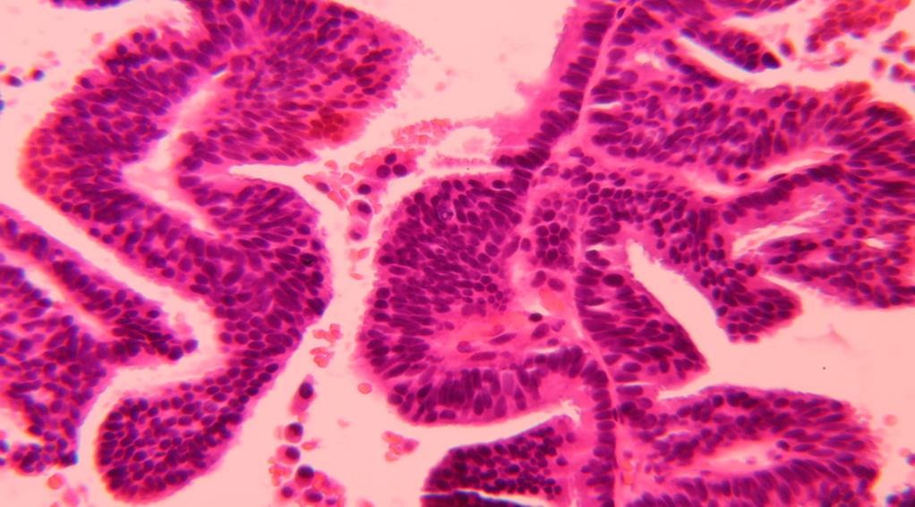
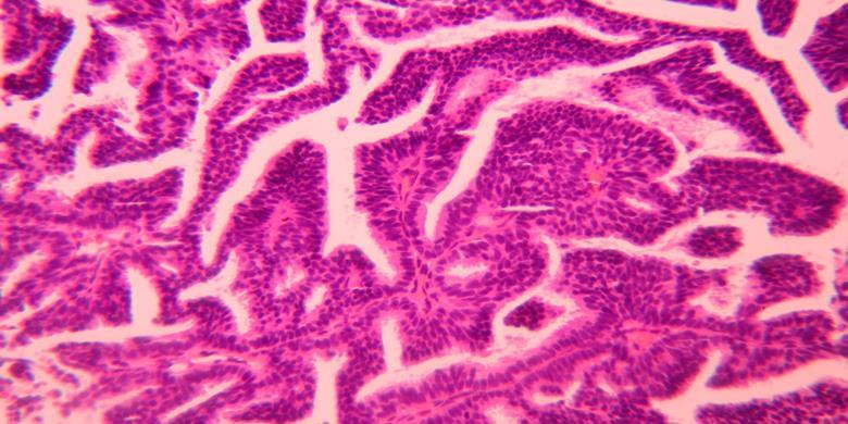
2 Papillary lesions of the breast are a heterogeneous group of disease, which are characterised by neoplastic epithelial proliferation supported by fibrovascular stalks Micropapillary structures (no stromal cores) Papillary cores


3 Typical papillary structure Thick fibrovascular cores Solid papillary growth with indiscernible cores

4 Classification 1- Location and number of involved ducts Large ducts Small ducts Intraductal papilloma Papillary carcinoma Multiple papillomas Papillary DCIS


5 Classification 2- Presence of cytonuclear and architectural atypia Benign papilloma Papillary carcinoma (PC) Papilloma with Atypia/DCIS 3- Presence of an internal and/or external myoepithelial layer Benign papilloma Papillary DCIS Encapsulated/Solid PC Invasive PC DD EPC and SPC Certain histological features including cystic/solid architecture, NE and mucinous differentiation, thick peripheral capsule, nuclear palisading, lobulation,..etc
")


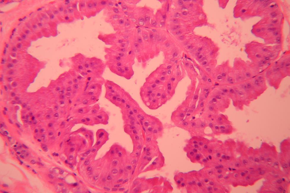
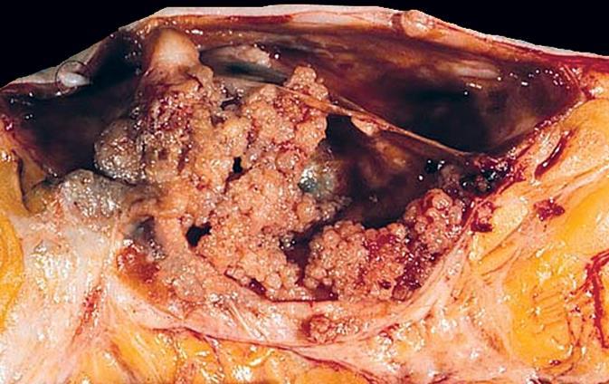
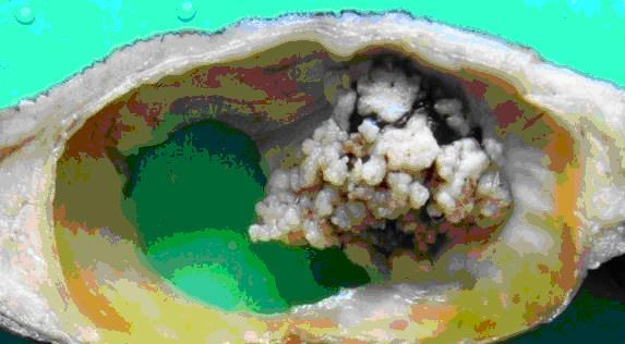
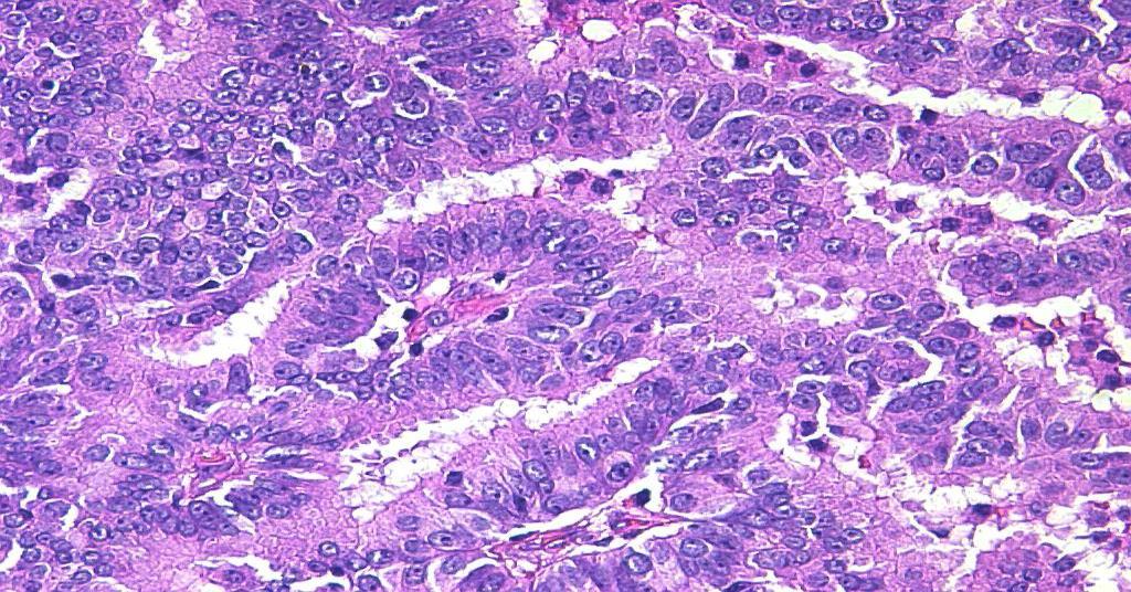
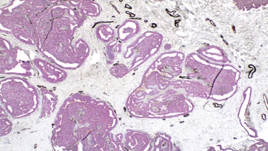
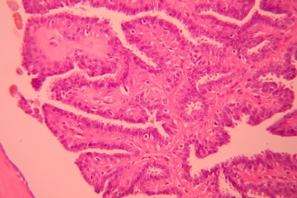
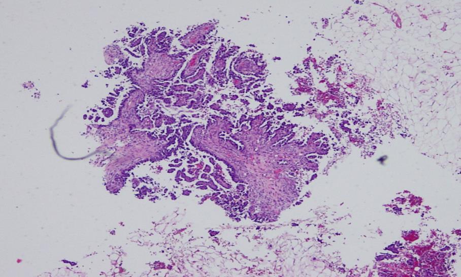
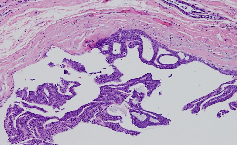
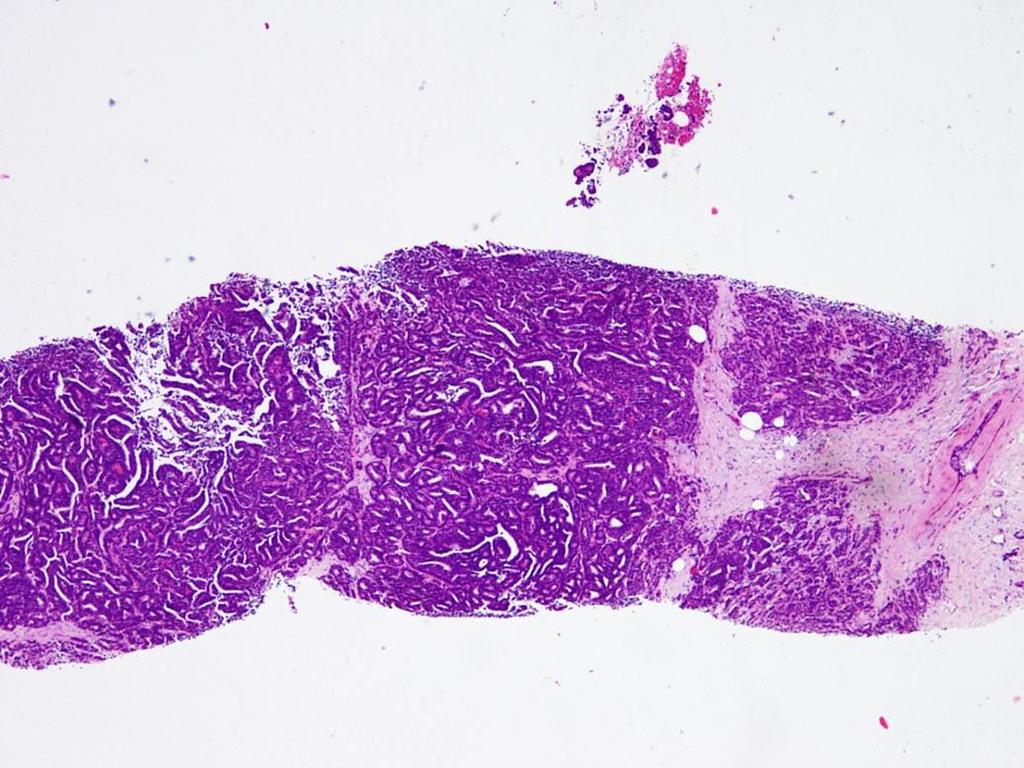
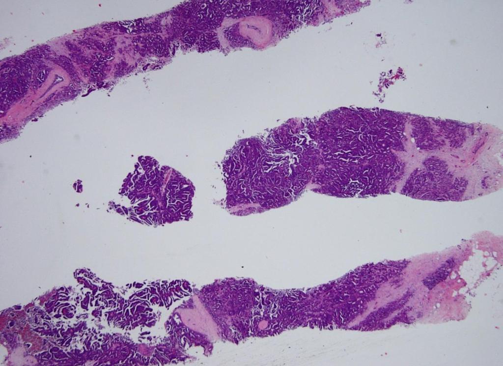
6 A-Large duct papilloma (Intraductal papilloma) Usually Central, solitary; well-defined margin, cystic +/-solid areas. usually present as a mass or nipple discharge


7 Well developed CT cores Myoepithelial cells throughout Epithelial cell layer single or multiple but no atypia Surrounding tissue is usually benign SMM


8 A-Large duct papilloma (Intraductal papilloma) May show benign apocrine metaplasia (when present, it favours benign)

9 Papillary apocrine hyperplasia


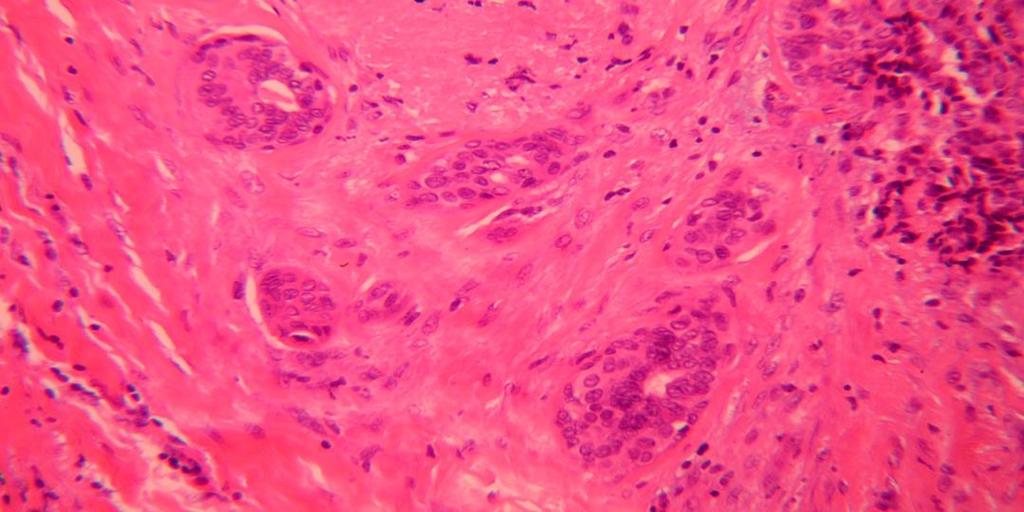
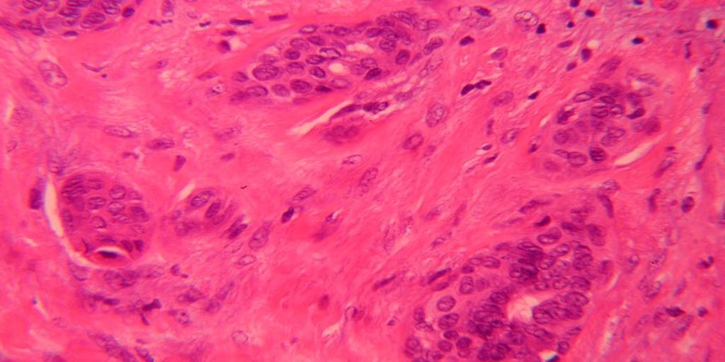
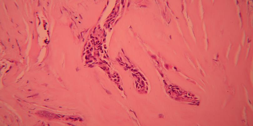
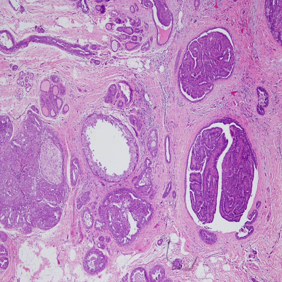
10 Papilloma may show florid adenotic and fibrotic pattern mimicking fibroepithelial lesion (FA)


11 Fibrosis with entrapped glands and epithelium
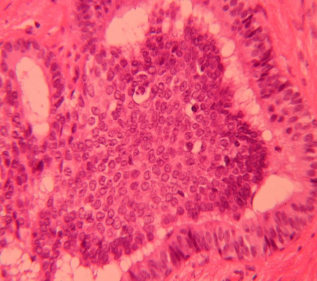
12


13 Benign papilloma with florid HUT


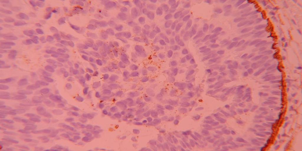
14 Papilloma with HUT- IHC ER CKs


15 * ME cells may show hyperplasia BUT if marked: DD from adenomyoepithelioma ME Adenomyoepithelioma

16 * Papilloma may show chondroid/chondromyxoid metaplasia: DD Pleomorphic Adenoma / MP MBC


17 Papilloma may show squamous or mucinous metaplasia: DD mucoepidermoid


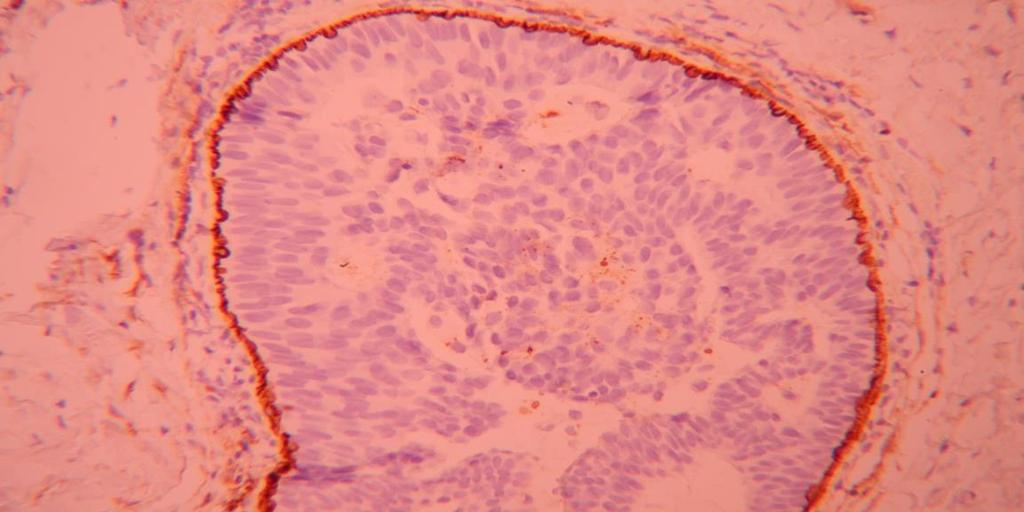
18 * Papillary lesion may show epithelial displacement May lack peripheral ME cells: DD from invasive: - Usually associated with biopsy site. - No DCIS or invasive carcinoma in surrounding tissue - Benign cytological features [ER, Ki67, Ck5/6, Ck14]

19 B-Multiple (microscopic) papillomas Peripheral multiple papillomas (TDLU), not mass forming. Less common than large duct papilloma but more frequently associated with epithelial hyperplasia and atypia Benign epithelium and myoepithelium +/- fibrocystic change, HUT
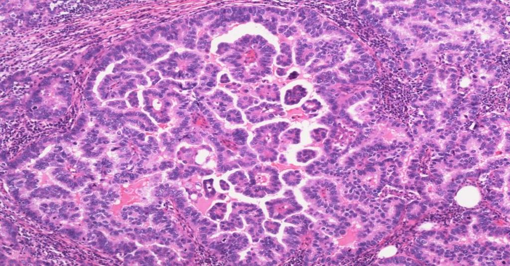
20
21 C-Benign papilloma with atypia 1- Low grade atypical Proliferation Benign papilloma containing solid or cribriform areas of uniform lowgrade nuclei devoid of basal/me cells akin to ADH/low grade DCIS <3mm (Papilloma with ADH) >3mm (Papilloma with low grade DCIS) 2- High grade atypical proliferation (Papilloma with DCIS regardless of the size of the atypical focus) Diagnosis: DCIS involving a Papilloma 3- CCC / Flat Eptheial Atypia (FEA)-type
 area of")

22 C-Benign papilloma with ADH Small (<3mm) area of atypical epithelial hyperplasia : ER+, basal CKs negative CK14

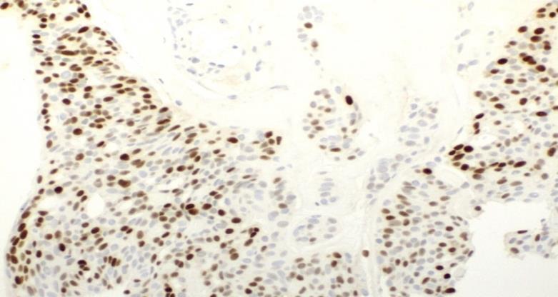
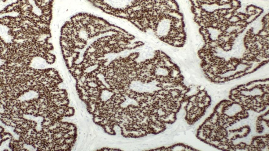
23 C-Benign papilloma with LG DCIS

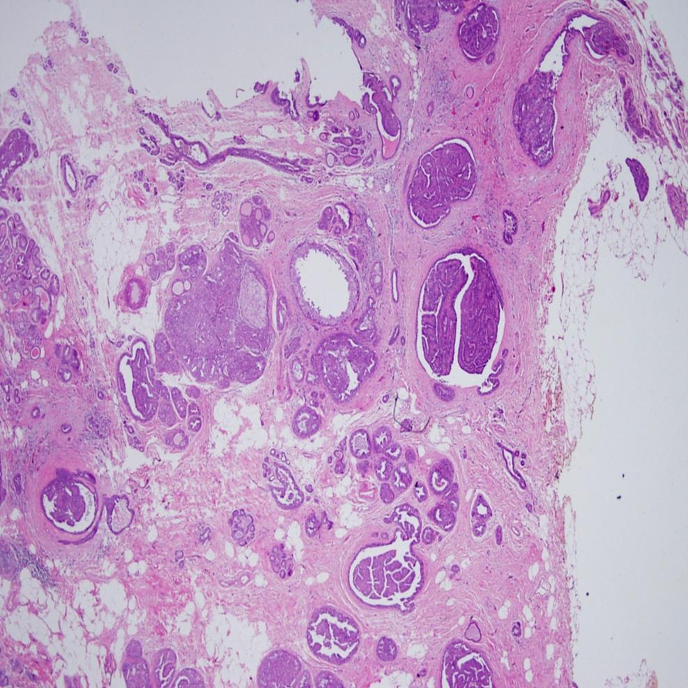
24


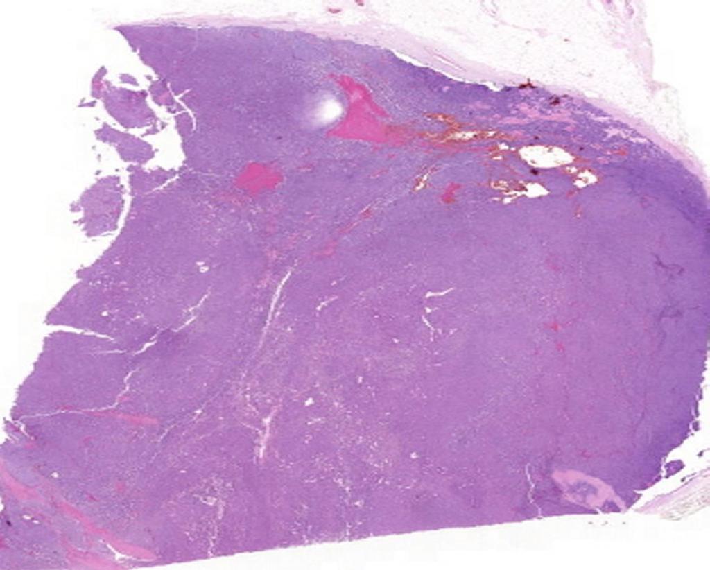
25 C-Benign papilloma with LG DCIS SMM ER staining CK5/6
26 However, extent of atypia within a papilloma is of limited clinical significant provided that there is complete excision and no DCIS in the surrounding breast tissue


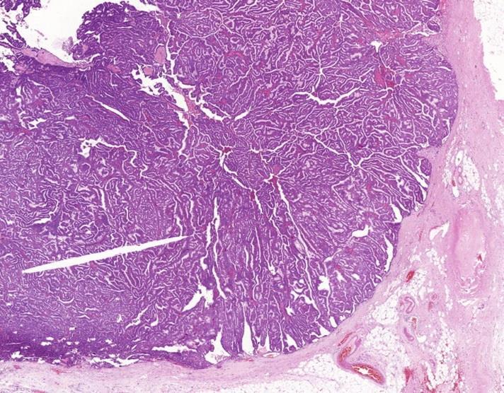
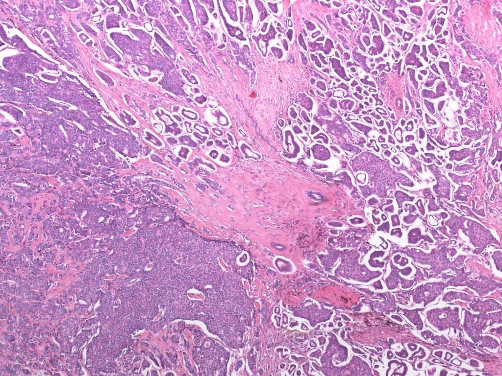
27 C-Benign papilloma with CCC or FEA (Uncertain significance) ER staining


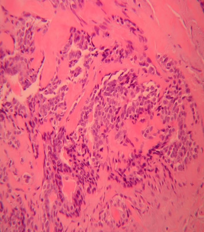
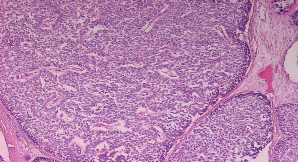
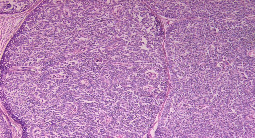
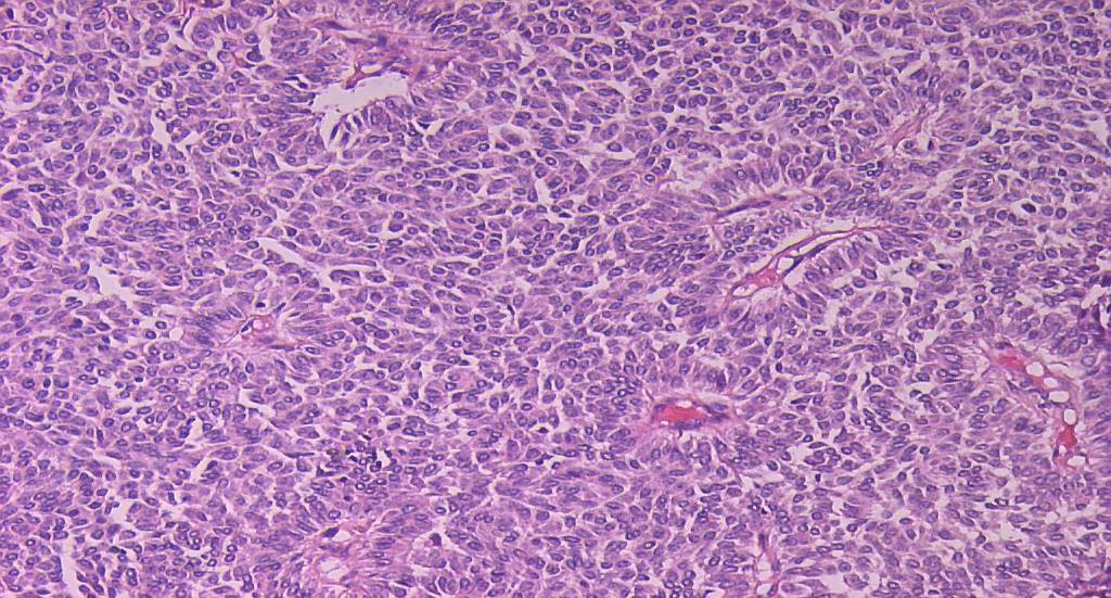
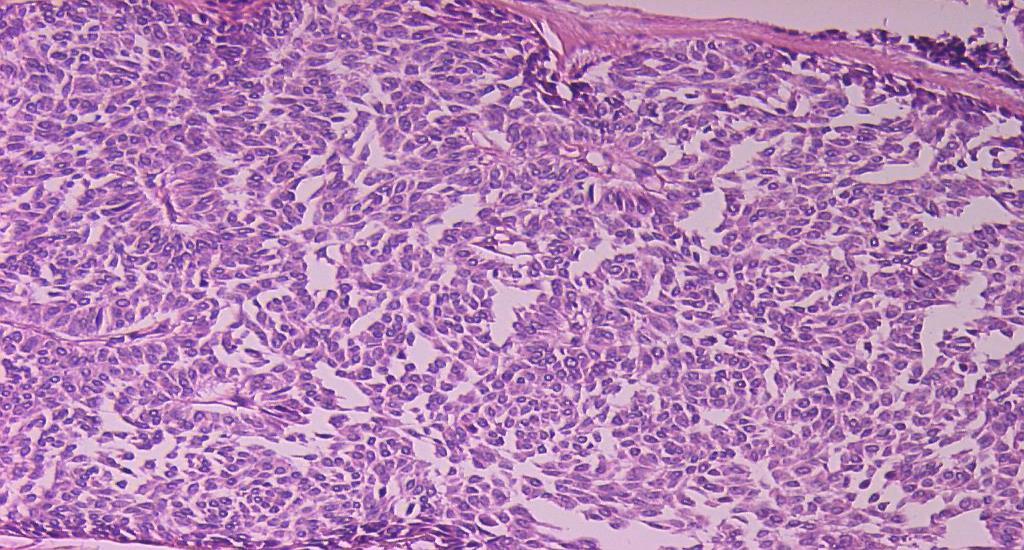
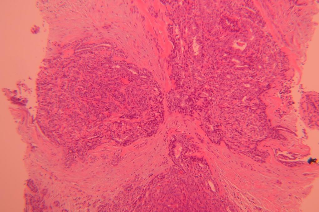
28 Papillary lesion involved by LCIS
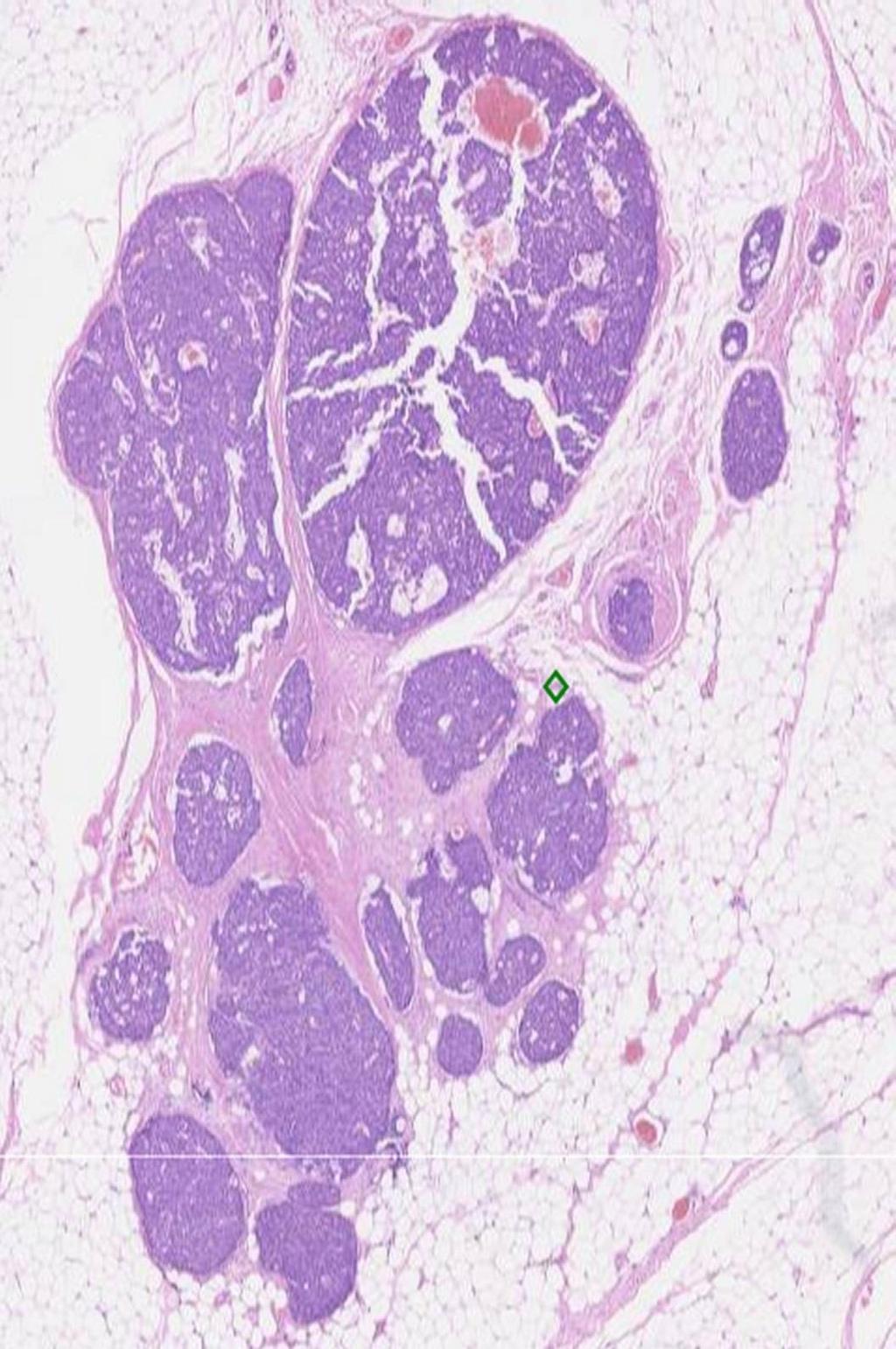
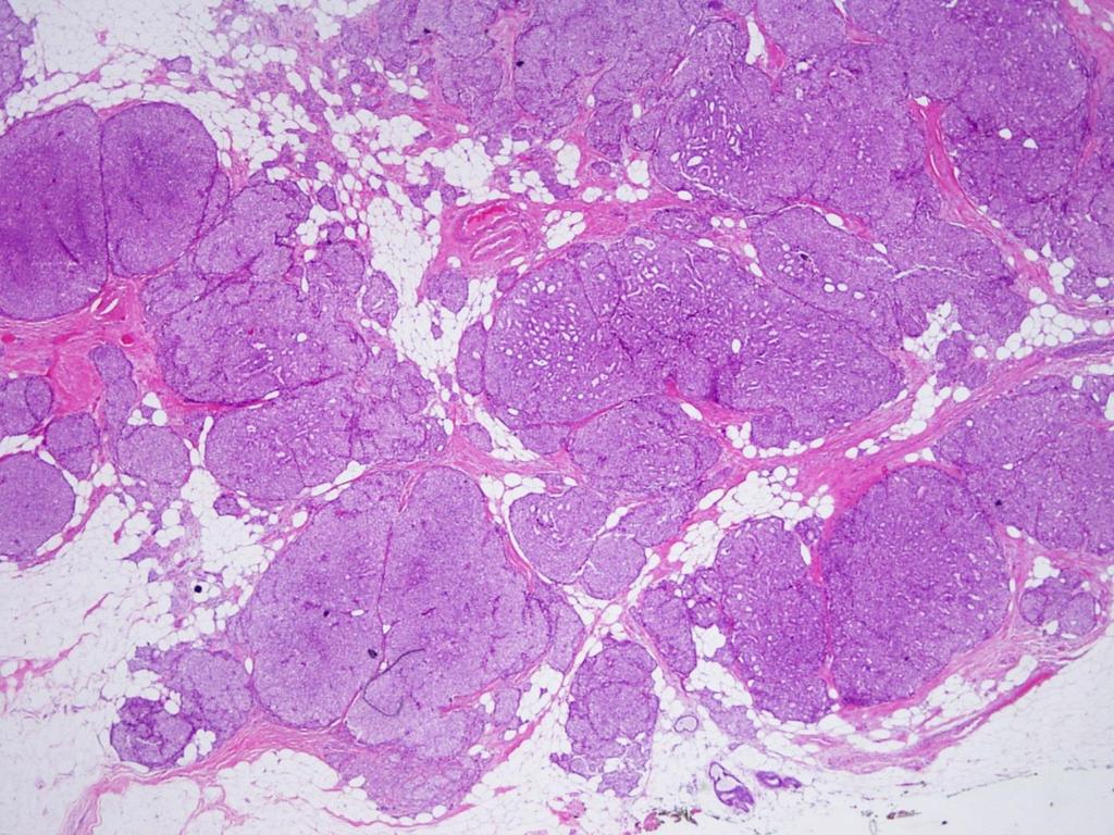
29 D-Papillary carcinoma (0.5-1% of breast cancer) Papillary DCIS (malignant counterpart of multiple papillomas) peripheral, multiple, associated with other types of DCIS Large duct papillary carcinoma (Central and usually solitary) Encapsulated (Intracystic/Encysted) PC Solid (SPC) invasive PC with or without in situ PC component
30 The diagnostic category distribution and number of papillary carcinomas Year Final diagnosis (Size in mm) Benign / atypical Malignant In-situ Invasive Total Total of readers Encapsulated PC (16) 45 (8%) 355 (65%) 147 (27%) 502 (92) Encapsulated PC (9) 54 (8%) 452 (70%) 145 (22%) 597 (92%) A- Solid PC with features of invasion(15) 32 (6%) 107 (19%) 425 (75%) 532 (94%) B- Solid PC with typical features (14) 30 (5%) 107 (18%) 466 (77%) 573 (95%) Papillary DCIS (12) 59 (12%) 384 (77%) 56 (11%) 440 (88%) Pure Invasive PC (19) 7 (1%) 186 (28%) 478 (71%) 664 (99%) 671 Rakha et al Histopathology. 2016, 69(5):
; of these 9 were papillary lesions. Rakha et al Histopathology.")
31 Review of 240 cases identified over- and under-diagnosis in 13 cases (5.4% of all cases); of these 9 were papillary lesions. Rakha et al Histopathology. 2017, 70(4):
 -")


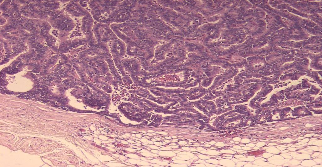
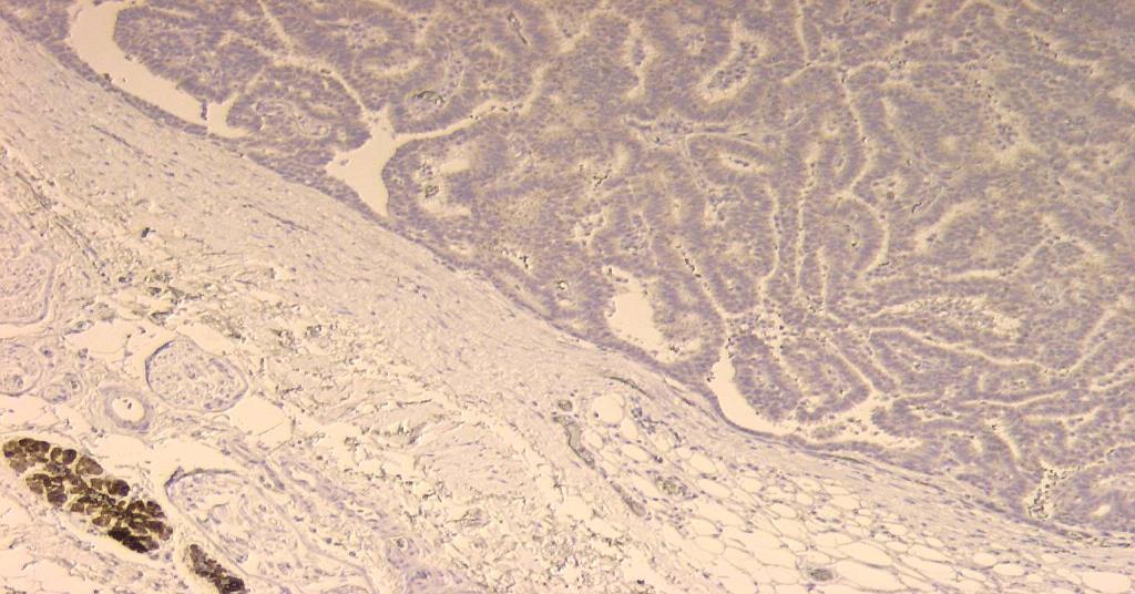
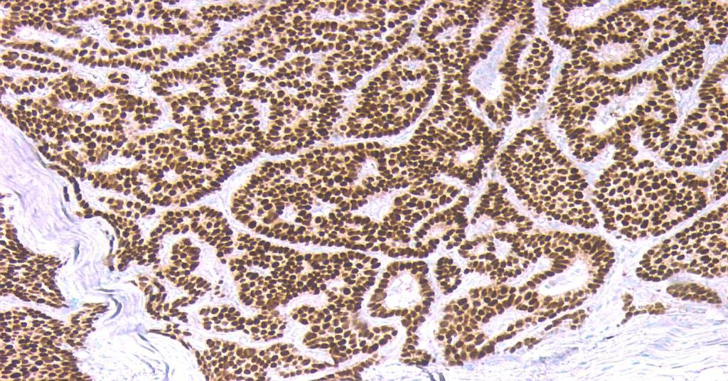
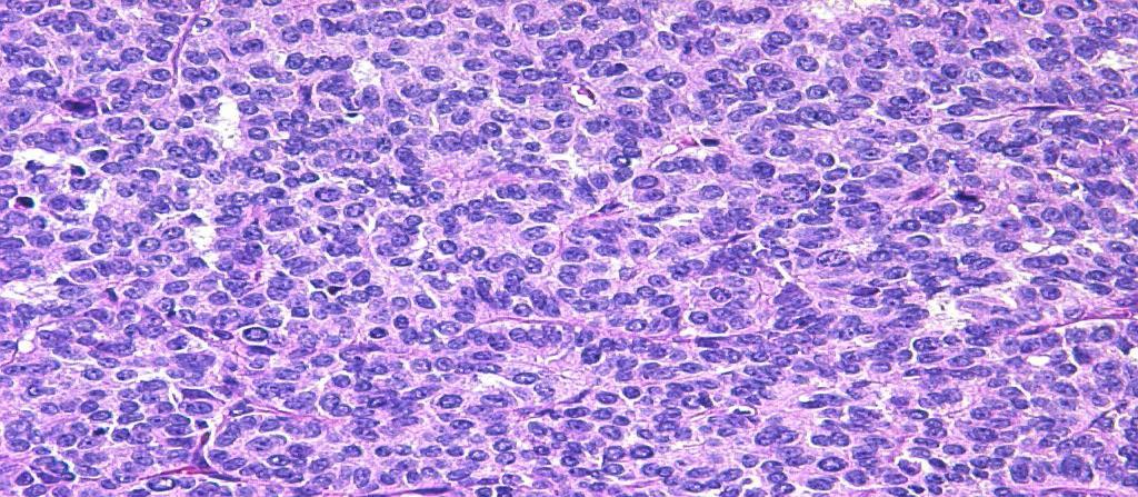
32 Papillary DCIS Less common form of PC. It is usually: - Peripheral and multiple (TDLU) - Malignant epithelium - ME absent in the papillae - ME preserved at epithelial stroma interface No evidence of pre-existing benign papilloma * Usually associated with other types of DCIS * Low to intermediate nuclear grade * No peripheral fibrous capsule
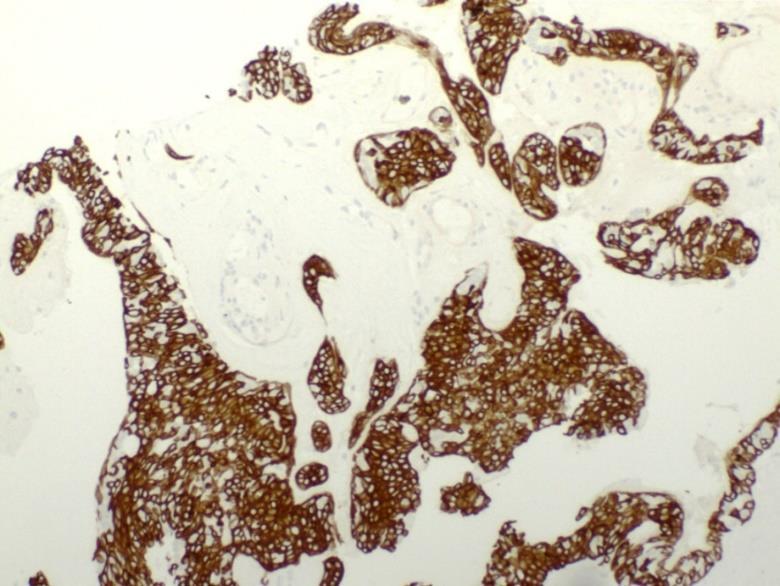
33 IHC for Myoepithelial cells

34 Encapsulated PC (encysted / intracystic PC)
 and periphery (complete absence in >80%) CT cores Cystic")
35 Encapsulated (encysted/intracystic) PC Usually central and solitary, cystically dilated ducts +/- solid areas Malignant/atypical cytology/architecture Surrounded by thick fibrous capsule Absent ME in centre (100%) and periphery (complete absence in >80%) CT cores Cystic area Fibrous capsule
36 ER CK5/6

37 Solid areas
 make it possible to recognize it as papillary carcinoma.")

38 Solid PC Composed almost entirely of solid epithelial proliferation Small cords of fibrovascular stroma (H&E or IHC) make it possible to recognize it as papillary carcinoma. Often central multinodular Spindle areas, focal mucin production, low-nuclear grade NE features (~70%), and peripheral palisading. Often lack thick fibrous capsule Frequently assoc. with invasion: mucinous carcinoma Synonyms: Spindle, NE / Endocrine DCIS
39
40 More often multi-nodular


41 Papillary DCIS: behave as conventional DCIS EPC and SPC have the potential to behave as an indolent form of invasive BC with low volume LN mets in few cases, but the excellent outcome justify managing them as a form of in-situ disease WHO recommends staging EPC and SPC as DCIS [ptis]
, it may be safer")
42 Exceptions: EPC and SPC showing intact peripheral ME cell layer (10-20%) is a true in-situ cancer both biologically and clinically and be confidently called PC in-situ Although microinvasion/suspicion of invasion in typical PC seems to be of little clinical significance, if you are in doubt (ie malignant glands outside the fibrous capsule lacking ME cells), it may be safer to call it: EPC with microinvasion or EPC, invasion cannot be excluded BUT still treated as DCIS


43 Exceptions in pure EPC: When PC shows high grade nuclei with frequent mitotic figures, we call them invasive as the number is insufficient to comment on behaviour


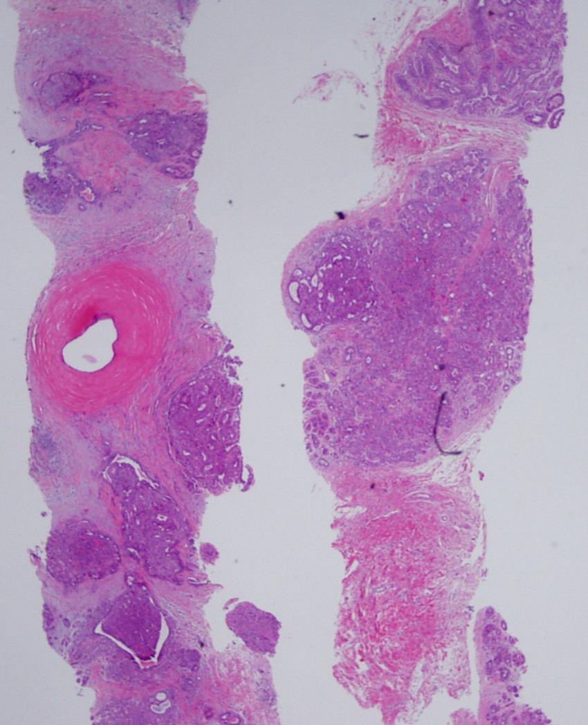
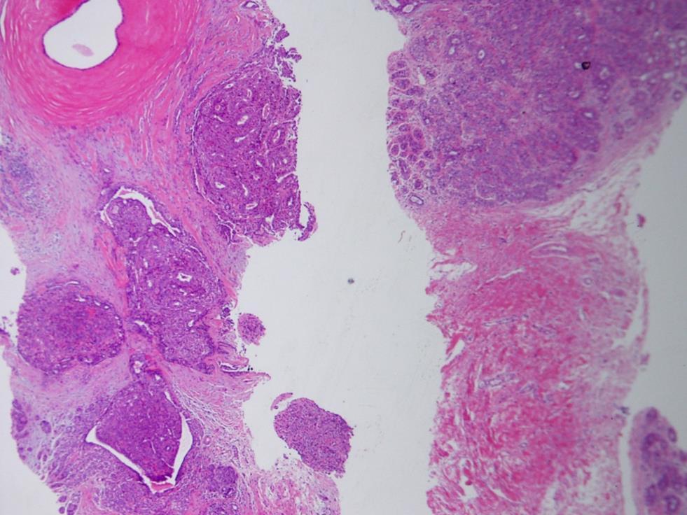
44 Exceptions in pure EPC: EPC containing foci of invasive micropapillary carcinoma, or invasive mucinous carcinoma call it invasive carcinoma
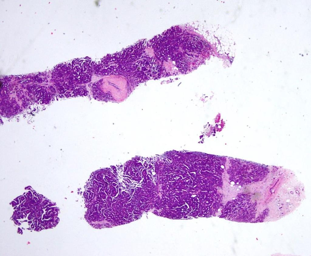
45 Invasive Papillary carcinoma: 1- EPC / SPC associated with conventional type invasion 2- EPC / SPC with invasive features 3- Invasive PC (NOS) without EPC/SPC
 Type, grade ER, PR and HER only on invasive foci and not EPC Invasive size = size of the invasive component while PC can be added")
46 EPC/SPC with frank conventional-type invasion It is usually easy to identify frank invasion associated with PC (e.g., usually conventional carcinoma such as mucinous, NST or cribriform) Type, grade ER, PR and HER only on invasive foci and not EPC Invasive size = size of the invasive component while PC can be added to the whole tumour size Few scattered foci, measure largest focus and call it multifocal Many scattered foci, measure the whole area and mention this in the text


47 Invasive solid PC WHO definition of invasion in SPC: Geographic jigsaw pattern with ragged and irregular margins, + absence of ME cells +/- infiltrating fat
48

49
50


51 Invasive PC Pure invasive PC is extremely rare - Papillary architecture (>90%) - Infiltrating outlines - Complete absence of ME cells Often low/intermediate grade


52 IHC Invasive Papillary Ca


53 Invasive papillary in LN
54 Variables Age Papilloma Y PC 60-70Y Appearance Eosinophilic (pink) Basophilic (blue) Fibrovascular Core Usually broad and frequently sclerotic Usually fine and less fibrotic Cell types Biphasic (Epithelial and ME) Single (Epithelial) Nuclei Normochromatic & variable in size and shape Hyperchromatic & usually uniform with increased N/C ratio Cell orientation Haphazard. May show HUT Orderly, solid or cribriform May show mucinous secretion, spindle, columnar or transitional epithelium Mitosis Infrequent/absent More frequent (>1 /10hpf) ± abnormal forms Apocrine metaplasia Frequent (~36%) benign Absent (<15%) / atypical Adjacent ducts Benign changes May be DCIS
55 ME cells in Papillae (core / epithelial interface) ME cells at Periphery (tumour / peripheral stroma interface) Papilloma Present Present Papilloma with atypia / DCIS Present but absent/ reduced in atypical areas Present Papillary DCIS Absent Present Encysted PC Absent Frequently absent (>85%) Solid PC Absent Usually Absent (>70%) Invasive PC Absent Absent
56 EQA case

57 Answer??? EQA case
58 Year Final diagnosis (Size in mm) Benign / atypical Malignant In-situ Invasive Total Total of readers Solid PC with typical features 30 (5%) 107 (18%) 466 (77%) 573 (95%) 603
 Basal/ME")
")


 (B5a) otherwise B3 with")

59 Core biopsy Benign papilloma: B3 (without atypia) Basal/ME markers Benign papillary lesion with Atypia: B3 (with atypia) Papillary lesion with extensive atypia (?DCIS): Unless it is sufficient for the diagnosis of DCIS (ie high grade or definitely large size) (B5a) otherwise B3 with comments that it is likely to represent DCIS on excision to be considered in the management ER p63 Basal CK


60 EPC: B5a -Well-defined margin, -Surrounded by capsule - No evidence of invasion Core biopsy ME cells EPC with suspicious area/not sure about invasion: B5?

61 B3
62 B5a
63 IHC
64 IHC
65 B5
66 B5

67 Papillary, ER-, basal CKs +, No ME cells. SPCRP IDH2 gene mutations


68 Thank you EAR
Papillary Lesions of the Breast A Practical Approach to Diagnosis. (Arch Pathol Lab Med. 2016;140: ; doi: /arpa.
 Papillary Lesions of the Breast A Practical Approach to Diagnosis (Arch Pathol Lab Med. 2016;140:1052 1059; doi: 10.5858/arpa.2016-0219-RA) Papillary lesions of the breast Span the spectrum of benign,
04/10/2018. Intraductal Papillary Neoplasms Of Breast INTRADUCTAL PAPILLOMA
 Intraductal Papillary Neoplasms Of Breast Savitri Krishnamurthy MD Professor of Pathology Deputy Division Head The University of Texas MD Anderson Cancer Center 25 th Annual Seminar in Pathology Pittsburgh,
Proliferative Epithelial lesions of the Breast. Sami Shousha, MD, FRCPath Charing Cross Hospital & Imperial College, London
 Proliferative Epithelial lesions of the Breast Sami Shousha, MD, FRCPath Charing Cross Hospital & Imperial College, London Amman, November2013 Proliferative Epithelial Lesions of the Breast Usual type
Papillary Lesions of the Breast
 Papillary Lesions of the Breast Laura C. Collins, M.D. Associate Professor of Pathology Associate Director, Division of Anatomic Pathology Beth Israel Deaconess Medical Center and Harvard Medical School
Papillary Lesions of the Breast: WHO Update
 Papillary Lesions of the Breast: WHO Update Stuart J. Schnitt, M.D. Department of Pathology Beth Israel Deaconess Medical Center and Harvard Medical School Boston, MA, USA Papillary Lesions of the Breast
Breast pathology. 2nd Department of Pathology Semmelweis University
 Breast pathology 2nd Department of Pathology Semmelweis University Breast pathology - Summary - Benign lesions - Acute mastitis - Plasma cell mastitis / duct ectasia - Fat necrosis - Fibrocystic change/
Papillary Lesions of the Breast
 Papillary Lesions of the Breast Texas Society of Pathologists 2013 Laura C. Collins, M.D. Associate Professor of Pathology Associate Director, Division of Anatomic Pathology Beth Israel Deaconess Medical
CLINICAL SIGNIFICANCE OF BENIGN EPITHELIAL CHANGES
 Papillomas. Papillomas are composed of multiple branching fibrovascular cores, each having a connective tissue axis lined by luminal and myoepithelial cells ( Fig. 23-11 ). Growth occurs within a dilated
Diseases of the breast (1 of 2)
") Diseases of the breast (1 of 2) Introduction A histology introduction Normal ducts and lobules of the breast are lined by two layers of cells a layer of luminal cells overlying a second layer of myoepithelial
Diseases of the breast (1 of 2) Introduction A histology introduction Normal ducts and lobules of the breast are lined by two layers of cells a layer of luminal cells overlying a second layer of myoepithelial
A712(19)- Test slide, Breast cancer tissues with corresponding normal tissues
- Test slide, Breast cancer tissues with corresponding normal tissues") A712(19)- Test slide, Breast cancer tissues with corresponding normal tissues (formalin fixed) For research use only Specifications: No. of cases: 12 Tissue type: Breast cancer tissues with corresponding
A712(19)- Test slide, Breast cancer tissues with corresponding normal tissues (formalin fixed) For research use only Specifications: No. of cases: 12 Tissue type: Breast cancer tissues with corresponding
Breast Pathology. Breast Development
 Breast Pathology Lecturer: Hanina Hibshoosh, M.D. Reading: Kumar, Cotran, Robbins, Basic Pathology, 6th Edition, pages 623-635 Breast Development 5th week - thickening of the epidermis - milk line 5th
Treatment options for the precancerous Atypical Breast lesions. Prof. YOUNG-JIN SUH The Catholic University of Korea
 Treatment options for the precancerous Atypical Breast lesions Prof. YOUNG-JIN SUH The Catholic University of Korea Not so benign lesions? Imaging abnormalities(10% recall) lead to diagnostic evaluation,
Salivary Glands 3/7/2017
 Salivary Glands 3/7/2017 Goals and objectives Focus on the entities unique to H&N Common board type facts Information for your future practice Salivary Glands Salivary Glands Major gland. Paratid. Submandibular.
Papillary Lesions in Breast Pathology Practice: Diagnostic Challenges and Practical Approach. A Six- Year Experience from a Tertiary Care Hospital
 Open Access Journal Research Article DOI: 10.23958/ijirms/vol02-i05/12 Papillary Lesions in Breast Pathology Practice: Diagnostic Challenges and Practical Approach. A Six- Year Experience from a Tertiary
Columnar Cell Lesions
 Columnar Cell Lesions Laura C. Collins, M.D. Department of Pathology Beth Israel Deaconess Medical Center and Harvard Medical School Boston, MA Question? Columnar cell lesions are: a) Annoying lesions
Spindle Cell Lesions Of The Breast. Emad Rakha Professor of Breast Pathology and Consultant Pathologist
 Spindle Cell Lesions Of The Breast Emad Rakha Professor of Breast Pathology and Consultant Pathologist * SCLs comprise a wide spectrum of diseases, ranging from reactive processes to aggressive malignant
1 NORMAL HISTOLOGY AND METAPLASIAS
 1 NORMAL HISTOLOGY AND METAPLASIAS, MD Anatomy and Histology 1 Metaplasias 2 ANATOMY AND HISTOLOGY The female breast is composed of a branching duct system, which begins at the nipple with the major lactiferous
Flat Epithelial Atypia
 Flat Epithelial Atypia Richard Owings, M.D. University of Arkansas for Medical Sciences Department of Pathology Flat epithelial atypia can be a difficult lesion May be a subtle diagnosis Lots of changes
Cytyc Corporation - Case Presentation Archive - March 2002
 FirstCyte Ductal Lavage History: 68 Year Old Female Gail Index: Unknown Clinical History: Negative Mammogram in 1995 6 yrs. later presents with bloody nipple discharge Subsequent suspicious mammogram Suspicious
Ductal Carcinoma in Situ. Laura C. Collins, M.D. Department of Pathology Beth Israel Deaconess Medical Center and Harvard Medical School Boston, MA
 Ductal Carcinoma in Situ Laura C. Collins, M.D. Department of Pathology Beth Israel Deaconess Medical Center and Harvard Medical School Boston, MA Definition of DCIS WHO 2012 A neoplastic proliferation
EQA Circulation 43 Educational Cases
 EQA Circulation 43 Educational Cases E1-E2 Monica Agarwal Monklands Hospital E1 38 yrs male Submandibular gland tumour E1 Formal excision following diagnosis of poorly differentiated carcinoma on core
Columnar Cell Lesions. Columnar Cell Lesions and Flat Epithelial Atypia
 Columnar Cell Lesions and Stuart J. Schnitt, M.D. Beth Israel Deaconess Medical Center and Harvard Medical School Boston, MA, USA Columnar Cell Lesions Lesions characterized by columnar epithelial cells
BREAST PATHOLOGY. Fibrocystic Changes
 BREAST PATHOLOGY Lesions of the breast are very common, and they present as palpable, sometimes painful, nodules or masses. Most of these lesions are benign. Breast cancer is the 2 nd most common cause
3/28/2017. Disclosure of Relevant Financial Relationships. GU Evening Subspecialty Case Conference. Differential Diagnosis:
 GU Evening Subspecialty Case Conference Rajal B. Shah, M.D. VP, Medical Director, Urologic Pathology Miraca Life Sciences, Irving, Texas Clinical Associate Professor of Pathology Baylor College of Medicine,
Gross appearance of nodular hyperplasia in material obtained from suprapubic prostatectomy. Note the multinodular appearance and the admixture of
 Tiền liệt tuyến Tiền liệt tuyến Gross appearance of nodular hyperplasia in material obtained from suprapubic prostatectomy. Note the multinodular appearance and the admixture of solid and microcystic areas.
Epithelial Columnar Breast Lesions: Histopathology and Molecular Markers
 29th Annual International Conference Advances in the Application of Monoclonal Antibodies in Clinical Oncology and Symposium on Cancer Stem Cells 25 th -27t h June, 2012, Mykonos, Greece Epithelial Columnar
Lesions Mimicking Adenoid Cystic Carcinoma. Diagnostic Problems in Salivary Gland Pathology An Update 5/29/2009
 Diagnostic Problems in Salivary Gland Pathology An Update Lesions Mimicking Adenoid Cystic Carcinoma Stacey E. Mills, M.D. W.S. Royster Professor of Pathology Director of Surgical and Cytopathology University
Recent advances in breast cancers
 Recent advances in breast cancers Breast cancer is a hetrogenous disease due to distinct genetic alterations. Similar morphological subtypes show variation in clinical behaviour especially in response
A712(18)- Test slide, Breast cancer tissues with corresponding normal tissues
- Test slide, Breast cancer tissues with corresponding normal tissues") A712(18)- Test slide, Breast cancer tissues with corresponding normal tissues (formalin fixed) For research use only Specifications: No. of cases: 12 Tissue type: Breast cancer tissues with corresponding
A712(18)- Test slide, Breast cancer tissues with corresponding normal tissues (formalin fixed) For research use only Specifications: No. of cases: 12 Tissue type: Breast cancer tissues with corresponding
COMPANION MEETING BREAST. Auditorium 11:15 1:00 am. Convenor: A/Professor Gelareh Farshid, SA Pathology, SA
 Australasian Division of the International Academy of Pathology Limited ABN 73 008 593 815 36 TH Annual Scientific Meeting Darling Harbour Convention Centre, Sydney, Australia June 3-5, 2011 COMPANION
Update in Salivary Gland Pathology. Benjamin L. Witt University of Utah/ARUP Laboratories February 9, 2016
 Update in Salivary Gland Pathology Benjamin L. Witt University of Utah/ARUP Laboratories February 9, 2016 Objectives Review the different appearances of a selection of salivary gland tumor types Establish
ARTHUR PURDY STOUT SOCIETY COMPANION MEETING: DIFFICULT NEW DIFFERENTIAL DIAGNOSES IN PROSTATE PATHOLOGY. Jonathan I. Epstein.
 1 ARTHUR PURDY STOUT SOCIETY COMPANION MEETING: DIFFICULT NEW DIFFERENTIAL DIAGNOSES IN PROSTATE PATHOLOGY Jonathan I. Epstein Professor Pathology, Urology, Oncology The Reinhard Professor of Urological
6/3/2010. Outline of Talk. Lobular Breast Cancer: Definition of lobular differentiation. Common Problems in Diagnosing LCIS in Core Biopsies
 Outline of Talk Lobular Breast Cancer: Common Problems in Diagnosing LCIS in Core Biopsies Definition of lobular differentiation Variants of LCIS that: carry risk for unsampled invasive cancer mimic DCIS
Disclosures 5/27/2012. Outline of Talk. Outline of Talk. When Is LCIS Clinically Significant? Classic LCIS. Classic LCIS
 When Is LCIS Clinically Significant? Disclosures I have nothing to disclose Yunn-Yi Chen, MD, PhD Professor Outline of Talk Outline of Talk Classic LCIS Classic LCIS Definition of lobular differentiation
Mody. AIS vs. Invasive Adenocarcinoma of the Cervix
 Common Problems in Gynecologic Pathology Michael T. Deavers, M.D. Houston Methodist Hospital, Houston, Texas Common Problems in Gynecologic Pathology Adenocarcinoma in-situ (AIS) of the Cervix vs. Invasive
Benign Mimics of Malignancy in Breast Pathology
 Arthur Purdy Stout Society of Surgical Pathologists Companion Meeting Benign Mimics of Malignancy in Breast Pathology Stuart J. Schnitt, M.D. Beth Israel Deaconess Medical Center and Harvard Medical School,
04/10/2018 HIGH RISK BREAST LESIONS. Pathology Perspectives of High Risk Breast Lesions ELEVATED RISK OF BREAST CANCER HISTORICAL PERSPECTIVES
 Pathology Perspectives of High Risk Breast Lesions Savitri Krishnamurthy MD Professor of Pathology Deputy Division Head Director of Clinical Trials, Research and Development The University of Texas MD
Papillary lesions of the breast: selected diagnostic and management issues
 Histopathology 2008, 52, 20 29. DOI: 10.1111/j.1365-2559.2007.02898.x REVIEW Papillary lesions of the breast: selected diagnostic and management issues L C Collins & S J Schnitt Department of Pathology,
Enterprise Interest None
 Enterprise Interest None B3 lesions of the breast What are they at surgery? Case 4 Edi Brogi MD PhD Attending Pathologist - Director of Breast Pathology Memorial Sloan Kettering Cancer Center New York
Image guided core biopsies:
 Recommendations on the Surgical, Radiologic and Pathologic Approaches to Breast Disease: Using best practices based on multidisciplinary methodologies developed through the Allina Breast Committee. Image
Protocol for the Examination of Biopsy Specimens From Patients With Invasive Carcinoma of the Breast
 Protocol for the Examination of Specimens From Patients With Invasive Carcinoma of the Breast Version: BreastInvasive 1.0.0.0 Protocol Posting Date: February 2019 Accreditation Requirements The use of
CPC 4 Breast Cancer. Rochelle Harwood, a 35 year old sales assistant, presents to her GP because she has noticed a painless lump in her left breast.
 CPC 4 Breast Cancer Rochelle Harwood, a 35 year old sales assistant, presents to her GP because she has noticed a painless lump in her left breast. 1. What are the most likely diagnoses of this lump? Fibroadenoma
Proliferative Breast Disease: implications of core biopsy diagnosis. Proliferative Breast Disease
 Proliferative Breast Disease: implications of core biopsy diagnosis Jean F. Simpson, M.D. Breast Pathology Consultants, Inc. Nashville, TN Proliferative Breast Disease Must be interpreted in clinical and
PLEOMORPHIC ADENOMA ( BENIGN MIXED TUMOR )
") ( BENIGN MIXED TUMOR ) Grossly, the tumor is freely movable, solid, sometimes lobulated and occasionally cystic. If recurrent, multinodular masses are common. Histologically, within a fibrous capsule,
( BENIGN MIXED TUMOR ) Grossly, the tumor is freely movable, solid, sometimes lobulated and occasionally cystic. If recurrent, multinodular masses are common. Histologically, within a fibrous capsule,
IBCM 2, April 2009, Sarajevo, Bosnia and Herzegovina
 Preoperative diagnosis and treatment planning in breast cancer The pathologist s perspective L. Mazzucchelli Istituto Cantonale di Patologia Locarno, Switzerland IBCM 2, 23-25 April 2009, Sarajevo, Bosnia
High risk lesions of the breast : Review of the current diagnostic and management strategies
 High risk lesions of the breast : Review of the current diagnostic and management strategies Poster No.: C-1204 Congress: ECR 2016 Type: Educational Exhibit Authors: P. Jagmohan, F. J. Pool, P. G. Pillay,
Good afternoon everyone. First of all many thanks to Dr. Bonaventura and Dr. Arn for inviting
 PATHOLOGY IN-SITU CARCINOMA, ROHIT BHARGAVA, MD 1 Good afternoon everyone. First of all many thanks to Dr. Bonaventura and Dr. Arn for inviting me here, it s great to be here and I m going to talk about
Histological Type. Morphological and Molecular Typing of breast Cancer. Nottingham Tenovus Primary Breast Cancer Study. Survival (%) Ian Ellis
 Ian Ellis") Morphological and Molecular Typing of breast Cancer Ian Ellis Molecular Medical Sciences, University of Nottingham Department of Histopathology, Nottingham University Hospitals NHS Trust Histological Type
Morphological and Molecular Typing of breast Cancer Ian Ellis Molecular Medical Sciences, University of Nottingham Department of Histopathology, Nottingham University Hospitals NHS Trust Histological Type
CASE REPORT Malignant transformation of breast ductal adenoma: a diagnostic pitfall
 Malaysian J Pathol 2015; 37(3) : 281 285 CASE REPORT Malignant transformation of breast ductal adenoma: a diagnostic pitfall Hiroko HAYASHI, Hiroshi OHTANI,* Junzo YAMAGUCHI,** and Isao SHIMOKAWA Department
Title malignancy. Issue Date Right 209, 12, (2013)
") NAOSITE: Nagasaki University's Ac Title Author(s) A case of intracystic apocrine papi malignancy Hayashi, Hiroko; Ohtani, Hiroshi; Y Citation Pathology - Research and Practice, Issue Date 2013-12 URL Right
NAOSITE: Nagasaki University's Ac Title Author(s) A case of intracystic apocrine papi malignancy Hayashi, Hiroko; Ohtani, Hiroshi; Y Citation Pathology - Research and Practice, Issue Date 2013-12 URL Right
3/27/2017. Disclosure of Relevant Financial Relationships. Papilloma???
 Management of Papillary Lesions Diagnosed at Rad Path Concordant Core Biopsy (CNB) Disclosure of Relevant Financial Relationships USCAP requires that all planners (Education Committee) in a position to
ACRIN 6666 Therapeutic Surgery Form
 S1 ACRIN 6666 Therapeutic Surgery Form 6666 Instructions: Complete a separate S1 form for each separate area of each breast excised with the intent to treat a cancer (e.g. each lumpectomy or mastectomy).
Enterprise Interest None
 Enterprise Interest None What are triple negative breast cancers? A synopsis of their histological patterns Ian Ellis Molecular Medical Sciences, University of Nottingham Department of Histopathology,
Overview of Pathology Evaluation of Breast Lesions and Quality Assurance
 Overview of Pathology Evaluation of Breast Lesions and Quality Assurance 2 Michael O. Idowu, Jaime A. Singh, and Margaret M. Grimes Masses/Densities/Distortions: General Considerations Radiologic evaluation
HISTOPATHOLOGICAL EVALUATION OF BENIGN PROLIFERATIVE BREAST LESIONS
 7 ORIGINAL ARTICLE HISTOPATHOLOGICAL EVALUATION OF BENIGN PROLIFERATIVE BREAST LESIONS DR. VIBHUTI H. CHIHLA*, DR. N N. JAGRIT **, DR. JAYASHREE M. SHAH*** *3 rd year Pathology Resident, **Associate Professor,
Synonyms. Nephrogenic metaplasia Mesonephric adenoma
 Nephrogenic Adenoma Synonyms Nephrogenic metaplasia Mesonephric adenoma Definition Benign epithelial lesion of urinary tract with tubular, glandular, papillary growth pattern Most frequently in the urinary
Basement membrane in lobule.
 Bahram Memar, MD Basement membrane in lobule. Normal lobule-luteal phase Normal lobule-follicular phase Lactating breast Greater than 95% are adenocarcinomas in situ carcinomas and invasive carcinomas.
Columnar Cell Lesions and Flat Epithelial Atypia
 Columnar Cell Lesions and Flat Epithelial Atypia Laura C. Collins, M.D. Department of Pathology Beth Israel Deaconess Medical Center and Harvard Medical School, Boston, MA Terminology for Columnar Cell
Mousa. Israa Ayed. Abdullah AlZibdeh. 0 P a g e
 1 Mousa Israa Ayed Abdullah AlZibdeh 0 P a g e Breast pathology The basic histological units of the breast are called lobules, which are composed of glandular epithelial cells (luminal cells) resting on
Case study 1. Rie Horii, M.D., Ph.D. Division of Pathology Cancer Institute Hospital, Japanese Foundation for Cancer Research
 NCCN/JCCNB Seminar in Japan April 15, 2012 Case study 1 Rie Horii, M.D., Ph.D. Division of Pathology Cancer Institute Hospital, Japanese Foundation for Cancer Research Present illness: A 50y.o.premenopausal
Case Report Synchronous Bilateral Solid Papillary Carcinomas of the Breast
 Case Reports in Surgery Volume 2013, Article ID 812129, 4 pages http://dx.doi.org/10.1155/2013/812129 Case Report Synchronous Bilateral Solid Papillary Carcinomas of the Breast Noriko Yoshimura, 1 Shigeru
Lesion Imaging Characteristics Mass, Favoring Benign Circumscribed Margins Intramammary Lymph Node
 Lesion Imaging Characteristics Mass, Favoring Benign Circumscribed Margins Intramammary Lymph Node Oil Cyst Mass, Intermediate Concern Microlobulated Margins Obscured Margins Mass, Favoring Malignant Indistinct
Neoplasia 2018 Lecture 2. Dr Heyam Awad MD, FRCPath
 Neoplasia 2018 Lecture 2 Dr Heyam Awad MD, FRCPath ILOS 1. List the differences between benign and malignant tumors. 2. Recognize the histological features of malignancy. 3. Define dysplasia and understand
Diagnosis of Fibroepithelial and Mesenchymal Lesions on Core Needle Biopsy
 Diagnosis of Fibroepithelial and Mesenchymal Lesions on Core Needle Biopsy Emmanuel Agosto-Arroyo, MD Assistant Member Department of Anatomic Pathology 3/3/2018 Disclosure There are no conflicts of interest.
PSA. HMCK, p63, Racemase. HMCK, p63, Racemase
 Case 1 67 year old male presented with gross hematuria H/o acute prostatitis & BPH Urethroscopy: small, polypoid growth with a broad base emanating from the left side of the verumontanum Serum PSA :7 ng/ml
Follicular Derived Thyroid Tumors
 Follicular Derived Thyroid Tumors Jennifer L. Hunt, MD, MEd Aubrey J. Hough Jr, MD, Endowed Professor of Pathology Chair of Pathology and Laboratory Medicine University of Arkansas for Medical Sciences
Diagnostic Problems in Breast Pathology How to avoid the pitfalls
 FORUM OF PATHOLOGY Diagnostic Problems in Breast Pathology How to avoid the pitfalls Professor C W Elston City Hospital Nottingham, United Kingdom Introduction Almost any breast lesion may produce diagnostic
The management of B3 lesions with emphasis on lobular neoplasia
 The management of B3 lesions with emphasis on lobular neoplasia Abeer Shaaban Queen Elizabeth Hospital Birmingham NHSBSP core biopsy categories B1 - Normal B2 - Benign B3 Uncertain malignant potential
Pleomorphic adenoma of breast - a case report and distinction with metaplastic carcinoma D Gupta, S Agrawal, N Trivedi, A Tewari
 of breast - a case report and distinction with metaplastic carcinoma D Gupta, S Agrawal, N Trivedi, A Tewari Introduction, also known as mixed tumour, is a benign tumour which typically presents as a painless,
Management of B3 lesions
 Management of B3 lesions Pathological view Abeer Shaaban Queen Elizabeth Hospital Birmingham FEA AIDP B3 lesions In situ Lobular neoplasia Papilloma Radial scar Fibroaepithelial lesion Mucocoele like lesion
Slide seminar. Asist. Prof. Jože Pižem, MD, PhD Institute of Pathology Medical Faculty, University of Ljubljana
 Slide seminar Asist. Prof. Jože Pižem, MD, PhD Institute of Pathology Medical Faculty, University of Ljubljana Case 5 A 57-year-old man with a dermal/subcutaneous lesion on the scalp, which was interpreted
Guidance on the management of B3 lesions
 Guidance on the management of B3 lesions Lesion diagnosed on 14g or vacuumassisted biopsy (VAB) Risk of upgrade Recommended investigation Suggested approach for follow-up if no malignancy on VAE awaiting
Breast: Difficulties in Core Biopsies
 Breast: Difficulties in Core Biopsies Anna Marie Mulligan, MB, MSc, FRCPath University Health Network and University of Toronto E-mail: annamarie.mulligan@uhn.ca No conflicts of interest Role of Core Needle
Biliary tract tumors
 Short Course 2010 Annual Fall Meeting of the Korean Society for Pathologists Biliary tract tumors Joon Hyuk Choi, M.D., Ph.D. Professor, Department of Pathology, Yeungnam Univ. College of Medicine, Daegu,
EQA circulation 35 educational cases. Dr. A Graham Aberdeen Royal Infirmary
 EQA circulation 35 educational cases Dr. A Graham Aberdeen Royal Infirmary Case E1 Female 52 Polypoid mass right side of cervix, adjacent to os 70 Biphasic lesion 4 No answer 3 Prolapsed tube 2 Endometriosis
Evaluation of Breast Specimens Removed by Needle Localization Technique
 Evaluation of Breast Specimens Removed by Needle Localization Technique Specimen Handling: The breast specimen when received should be measured and grossly inspected for any orientation designated by the
LYMPHATIC DRAINAGE AXILLARY (MOSTLY) INTERNAL MAMMARY SUPRACLAVICULAR
 INTERNAL MAMMARY SUPRACLAVICULAR") BREAST LYMPHATIC DRAINAGE AXILLARY (MOSTLY) INTERNAL MAMMARY SUPRACLAVICULAR HISTOLOGY LOBE: (10 in whole breast) LOBULE: (many per lobe) ACINUS/I, aka ALVEOLUS/I: (many per lobule) DUCT(S): INTRA- or
BREAST LYMPHATIC DRAINAGE AXILLARY (MOSTLY) INTERNAL MAMMARY SUPRACLAVICULAR HISTOLOGY LOBE: (10 in whole breast) LOBULE: (many per lobe) ACINUS/I, aka ALVEOLUS/I: (many per lobule) DUCT(S): INTRA- or
Diagnostically Challenging Cases in Gynecologic Pathology
 Diagnostically Challenging Cases in Gynecologic Pathology Eric C. Huang, M.D., Ph.D. Department of Pathology and Laboratory Medicine University of California, Davis Medical Center Case 1 Presentation 38
INTRADUCTAL LESIONS OF THE PROSTATE. Jonathan I. Epstein
 INTRADUCTAL LESIONS OF THE PROSTATE Jonathan I. Epstein Topics Prostatic intraepithelial neoplasia (PIN) Intraductal adenocarcinoma (IDC-P) Intraductal urothelial carcinoma Ductal adenocarcinoma High Prostatic
Low-grade serous neoplasia. Robert A. Soslow, MD
 Low-grade serous neoplasia Robert A. Soslow, MD soslowr@mskcc.org Outline Orientation Ovarian tumor overview Non serous borderline tumors Serous borderline tumors Clinical summary Morphologic description
04/09/2018. Squamous Cell Neoplasia and Precursor Lesions. Agenda. Squamous Dysplasia. Squamo-proliferative lesions. Architectural features
 Squamous Cell Neoplasia and Precursor Lesions Jennifer L. Hunt, MD, MEd Aubrey J. Hough Jr, MD, Endowed Professor of Pathology Chair of Pathology and Laboratory Medicine University of Arkansas for Medical
Normal thyroid tissue
 Thyroid Pathology Overview Normal thyroid tissue Normal thyroid tissue with follicles filled with colloid. Thyroid cells form follicles, spheres of epithelial cells (always single layered in health, usually
Macro- and microacinar proliferations of the prostate
 Macro- and microacinar proliferations of the prostate (with emphasis on cancer mimics) Rodolfo Montironi, MD (IT), FRCPath (UK), IFCAP (USA) Polytechnic University of Marche Region (Ancona) School of Medicine,
ACCME/Disclosures. Cribriform Lesions of the Prostate. Case
 Cribriform Lesions of the Prostate Ming Zhou, MD, PhD Departments of Pathology and Urology New York University Langone Medical Center New York, NY Ming.Zhou@NYUMC.ORG ACCME/Disclosures The USCAP requires
American Journal of Cancer Case Reports. Invasive Papillary Carcinoma of Male Breast: A Rare Case Report
 American Journal of Cancer Case Reports http://ivyunion.org/index.php/ajccr SantraAetal. American Journal of Cancer Case Reports 2014, 3:56-61 Page 1 of 6 Vol 3 Article ID 20140617, 6 pages Case Report
Salivary gland Workshop Trondheim 31th may 2012
 Salivary gland Workshop Trondheim 31th may 2012 Peter Jebsen cytopathologist Oslo University Hospital Rikshospitalet Anna Bofin ass. Professor St. Olavs Hospital, Trondheim Drying artifacts Lymfocytes
Update on 2015 WHO Classification of Lung Adenocarcinoma 1/3/ Mayo Foundation for Medical Education and Research. All rights reserved.
 1 Our speaker for this program is Dr. Anja Roden, an associate professor of Laboratory Medicine and Pathology at Mayo Clinic as well as consultant in the Anatomic Pathology Laboratory and co-director of
Disclosures. Parathyroid Pathology. Objectives. The normal parathyroid 11/10/2012
 Disclosures Parathyroid Pathology I have nothing to disclose Annemieke van Zante MD/PhD Assistant Professor of Clinical Pathology Associate Chief of Cytopathology Objectives 1. Review the pathologic features
Benign Breast Disease and Breast Cancer Risk
 Benign Breast Disease and Breast Cancer Risk Jean F. Simpson, M.D. Vanderbilt University Nashville, Tennessee December 1, 2011 Nashville Nashville Lebanon 1 Cedars of Lebanon State Park The American University
They Do Look Alike : Mimics of Prostate Cancer in Biopsy Samples
 They Do Look Alike : in Biopsy Samples Gladell P. Paner, MD Departments of Pathology and Surgery (Urology) University of Chicago, IL USA Gladell.paner@uchospitals.edu Benign in Needle Biopsy 1. Benign
PROSTATIC ADENOCARCINOMA: DIAGNOSTIC CRITERIA AND IMPORTANT MIMICKERS PROSTATIC ADENOCARCINOMA: DIAGNOSTIC CRITERIA
 PROSTATIC ADENOCARCINOMA: DIAGNOSTIC CRITERIA AND IMPORTANT MIMICKERS PROSTATIC ADENOCARCINOMA: DIAGNOSTIC CRITERIA 1 A good H & E helps! ADENOCARCINOMA DIAGNOSTIC CRITERIA Relatively uniform proliferation
Incidence of ductal lesions
 Ductal Proliferative Lesions of the Breast: From FEA to ADH to DCIS Incidence of ductal lesions Pre-mammography: DCIS < 3% of breast cancers, large palpable masses, with invasion Mammography: DCIS 25%
FIBROEPITHELIAL LESIONS
 DEFINITIONS FIBROEPITHELIAL LESIONS Suzanne Moore FIBROADENOMA- A discrete benign tumour showing evidence of connective tissue and epithelial proliferation- WHO Fibrous stromal element of these tumours
Intraductal carcinoma of the prostate on needle biopsy: histologic features and clinical significance
 & 2006 USCAP, Inc All rights reserved 0893-3952/06 $30.00 www.modernpathology.org Intraductal carcinoma of the prostate on needle biopsy: histologic features and clinical significance Charles C Guo 1 and
Low-grade Adenosquamous Carcinoma Coexisting with Sclerosing Adenosis of the Breast: A Case Report
 31 Case Report J. St. Marianna Univ. Vol. 8, pp. 31 35, 2017 Low-grade Adenosquamous Carcinoma Coexisting with Sclerosing Adenosis of the Breast: A Case Report Ryoko Oi 1, 2, Ichiro Maeda 1, Yoshio Aida
Carcinoma mammario: le istologie non frequenti. Valentina Guarneri Università di Padova IOV-IRCCS
 Carcinoma mammario: le istologie non frequenti Valentina Guarneri Università di Padova IOV-IRCCS Histological diversity of breast adenocarcinomas Different histological types are defined according to specific
Educational Cases EQA November T.J. Palmer Raigmore Hospital Inverness
 Educational Cases EQA November 2013 T.J. Palmer Raigmore Hospital Inverness Case 2 Clinical Details Dob 11 February 1951 PMH: 1964 Extraction of 45 aet 13 yr 1966 Cyst between 44 and 46 enucleated 1973
COMMON CONSULTATION CONUNDRUMS IN BREAST PATHOLOGY
 COMMON CONSULTATION CONUNDRUMS IN BREAST PATHOLOGY SANDRA J. SHIN CHIEF OF BREAST PATHOLOGY ASOCIATE PROFESSOR OF PATHOLOGY AND LABORATORY MEDICINE NEW YORK PRESBYTERIAN HOSPITAL-WEILL CORNELL MEDICAL
Can True Papillary Neoplasms of Breast and Their Mimickers Be Accurately Classified by Cytology?
 92 CANCER CYTOPATHOLOGY Can True Papillary Neoplasms of Breast and Their Mimickers Be Accurately Classified by Cytology? Claire W. Michael, M.D. 1 Bruce Buschmann, C.T. 2 1 University of Michigan, Department
Differential Diagnosis of Oral Masses. Palatal Lesions
 Differential Diagnosis of Oral Masses Palatal Lesions Palatal Masses Periapical Abscess Torus Palatinus Mucocele Lymphoid Hyperplasia Adenomatous Hyperplasia Benign Salivary Neoplasms Malignant Salivary