Case 18. M75. Excision of mass on scalp. Clinically SCC. The best diagnosis is:
|
|
|
- Ellen Stone
- 6 years ago
- Views:
Transcription


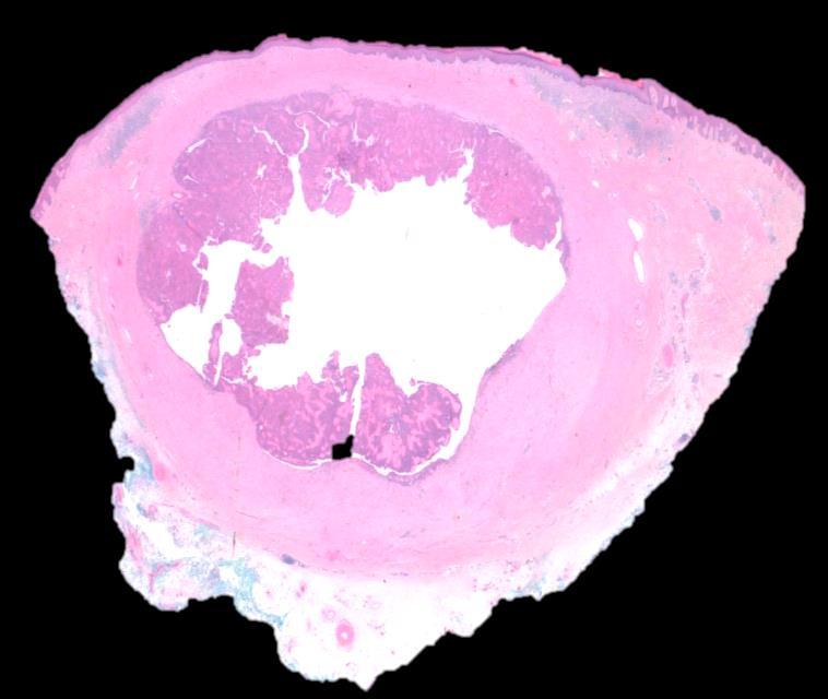
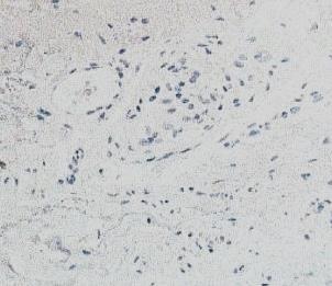
1 Case 18 M75. Excision of mass on scalp. Clinically SCC. The best diagnosis is: A. Pilomatrical carcinoma B. Adnexal carcinoma NOS C. Metastatic squamous cell carcinoma D.Primary squamous cell carcinoma

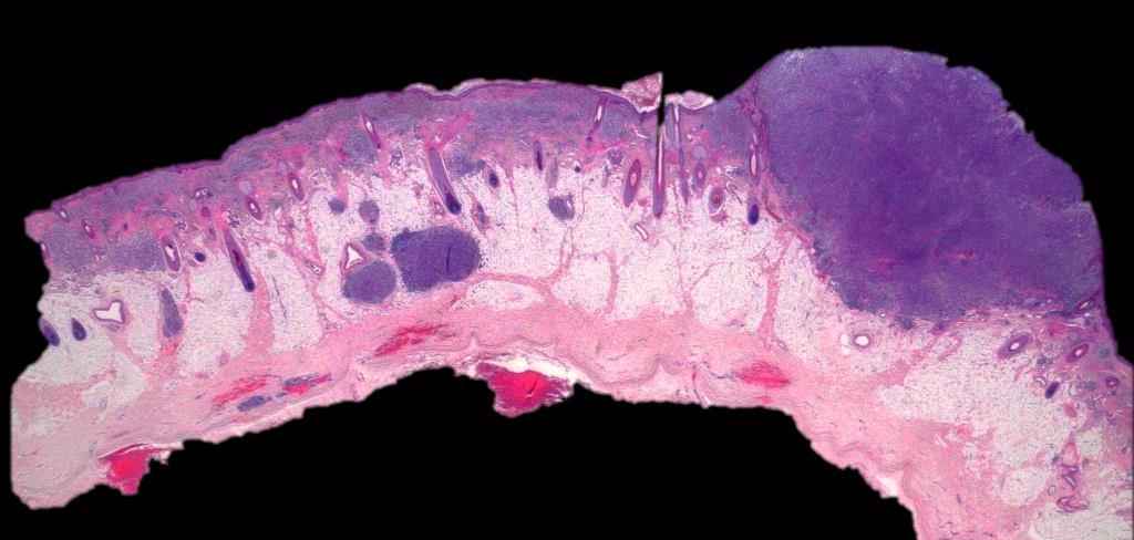
2 Case 18 M75. Excision of mass on scalp. Clinically SCC.
3 Case 18 M75. NOTE: UNUSUAL PATTERN OF IN SITU CARCINOMA Excision of mass on scalp. Clinically SCC. The best diagnosis is:
4 PC2a. Male 85, mass on scalp. CK5, p63 positive. Answer C. metastatic squamous cell carcinoma (of lung)
5 Primary sites of origin of skin metastases: Breast carcinoma (female) > lung carcinoma > colorectal carcinoma > metastatic mucosal SCC from head and neck Less common gastric carcinoma, renal cell carcinoma, ovarian carcinoma, oesophageal carcinoma, other carcinomas and sarcomas Children neuroblastoma, rhabdomyosarcoma
6 Clues to a metastasis - Clinical: Site: scalp is the commonest site for cutaneous metastases other than breast carcinoma (anterior chest) neck (primary head and neck carcinomas especially SCC) and face. Trunk less common. often close to the internal primary malignancy (& locoregional skin in melanoma) e.g. Carcinomas of intra-abdominal origin metastasise often to the anterior abdominal wall & umbilicus (Sister Mary Joseph s nodule), ovarian carcinoma - perineum.
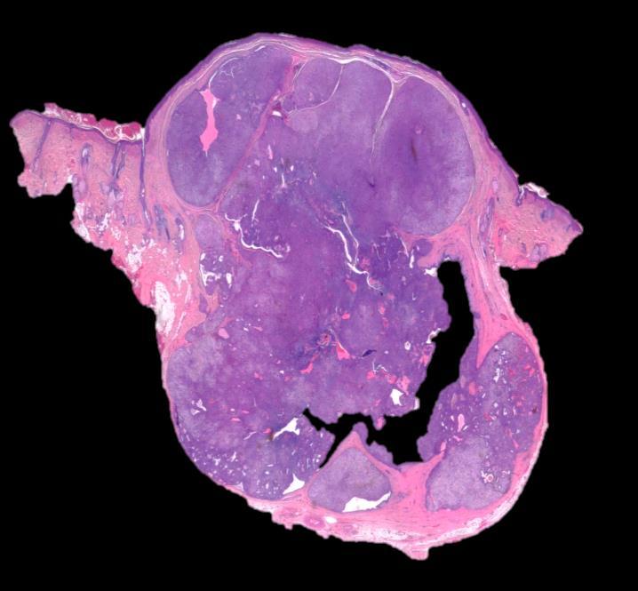
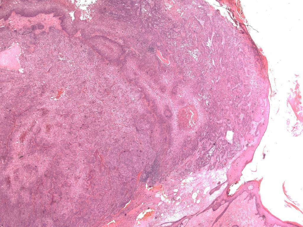
7 National Specialist Dermatopathology EQA Scheme 2017 case U337 Female 88 years. Skin ellipse excision, scalp. Skin tumour scalp. No other history available.
8 National Specialist Dermatopathology EQA Scheme 2017 case U337 Female 88 years. Skin ellipse excision, scalp. Skin tumour scalp. No other history available. Diagnosis: metastatic carcinoma

9 National Specialist Dermatopathology EQA Scheme 2017 case U337 Female 88 years. Skin ellipse excision, scalp. Skin tumour scalp. No other history available. NSD EQA Case U337 Case 18
10 National Specialist Dermatopathology EQA Scheme 2017 case U337 Diagnostic categories: Popularity: Score : 1 Metastatic carcinoma 2.55 REMOVED 2 Porocarcinoma 2.22 FROM 3 Neuroendocrine carcinoma / metastasis 2.20 SCORING 4 Basal cell carcinoma (+/- basosquamous) Sweat gland carcinoma/cutaneous adenoca Squamous cell carcinoma (+/- basaloid) Other diagnosis Poorly differentiated carcinoma 0.66 Other diagnoses : Sebaceous carcinoma (x4), trichilemmal carcinoma (x2), trichoblastic carcinoma, pilomatrical carcinoma (x3), No response, synovial sarcoma, melanoma. Inapproritae case (multiple). Original report : Metastatic carcinoma. (Subsequently found to have large cell neuroendocrine carcinoma of lung.)
11 Clues to a metastasis - Clinical: Age: Elderly patients (not a very helpful clue!) Sex: Female metastatic breast carcinoma accounts for 69% of cutaneous metastases in females
12 Clues to a metastasis - Clinical: Clinical history: Always check if unusual clinical findings or unusual histological features check for history of previous malignancy Clinical presentation: cluster of nodules, but often solitary nodule often rapidly growing and large Breast sclerotic areas on anterior chest wall, erythema
13 Presentation of metastasis many mimics: cyst pyogenic granuloma, hemangioma papular eruptions herpes zoster eruption, Rapidly infiltrating plaques, alopecic patches Cellulitis, erysipelas Hussein MR. Skin metastasis: a pathologist's perspective. J Cutan Pathol Sep;37(9):e1-20


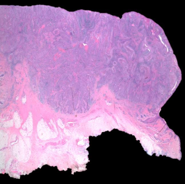
14 Clues to a metastasis - Histopathology: Well-circumscribed nodule Presence of necrosis or haemorrhage dirty necrosis in colorectal carcinoma Lack of connection to the epidermis Lack of in situ component Unusual pattern of in situ component

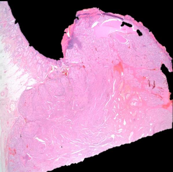
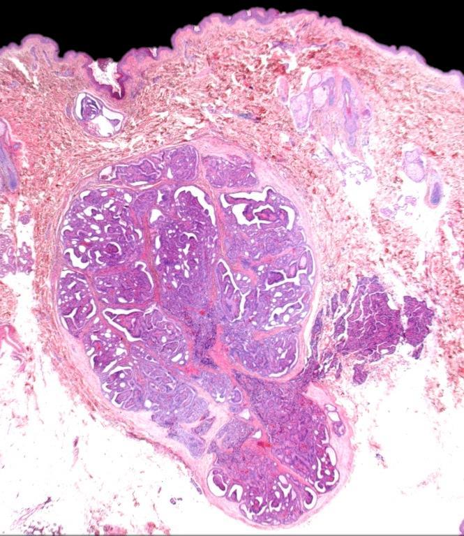
15 Primary squamous cell carcinoma attached to epidermis

16 No connection to epidermis: metastatic SCC

17 No connection to epidermis: metastatic SCC Primary SCC of larynx


18 Male 82 solitary nodule right post-auricular region no connection to epidermis metastatic SCC from previous primary SCC skin of neck


19 Clues to a metastasis - Histopathology: Multifocality Vascular invasion

20 Unusual pattern of in situ carcinoma think epidermotropic metastasis rather than primary in situ carcinoma


21 Unusual pattern of in situ carcinoma focal
22 Solitary circumscribed nodules can be primary or metastatic carcinoma or primary benign adnexal neoplasm


23 Solitary circumscribed nodules always have metastasis in differential diagnosis
24 Male 57 solitary circumscribed nodule on trunk


25 Pitfall! Nodular hidradenoma common but has variable morphology: Mucinous metaplasia not adenocarcinoma
26 Growth pattern and glandular/ductal morphology not typical for primary carcinoma? think metastasis!
27 Metastatic adenocarcinoma of pancreas positive for CK7, CK20, CA19-9 negative for WT1, CDX2, p53
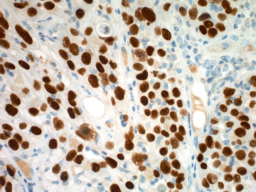
28 Immunohistochemistry for suspected metastasis or to exclude metastasis: If any of the clinical or histological features are unusual for a primary then consider immunohistochemistry for metastasis need to choose an immunohistochemical panel based on the histopathology and the clinical features. dependent on immunophenotype of specific primary or metastatic tumour Consider panel to include most common cancers: pancytokeratin, CK7, CK20 TTF-1, GATA3 (breast, urothelial), oestrogen receptor, PAX8 (females) PSA (males)
")
29 Case 18: Differential diagnosis: pilomatrical carcinoma Areas reminiscent of pilomatricoma but frankly carcinomatous (rare)
30 Case 18: Differential diagnosis: adnexal carcinoma NOS Primary skin adnexal neoplasms: usually p63 and podoplanin (D2-40) positive follicular lesions are usually also CK15 positive and ductal lesions are normally also basal cytokeratin (CK5, CK14, and CK17) positive unlike most metastases. therefore p63, D2-40, CK15 and CK5 may be helpful in select cases Positivity for all 3 of p63, D2-40, CK15 = good evidence for primary skin adnexal carcinoma but not definite evidence for a primary origin correlate with morphology and clinical features.
31 p63, podoplanin and cytokeratin 15 positive in most primary skin adnexal neoplasms and negative in metastatic adenocarcinoma Liang H, Wu H, Giorgadze TA, Sariya D, Bellucci KS, Veerappan R, Liegl B, Acs G, Elenitsas R, Shukla S, Youngberg GA, Coogan PS, Pasha T, Zhang PJ, Xu X. Podoplanin is a highly sensitive and specific marker to distinguish primary skin adnexal carcinomas from adenocarcinomas metastatic to skin. Am J Surg Pathol Feb;31(2): Plaza JA, Ortega PF, Stockman DL, Suster S. Value of p63 and podoplanin (D2-40) immunoreactivity in the distinction between primary cutaneous tumors and adenocarcinomas metastatic to the skin: a clinicopathologic and immunohistochemical study of 79 cases. J Cutan Pathol Apr;37(4): Mahalingam M, Nguyen LP, Richards JE, Muzikansky A, Hoang MP. The diagnostic utility of immunohistochemistry in distinguishing primary skin adnexal carcinomas from metastatic adenocarcinoma to skin: an immunohistochemical reappraisal using cytokeratin 15, nestin, p63, D2-40, and calretinin. Mod Pathol May;23(5):713-9.
32 Primary skin carcinoma follicular/trichilemmal

33 Primary skin carcinoma follicular/trichilemmal D2-40 (podoplanin) p63


34 Male 62. Skin nodule on neck

35 P63 negative podoplanin (D2-40) negative

36 Growth pattern and glandular/ductal morphology not typical for primary carcinoma? so Check for previous history of cancer Report your concern The diagnosis is uncertain; could be a primary or secondary epithelial neoplasm in the skin suggest full examination of patient may require radiological investigation eg CT scan
BSD 2015 Case 19. Female 21. Nodule on forehead. The best diagnosis is:
 BSD 2015 Case 19 Female 21. Nodule on forehead. The best diagnosis is: A. mixed tumour of skin B. porocarcinoma C. nodular hidradenoma D. metastatic adenocarcinoma BSD 2015 Case 19 Female 21 Nodule on
Cutaneous metastases. Thaddeus Mully. University of California, San Francisco Professor, Departments of Pathology and Dermatology
 Cutaneous metastases Thaddeus Mully University of California, San Francisco Professor, Departments of Pathology and Dermatology DISCLOSURE OF RELATIONSHIPS WITH INDUSTRY Thaddeus Mully Course C005 Essential
- Selected Tumors of the Skin Appendages - Primary vs. Metastasis
 - Selected Tumors of the Skin Appendages - Primary vs. Metastasis Napa Valley 2018 Victor G. Prieto, MD, PhD Chair of Pathology UT MD Anderson Cancer Center vprieto@mdanderson.org Napa Valley in May Introduction
Slide seminar. Asist. Prof. Jože Pižem, MD, PhD Institute of Pathology Medical Faculty, University of Ljubljana
 Slide seminar Asist. Prof. Jože Pižem, MD, PhD Institute of Pathology Medical Faculty, University of Ljubljana Case 5 A 57-year-old man with a dermal/subcutaneous lesion on the scalp, which was interpreted
confusing, especially in small, limited biopsies. One such case of digital papillary adnexal adenocarcinoma with unusual histologic features will be
 1 Cutaneous Adnexal Lesions: The Must-Know Stuff 2011 USCAP Annual Meeting Dermatopathology Companion Meeting Doina Ivan, M.D. University of Texas MD Anderson Medical Center, Houston, TX The diagnosis
Whitney A. High, MD, JD, MEng
 ADS Dermatopathology Meeting 2014 Selected Adnexal Tumors Whitney A. High, MD, JD, MEng Associate Professor, Dermatology & Pathology Director of Dermatopathology (Dermatology) University of Colorado School
Introduction. Results. Discussion. Histopathologic and immunohistochemical findings. Results. conclusions,
 1/5 2/5 Carcinoma distinctive carcinoma. form erysipeloides (CE), metastasis. which clinically Itfrom has resembles been termed erysipelas, is an uncommon, but may extend It164 toclassically back, presents
Adnexal primary or Melanocy+c prolifera+ons in sundamaged metastatic carcinoma?
 Adnexal primary or Melanocy+c prolifera+ons in sundamaged metastatic carcinoma? skin Jane L. Messina, MD Interna0onal Melanoma Pathology Working Group 4 th annual mee0ng Tampa, Florida November 14, 2011
Mesothelioma: diagnostic challenges from a pathological perspective. Naseema Vorajee August 2016
 Mesothelioma: diagnostic challenges from a pathological perspective Naseema Vorajee August 2016 Naseema.vorajee@nhls.ac.za Pleural diseases (whether neoplastic, reactive or infective) may have similar
Cancers of unknown primary : Knowing the unknown. Prof. Ahmed Hossain Professor of Medicine SSMC
 Cancers of unknown primary : Knowing the unknown Prof. Ahmed Hossain Professor of Medicine SSMC Definition Cancers of unknown primary site (CUPs) Represent a heterogeneous group of metastatic tumours,
Basal cell carcinoma 5/28/2011
 Goal of this Presentation A practical approach to the diagnosis of cutaneous carcinomas and their mimics Thaddeus Mully, MD University of California San Francisco To review common non-melanoma skin cancers
The clinically challenging entity of liver metastasis from tumors of unknown primary
 The clinically challenging entity of liver metastasis from tumors of unknown primary Xuchen Zhang, MD, PhD Associate Professor of Pathology Department of Pathology Yale University School of Medicine Liver
SEBACEOUS NEOPLASMS. Dr. Prachi Saraogi Clinical Fellow in Dermatology
 SEBACEOUS NEOPLASMS Dr. Prachi Saraogi Clinical Fellow in Dermatology Sebaceous neoplasms Sebaceous adenoma (Benign) Sebaceous carcinoma (Malignant) SEBACEOUS ADENOMA Benign tumours composed of incompletely
Update on Cutaneous Mesenchymal Tumors. Thomas Brenn
 Update on Cutaneous Mesenchymal Tumors Thomas Brenn Cutaneous Mesenchymal Tumours Wide morphological and biological spectrum Myofibroblastic, smooth muscle, neural, vascular, apidocytic, undifferentiated;
Differential diagnosis of HCC
 Hepatocellular Carcinoma Quest for an Ideal Immunohistochemical Panel Sanjay Kakar, MD UCSF Differential diagnosis of HCC Hepatocellular lesions Adenoma, FNH, HG dysplasia Adenocarcinoma CholangioCA, metastasis
Journal of International Academy of Forensic Science & Pathology (JIAFP)
") Journal of International Academy of Forensic Science & Pathology (JIAFP) ISSN 2395-0722 MICROCYSTIC ADNEXAL CARCINOMA-A CASE REPORT WITH REVIEW OF LITERATURE Case Report Sulakshana M S 1,Natarajan M 2
Journal of International Academy of Forensic Science & Pathology (JIAFP) ISSN 2395-0722 MICROCYSTIC ADNEXAL CARCINOMA-A CASE REPORT WITH REVIEW OF LITERATURE Case Report Sulakshana M S 1,Natarajan M 2
Pathology Mystery and Surprise
 Pathology Mystery and Surprise Tim Smith, MD Director Anatomic Pathology Medical University of South Carolina Disclosures No conflicts to declare Some problem cases Kidney tumor Scalp tumor Bladder tumor
CASE REPORT SOLITARY SEBACEOUS NEVUS OF JADASSOHN COMPLICATED BY SQUAMOUS CELL CARCINOMA AND BASAL CELL CARCINOMA
 CASE REPORT Dennis H. Kraus, MD, Section Editor SOLITARY SEBACEOUS NEVUS OF JADASSOHN COMPLICATED BY SQUAMOUS CELL CARCINOMA AND BASAL CELL CARCINOMA Ahmad Ridzwan Arshad, FRCS, 1 Wan S. Azman, MS, 1 Ayadurai
Urinary Bladder: WHO Classification and AJCC Staging Update 2017
 Urinary Bladder: WHO Classification and AJCC Staging Update 2017 Houston Society of Clinical Pathologists 58 th Annual Spring Symposium Houston, TX April 8, 2017 Jesse K. McKenney, MD Classification
ARIZONA SOCIETY OF PATHOLOGISTS 13 TH APRIL 2013 HEAD AND NECK CYTOPATHOLOGY. F ZAHRA ALY, MD, PhD
 ARIZONA SOCIETY OF PATHOLOGISTS 13 TH APRIL 2013 HEAD AND NECK CYTOPATHOLOGY F ZAHRA ALY, MD, PhD The main areas sites amenable for cytopathology include lymph nodes, thyroid, major salivary glands especially
Reporting of carcinoma of unknown primary tumour (CUP)
") Reporting of carcinoma of unknown primary tumour (CUP) Prof John Schofield Kent Oncology Centre with grateful thanks to Dr Karin Oien University of Glasgow Royal College of Pathologists Cancer datasets
Reporting of carcinoma of unknown primary tumour (CUP) Prof John Schofield Kent Oncology Centre with grateful thanks to Dr Karin Oien University of Glasgow Royal College of Pathologists Cancer datasets
performed to help sway the clinician in what the appropriate diagnosis is, which can substantially alter the treatment of management.
 Hello, I am Maura Polansky at the University of Texas MD Anderson Cancer Center. I am a Physician Assistant in the Department of Gastrointestinal Medical Oncology and the Program Director for Physician
Disclosure of Relevant Financial Relationships
 Squamous entities of the thyroid: Reactive to Neoplastic Michelle D. Williams Associate Professor Dept of Pathology, Head & Neck Section University of Texas MD Anderson Cancer Center Disclosure of Relevant
Papillary Lesions of the Breast A Practical Approach to Diagnosis. (Arch Pathol Lab Med. 2016;140: ; doi: /arpa.
 Papillary Lesions of the Breast A Practical Approach to Diagnosis (Arch Pathol Lab Med. 2016;140:1052 1059; doi: 10.5858/arpa.2016-0219-RA) Papillary lesions of the breast Span the spectrum of benign,
Histopathological diagnosis of CUP
 Histopathological diagnosis of CUP Dr Karin Oien karin.oien@glasgow.ac.uk Disclosure slide Dr Karin Oien has no financial interests in any company mentioned in this presentation. Dr Karin Oien is conducting
The Panel Approach to Diagnostics. Lauren Hopson International Product Specialist Cell Marque Corporation
 The Panel Approach to Diagnostics Lauren Hopson International Product Specialist Cell Marque Corporation Cell Marque Rocklin, California About Cell Marque: IVD primary antibody manufacturer Distributors
Radiology Pathology Conference
 Radiology Pathology Conference Sharlin Johnykutty,, MD, Cytopathology Fellow Sara Majewski, MD, Radiology Resident Friday, August 28, 2009 Presentation material is for education purposes only. All rights
Cutaneous Adnexal Tumors
 Cutaneous Adnexal Tumors Lesions with Predominant Follicular Differentiation Special Emphasis on Basal Cell Carcinoma 2014-04-01 Prof. Dr. med. Katharina Glatz Pathologie Cutaneous Adnexal Tumors Hair
American College of Mohs Surgery. Diagnostic Quality Control Program (Review of Answers)
") American College of Mohs Surgery Diagnostic Quality Control Program 2010 (Review of Answers) Question 1 A flesh-colored plaque on the right ala of an otherwise healthy 57 year-old male is referred for
American College of Mohs Surgery Diagnostic Quality Control Program 2010 (Review of Answers) Question 1 A flesh-colored plaque on the right ala of an otherwise healthy 57 year-old male is referred for
4/12/2018. MUSC Pathology Symposium Kiawah Island April 18, Jesse K. McKenney, MD
 MUSC Pathology Symposium Kiawah Island April 18, 2018 Jesse K. McKenney, MD 1 Urothelial Carcinoma with Alternative Differentiation 2 Urothelial Carcinoma with Alternative Differentiation Recognition as
South East England General Histopathology EQA Scheme
 South East England General Round a Final Case Analyses Cases 635 to 646 Circulated January February 2016 131 responses (89.73%) Prepared April 2016 Authorised by: Prof J Schofield Date: 19/4/2016 With
A 5 Year Histopathological Study of Skin Adnexal Tumors at a Tertiary Care Hospital
 IOSR Journal of Dental and Medical Sciences (IOSR-JDMS) e-issn: 2279-0853, p-issn: 2279-0861.Volume 14, Issue 4 Ver. VII (Apr. 2015), PP 01-05 www.iosrjournals.org A 5 Year Histopathological Study of Skin
Immunohistochemistry in Bone and Soft Tissue Tumors. Sahar Rassi Zankoul, MD
 Immunohistochemistry in Bone and Soft Tissue Tumors Sahar Rassi Zankoul, MD Introduction Bone tumors represent a wide variety of tumors of various origins and malignant potentials. These different tumor
57th Annual HSCP Spring Symposium 4/16/2016
 An Unusual Malignant Spindle Cell Lesion to Involve the Breast Erinn Downs-Kelly, D.O. Associate Professor of Pathology University of Utah & ARUP Laboratories No disclosures Case 39 y/o female with no
Dermatopathology. Dr. Rafael Botella Estrada. Hospital La Fe de Valencia
 Dermatopathology Dr. Rafael Botella Estrada. Hospital La Fe de Valencia DERMATOPATHOLOGY CASE CHALLENGE: RECOGNIZING MIMIS AND MASQUERADERS Rosalie Elenitsas. University of Pennsylvania Spectrum Lupus
What I Learned from 3 Cases and 3 Antibodies
 What I Learned from 3 Cases and 3 Antibodies Melinda Sanders, M.D Vanderbilt University Medical Center Professor of Pathology Consultant in Breast Pathology Disclosure of Relevant Financial Relationships
3/27/2017. Pulmonary Pathology Specialty Conference. Disclosure of Relevant Financial Relationships. Clinical History:
 Pulmonary Pathology Specialty Conference Saul Suster, M.D. Medical College of Wisconsin Disclosure of Relevant Financial Relationships USCAP requires that all planners (Education Committee) in a position
ACCME/Disclosures. 52 year old man who consulted for a long-standing mass on the distal penis 4/13/2016
 ACCME/Disclosures United States and Canadian Academy of Pathology Seattle, WA 2016 Elsa F Velazquez, MD Director of Dermatopathology, V.P. Clinical Assistant Professor of Dermatology Tufts University,
Aggressive Malignant Mesothelioma In A Patient Without Previous Asbestos Exposure
 ISPUB.COM The Internet Journal of Pathology Volume 19 Number 1 Aggressive Malignant Mesothelioma In A Patient Without Previous Asbestos Exposure A Julai, G Wathuge Citation A Julai, G Wathuge. Aggressive
CASE year old male with a PET avid nodule in the left adrenal gland
 CASE 1 55 year old male with a PET avid nodule in the left adrenal gland Case 1 Adrenal gland parenchyma partly replaced by a spindle cell tumour with mild nuclear pleomorphism Atypical mitoses present
Case Report A Rare Cutaneous Adnexal Tumor: Malignant Proliferating Trichilemmal Tumor
 Case Reports in Medicine Volume 2015, Article ID 742920, 4 pages http://dx.doi.org/10.1155/2015/742920 Case Report A Rare Cutaneous Adnexal Tumor: Malignant Proliferating Trichilemmal Tumor Omer Alici,
Research Article A Clinicopathological and Immunohistochemical Correlation in Cutaneous Metastases from Internal Malignancies: A Five-Year Study
 Skin Cancer, Article ID 793937, 5 pages http://dx.doi.org/10.1155/2014/793937 Research Article A Clinicopathological and Immunohistochemical Correlation in Cutaneous Metastases from Internal Malignancies:
NEOPLASIA. 3. Which of the following tumour is benign a. Chondrosarcoma b. Osteochondroma c. Chondroblastoma d. Ewing s tumour e.
 NEOPLASIA 1. malignant neoplasms a. are independent of hormonal influence b. are always composed of homogenous cell lines c. arise from differentiated cells by a process of anaplasia d. display abnormal
64 y.o. F with CLL and leg tumour
 64 y.o. F with CLL and leg tumour Case History Excision with split-skin grafting Histology moderately differentiated squamous cell carcinoma with large areas of necrosis and brisk mitotic activity.
Biopsy Interpretation of Spindle cell proliferations of the Serosa
 Biopsy Interpretation of Spindle cell proliferations of the Serosa Richard Attanoos, Cardiff. U.K. Disclosure of Relevant Financial Relationships USCAP requires that all planners (Education Committee)
Update in Salivary Gland Pathology. Benjamin L. Witt University of Utah/ARUP Laboratories February 9, 2016
 Update in Salivary Gland Pathology Benjamin L. Witt University of Utah/ARUP Laboratories February 9, 2016 Objectives Review the different appearances of a selection of salivary gland tumor types Establish
Objectives. Salivary Gland FNA: The Milan System. Role of Salivary Gland FNA 04/26/2018
 Salivary Gland FNA: The Milan System Dr. Jennifer Brainard Section Head Cytopathology Cleveland Clinic Objectives Introduce the Milan System for reporting salivary gland cytopathology Define cytologic
The role of immunohistochemistry in surgical pathology of the uterine corpus and cervix
 The role of immunohistochemistry in surgical pathology of the uterine corpus and cervix Prof. Ben Davidson, MD PhD Department of Pathology, Norwegian Radium Hospital, Oslo University Hospital, Oslo, Norway
Enterprise Interest Nothing to declare
 Enterprise Interest Nothing to declare Diagnoses one would not like to miss in soft tissue pathology early in your career Marta Sbaraglia, MD Department of Pathology Hospital of Treviso University of Padua
Wendy L Frankel. Chair and Distinguished Professor
 1 Wendy L Frankel Chair and Distinguished Professor Case 1 59 y/o woman Abdominal pain No personal or family history of cancer History of colon polyps Colonoscopy Polypoid rectosigmoid mass Biopsy 3 4
Neuroendocrine Carcinoma. Lebanon Neuroendocrine Neoplasms of H&N Nov /7/2011. Broad Classification:
 H&N Neuroendocrine Neoplasms: Classification and Diagnostic Considerations Adel K. El-Naggar, M.D., Ph.D. The University of Texas MD Anderson Cancer Center, Houston, Texas Broad Classification: A. Epithelial:
Expression of Cytokeratin 5/6 in Epithelial Neoplasms: An Immunohistochemical Study of 509 Cases
 Expression of Cytokeratin 5/6 in Epithelial Neoplasms: An Immunohistochemical Study of 509 Peiguo G. Chu, M.D., Ph.D., Lawrence M. Weiss, M.D. Department of Pathology, City of Hope National Medical Center,
FINALIZED SEER SINQ QUESTIONS January June, 2016
 20160046 MP/H Rules/Multiple primaries/histology--rectum: How many primaries does this person have and what is the correct histology? See discussion. Rectal polyp excised in June, 2012, found to have adenocarcinoma
Classification (1) Classification (3) Classification (2) Spindle cell lesions. Spindle cell lesions of bladder (Mills et al.
 Classification (3) Classification (2) Spindle cell lesions. Spindle cell lesions of bladder (Mills et al.") Non-epithelial tumours and nonepithelial tumour-like lesions of the bladder Dr Jonathan H Shanks The Christie NHS Foundation Trust, Manchester, UK Classification (1) Myofibroblastic proliferations and
Non-epithelial tumours and nonepithelial tumour-like lesions of the bladder Dr Jonathan H Shanks The Christie NHS Foundation Trust, Manchester, UK Classification (1) Myofibroblastic proliferations and
Gastric and Oesophageal Neuroendocrine tumours. Dr Tim Bracey, Consultant Pathologist MBChB PhD MRCS FRCPath
 Gastric and Oesophageal Neuroendocrine tumours Dr Tim Bracey, Consultant Pathologist MBChB PhD MRCS FRCPath Intestinal (and BO) endocrine cells in crypt bases NE cell (granules towards vessels) Paneth
Diploma Examination. Dermatopathology: First paper. Tuesday 20 March Candidates must answer FOUR questions. Time allowed: 3 hours
 Dermatopathology: First paper Tuesday 20 March 2018 Candidates must answer FOUR questions Time allowed: 3 hours 1. Give an account of the genetic aberrations encountered in Spitzoid neoplasms and how these
Radiology Pathology Conference
 Radiology Pathology Conference Nadia F. Yusaf, M.D. PGY-3 1/29/2010 Presentation material is for education purposes only. All rights reserved. 2010 URMC Radiology Page 1 of 90 Case 1 60 year- old man presents
Carcinoma of Unknown Primary site (CUP) in HEAD & NECK SURGERY
 in HEAD & NECK SURGERY") Carcinoma of Unknown Primary site (CUP) in HEAD & NECK SURGERY SEARCHING FOR THE PRIMARY? P r o f J P P r e t o r i u s H e a d : C l i n i c a l U n i t C r i t i c a l C a r e U n i v e r s i t y O f
Carcinoma of Unknown Primary site (CUP) in HEAD & NECK SURGERY SEARCHING FOR THE PRIMARY? P r o f J P P r e t o r i u s H e a d : C l i n i c a l U n i t C r i t i c a l C a r e U n i v e r s i t y O f
Immunohistochemical Distinction of Primary Sweat Gland Carcinoma and Metastatic Breast Carcinoma
 AJCP / Original Article Immunohistochemical Distinction of Primary Sweat Gland Carcinoma and Metastatic Breast Carcinoma Can It Always Be Accomplished Reliably? Mark J. Mentrikoski, MD, and Mark R. Wick,
Cerebral Parenchymal Lesions: I. Metastatic Neoplasms
 Chapter 4 Cerebral Parenchymal Lesions: I. Metastatic Neoplasms After one has reasonably ruled out the possibility of a nonneoplastic diagnosis (see Chap. 3), one is left with considering a diagnosis of
Appendageal skin tumors
 Appendageal skin tumors Ibrahim Khalifeh, M.D. Associate Professor Department of Pathology American University of Beirut Medical Center Beirut, Lebanon Appendageal tumors Neoplasms whose differentiation
When Immunostains Can Get You in Trouble: Gynecologic Pathology p16: Panacea or Pandora s Box?
 When Immunostains Can Get You in Trouble: Gynecologic Pathology p16: Panacea or Pandora s Box? Teri A. Longacre, MD Stanford Medicine Stanford California pi6 in Gynecologic Pathology: Panacea or Pandora
Liver Specialty Evening Conference. Matthew M. Yeh, MD, PhD Professor of Pathology Adjunct Professor of Medicine University of Washington, Seattle
 Liver Specialty Evening Conference Matthew M. Yeh, MD, PhD Professor of Pathology Adjunct Professor of Medicine University of Washington, Seattle Case History A 65 year-old man presents with abdominal
New Developments in Immunohistochemistry for Gynecologic Pathology
 New Developments in Immunohistochemistry for Gynecologic Pathology Michael T. Deavers, M.D. Professor, Departments of Pathology and Gynecologic Oncology Immunohistochemistry in Gynecologic Pathology Majority
Case Report Clear Cell Basal Cell Carcinoma
 SAGE-Hindawi Access to Research Volume 2011, Article ID 386921, 4 pages doi:10.4061/2011/386921 Case Report Clear Cell Basal Cell Carcinoma Deba P. Sarma, 1 Daniel Olson, 1 Jennifer Olivella, 1 Tracey
CODING TUMOUR MORPHOLOGY. Otto Visser
 CODING TUMOUR MORPHOLOGY Otto Visser INTRODUCTION The morphology describes the tissue of the tumour closest to normal tissue Well differentiated tumours are closest to normal Undifferentiated tumours show
Cluster designation 5 staining of normal and non-lymphoid neoplastic skin*
 J Cutan Pathol 2005: 32: 50 54 Copyright # Blackwell Munksgaard 2005 Blackwell Munksgaard. Printed in Denmark Journal of Cutaneous Pathology Cluster designation 5 staining of normal and non-lymphoid neoplastic
FINALIZED SEER SINQ QUESTIONS
 0076 Source 1: WHO Class CNS Tumors pgs: 33 MP/H Rules/Histology--Brain and CNS: What is the histology code for a tumor originating in the cerebellum and extending into the fourth ventricle described as
Uropathology January Jon Oxley
 Uropathology January 2012 Jon Oxley Background to seminar These slides were available to view via the web from scanned slides The junior pathologists answered questions on them via the web The answers
NEW IHC A n t i b o d i e s
 NEW IHC Antibodies TABLE OF CONTENTS NEW IHC ANTIBODIES from Cell Marque CITED1 (5H6).... 1 Claudin 7 (5D10F3).... 1 GATA1 (4F5).... 1 Transgelin (2A10C2).... 1 NEW IHC ANTIBODIES using RabMAb Technology
DATA STANDARDS AND QUALITY CONTROL MEMORANDUM DSQC #
 DATA STANDARDS AND QUALITY CONTROL MEMORANDUM DSQC #2006-01 CATEGORY: CLARIFICATION SUBJECT: RESCINDMENT - DSQC MEMORANDUM 2002-08 Coding Complex Morphologic Diagnoses (revised 8/02) EFFECTIVE: For Cases
Unknown Slides Conference
 Unknown Slides Conference Jae Y. Ro, MD, PhD Weill Medical College of Cornell Univ. The Methodist Hospital, and UT MD Anderson Cancer Center Houston, TX November 9, 2013 Amman, Jordan 25 th Congress of
Case history: Figure 1. H&E, 5x. Figure 2. H&E, 20x.
 1 Case history: A 49 year-old female presented with a 5 year history of chronic anal fissure. The patient s past medical history is otherwise unremarkable. On digital rectal examination there was a very
BAP-oma & BEYOND MICHAEL A NOWAK, MD
 BAP-oma & BEYOND MICHAEL A NOWAK, MD CONFLICTS No conflicts with the content of this lecture BAP-oma Wiesner 2011: Families with multiple tan dome-shaped papules of head, neck, trunk, and extremities.
Applications of IHC. Determination of the primary site in metastatic tumors of unknown origin
 Applications of IHC Determination of the primary site in metastatic tumors of unknown origin Classification of tumors that appear 'undifferentiated' by standard light microscopy Precise classification
Carcinoma of Unknown Primary (CUP)
") Metasta c Carcinoma of Unknown Primary: Diagnos c Approach Using Immunohistochemistry James R. Conner, MD, PhD Mount Sinai Hospital Toronto, ON Carcinoma of Unknown Primary (CUP) 3-5% of all new malignant
Metasta c Carcinoma of Unknown Primary: Diagnos c Approach Using Immunohistochemistry James R. Conner, MD, PhD Mount Sinai Hospital Toronto, ON Carcinoma of Unknown Primary (CUP) 3-5% of all new malignant
5/21/2018. Prostate Adenocarcinoma vs. Urothelial Carcinoma. Common Differential Diagnoses in Urological Pathology. Jonathan I.
 Common Differential Diagnoses in Urological Pathology Jonathan I. Epstein Prostate Adenocarcinoma vs. Urothelial Carcinoma 1 2 NKX3.1 NKX3.1 3 4 5 6 Proposed ISUP Recommendations Option to use PSA as a
Research Article Histopathological Study of Skin Adnexal Tumours Institutional Study in South India
 Skin Cancer, Article ID 543756, 4 pages http://dx.doi.org/10.1155/2014/543756 Research Article Histopathological Study of Skin Adnexal Tumours Institutional Study in South India Ankit Sharma, 1 Deepak
Greater Manchester & Cheshire Guidelines for Pathology Reporting for Oesophageal and Gastric Malignancy
 Greater Manchester & Cheshire Guidelines for Pathology Reporting for Oesophageal and Gastric Malignancy Authors: Dr Gordon Armstrong, Dr Sue Pritchard 1. General Comments 1.1 Cancer reporting: Biopsies
Lung Tumor Cases: Common Problems and Helpful Hints
 Lung Tumor Cases: Common Problems and Helpful Hints Brandon T. Larsen, MD, PhD Senior Associate Consultant Department of Laboratory Medicine and Pathology Mayo Clinic Arizona Arizona Society of Pathologists
Take Home Quiz 1 Please complete the quiz below prior to the session. Use the Multiple Primary and Histology Rules
 Take Home Quiz 1 Please complete the quiz below prior to the session. Use the Multiple Primary and Histology Rules Case 1 72 year old white female presents with a nodular thyroid. This was biopsied in
ACCME/Disclosures. Diagnosing Mesothelioma in Limited Tissue Samples. Papanicolaou Society of Cytopathology Companion Meeting March 12 th, 2016
 Diagnosing Mesothelioma in Limited Tissue Samples Papanicolaou Society of Cytopathology Companion Meeting March 12 th, 2016 Sanja Dacic, MD, PhD University of Pittsburgh ACCME/Disclosures GENERAL RULES
Metastases to the kidney: a clinicopathological study of 43 cases with an emphasis on deceptive features
 Histopathology 2015, 66, 587 597. DOI: 10.1111/his.12524 Metastases to the kidney: a clinicopathological study of 43 cases with an emphasis on deceptive features Angela J Wu, Rohit Mehra, Khaled Hafez,
Primary Cutaneous Apocrine Carcinoma of Sweat Glands: A Rare Case Report
 This is an Open Access article licensed under the terms of the Creative Commons Attribution-NonCommercial-NoDerivs 3.0 License (www.karger.com/oa-license), applicable to the online version of the article
Maligna Melanoma and Atypical Fibroxanthoma: An Unusual Collision Tumour G Türkcü 1, A Keleş 1, U Alabalık 1, D Uçmak 2, H Büyükbayram 1 ABSTRACT
 Maligna Melanoma and Atypical Fibroxanthoma: An Unusual Collision Tumour G Türkcü 1, A Keleş 1, U Alabalık 1, D Uçmak 2, H Büyükbayram 1 ABSTRACT Two different neoplasia in the same biopsy material called
Financial disclosures
 Cutaneous Mesenchymal Neoplasms with EWSR1 Rearrangement By Konstantinos Linos MD, FCAP, FASDP Bone, Soft Tissue and Dermatopathology Assistant Professor of Pathology Dartmouth-Hitchc Geisel School of
Salivary Gland Cytology
 Salivary Gland Cytology Diagnostic challenges and potential pitfalls Tarik M. Elsheikh, MD Professor and Medical Director Anatomic Pathology Cleveland Clinic FNA Salivary Gland Lesions Indications Distinguish
Cancer of Unknown Primary (CUP)
") Cancer of Unknown Primary (CUP) Pathways and Guidelines V1.0 London Cancer September 2013 The following pathways and guidelines document has been compiled by the London Cancer CUP technical subgroup and
Cancer of Unknown Primary (CUP) Pathways and Guidelines V1.0 London Cancer September 2013 The following pathways and guidelines document has been compiled by the London Cancer CUP technical subgroup and
Impact of immunostaining of pulmonary and mediastinal cytology
 Impact of immunostaining of pulmonary and mediastinal cytology Harman Sekhon MD, PhD Director of Cytopathology Head of Ottawa-site Ontario Tumour Bank June 20, 2014 Disclaimer Pfizer: Honorarium-Advisory
Microcystic Squamous Cell Carcinoma of the Lung A Clinicopathologic Study of Three Cases
 Anatomic Pathology / Microcystic SCC of the Lung Microcystic Squamous Cell Carcinoma of the Lung A Clinicopathologic Study of Three Cases Annikka Weissferdt, MD, and Cesar A. Moran, MD Key Words: Squamous
Coordinate Expression of Cytokeratins 7 and 20 in Prostate Adenocarcinoma and Bladder Urothelial Carcinoma
 Anatomic Pathology / CYTOKERATINS 7 AND 20 IN PROSTATE AND BLADDER CARCINOMAS Coordinate Expression of Cytokeratins 7 and 20 in Prostate Adenocarcinoma and Bladder Urothelial Carcinoma Nader H. Bassily,
Case Report Aggressive invasive micropapillary salivary duct carcinoma of the parotid gland
 Pathology International 2008; 58: 322 326 doi:10.1111/j.1440-1827.2008.02231.x Case Report Aggressive invasive micropapillary salivary duct carcinoma of the parotid gland Hidetaka Yamamoto, 1 Hideoki Uryu,
Glycogen-rich adenocarcinoma in the lower lip: report of a case with particular emphasis on differential diagnosis
 Open Journal of Stomatology, 2011, 1, 109-113 doi:10.4236/ojst.2011.13017 Published Online September 2011 (http://www.scirp.org/journal//). Glycogen-rich adenocarcinoma in the lower lip: report of a case
Circulation: X Case number: 501 Number of responses: 84 Date: 4 MAY 12
 Circulation: X Case number: 500 Number of responses: 81 Date: 4 MAY 12 Female, aged 65 TAH and BSO for G1 endometrioid adenocarcinoma. Tumour positive with inhibin, vimentin, CD56 and SMA. Negative with
Synonyms. Nephrogenic metaplasia Mesonephric adenoma
 Nephrogenic Adenoma Synonyms Nephrogenic metaplasia Mesonephric adenoma Definition Benign epithelial lesion of urinary tract with tubular, glandular, papillary growth pattern Most frequently in the urinary
List of Available TMAs in the PRN
 TMA RPCI_BrainCa01 RPCI_BrCa03 RPCI_BrCa04 RPCI_BrCa05 RPCI_BrCa0 RPCI_BrCa07 RPCI_BrCa08 RPCI_BrCa15 RPCI_BrCa1 RPCI_BrCa17 RPCI_BrCa18 RPCI_BrCa19 RPCI_BrCa20 RPCI_BrCa21 RPCI_BrCa24 RPCI_BrCa25 RPCI_BrCa2
Melanoma Update: 8th Edition of AJCC Staging System
 Melanoma Update: 8th Edition of AJCC Staging System Rosalie Elenitsas, M.D. Professor of Dermatology Director, Dermatopathology University of Pennsylvania DISCLOSURE OF RELATIONSHIPS WITH INDUSTRY None
Immunohistochemical classification of lung carcinomas and mesotheliomas. Prof. Mogens Vyberg NordiQC Institute of Pathology Aalborg, Denmark
 Immunohistochemical classification of lung carcinomas and mesotheliomas Prof. Mogens Vyberg NordiQC Institute of Pathology Aalborg, Denmark Endobronchial ultrasound guided transbronchial needle biopsy
Desmoplastic Melanoma R/O BCC. Clinical Information. 74 y.o. man with lesion on left side of neck r/o BCC
 R/O BCC Sabine Kohler, M.D. Professor of Pathology and Dermatology Dermatopathology Service Stanford University School of Medicine Clinical Information 74 y.o. man with lesion on left side of neck r/o
Cutaneous Mesenchymal Neoplasms with EWSR1 Rearrangement
 Cutaneous Mesenchymal Neoplasms with EWSR1 Rearrangement By Konstantinos Linos MD, FCAP, FASDP Bone, Soft Tissue and Dermatopathology Assistant Professor of Pathology Dartmouth-Hitchcock Medical Center