Breast lumps to refer or not to refer? Simon Cawthorn Breast Specialist
|
|
|
- Laura Dawson
- 5 years ago
- Views:
Transcription
1 Breast lumps to refer or not to refer? Simon Cawthorn Breast Specialist
2 Learning objectives Know the indications to refer urgently Who to reassure and review How to reassure patients with non-urgent symptoms and signs
3 Learning Objectives 1 After completing this module you should know: How to assess a patient reporting a breast lump - Consider what should be covered in the 10 minute consultation The differential diagnosis of breast lumps How to recognise red flag symptoms and signs -Who should I be worried about? When to refer to secondary care and the urgency of this Highlight the new NICE cancer referral guidance Any other referral guidance for patients not meeting NICE criteria for 2 week wait referral? What a patient should expect to happen when referred to breast clinic
4 Learning Objectives 2 Understand the possible causes other than cancer which can lead patients to seek advice in primary care about a breast lump, and how to manage those in whom cancer is unlikely (in less than 3% of cases ) where urgent referral is not recommended by NICE
5 Managing the Breast TWW s 1 in 7 of NHS out patient referrals are to the One-stop Breast clinic
6 Number of Patients Performance Increasing TWW referrals in 5 years-(nhs Eng) 450, , , , , , , ,000 50,000 0 Two Week Wait From GP Urgent Referral to First Consultant Appointment 97.0% 96.0% 95.0% 94.0% 93.0% 92.0% 91.0% Financial Quarter Total Patients Seen - Quarterly Percentage of Patients Seen Within 2 weeks Operational Standard
7 Number of Patients Performance No of cancers 68,000 66,000 64,000 62,000 One Month Wait from a Decision to Treat to a First Treatment for Cancer 99.0% 98.5% 98.0% 97.5% 97.0% 60, % 58,000 56, % 95.5% 95.0% 54,000 11/12 Q1 11/12 Q2 11/12 Q3 11/12 Q4 12/13 Q1 12/13 Q2 12/13 Q3 12/13 Q4 13/14 Q1 Financial Quarter Total Patients Treated - Quarterly Percentage of Patients Treated Within 31 Days Operational Standard 13/14 Q2 13/14 Q3 13/14 Q4 14/15 Q1 14/15 Q2 14/15 Q3 14/15 Q4 15/16 Q1 94.5%

8
9 Breast cancer NICE Ref Guidelines 2015 Refer people using a suspected cancer pathway referral (for an appointment within 2 weeks) for breast cancer if they are: aged 30 and over and have an unexplained breast lump with or without pain Over 50 with nipple discharge, retraction or symptoms of concern eczema aged 30 and over with an unexplained lump in the axilla
10 Breast Cancer 2015 NICE guidance Non-urgent referral Consider non-urgent referral in people aged under 30 with an unexplained breast lump with or without pain. (nothing about all lumps should be referred-nothing about breast pain!)
11
12 Breast Examinationwhat is your duty of care? In line with good practice and GMC guidance, explain to the patient what you intend to do and why. Obtain consent for the examination and document this. Offer a chaperone and document the discussion. Breast inspection Inspect with the patient sitting and then with their hands raised above head, and then pressed down on their hips producing pectoralis major muscle contraction A lump may be visible.. This may result in indentation of the skin, or indrawing of the nipple, with muscle contraction, a sign of a probable underlying cancer/

13

14
15 Breast Examinationwhat is your duty of care? Look for: -Variations in breast size and contour. -Whether there is an inverted nipple (nipple retraction) and, if so, is it unilateral or bilateral? -Redness associated with brawny induration of the skin. (called peau d'orange -orange peelcaused by lymphatic invasion - inflammatory breast cancer). -Any sign of visible ulceration is a sign of skin infiltration -eczema of the nipple skin a sign of Paget s disease, a form of in-situ carcinoma associated in 50% of cases with an underlying breast cancer

16
17 Breast Examination Breast palpation The next next stage is palpation, and a systematic search pattern improves the rate of detection. There is no proven "best method" Technique for palpation of the breast [13] to feel for breast lumps. Ask the patient to lie supine with their hands above their head. Examine from the clavicle medially to the midsternum, laterally to the mid-axillary line and to the inferior portion of the breast. Remember the axillary tail of breast tissue. Examine the axilla for palpable lymphadenopathy.
18 Breast Examination Breast palpation Examine with the hand flat to avoid pinching or probing the breast tissue. Press the breast flat with the palmar surfaces of the second, third and fourth fingers held together. The breast tissue is often lumpy, and the purpose of the press test is to identify the lump which persists and becomes more obvious with compression. A lump will have defined margins separate and distinct from the surrounding tissue. The consistency is the most important clinical parameter in diagnosing the presence of a lump hardness as the tissue is pressed a resistance to compression also the contour characteristics of smoothness versus irregularity.
19 Breast Examination Breast palpation is there a lump? The clinical features also of importance in addition to those in the skin described above -the tethering to the underlying tissues or skin, and the mobility of the lump when palpated. If you have difficulty finding a discrete lump, ask the patient to demonstrate it for you. A discrete mass should be described in terms of location, size, mobility and texture. thickening if localised and is asymetric = LUMP- avoid words like fibroadenosis Examine both breasts, the axillae, the infra-clavicular and supra-clavicular areas if there is no lump, state this- no lump found- normal breasts

20
21 Breast Examination If there is a history of discharge from the nipple, with permission apply gentle pressure to the areolar skin pressing with the index finger tip over the 4 quadrants. This is to distinguish the presence of single or multi-duct discharge, and the type of fluid seen, of note clear or blood-stained. If there is no such history, it is inappropriate to attempt to demonstrate a discharge. Breast examination should be thorough and take about three minutes each side.

22
23
24 Answer NO this is a reason for an urgent 2WW referral
25 Question A woman aged 31 complains of a lump in her breast, without any other symptoms other than discomfort. On both visual examination and palpation you cannot confirm the presence of a lump in the left breast in the area where the patient had felt a lump and no skin changes are seen. The opposite breast and both axillae are also normal. Q. Do you reassure her but ask her to return after her next period?
26 Answer Yes if the GP is unable to find a discrete lump (including localised thickening not present in the opposite breast) the duty of care has been undertaken, and no action is needed you don t need to see her after the next period her breast was normal
27 But does that work? How often does the patient come back still worried with the same feeling that there is a lump? But would they if in addition you gave them the offer of a full explanation about why women s breasts get painful and lumpy, and that this is not a sign of cancer in 99% Plus show them how to do BSE? All that in 10 mins (inc finding a chaperone!)
28 Same patient? Lump age 31 You are unable to confirm the presence of a lump. The breast examination as described above is normal- The breast is generally tender and lumpy. It is day 20 of her menstrual cycle, and this is irregular. She is very anxious as her mother had breast cancer at the age of 64, and is the only relative with the disease. The patient has no other risk factors. What should you do?
29 Answers? Tell the patient you are pretty sure it s OK but refer her as a non-urgent referral to the breast clinic 2. Tell her you are not an expert, and refer her as an urgent 2ww 3. Ask her to come back after her next period explaining you are confident it s nothing to worry about 4. Inform the patient there is no lump, and that the area she is worried about is swollen but normal tissue. 5. offer to refer the patient over 40 years of age for a mammogram or if under 40 for an U/S scan
30 Tests Where patients have symptoms suggestive of breast cancer, NICE recommends that tests should not be carried out before referral to a specialist. So ask her to come back after her next period explaining you are confident it s nothing to worry about
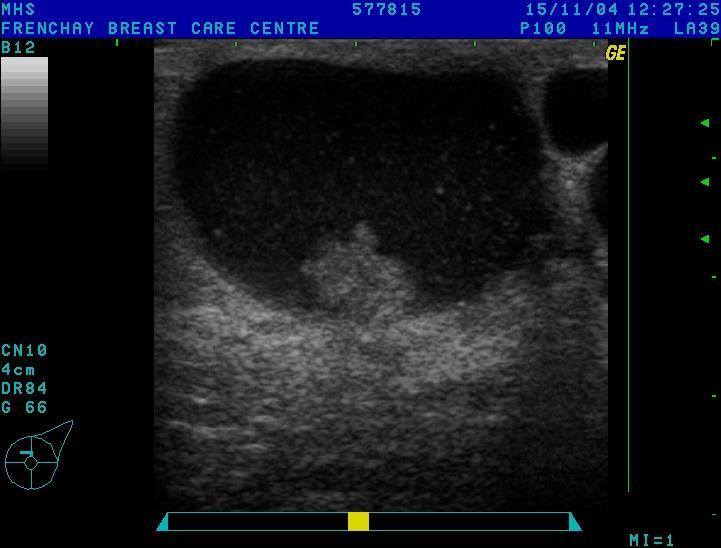
31 Imaging - Ultrasound Sensitivity high - may be the only modality to show a cancer Specificity fairly high Quick, Cheap, Safe Particularly safe in young women, pregnant women and lactating women Can be used for guided needle biopsy of impalpable lesions Frenchay Breast Care Centre 2002
32 Mammography Sensitivity fairly high - about 10-20% of cancers not seen on mammogram Specificity high Quick but more expensive, dose of radiation, and can be uncomfortable Not useful in young women - dense breasts and increased sensitivity to radiation Frenchay Breast Care Centre 2002
33 Breast Cancer 2015 NICE guidance Non-urgent referral Consider non-urgent referral in people aged under 30 with an unexplained breast lump with or without pain. (nothing about all lumps should be referred-nothing about breast pain!)
34 Same patient Q. The same patient aged 31 in whom no lump was detected did however have a localised area of tenderness in the site where she had felt the lump. Do you refer her for a focal Breast U/S scan?
35 Answer Yes Best practice diagnostic guidelines for patients presenting with breast symptoms Editors Alexis M Willett, Michael J Michell, Martin J R Lee November 2010 Patients in a one stop clinic with no lump, but an area of localised tenderness should have a breast U/S
36 A 20 year old woman presents with a painless lump in her breast. Breast examination reveals a tender well defined firm smooth mobile mass 2 cms in diameter with no skin or other worrying signs. She is taking microgynon. Do you 1.refer as urgent 2ww referral? 2. Refer as a non-urgent referral? 3. Refer her for an U/S scan 4. Reassure her it is harmless and review after the next period 5. Switch her to a progesterone only pill 6. Inform her it is a fibro-adenoma and it is harmless
37 Ans no 6 is correct This is likely to be a fibro-adenoma BUT reassurance without imaging is unlikey to be effective in allaying her anxieties in which case a non-urgent referral to a breast clinic is appropriate. Biopsy is not indicated unless the U/S has specific worrying features under 25 less than 3 cms (Best Practice guidelines)
38 Smith et al. Bradford Royal Infirmary a palpable lump with ultrasound characteristics consistent with a fibroadenoma need not be biopsied unless there is overriding clinical concern. They suggested the patients to be reassured, discharged and advised them to return for further evaluation only if they detected a change in the palpable abnormality

39 52

40 Fibroadenomas True Benign tumour Commonest age Common and multiple in Africans Most remain unchanged Some, particularly in very young grow rapidly giant fibroadenomas
41 a painless lump in her breast. She is 20 weeks pregnant Breast examination reveals a tender well defined firm smooth mobile mass 2 cms in diameter with no skin or other worrying signs. 1. refer as urgent 2ww referral? 2. Refer as a non-urgent referral? 3. Refer her for an U/S scan Reassure her it is harmless Refer her to the antenatal clinic
42 Answer 1. Urgent 2WW
43 A 48 year old woman presents with a vague history of a lumpy tender right breast for several months Her periods are irregular, she is nulliparous, and her mother had breast Cancer at the same age. Breast examination reveals both breast have a normal appearance. The right breast is different to the left in that there is thickening in the upper outer quadrant which although not a discrete lump feels firmer than the left breast which is soft and normal. Do you 1.refer as urgent 2ww referral? 2. Refer as a non-urgent referral? 3. Refer her for an mammogram 4. Reassure her it is harmless and review after the next period 5. Refer her for an U/S scan
44 Answer. 1 urgent 2ww
45 Same patient but history of cysts examination smooth tender lump Do you 1. refer as urgent 2ww 2.Refer as non-urgent probable cyst 3. Reassure her it feels like another cyst stop worrying and come back after next period 4.Refer for an U/S 5. refer for a mammogram
46 Answer Ask her to come back after her next period explaining you are confident it s nothing to worry about
47
48 Cysts Common in year olds Stop after menopause unless taking HRT Appears quickly, painful at first Gets smaller and less painful over a week Degenerative?glandular tissue degenerates before the ducts
49 Breast Pain A forty 43 year old woman presents with 6 months of breast pain in the right breast, which is non-cyclical. She has no other symptoms, and breast examination bilaterally is normal. (no focal tenderness) Do you 1. refer an non-urgent 2ww 2.Reassure and offer to see her after her next period or refer to the well-woman Practice Nurse who has been trained to talk to women about breast pain, lumps and how to perform breast examination
50 Answer 5 if you have a practice nurse who has been trained to talk to women about breast pain, lumps and how to perform breast examination if not 2.
51 Same patient Q. The same patient aged 31 in whom no lump was detected did however have a localised area of tenderness in the site where she had felt the lump. Do you refer her for a non-urgent breast Clinic appointment?
52 Answer Yes Best practice diagnostic guidelines for patients presenting with breast symptoms Editors Alexis M Willett, Michael J Michell, Martin J R Lee November 2010 Patients in a one stop clinic with no lump, but an area of localised tenderness should have a breast U/S
53 Breast Pain in the absence of a lump Pre-menopausal Commom Not linked to breast cancer risk Duct hypertension milk leaking into breast Caused by oestrogen Made worse by stress Stress increases prolactin Prolactin up-reg ER Usually worse in one breast It become a viscious cycle

54
55 Premenopausal Breast Pain (no lump) how to manage? Explain Reassure Identify factors ie irregular period Stress Don t offer treatment Refer if fails to settle
56 Post-menopausal Breast Pain (no lump) how to manage? Refer (even if on HRT)

57
58 Abscesses Common in early lactation Uncommon in other women except periductal mastitis (usually young smokers) Refer urgently by phone, fax, for an urgent scan that day - Southmead Hospital ext 47035

59

60 Multi-duct Discharge
61 Non Suspicious Discharge Multi-duct, Not Spontaneous, Not Bloodstained No Treatment unless troublesome
62 Causes of Nipple Discharge Physiological Duct Papilloma Duct Ectasia Older women - dilated ducts Periductal Mastitis Younger women Cancer Invasive or DCIS Decreasing Frequency Galactorrhoea Raised Prolactin levels
63 Other Benign Conditions- Periductal Mastitis Peri-areolar inflammation Young women Smokers Nipple discharge, nipple inversion Recurrent infections
64 Suspicious Discharge Single Duct, Bloodstained, Spontaneous Needs further investigation Usual Investigation is ultra-sound and mammography Diagnosis and treatment of discharge can also be achieved by mammotome

65 163
66 Investigations Physical Examination Ultrasound Mammography (if over age 35) Single duct profuse or blood-stained discharge do a mammotome excision biopsy
67
68 questions?
Disclaimer no conflict of interest
 Disclaimer no conflict of interest Benign Breast Disease Alison Hayes FRACS Content Clinical assessment of the breast Triple assessment Focal nodularity Breast pain Cysts Infection Nipple discharge Gynaecomastia
Breast Cancer Screening and Treatment Mrs Belinda Scott Breast Surgeon Breast Associates Auckland
 Breast Cancer Screening and Treatment 2009 Mrs Belinda Scott Breast Surgeon Breast Associates Auckland BREAST CANCER THE PROBLEM 1.1 million women per year 410,000 deaths each year Increasing incidence
Imaging the Symptomatic Patient. Avice M.O Connell MD,FACR,FSBI Professor of Imaging Sciences Director, Women s Imaging University of Rochester
 Imaging the Symptomatic Patient Avice M.O Connell MD,FACR,FSBI Professor of Imaging Sciences Director, Women s Imaging University of Rochester The four most common symptoms Mass Pain Discharge Infection
Case Scenario 1 History and Physical 3/15/13 Imaging Pathology
 Case Scenario 1 History and Physical 3/15/13 The patient is an 84 year old white female who presented with an abnormal mammogram. The patient has a five year history of refractory anemia with ringed sideroblasts
Your Breasts. Common Questions and Answers
 Your Breasts Common Questions and Answers Your Breasts What do I need to know about my breasts? Your breasts go through many changes over your lifetime. They change in your early teens as you go through
DISORDERS OF THE BREAST Dated. FIBROADENOSIS Other common names: mastitis, fibrocystic disease, cystic mammary dysplasia.
 DISORDERS OF THE BREAST Dated BENIGN BREAST DISORDERS (Essential Surg 2 nd Ed, pp 540) FIBROADENOSIS Other common names: mastitis, fibrocystic disease, cystic mammary dysplasia. Fibroadenosis is the distortion
Fat necrosis Benign breast conditions information
 Fat necrosis This leaflet tells you about fat necrosis. It explains what fat necrosis is, how it s diagnosed and what will happen if it needs to be followed up or treated. Benign breast conditions information
Imaging in breast cancer. Mammography and Ultrasound Donya Farrokh.MD Radiologist Mashhad University of Medical Since
 Imaging in breast cancer Mammography and Ultrasound Donya Farrokh.MD Radiologist Mashhad University of Medical Since A mammogram report is a key component of the breast cancer diagnostic process. A mammogram
The best treatment Your guide to breast cancer treatment in England and Wales
 The best treatment Your guide to breast cancer treatment in England and Wales If you are looking for information on the treatment of secondary breast cancer (also known as advanced or metastatic breast
Breast Health. Learning Objectives. Breast Anatomy. Poll Question. Breast Anatomy
 Learning Objectives Describe breast anatomy to a patient Breast Health Answer questions about causes of breast pain and masses Explain breast cancer screening/diagnostic modalities Appropriately triage
Detecting and Treating Breast Problems
 WOMENCARE A Healthy Woman is a Powerful Woman (407) 898-1500 Detecting and Treating Breast Problems A woman's breasts are always changing. They change during the menstrual cycle, pregnancy, breastfeeding,
A study on the usefulness of triple assessment in lumpy breasts in peri-menopausal women
 International Surgery Journal http://www.ijsurgery.com pissn 2349-3305 eissn 2349-2902 Research Article DOI: http://dx.doi.org/10.18203/2349-2902.isj20151249 A study on the usefulness of triple assessment
Health Bites Breast Cancer. Breast Cancer. Normal breast
 Health Bites Breast Cancer Breast Cancer Normal breast The normal breast tissue varies in size and shape. The breasts rest in front of the rib cage. The breasts are made up of fatty tissue, milk ducts
Breast Cancer. Common kinds of breast cancer are
 Breast Cancer A breast is made up of three main parts: glands, ducts, and connective tissue. The glands produce milk. The ducts are passages that carry milk to the nipple. The connective tissue (which
Benign phyllodes tumour
 Benign phyllodes tumour This leaflet tells you about benign phyllodes tumours. It explains what a benign phyllodes tumour is, and how it is diagnosed and treated. Benign breast conditions information provided
Mammography and Other Screening Tests. for Breast Problems
 301.681.3400 OBGYNCWC.COM Mammography and Other Screening Tests What is a screening test? for Breast Problems A screening test is used to find diseases, such as cancer, in people who do not have signs
Information leaflet for women with a slightly increased risk of breast cancer. Breast cancer in the family
 Information leaflet for women with a slightly increased risk of breast cancer Breast cancer in the family Breast cancer in the family what does this mean? Breast cancer is the most common cancer affecting
Mousa. Israa Ayed. Abdullah AlZibdeh. 0 P a g e
 1 Mousa Israa Ayed Abdullah AlZibdeh 0 P a g e Breast pathology The basic histological units of the breast are called lobules, which are composed of glandular epithelial cells (luminal cells) resting on
CPC 4 Breast Cancer. Rochelle Harwood, a 35 year old sales assistant, presents to her GP because she has noticed a painless lump in her left breast.
 CPC 4 Breast Cancer Rochelle Harwood, a 35 year old sales assistant, presents to her GP because she has noticed a painless lump in her left breast. 1. What are the most likely diagnoses of this lump? Fibroadenoma
Breast Care Unit. 1. The triple assessment means that your breast will be examined by a doctor trained in breast disease.
 Breast Care Unit Information for patients visiting a symptomatic breast clinic at the Medway NHS Foundation Trust Breast Care Unit, with breast symptoms. Your doctor has referred you to our symptomatic
Breast Evaluation & Management Guidelines
 Breast Evaluation & Management Guidelines Pamela L. Kurtzhals, M.D. F.A.C.S. Head, Dept. of General Surgery Scripps Clinic, La Jolla Objective Review screening & diagnostic guidelines Focused patient complaints
Breast Health and Imaging Glossary
 Contact: Lorna Vaughan HerSpace Breast Imaging & Biopsy Associates 300 State Route 35 South W. Long Branch, NJ 07764 732-571-9100, ext. 104 lorna@breast-imaging.com Breast Health and Imaging Glossary Women
MENNONITE COLLEGE OF NURSING AT ILLINOIS STATE UNIVERSITY Diagnostic Reasoning for Advanced Practice Nursing 431
 MENNONITE COLLEGE OF NURSING AT ILLINOIS STATE UNIVERSITY Diagnostic Reasoning for Advanced Practice Nursing 431 MODULE: BREASTS AND AXILLAE OBJECTIVES: Upon completion of this module, the student will
F r e q u e n t l y A s k e d Q u e s t i o n s. Mammograms
 Mammograms Q: What is a mammogram? A: A mammogram is a safe, low-dose x-ray exam of the breasts to look for changes that are not normal. The results are recorded on x-ray film or directly into a computer
Current issues and controversies in breast imaging. Kate Brown, South GP CME 2015
 Current issues and controversies in breast imaging Kate Brown, South GP CME 2015 JUDICIOUS USE OF RESOURCES IN REFERRALS FOR BREAST IMAGING THE DILEMMA How do target referrals for breast imaging? Want
Breast Cancer Screening and Surgery. April 26, 2018 Ashley B. Simpson, DO
 Breast Cancer Screening and Surgery April 26, 2018 Ashley B. Simpson, DO Objectives Breast cancer screening Common breast complaints Surgical management of breast cancer Breast Screening Question 1 At
Mary Smania, DNP, FNP-BC Clinical Practice Champion Assistant Professor Michigan State University College of Nursing
 Mary Smania, DNP, FNP-BC Clinical Practice Champion Assistant Professor Michigan State University College of Nursing Identify evidence based routine screening guidelines for women of all ages Identify
Mammography. The Lebanese Society of Obstetrics and Gynecology. Women s health promotion series
 The Lebanese Society of Obstetrics and Gynecology Women s health promotion series Mammography When breast cancer is diagnosed at an early stage it could be treated and the patient would have a high chance
Follow-up of Abnormal Breast Findings. E.J. Siegl RN, OCN, MA, CBCN BCCCP Nurse Consultant January 2012
 Follow-up of Abnormal Breast Findings E.J. Siegl RN, OCN, MA, CBCN BCCCP Nurse Consultant January 2012 Abnormal Breast Findings include the following: CBE results of: Nipple discharge, no palpable mass
NHS breast screening Helping you decide
 NHS breast screening Helping you decide 1 What is breast cancer? 2 What is breast screening? 3 Breast screening results 6 Making a choice the possible benefits 9 and risks of breast screening What are
BREAST HEALTH AWARENESS PANTONE 4515 (current) PANTONE 4505 PANTONE 4495
 PANTONE 4505 PANTONE 4495") BREAST HEALTH AWARENESS African American Breast Cancer Facts** In 2013, an estimated 27,060 new cases of breast cancer and 6,080 deaths are expected to occur among African American women.** Breast cancer
BREAST HEALTH AWARENESS African American Breast Cancer Facts** In 2013, an estimated 27,060 new cases of breast cancer and 6,080 deaths are expected to occur among African American women.** Breast cancer
Breast Cancer in Women
 The Crawford Clinic 1900 Leighton Avenue Suite 101 Anniston, Alabama 36207 Phone: 256-240-7272 Fax: 256-240-7242 Breast Cancer in Women What is breast cancer? When abnormal cells grow uncontrollably, they
Your breasts, your health throughout your life
 Your breasts, your health throughout your life Contents Introduction 04 About your breasts 05 Normal breast changes 07 Before a period 07 During pregnancy 08 When breastfeeding 08 Before, during and after
Common Breast Problems: Breast Pain
 Common Breast Problems: Breast Pain Breast pain is the most common symptom that brings women to their physician. In general, there are two common presentations of breast pain: cyclic and noncyclic. Breast
PRINCIPLES OF BREAST SURGERY & COMPLICATIONS
 PRINCIPLES OF BREAST SURGERY & COMPLICATIONS Adam Cichowitz The Royal Melbourne Hospital ANATOMY Lies in subcutaneous tissue Base: midline to midaxillary line, 2nd to 6th rib Overlies pec major, serratus
Breast Health. Breast Health. A Guide to Self-Care. Early Detection. Living a Healthier Lifestyle PROTECTING YOUR HEALTH
 Breast Health Breast Health A Guide to Self-Care Early Detection Living a Healthier Lifestyle PROTECTING YOUR HEALTH Lifelong Breast Health Most breast conditions are not life threatening. But with any
Diseases of the breast (1 of 2)
") Diseases of the breast (1 of 2) Introduction A histology introduction Normal ducts and lobules of the breast are lined by two layers of cells a layer of luminal cells overlying a second layer of myoepithelial
Diseases of the breast (1 of 2) Introduction A histology introduction Normal ducts and lobules of the breast are lined by two layers of cells a layer of luminal cells overlying a second layer of myoepithelial
Your breasts, your health throughout your life
 Your breasts, your health throughout your life Contents Introduction 04 About your breasts 05 Normal breast changes 07 During the menstrual cycle 07 During pregnancy 08 When breastfeeding 08 Before, during
NHS Cancer Screening Programmes. with breast problems. Second edition. (with amendments) revised by. Joan Austoker. Robert Mansel
 revised by. Joan Austoker. Robert Mansel") NHS Cancer Screening Programmes with breast problems Second edition (with amendments) revised by Joan Austoker Robert Mansel Authors Dr Joan Austoker Director CRC Primary Care Education Research Group
NHS Cancer Screening Programmes with breast problems Second edition (with amendments) revised by Joan Austoker Robert Mansel Authors Dr Joan Austoker Director CRC Primary Care Education Research Group
Breast Imaging & You
 Breast Imaging & You What s Inside: Breast Imaging... 2 Digital Breast Tomosynthesis (DBT) mammograms... 4 Breast cancer screening... 6 Dense breast tissue... 8 Automated breast ultrasound (ABUS)... 9
Wellness Along the Cancer Journey: Cancer Types Revised October 2015 Chapter 2: Breast Cancer
 Wellness Along the Cancer Journey: Cancer Types Revised October 2015 Chapter 2: Breast Cancer Cancer Types Rev. 10.20.15 Page 19 Breast Cancer Group Discussion True False Not Sure 1. Breast cancer is not
Plan. Lumps, Bumps, Leaking and Pain. Breast Cancer. Management of Breast Conditions. Palpable breast mass. Non-Palpable breast mass
 Lumps, Bumps, Leaking and Pain Management of Breast Conditions Rebecca A. Jackson, MD Obstetrics, Gynecology & Reproductive Sciences Epidemiology & Biostatistics University of California, San Francisco
WOMENCARE A Healthy Woman is a Powerful Woman (407) Mammography
 Mammography") Mammography WOMENCARE A Healthy Woman is a Powerful Woman (407) 898-1500 Mammography is an X-ray technique used to study the breasts. It can help doctors find breast cancer at an early stage (when treatment
Mammography WOMENCARE A Healthy Woman is a Powerful Woman (407) 898-1500 Mammography is an X-ray technique used to study the breasts. It can help doctors find breast cancer at an early stage (when treatment
BREAST CANCER PATHOLOGY
 BREAST CANCER PATHOLOGY FACT SHEET Version 4, Aug 2013 This fact sheet was produced by Breast Cancer Network Australia with input from The Royal College of Pathologists of Australasia I m a nurse and know
Screening Mammograms: Questions and Answers
 CANCER FACTS N a t i o n a l C a n c e r I n s t i t u t e N a t i o n a l I n s t i t u t e s o f H e a l t h D e p a r t m e n t o f H e a l t h a n d H u m a n S e r v i c e s Screening Mammograms:
Information leaflet for women with an increased lifetime risk of breast and ovarian cancer. Hereditary Breast and Ovarian Cancer (HBOC)
") Information leaflet for women with an increased lifetime risk of breast and ovarian cancer Hereditary Breast and Ovarian Cancer (HBOC) What is Hereditary Breast and Ovarian Cancer (HBOC)? Hereditary Breast
Information leaflet for women with an increased lifetime risk of breast and ovarian cancer Hereditary Breast and Ovarian Cancer (HBOC) What is Hereditary Breast and Ovarian Cancer (HBOC)? Hereditary Breast
Breast Imaging & You
 Breast Imaging & You What s Inside: Breast Imaging... 2 Digital Breast Tomosynthesis (DBT) mammograms... 4 Breast cancer screening... 6 Dense breast tissue... 8 Automated Breast Ultrasound (ABUS)... 9
Pathology & Presentation of Benign Breast Disease Zdenek Dubrava - February 2006
 Pathology & Presentation of Benign Breast Disease Zdenek Dubrava - February 2006 Presentation Lump Pain Nipple Discharge Breast Shape/Size Richard J Santen, Robert Mansel. The New England Journal of Medicine.
BREAST PATHOLOGY MCQS
 BREAST PATHOLOGY MCQS 1) :The most important factor in breast enlargement during pregnancy is A. stromal edema B. secretion of chorionic gonadotropin C. glandular hyperplasia D. proliferation of stroma
NHS breast screening NHS BCS Fact booklet_aw_cs4.indd 1 29/12/ :51
 NHS breast screening 2 What is the aim of this leaflet? This leaflet tells you about screening for breast cancer. It aims to help you choose whether or not you take part in the NHS Breast Screening Programme.
Breast Thermography A radiation-free breast health assessment suitable for young women By Janet van Dam Medical Thermographer/ Member ACCT
 Breast Thermography A radiation-free breast health assessment suitable for young women By Janet van Dam Medical Thermographer/ Member ACCT I often get the question: at what age should I start worrying
Imaging Guidelines for Breast Cancer Screening
 Imaging Guidelines for Breast Cancer Screening Sarah Colwick, MD Dr. Sarah Colwick was born and raised in Sikeston, MO. She attended college and medical school at the University of Missouri-Kansas City
SAMPLE. Do Not Reproduce. Breast Lumps & Breast Cancer. Breast Lumps. Breast Cancer. Treatment. Signs, Symptoms, and Causes. Signs and Symptoms
 Breast Lumps & Breast Cancer Feeling a lump in a breast can be scary. For a lot of women, the first thought is cancer. The good news is that 80% to 90% of breast lumps are not cancer. If you feel a lump
A GP S APPROACH TO BREAST LUMPS AND SYMPTOMS DR KK CHEUNG GPGC WORKSHOP
 A GP S APPROACH TO BREAST LUMPS AND SYMPTOMS DR KK CHEUNG GPGC WORKSHOP 18.08.18 HAVE A SYSTEM HISTORY EXAMINATION INVESTIGATION FOLLOW UP BREAST SYMPTOMS HISTORY DON T FORGET SKIN CHANGES AND NIPPLE CHANGES
The exact cause of breast cancer remains unknown, yet certain factors are linked to the chance of getting the disease. They are as below:
 Published on: 9 Feb 2013 Breast Cancer What Is Cancer? The body is made up of cells that grow and die in a controlled way. Sometimes, cells keep dividing and growing without normal controls, causing an
The radiologic workup of a palpable breast mass
 Imaging in Practice CME CREDIT EDUCTIONL OJECTIVE: The reader will consider which breast masses require further workup and which imaging study is most appropriate Lauren Stein, MD Imaging Institute, Cleveland
Breast Cancer Screening
 Scan for mobile link. Breast Cancer Screening What is breast cancer screening? Screening examinations are tests performed to find disease before symptoms begin. The goal of screening is to detect disease
Breast Cancer How to reduce your risk
 Prevention Series Breast Cancer How to reduce your risk Let's Make Cancer History 1 888 939-3333 www.cancer.ca Breast Cancer How to reduce your risk Breast cancer develops in abnormal cells in the breast
SIGNIFICANT OTHERS. Miscellaneous Benign Breast Conditions
 SIGNIFICANT OTHERS Miscellaneous Benign Breast Conditions Epworth HealthCare 1 FAT NECROSIS TRAUMATIC Cell rupture Seat-Belt injury Blunt trauma Iatrogenic injury Surgery, Flaps, Radiotherapy Pathology
Gynaecology Cancer Red Flags. Dr Dina Bisson Consultant Obstetrician and Gynaecologist Southmead Hospital North Bristol NHS Trust 27 April 2017
 Gynaecology Cancer Red Flags Dr Dina Bisson Consultant Obstetrician and Gynaecologist Southmead Hospital North Bristol NHS Trust 27 April 2017 Gynaecological Cancers Endometrial Cancer Ovarian Cancer Cervical
Breast cancer is the second most common cancer affecting South African women
 Breast cancer is the second most common cancer affecting South African women Breast cancer What are the symptoms? Early breast cancer usually doesn't show symptoms, but as the tumour grows, it can change
A Guide for Women with Breast Cancer
 INFORMATION SHEET The sheet has information about diagnosis, treatment, practical support and the emotional impact of breast cancer. We hope this information sheet will answer some of your questions and
Breast Cancer and Screening Awareness.
 Midlands and East (East) Breast Cancer and Screening Awareness. ABOUT BREAST CANCER Breast cancer is the most common type of cancer in the UK. About 12,000 in the UK die of breast cancer every year. Survival
BRCA genes and inherited breast and ovarian cancer. Information for patients
 BRCA genes and inherited breast and ovarian cancer Information for patients This booklet has been written for people who have a personal or family history of breast and/or ovarian cancer that could be
Cairo/EG, Khartoum/SD, London/UK Biological effects, Diagnostic procedure, Ultrasound, Mammography, Breast /ecr2015/C-0107
 Role of sono-mammography in the evaluation of clinically palapble breast masses during pregnancy & lactation with differentaition between true patholgical & false physiological lobular hyperlpasia.sudanese
PATIENT INFORMATION. about BREAST CANCER
 PATIENT INFORMATION about BREAST CANCER What is Breast Cancer? The female breast is made up mainly of: Lobules (milk-producing glands) Ducts (tiny tubes that carry the milk from the lobules to the nipple)
Breast and Ovarian Cancer
 Patient Education Breast and Ovarian Cancer Screening and detection The goal of screening for cancer is to find it as early as possible, when it is easiest to cure. This handout describes the symptoms
Benign Breast Conditions. Dr. Kim Kelly, CCFP, FCFP Breast Expert, CBCP February, 2015
 Benign Breast Conditions Dr. Kim Kelly, CCFP, FCFP Breast Expert, CBCP February, 2015 Kim Kelly M.D. U of A Family Medicine 1996 2011 Special Interest in Breast Medicine designation, CP&S 2012 15, Breast
Lumps, Bumps, Leaking and Pain. Plan. Management of Breast Conditions. Palpable breast mass. Non-Palpable breast mass. Perceived. Actual.
 Lumps, Bumps, Leaking and Pain Management of Breast Conditions Rebecca A. Jackson, MD Professor & Chief Department of Obstetrics, Gynecology and Reproductive Sciences San Francisco General Hospital University
RVP Medical Director Anthem Blue Cross. Provider Clinical Liaison, Oncology Solutions
 David Pryor MD, MPH RVP Medical Director Anthem Blue Cross Leora Fogel Provider Clinical Liaison, Oncology Solutions Remember these key facts: There are things you can do to lower your risk. Progress is
Presented by: Lillian Erdahl, MD
 Presented by: Lillian Erdahl, MD Learning Objectives What is Breast Cancer Types of Breast Cancer Risk Factors Warning Signs Diagnosis Treatment Options Prognosis What is Breast Cancer? A disease that
Breast Cancer Diagnosis, Treatment and Follow-up
 Breast Cancer Diagnosis, Treatment and Follow-up What is breast cancer? Each of the body s organs, including the breast, is made up of many types of cells. Normally, healthy cells grow and divide to produce
BREAST PATHOLOGY. Fibrocystic Changes
 BREAST PATHOLOGY Lesions of the breast are very common, and they present as palpable, sometimes painful, nodules or masses. Most of these lesions are benign. Breast cancer is the 2 nd most common cause
Breast Pain CANCER INFORMATION FACTSHEET. National Cancer Helpline: What is breast pain?
 CANCER INFORMATION FACTSHEET Breast Pain This factsheet gives information on breast pain in women. It explains the different types and causes of breast pain and how it can be diagnosed and treated. We
Breast Cancer case study workshop
 Breast Cancer case study workshop GP Education Programme 15 March 2013 Breast cancer case study Presented by Fiona MacNeill - Consultant breast surgeon Panel Fiona MacNeill - Consultant breast surgeon
Criteria of Malignancy. Evaluation Score
 30 5 Diagnostic Criteria Criteria of Malignancy Table 5.2 lists criteria in contrast-enhancing MR mammography that strongly indicate the presence of malignancy or are unspecific. Unifactorial evaluation
Case Scenario 1: This case has been slightly modified from the case presented during the live session to add clarity.
 Case Scenario 1: This case has been slightly modified from the case presented during the live session to add clarity. Background: 46 year old married premenopausal female with dense breasts has noticed
BREAST SELF-AWARENESS
 Many of the symptoms of breast cancer are invisible and not noticeable without a professional screening like a mammogram or ultrasound. There are other symptoms, however, that can be felt or observed when
MEDICAL IMAGING AND BREAST DISEASE HOW CAN WE HELP YOU?
 MEDICAL IMAGING AND BREAST DISEASE HOW CAN WE HELP YOU? Barbara M. Preston, M.D. SCREENING MAMMOGRAPHY AVERAGE RISK PATIENTS KAISER RECOMMENDATION: ALL WOMEN (INCLUDING TRANSGENDER FEMALES) Every 1-21
Common Problem of the Breast
 Common Problem of the Breast Breast Lumps, Nipple Discharge, and Pain 本講義表格資料取自 Dains, J.E., Baumann, L.C., & Scheibel, P. (2007). Advanced assessment and clinical diagnosis in primary care. (3rd ed).
COPE Library Sample
 Breast Anatomy LOBULE LOBE ACINI (MILK PRODUCING UNITS) NIPPLE AREOLA COMPLEX ENLARGEMENT OF DUCT AND LOBE LOBULE SUPRACLAVICULAR NODES INFRACLAVICULAR NODES DUCT DUCT ACINI (MILK PRODUCING UNITS) 8420
Scans in Neurofibromatosis
 Scans in Neurofibromatosis A scan creates an image or picture of internal organs of the body such as bone or soft tissue. Scans are used by doctors to help to identify the cause of your symptoms. Your
So, we already talked about that recognition is the key to optimal treatment and outcome.
 Hi, I m Dr. Anthony Lucci from the University of Texas MD Anderson Cancer Center in Houston. And today, I d like to talk to you about the role of surgery in inflammatory breast cancer patients. So, there
ACRIN 6666 IM Additional Evaluation: Additional Views/Targeted US
 Additional Evaluation: Additional Views/Targeted US For revised or corrected form check box and fax to 215-717-0936. Instructions: The form is completed based on recommendations (from ID form) for additional
Breast Cancer Task Force of the Greater Miami Valley A collaborative effort of health care professionals and breast cancer survivors in the Greater
 Breast Cancer Task Force of the Greater Miami Valley A collaborative effort of health care professionals and breast cancer survivors in the Greater Dayton Area Last Updated Fall 2014 TABLE OF CONTENTS
CANCER AND THE AFRICAN WOMAN.
 Africa Cancer Care Inc. CANCER AND THE AFRICAN WOMAN. Authored By Dr. Eucharia Iwuanyanwu, PA-C April 23, 2012 For a number of years now, immigrant men and women in the Houston communities have died of
Lumps, Bumps, Leaking and Pain
 Lumps, Bumps, Leaking and Pain Management of Breast Conditions Rebecca A. Jackson, MD Professor & Chief Department of Obstetrics, Gynecology and Reproductive Sciences San Francisco General Hospital University
Your breasts, your health throughout your life
 Your breasts, your health throughout your life This booklet aims to help you understand how your breasts develop and age, and the normal changes to the breasts that can occur at different times throughout
Your breast clinic appointment
 Your breast clinic appointment This booklet explains what to expect if you ve been asked to attend a breast clinic and the different tests you may have. This information is by Breast Cancer Care. We are
Invasive Papillary Breast Carcinoma
 410 This is an Open Access article licensed under the terms of the Creative Commons Attribution- NonCommercial-NoDerivs 3.0 License (www.karger.com/oa-license), applicable to the online version of the
NHS breast screening Helping you decide
 NHS breast screening Helping you decide What is breast cancer?... 3 What is breast screening?... 4 Breast screening results... 7 Making a choice the possible benefits and risks of breast screening... 11
NHS breast screening Helping you decide
 NHS breast screening Helping you decide What is breast cancer?... 3 What is breast screening?... 3 Breast screening results... 5 Making a choice the possible benefits and risks of breast screening... 8
Q: Why is breast cancer a big deal?
 I hate breast cancer. As a radiologist who specializes in breast imaging, my career is devoted to the detection and diagnosis of breast cancer. I am passionate about women s health and my goal is to find
Giant Ulcerative Lactating Nodule of Ectopic Breast Mimicking Malignancy.
 57 Giant Ulcerative Lactating Nodule of Ectopic Breast Mimicking Malignancy. I. Roy 1 FRCS(C), E Othieno 2 MMed (Path), R Ssentongo 3 FCS (ECSA) 1 Department of Pathology, St. Raphael of St. Francis Hospital,
Mammatome procedure explained
 Mammatome procedure explained Exceptional healthcare, personally delivered Your surgeon has suggested or recommended that you have a procedure on your breast using the Mammotome vacuum biopsy method. You
Intracystic papillary carcinoma of the breast
 Intracystic papillary carcinoma of the breast Poster No.: C-1932 Congress: ECR 2011 Type: Educational Exhibit Authors: V. Dimarelos, F. TZIKOS, N. Kotziamani, G. Rodokalakis, 1 2 3 1 1 1 2 T. MALKOTSI
Northern Ireland breast screening. Helping you decide
 Northern Ireland breast screening Helping you decide What is breast cancer? 4 What is breast screening? 5 Breast screening results 8 Making a choice the possible benefits 11 and risks of breast screening
What You Need to Know About. Breast Cancer. At Every Age. Partners for your health.
 What You Need to Know About Breast Cancer At Every Age Partners for your health. Like many diseases, the risk of breast cancer increases as you age. About one out of eight invasive breast cancers develop
Breast Self Examination (BSE)
") Breast Self Examination (BSE) 1 Breast Self Examination (BSE) Do It For Yourself Breast self examination is an easy way and can be a useful tool in detecting for breast cancer early when it is most treatable
Breast Self Examination (BSE) 1 Breast Self Examination (BSE) Do It For Yourself Breast self examination is an easy way and can be a useful tool in detecting for breast cancer early when it is most treatable
Diseases of the breast (2 of 2) Breast cancer
 Breast cancer") Diseases of the breast (2 of 2) Breast cancer Epidemiology & etiology The most common type of cancer & the 2 nd most common cause of cancer death in women 1 of 8 women in USA Affects 7% of women Peak at
Diseases of the breast (2 of 2) Breast cancer Epidemiology & etiology The most common type of cancer & the 2 nd most common cause of cancer death in women 1 of 8 women in USA Affects 7% of women Peak at