SIGNIFICANT OTHERS. Miscellaneous Benign Breast Conditions
|
|
|
- Rosalyn Byrd
- 5 years ago
- Views:
Transcription
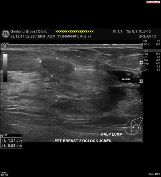
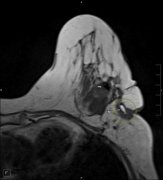
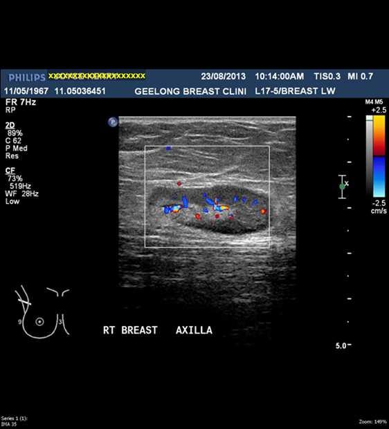
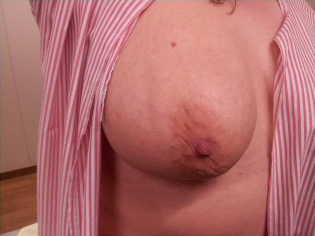
1 SIGNIFICANT OTHERS Miscellaneous Benign Breast Conditions Epworth HealthCare 1 FAT NECROSIS TRAUMATIC Cell rupture Seat-Belt injury Blunt trauma Iatrogenic injury Surgery, Flaps, Radiotherapy Pathology Single droplet of Triglyceride per adipocyte Extra-cellular oil after cell rupture causes chronic inflammation and granuloma formation Hormone Sensitive Lipase - high in adipocytes, low in macrophages Presentation Palpable lump - irregular, firm, producing distortion or dimpling History of trauma, often months 2 years before Epworth HealthCare 2 1
2 2
3 Epworth HealthCare 6 3

4 Epworth HealthCare 7 Epworth HealthCare 8 4

5 Epworth HealthCare 9 Epworth HealthCare 10 5
6 6

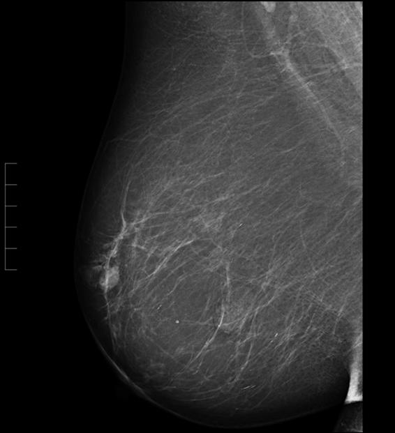
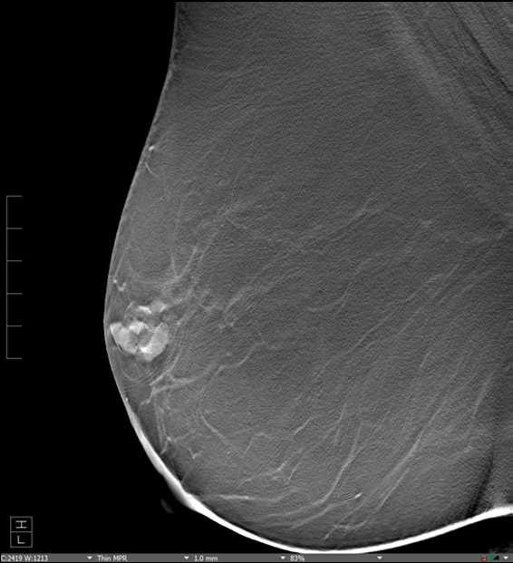
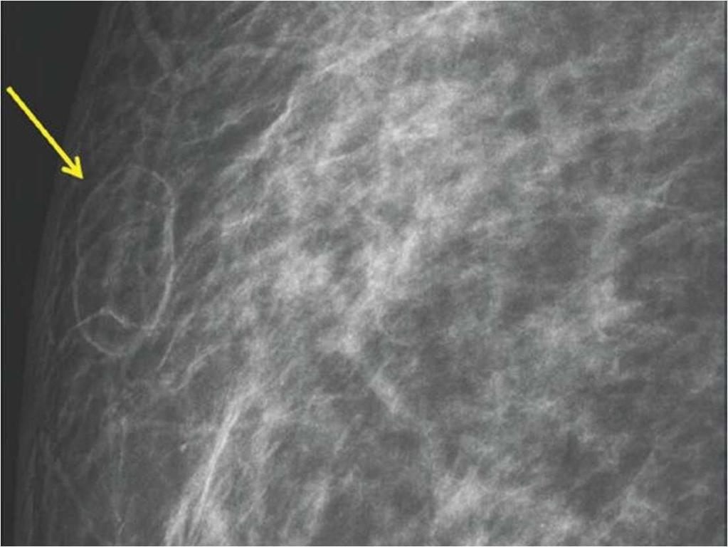
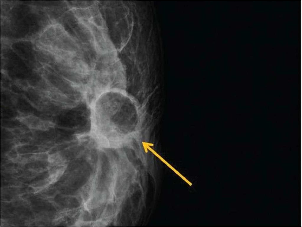
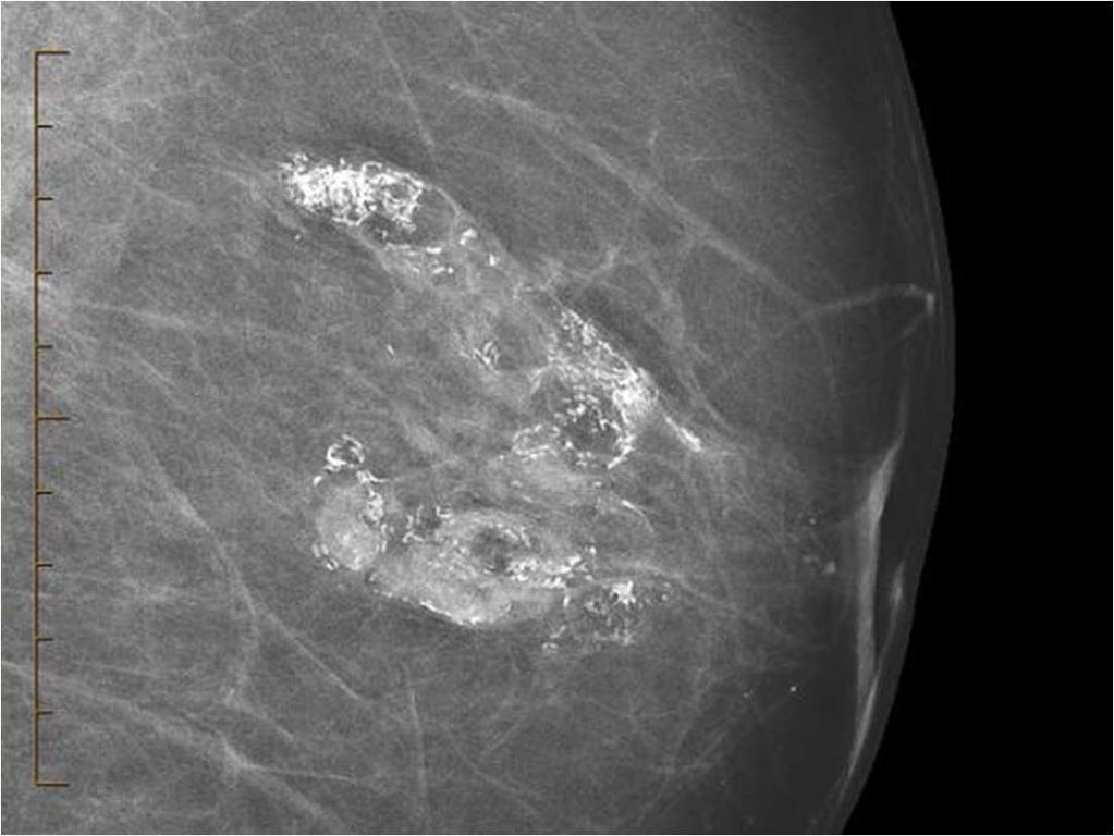
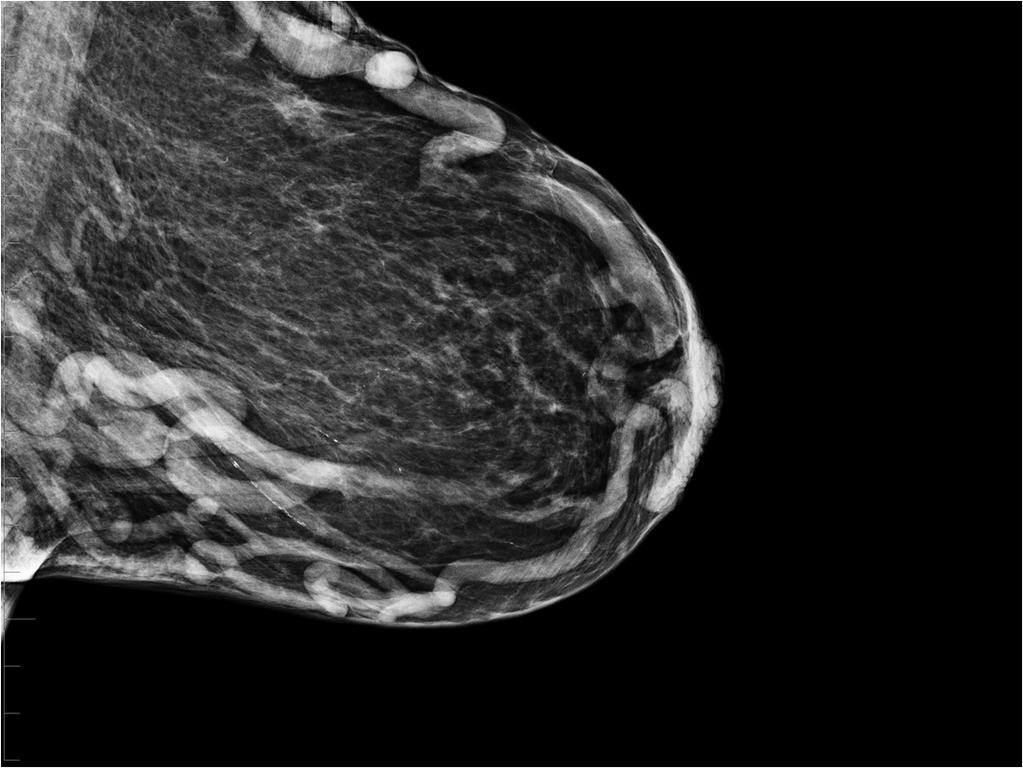
7 Epworth HealthCare 13 FAT NECROSIS Imaging findings Early - haematoma a superficial mass on mammography Tomo shows lobulation with well-defined margins echogenic mass with cystic spaces on U/S Later radiolucent, well-defined cyst progressive fibrosis and calcification Ultrasound - Echogenic band or mass in subcutaneous tissues Cysts with echogenic calcified wall and acoustic shadowing Diagnosis History and Imaging Biopsy is it necessary, will it make it worse? Consider FNA Management Reassurance? Aspirate oil cysts Epworth HealthCare 14 7

8 IATROGENIC INJURY After simple to complex surgery Around margins of flaps Late radiotherapy effect Risk factors are smoking, BMI >30, radiotherapy and ischaemia time Peak incidence is 2-3 years post-op Epworth HealthCare 15 8
9 9
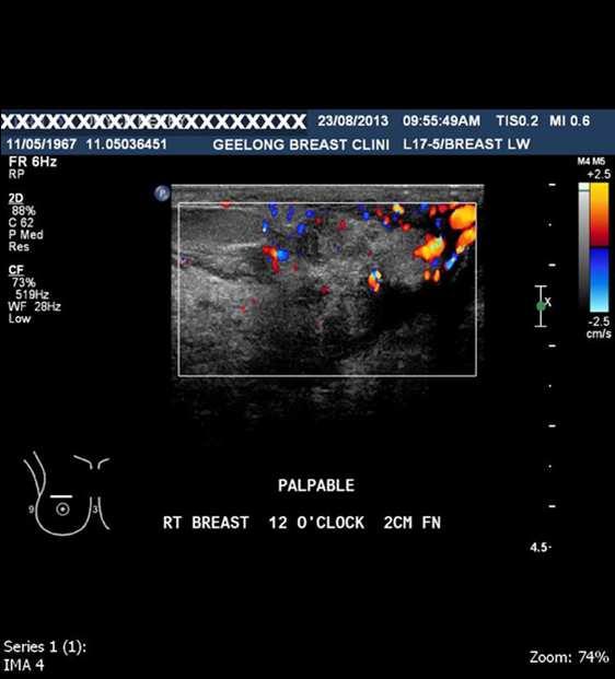
10 IATROGENIC INJURY After simple to complex surgery Around margins of flaps Late radiotherapy effect Risk factors are smoking, BMI >30, radiotherapy and ischaemia time Peak incidence is 2-3 years post-op Fat Grafting Adipose-derived stem cells Ischaemic fat Traumatic acquisition/delivery and cell rupture Results in increased vascularity and fat volume or fibrosis and calcification? Epworth HealthCare 19 IDIOPATHIC GRANULOMATOUS MASTITIS Presentation Palpable breast mass, often tender and peripheral DD inflammatory cancer, Lobular cancer Imaging findings Mammogram - Unilateral focal or regional asymmetry Ultrasound hypoechoic mass(es) with indistinct or irregular margins, increased vascularity, sinus tracts MRI ill-defined masses and non-mass enhancement, intense parenchymal enhancement Epworth HealthCare 20 10
11 11
12 12
13 IDIOPATHIC GRANULOMATOUS MASTITIS Diagnosis of exclusion - Core Biopsy and Cultures Exclude: TB, Sarcoid, Fungal infection Periductal Mastitis, Fat necrosis, Vaccination Management Medical antibiotics steroids, methotrexate Surgical excision ( consider ducts) BUT Wound infection, delayed healing, recurrence Followup For wound infection and recurrence Epworth HealthCare 25 MONDOR S DISEASE Presentation Dull pain associated with an elongated mass History of nothing Or recent direct trauma (males) or surgery Or vigorous arm exercise Epworth HealthCare 26 13

14 Epworth HealthCare 27 Epworth HealthCare 28 14


15 Epworth HealthCare 29 Epworth HealthCare 15

16 Epworth HealthCare 31 Epworth HealthCare 32 16

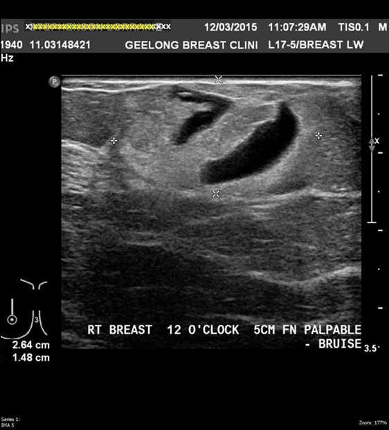
17 Epworth HealthCare MONDOR S DISEASE Pathology Palpable tender cord just beneath the skin of the breast Bowstring Imaging findings Mammography shows a tubular structure U/S shows a superficial vessel without flow +/- intraluminal thrombus Diagnosis Clinical +/- imaging Management Analgaesia? divide under LA Follow-up There is an incidence of cancer diagnosis in the next 2 years Anatomy Usually the Thoraco-Epigastric vein, sometimes Lateral Thoracic or Superior Epigastric Involves the epigastric plexus to the inguinal vessels Epworth HealthCare

18 PASH Pseudo Angiomatous Stromal Hyperplasia 1986 Presentation Breast mass palpable or on screening Often painful or tender 1/3 Pre-menopausal women Pathology Hormone-dependent collagenous expansion of the stroma Benign proliferation of myofibroblasts which are PR positive CD34 positive DD angiosarcoma, phyllodes tumour Imaging findings Round or Oval circumscribed mass, DD Fibroadenoma Diagnosis Core biopsy or excision Management Excise or observe, depending on the size and FH Epworth HealthCare
19 Epworth HealthCare 37 19
20 20

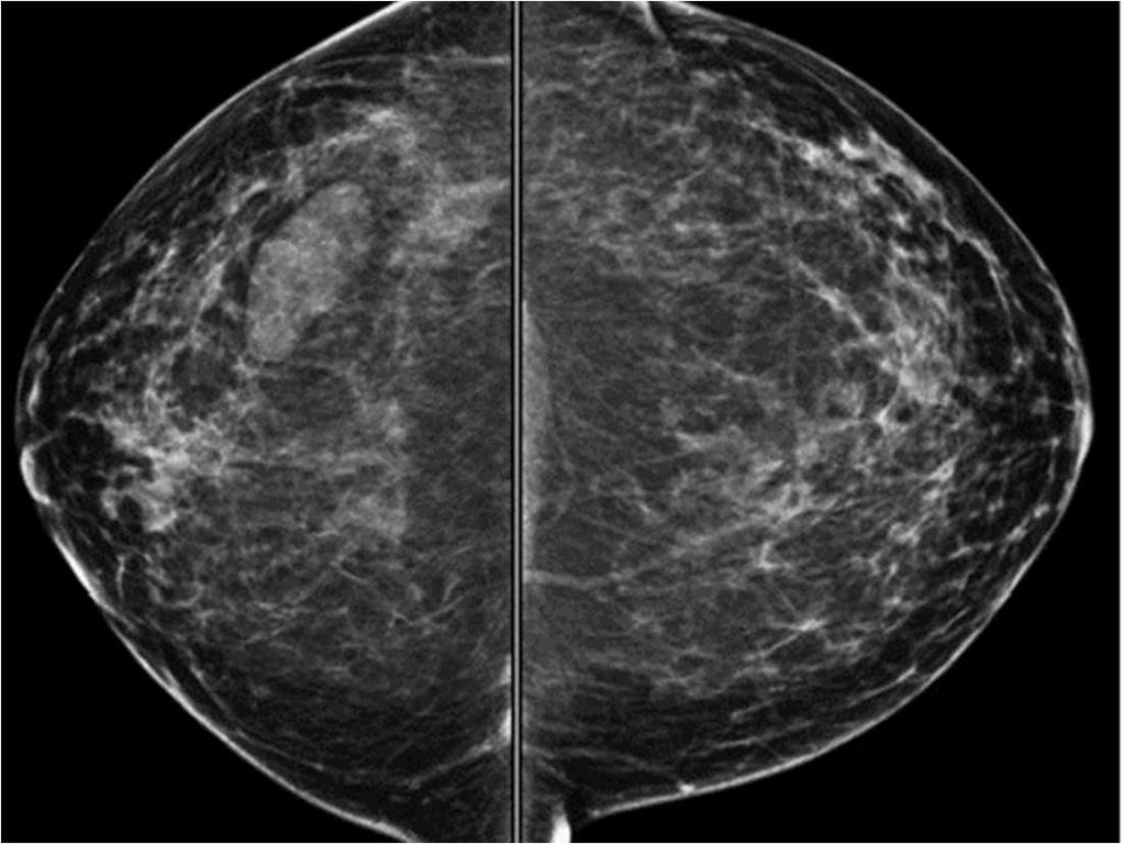
21 DIABETIC MASTOPATHY Sclerosing Lymphocytic Lobulitis Presentation Premenopausal diabetics Usually a large hard painless breast mass Imaging findings Mammogram - ill-defined masses/densities Ultrasound - irregular hypoechoic masses, marked posterior shadowing MRI - variable Epworth HealthCare 41 Epworth HealthCare 42 21
22 22
23 23

24 DIABETIC MASTOPATHY Pathology Prominent keloidal fibrosis and lymphocytic infiltrate DD Extra-nodal MALT lympho`ma Diagnosis Core biopsy Management Followup Sclerosing Lymphocytic Lobulitis - conservative - none Epworth HealthCare 48 24
 Atypia in 10% Presentation Children and adolescents, but mean age 20 Firm, mobile Lump, 4 cm, suggesting Fibroadenoma FH breast cancer 1/4 Imaging findings Ultrasound - small masses with")
25 JUVENILE PAPILLOMATOSIS Swiss cheese disease Pathology Usually a single cystic mass in UOQ Papillary epithelial hyperplasia, numerous cysts and dilated ducts with dense stroma (No papillomas!) Atypia in 10% Presentation Children and adolescents, but mean age 20 Firm, mobile Lump, 4 cm, suggesting Fibroadenoma FH breast cancer 1/4 Imaging findings Ultrasound - small masses with heterogenous echotexture Management/Diagnosis surgical excision Followup Yes - Note - significant subsequent cancer risk if FH and bilateral or recurrent Epworth HealthCare 49 Epworth HealthCare 50 25

26 Epworth HealthCare 51 26
University of Washington Radiology Review Course: Strange and Specific Diagnoses. Case #1
 University of Washington Radiology Review Course: Strange and Specific Diagnoses Katherine E. Dee, MD Seattle Breast Center Via Radiology 2014 Case #1 37 year old presents with bilateral palpable lumps.
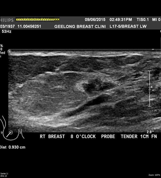
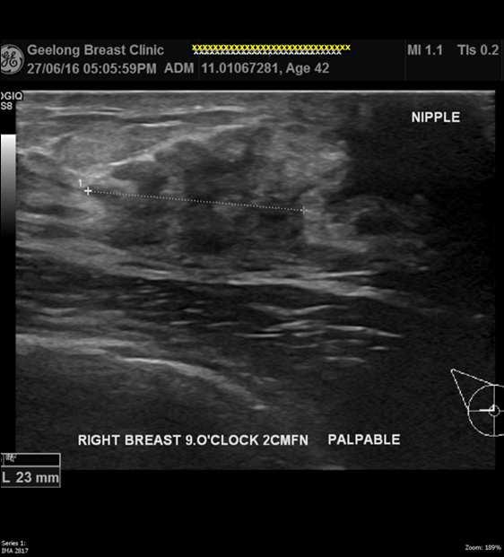
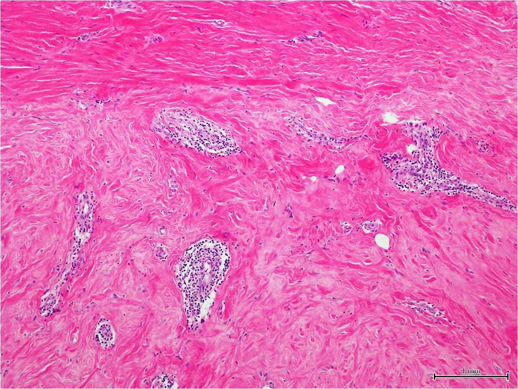
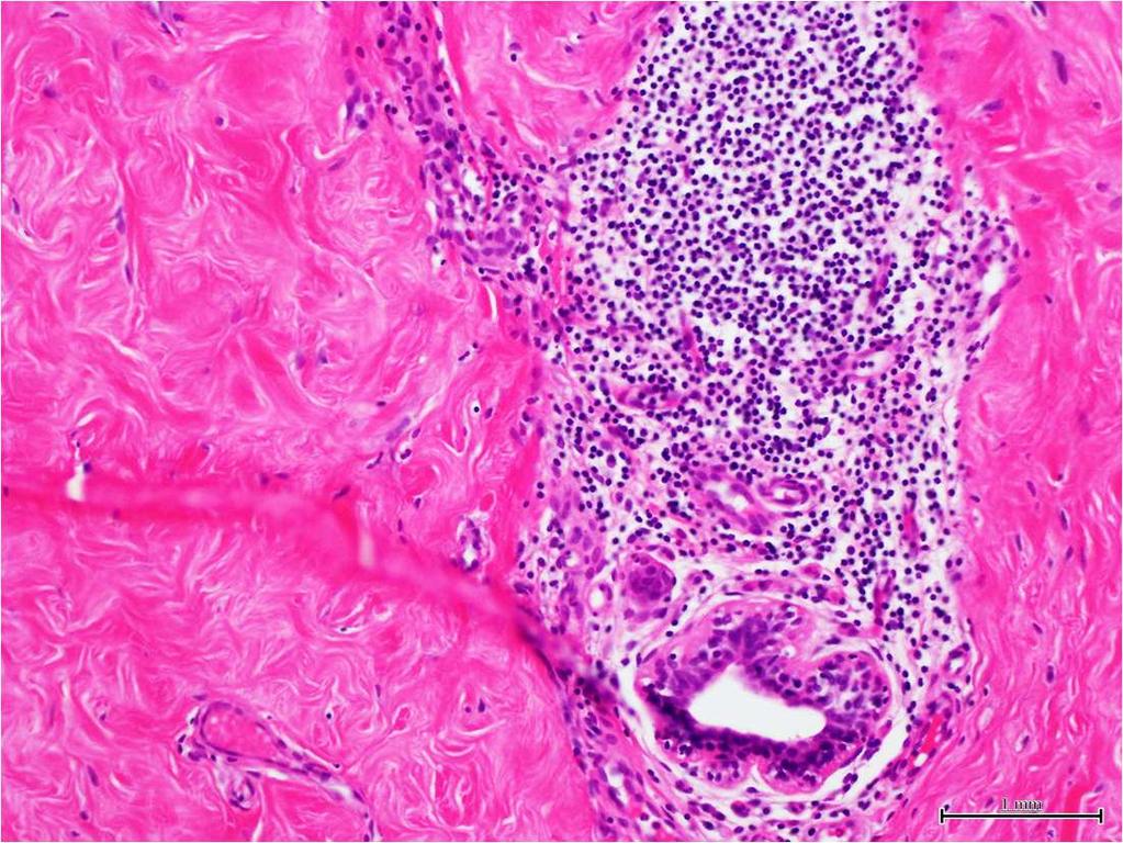
Case year old Chinese female. Radiological echo-distortion in the right breast at o clock. Core biopsy of the o clock lesion.
 Case 3 64 year old Chinese female. Radiological echo-distortion in the right breast at 10-12 o clock. Core biopsy of the 11-12 o clock lesion. Division of Pathology Courtesty of Dr Lester Leong ill-defined,
Diseases of the breast (1 of 2)
") Diseases of the breast (1 of 2) Introduction A histology introduction Normal ducts and lobules of the breast are lined by two layers of cells a layer of luminal cells overlying a second layer of myoepithelial
Diseases of the breast (1 of 2) Introduction A histology introduction Normal ducts and lobules of the breast are lined by two layers of cells a layer of luminal cells overlying a second layer of myoepithelial
Lesion Imaging Characteristics Mass, Favoring Benign Circumscribed Margins Intramammary Lymph Node
 Lesion Imaging Characteristics Mass, Favoring Benign Circumscribed Margins Intramammary Lymph Node Oil Cyst Mass, Intermediate Concern Microlobulated Margins Obscured Margins Mass, Favoring Malignant Indistinct
DISORDERS OF THE BREAST Dated. FIBROADENOSIS Other common names: mastitis, fibrocystic disease, cystic mammary dysplasia.
 DISORDERS OF THE BREAST Dated BENIGN BREAST DISORDERS (Essential Surg 2 nd Ed, pp 540) FIBROADENOSIS Other common names: mastitis, fibrocystic disease, cystic mammary dysplasia. Fibroadenosis is the distortion
Abid Irshad, MD Director Breast Imaging. Medical University of South Carolina Charleston
 Abid Irshad, MD Director Breast Imaging Medical University of South Carolina Charleston Cases Financial disclosure: I or my family have no financial interest related to the material discussed in this presentation
Benign, Reactive and Inflammatory Lesions of the Breast
 Benign, Reactive and Inflammatory Lesions of the Breast Marilin Rosa, MD Associate Member Section Head of Breast Pathology Department of Anatomic Pathology Program Director, Breast Pathology Fellowship
LYMPHATIC DRAINAGE AXILLARY (MOSTLY) INTERNAL MAMMARY SUPRACLAVICULAR
 INTERNAL MAMMARY SUPRACLAVICULAR") BREAST LYMPHATIC DRAINAGE AXILLARY (MOSTLY) INTERNAL MAMMARY SUPRACLAVICULAR HISTOLOGY LOBE: (10 in whole breast) LOBULE: (many per lobe) ACINUS/I, aka ALVEOLUS/I: (many per lobule) DUCT(S): INTRA- or
BREAST LYMPHATIC DRAINAGE AXILLARY (MOSTLY) INTERNAL MAMMARY SUPRACLAVICULAR HISTOLOGY LOBE: (10 in whole breast) LOBULE: (many per lobe) ACINUS/I, aka ALVEOLUS/I: (many per lobule) DUCT(S): INTRA- or
BREAST PATHOLOGY. Fibrocystic Changes
 BREAST PATHOLOGY Lesions of the breast are very common, and they present as palpable, sometimes painful, nodules or masses. Most of these lesions are benign. Breast cancer is the 2 nd most common cause
PRINCIPLES OF BREAST SURGERY & COMPLICATIONS
 PRINCIPLES OF BREAST SURGERY & COMPLICATIONS Adam Cichowitz The Royal Melbourne Hospital ANATOMY Lies in subcutaneous tissue Base: midline to midaxillary line, 2nd to 6th rib Overlies pec major, serratus
Imaging in breast cancer. Mammography and Ultrasound Donya Farrokh.MD Radiologist Mashhad University of Medical Since
 Imaging in breast cancer Mammography and Ultrasound Donya Farrokh.MD Radiologist Mashhad University of Medical Since A mammogram report is a key component of the breast cancer diagnostic process. A mammogram
Breast pathology. 2nd Department of Pathology Semmelweis University
 Breast pathology 2nd Department of Pathology Semmelweis University Breast pathology - Summary - Benign lesions - Acute mastitis - Plasma cell mastitis / duct ectasia - Fat necrosis - Fibrocystic change/
Criteria of Malignancy. Evaluation Score
 30 5 Diagnostic Criteria Criteria of Malignancy Table 5.2 lists criteria in contrast-enhancing MR mammography that strongly indicate the presence of malignancy or are unspecific. Unifactorial evaluation
Imaging the Symptomatic Patient. Avice M.O Connell MD,FACR,FSBI Professor of Imaging Sciences Director, Women s Imaging University of Rochester
 Imaging the Symptomatic Patient Avice M.O Connell MD,FACR,FSBI Professor of Imaging Sciences Director, Women s Imaging University of Rochester The four most common symptoms Mass Pain Discharge Infection
Leonard M. Glassman MD
 BI-RADS The New BI-RADS Leonard M. Glassman MD FACR Former Chief of Breast Imaging American Institute for Radiologic Pathology Washington Radiology Associates, PC Breast Imaging Reporting and Data System
CPC 4 Breast Cancer. Rochelle Harwood, a 35 year old sales assistant, presents to her GP because she has noticed a painless lump in her left breast.
 CPC 4 Breast Cancer Rochelle Harwood, a 35 year old sales assistant, presents to her GP because she has noticed a painless lump in her left breast. 1. What are the most likely diagnoses of this lump? Fibroadenoma
Vesalius SCALpel : Benign breast disease (see also: breast folios)
") Vesalius SCALpel : Benign breast disease (see also: breast folios) Breast cancer risk Imaging Pain non-proliferative: only fibroadenoma may be associated with a slight risk of cancer proliferative: moderate
Vesalius SCALpel : Benign breast disease (see also: breast folios) Breast cancer risk Imaging Pain non-proliferative: only fibroadenoma may be associated with a slight risk of cancer proliferative: moderate
Ultrasound of the Breast BASICS FOR THE ORDERING CLINICIAN
 Ultrasound of the Breast BASICS FOR THE ORDERING CLINICIAN Breast Ultrasound Anatomy Skin Breast Parenchyma Pectoralis Fascia Pectoralis Breast Ultrasound Anatomy Indications for Breast Ultrasound Palpable
FIBROEPITHELIAL LESIONS
 DEFINITIONS FIBROEPITHELIAL LESIONS Suzanne Moore FIBROADENOMA- A discrete benign tumour showing evidence of connective tissue and epithelial proliferation- WHO Fibrous stromal element of these tumours
CLINICAL SIGNIFICANCE OF BENIGN EPITHELIAL CHANGES
 Papillomas. Papillomas are composed of multiple branching fibrovascular cores, each having a connective tissue axis lined by luminal and myoepithelial cells ( Fig. 23-11 ). Growth occurs within a dilated
Granulomatous mastitis: Radio-pathologic correlation and management
 Granulomatous mastitis: Radio-pathologic correlation and management Poster No.: C-1418 Congress: ECR 2014 Type: Educational Exhibit Authors: S. E. Song, B. K. Seo, K. R. Cho, O. H. Woo, Y.-S. Kim ; 1 1
Disclaimer no conflict of interest
 Disclaimer no conflict of interest Benign Breast Disease Alison Hayes FRACS Content Clinical assessment of the breast Triple assessment Focal nodularity Breast pain Cysts Infection Nipple discharge Gynaecomastia
Mousa. Israa Ayed. Abdullah AlZibdeh. 0 P a g e
 1 Mousa Israa Ayed Abdullah AlZibdeh 0 P a g e Breast pathology The basic histological units of the breast are called lobules, which are composed of glandular epithelial cells (luminal cells) resting on
BREAST PATHOLOGY MCQS
 BREAST PATHOLOGY MCQS 1) :The most important factor in breast enlargement during pregnancy is A. stromal edema B. secretion of chorionic gonadotropin C. glandular hyperplasia D. proliferation of stroma
Amammography report is a key component of the breast
 Review Article Writing a Mammography Report Amammography report is a key component of the breast cancer diagnostic process. Although mammographic findings were not clearly differentiated between benign
Armed Forces Institute of Pathology.
 Armed Forces Institute of Pathology www.radpath.com Armed Forces Institute of Pathology Breast Disease www.radpath.org Armed Forces Institute of Pathology Interpretation of Breast MRI Leonard M. Glassman
ACRIN 6666 IM Additional Evaluation: Additional Views/Targeted US
 Additional Evaluation: Additional Views/Targeted US For revised or corrected form check box and fax to 215-717-0936. Instructions: The form is completed based on recommendations (from ID form) for additional
PAAF vs Core Biopsy en Lesiones Mamarias Case #1
 5/19/2014 PAAF vs Core Biopsy en Lesiones Mamarias Case #1 Fine Needle Aspiration Cytology of Breast: Correlation with Needle Core Biopsy 64-year-old woman Mass in breast Syed Hoda, MD CD31 Post-Radiation
UNC Breast Imaging Division July 2018
 UNC Breast Imaging Division July 2018 This module is to educate residents on ACR BI-RADS Atlas 2 nd edition Ultrasound Sonomammographic anatomy Sonographic features most associated with malignancy Clinical
Table of Contents: Foreword Preface Acknowledgementsi Dedication
 Table of Contents: Foreword Preface Acknowledgementsi Dedication Chapter 1 Problems of concept and nomenclature of benign disorders of the breast The source of the problem History The present and the future
Treatment options for the precancerous Atypical Breast lesions. Prof. YOUNG-JIN SUH The Catholic University of Korea
 Treatment options for the precancerous Atypical Breast lesions Prof. YOUNG-JIN SUH The Catholic University of Korea Not so benign lesions? Imaging abnormalities(10% recall) lead to diagnostic evaluation,
Index. C Calcifications fat necrosis 1, 61 fat necrosis 4, 69 nipple/peri-areolar involvement 1, 165
 A ADH. See Atypical ductal hyperplasia (ADH) American College of Radiology (ACR), BI-RADS background parenchymal enhancement, 8, 9, 81, 82 fibroglandular tissue guidelines, 6 American Joint Committee on
Breast Disease: What PCPs Need to Know. Eunice Cho MD FACS
 Breast Disease: What PCPs Need to Know Eunice Cho MD FACS New Breast Cancer Screening Guideline for women with average risk Every other year AGE 40 AGE 45 AGE 55 AGE 55 + Talk with your doctor about when
The radiologic workup of a palpable breast mass
 Imaging in Practice CME CREDIT EDUCTIONL OJECTIVE: The reader will consider which breast masses require further workup and which imaging study is most appropriate Lauren Stein, MD Imaging Institute, Cleveland
Breast Evaluation & Management Guidelines
 Breast Evaluation & Management Guidelines Pamela L. Kurtzhals, M.D. F.A.C.S. Head, Dept. of General Surgery Scripps Clinic, La Jolla Objective Review screening & diagnostic guidelines Focused patient complaints
Mimickers of breast malignancy
 Mimickers of breast malignancy Poster No.: C-0477 Congress: ECR 2010 Type: Educational Exhibit Topic: Breast Authors: S. H. Park, H.-Y. Choi; Incheon/KR Keywords: mimickers, breast malignancy, ultrasound
Fat Necrosis: A Grand Imposter
 Fat Necrosis: A Grand Imposter Poster No.: C-0751 Congress: ECR 2015 Type: Educational Exhibit Authors: L. C. Flores Salinas, Y. A. Ramirez Galvan, A. Garza Báez, C. M. Ferrara Chapa; Monterrey/MX Keywords:
Pathology & Presentation of Benign Breast Disease Zdenek Dubrava - February 2006
 Pathology & Presentation of Benign Breast Disease Zdenek Dubrava - February 2006 Presentation Lump Pain Nipple Discharge Breast Shape/Size Richard J Santen, Robert Mansel. The New England Journal of Medicine.
Ultrasonography. Methods. Brief Description. Indications. Device-related Prerequisites. Technical Requirements. Evaluation Criteria
 1 Ultrasonography Brief Description Imaging modality using sound waves Tissue-specific wave reflection. Indications Evaluation of palpable breast nodules Evaluation of clinically occult mammographic findings
Breast Pathology. Breast Development
 Breast Pathology Lecturer: Hanina Hibshoosh, M.D. Reading: Kumar, Cotran, Robbins, Basic Pathology, 6th Edition, pages 623-635 Breast Development 5th week - thickening of the epidermis - milk line 5th
COMMON BENIGN DISORDERS AND DISEASES OF THE BREAST
 COMMON BENIGN DISORDERS AND DISEASES OF THE BREAST Aberrations of Normal Development and Involution (ANDI). The basic principles underlying the aberrations of normal development and involution (ANDI) classification
Diagnosis of Pseudoangiomatous Stromal Hyperplasia of the Breast: Ultrasonography Findings and Different Biopsy Methods
 Yonsei Med J 49(5):757-764, 2008 DOI 10.3349/ymj.2008.49.5.757 Diagnosis of Pseudoangiomatous Stromal Hyperplasia of the Breast: Ultrasonography Findings and Different Biopsy Methods Yoon Jung Choi, 1
BI-RADS Update. Martha B. Mainiero, MD, FACR, FSBI Brown University Rhode Island Hospital
 BI-RADS Update Martha B. Mainiero, MD, FACR, FSBI Brown University Rhode Island Hospital No Disclosures BI-RADS History 1980s Quality Issues ACR Accreditation BI-RADS 1994 2003 4 th Edition MRI, US January
Radiologic and pathologic correlation of non-mass like breast lesions on US and MRI: Benign, high risk, versus malignant
 Radiologic and pathologic correlation of non-mass like breast lesions on US and MRI: Benign, high risk, versus malignant Poster No.: C-1161 Congress: ECR 2013 Type: Educational Exhibit Authors: J. Kwak,
Case study 1. Rie Horii, M.D., Ph.D. Division of Pathology Cancer Institute Hospital, Japanese Foundation for Cancer Research
 NCCN/JCCNB Seminar in Japan April 15, 2012 Case study 1 Rie Horii, M.D., Ph.D. Division of Pathology Cancer Institute Hospital, Japanese Foundation for Cancer Research Present illness: A 50y.o.premenopausal
Imaging of giant breast masses with pathological correlation
 P i c t o r i a l E s s a y Singapore Med J 2004 Vol 45(3) : 132 Imaging of giant breast masses with pathological correlation M Muttarak, B Chaiwun ABSTRACT Ultrasonography (US) and mammography are the
Contents. Basic Ultrasound Principles and Terminology. Ultrasound Nodule Characteristics
 Contents Basic Ultrasound Principles and Terminology Basic Ultrasound Principles... 1 Ultrasound System... 2 Linear Transducer for Superficial Images and Ultrasound-Guided FNA... 3 Scanning Planes... 4
MEDICAL IMAGING AND BREAST DISEASE HOW CAN WE HELP YOU?
 MEDICAL IMAGING AND BREAST DISEASE HOW CAN WE HELP YOU? Barbara M. Preston, M.D. SCREENING MAMMOGRAPHY AVERAGE RISK PATIENTS KAISER RECOMMENDATION: ALL WOMEN (INCLUDING TRANSGENDER FEMALES) Every 1-21
Diabetic Mastopathy in Long Standing Type I Diabetic Mellitus; A Case Report
 CASE REPORT J Surg Ultrasound 2017;4:25-29 JSU Journal of Surgical Ultrasound Diabetic Mastopathy in Long Standing Type I Diabetic Mellitus; A Case Report Ra Mi Kim, Hunkyoung Lee 1, Heeboong Park 2 Kangnam
Classification System
 Classification System A graduate of the Breast Oncology training program should be able to care for all aspects of disease and/or provide comprehensive management. When referring to a discipline of training
Radiological Appearances of Male Breast Disease
 Radiological Appearances of Male Breast Disease Poster No.: C-1916 Congress: ECR 2017 Type: Educational Exhibit Authors: A. VALDIVIELSO, A. Guma Martinez, R. Ortega Martinez, L. 1 1 1 2 1 1 1 Perez tapia,
Non-mass Enhancement on Breast MRI. Aditi A. Desai, MD Margaret Ann Mays, MD
 Non-mass Enhancement on Breast MRI Aditi A. Desai, MD Margaret Ann Mays, MD Breast MRI Important screening and diagnostic tool, given its high sensitivity for breast cancer detection Breast MRI - Indications
TOTALS 30. Preliminary analysis of NNDEQA 004 (May 2013 TSL workshops) 1) 70 year old female with focal irregularity in the right breast.
 1) 70 year old female with focal irregularity in the right breast.") -- Preliminary analysis of NNDEQA 00 (May 0 TSL workshops) ) 70 year old female with focal irregularity in the right breast. Fat necrosis (with organising thrombus/foreign body granuloma/surgical site
-- Preliminary analysis of NNDEQA 00 (May 0 TSL workshops) ) 70 year old female with focal irregularity in the right breast. Fat necrosis (with organising thrombus/foreign body granuloma/surgical site
BREAST SURGERY PROGRESS TEST Name:
 General Surgery Residency Program Excellent surgeons BREAST SURGERY PROGRESS TEST Name: Choose the BEST answer for the following questions. 1. All of the following factors are associated with an increased
Gynecomastia and Its Mimics: Not All Male Breast Lesions are Benign
 Gynecomastia and Its Mimics: Not All Male Breast Lesions are Benign Poster No.: C-0139 Congress: ECR 2014 Type: Educational Exhibit Authors: S. A. Choudhery, P. Gupta, S. Foshee, F. Garcia-Morales, G.
Journal of Medical Imaging and Radiation Oncology
 Journal of Medical Imaging and Radiation Oncology 60 (2016) 506 513 MEDICAL IMAGING PICTORIAL ESSAY Malignant hyperechoic breast lesions at ultrasound: A pictorial essay Stephen Tiang, 1 Cecily Metcalf,
Breast Cancer Screening and Treatment Mrs Belinda Scott Breast Surgeon Breast Associates Auckland
 Breast Cancer Screening and Treatment 2009 Mrs Belinda Scott Breast Surgeon Breast Associates Auckland BREAST CANCER THE PROBLEM 1.1 million women per year 410,000 deaths each year Increasing incidence
Fat necrosis Benign breast conditions information
 Fat necrosis This leaflet tells you about fat necrosis. It explains what fat necrosis is, how it s diagnosed and what will happen if it needs to be followed up or treated. Benign breast conditions information
Benign Breast Disease. David Anderson, MD Assistant Professor of Clinical Surgery
 Benign Breast Disease David Anderson, MD Assistant Professor of Clinical Surgery Overview Nipple Discharge Breast infection Breast Pain Gynecomastia Fibroepithelial lesions High Risk Lesions-Papilloma,
Sonographic and Mammographic Features of Phyllodes Tumours of the Breast: Correlation with Histological Grade
 Sonographic and Mammographic Features of Phyllodes Tumours of the Breast: Correlation with Histological Grade Poster No.: C-0046 Congress: ECR 2014 Type: Authors: Keywords: DOI: Scientific Exhibit C. Y.
Breast Imaging Lexicon
 9//201 200 BI RADS th Edition 201 BI RADS th Edition Breast Imaging Lexicon Mammographic Pathology and Assessment Categories Deborah Thames, R.T.(R)(M)(QM) The Advanced Health Education Center Nonmember:
OPTO-ACOUSTIC BREAST IMAGING
 OPTO-ACOUSTIC BREAST IMAGING A Novel Fusion of Functional and Morphologic Imaging Reni S. Butler, MD A. Thomas Stavros, MD F. Lee Tucker, MD Michael J. Ulissey, MD PURPOSE 1. Explain opto-acoustic (OA)
Common Breast Problems: Breast Pain
 Common Breast Problems: Breast Pain Breast pain is the most common symptom that brings women to their physician. In general, there are two common presentations of breast pain: cyclic and noncyclic. Breast
Diabetic mastopathy: spectrum of radiological findings.
 Diabetic mastopathy: spectrum of radiological findings. Poster No.: C-1519 Congress: ECR 2014 Type: Educational Exhibit Authors: V. Navarro-Aguilar, G. Montoliu, R. M. Viguer Benavent, M. Á. Sánchez Fuster,
Papillary lesions of the breast - Imaging findings and diagnostic challenges
 Papillary lesions of the breast - Imaging findings and diagnostic challenges Poster No.: R-0146 Congress: RANZCR-AOCR 2012 Type: Educational Exhibit Authors: P. Jagmohan, F. J. Pool Keywords: Breast, Mammography,
THE MALE BREAST CARCINOMA: EARLY DETECTION HOPE. Author (s) Supreethi Kohli a, Pragya Garg b
 Supreethi Kohli a, Pragya Garg b") Case Report ABSTRACT - Male breast cancer is exceptionally rare and accounts for less than 0.25% of male malignancies and approximately 0.5-1% of all breast cancer (both genders). Mammography of the male
Case Report ABSTRACT - Male breast cancer is exceptionally rare and accounts for less than 0.25% of male malignancies and approximately 0.5-1% of all breast cancer (both genders). Mammography of the male
Mammographic imaging of nonpalpable breast lesions. Malai Muttarak, MD Department of Radiology Chiang Mai University Chiang Mai, Thailand
 Mammographic imaging of nonpalpable breast lesions Malai Muttarak, MD Department of Radiology Chiang Mai University Chiang Mai, Thailand Introduction Contents Mammographic signs of nonpalpable breast cancer
Cystic Masses of the Breast
 Residents Section Pattern of the Month Eisenberg ystic Masses of the reast Residents Section Pattern of the Month Residents inradiology Neely Hines 1 Priscilla J. Slanetz Ronald L. Eisenberg Hines N, Slanetz
BREAST IMAGING and NEW IMAGING MODALITIES- A Surgeons view
 BREAST IMAGING and NEW IMAGING MODALITIES- A Surgeons view DR CHANTEL THORNTON SPECIALIST BREAST CANCER SURGEON BMSc (hons) MBBS (hons) FRACS Epworth Hospital, Richmond- Agora Centre for Women s Health
NAME/ AGE/57 SEX/Female AREA/Australia Visit 1: 5/8/2011. Right Breast Cancer Infiltrating Ductal Carcinoma, no specific type, Grade 1 to 2.
 Breast Cancer-CAM Rx NAME/ AGE/57 SEX/Female AREA/Australia Visit 1: 5/8/2011 Case History Discussion Right Breast Cancer Infiltrating Ductal Carcinoma, no specific type, Grade 1 to 2. Patient visited
Breast Pathology in Men: Radiologic-Pathologic Correlation
 Breast Pathology in Men: Radiologic-Pathologic Correlation Poster No.: C-0243 Congress: ECR 2012 Type: Scientific Exhibit Authors: G. Garrido; Málaga/ES Keywords: Breast, Ultrasound, Mammography, Biopsy,
Ana Sofia Preto 19/06/2013
 Ana Sofia Preto 19/06/2013 Understanding the underlying pathophysiologic processes leading to the various types of calcifications Description and illustration of the several types of calcifications, according
8/31/2016 HIDING IN PLAIN SITE, ARCHITECTURAL DISTORTIONS AND BREAST ASYMMETRIES ARCHITECTURAL DISTORTIONS ARCHITECTURAL DISTORTIONS
 HIDING IN PLAIN SITE, ARCHITECTURAL DISTORTIONS AND BREAST ASYMMETRIES DEBORAH THAMES R.T. (R)(M)(QM) ARCHITECTURAL DISTORTIONS Definition is disruption of the natural flow of breast pattern towards the
Cairo/EG, Khartoum/SD, London/UK Biological effects, Diagnostic procedure, Ultrasound, Mammography, Breast /ecr2015/C-0107
 Role of sono-mammography in the evaluation of clinically palapble breast masses during pregnancy & lactation with differentaition between true patholgical & false physiological lobular hyperlpasia.sudanese
Breast masses in children and adolescents
 Breast masses in children and adolescents Marilyn J. Siegel, MD, and Ellen Chung, MD I n the pediatric and adolescent population, breast masses are nearly always benign, self-limited and managed conservatively,
Jeddah Breast Cancer Pilot Screening Program, KSA
 Jeddah Breast Cancer Pilot Screening Program, KSA 7 th Global Summit on Cancer Therapy, Oct 5-7, 2015 Dubai, Crown Plaza Hotel Muna Baslaim, MD Consultant Surgeon Head of the Breast Unit, King Fahd General
Diagnostic Dilemmas of Breast Imaging
 Diagnostic Dilemmas of Breast Imaging Common Causes of Error in Breast Cancer Detection By: Jason Cord, M.D. Mammography: Initial Imaging The standard for detection of breast cancer Screening mammography
Margaret Ann Mays, M.D. Clinical Instructor in Radiology Vanderbilt University Medical Center Tennessee Radiological Society February 25, 2017
 Breast Imaging in Adolescents and Young Women Margaret Ann Mays, M.D. Clinical Instructor in Radiology Vanderbilt University Medical Center Tennessee Radiological Society February 25, 2017 Disclosures
RADIOLOGIC EVALUATION OF BREAST CANCER
 RADIOLOGIC EVALUATION OF BREAST CANCER Orsolya Farkas, Gabriella Bodrogi and Gábor Szalai Department of Radiology, Pécs University Orsifarkas@yahoo.com Complex evaluation of the breast Patient history
Breast Health. Learning Objectives. Breast Anatomy. Poll Question. Breast Anatomy
 Learning Objectives Describe breast anatomy to a patient Breast Health Answer questions about causes of breast pain and masses Explain breast cancer screening/diagnostic modalities Appropriately triage
Inflammatory and Reactive Lesions of the Breast
 Inflammatory and Reactive Lesions of the Breast Laura C. Collins, M.D. Vice Chair of Anatomic Pathology Professor of Pathology Beth Israel Deaconess Medical Center and Harvard Medical School Boston, MA
HISTOPATHOLOGICAL EVALUATION OF BENIGN PROLIFERATIVE BREAST LESIONS
 7 ORIGINAL ARTICLE HISTOPATHOLOGICAL EVALUATION OF BENIGN PROLIFERATIVE BREAST LESIONS DR. VIBHUTI H. CHIHLA*, DR. N N. JAGRIT **, DR. JAYASHREE M. SHAH*** *3 rd year Pathology Resident, **Associate Professor,
Pseudoangiomatous Stromal Hyperplasia: Diagnosis, Treatment and Follow-Up; Description of a Case-Series
 18 The Open Breast Cancer Journal, 2011, 3, 18-23 Open Access Pseudoangiomatous Stromal Hyperplasia: Diagnosis, Treatment and Follow-Up; Description of a Case-Series W.A. Donk *, R.J. Oostenbroek, R.K.
Final analysis of NNDEQA 004 (May 2013 TSL workshops) 1) 70 year old female with focal irregularity in the right breast.
 1) 70 year old female with focal irregularity in the right breast.") Final analysis of NNDEQA 00 (May 0 TSL workshops) ) 70 year old female with focal irregularity in the right breast. CONSUL TANTS Fat necrosis (with organising thrombus/foreign body granuloma/surgical site
Final analysis of NNDEQA 00 (May 0 TSL workshops) ) 70 year old female with focal irregularity in the right breast. CONSUL TANTS Fat necrosis (with organising thrombus/foreign body granuloma/surgical site
04/10/2018. Intraductal Papillary Neoplasms Of Breast INTRADUCTAL PAPILLOMA
 Intraductal Papillary Neoplasms Of Breast Savitri Krishnamurthy MD Professor of Pathology Deputy Division Head The University of Texas MD Anderson Cancer Center 25 th Annual Seminar in Pathology Pittsburgh,
Pitfalls and Limitations of Breast MRI. Susan Orel Roth, MD Professor of Radiology University of Pennsylvania
 Pitfalls and Limitations of Breast MRI Susan Orel Roth, MD Professor of Radiology University of Pennsylvania Objectives Review the etiologies of false negative breast MRI examinations Discuss the limitations
1 NORMAL HISTOLOGY AND METAPLASIAS
 1 NORMAL HISTOLOGY AND METAPLASIAS, MD Anatomy and Histology 1 Metaplasias 2 ANATOMY AND HISTOLOGY The female breast is composed of a branching duct system, which begins at the nipple with the major lactiferous
Posterior Acoustic Shadowing in Benign Breast Lesions
 Image Presentation Posterior coustic Shadowing in enign reast Lesions Sonographic-Pathologic Correlation Susan P. Weinstein, MD, Emily F. Conant, MD, Carolyn Mies, MD, Geza cs MD, PhD, Steven Lee, MD,
A GP S APPROACH TO BREAST LUMPS AND SYMPTOMS DR KK CHEUNG GPGC WORKSHOP
 A GP S APPROACH TO BREAST LUMPS AND SYMPTOMS DR KK CHEUNG GPGC WORKSHOP 18.08.18 HAVE A SYSTEM HISTORY EXAMINATION INVESTIGATION FOLLOW UP BREAST SYMPTOMS HISTORY DON T FORGET SKIN CHANGES AND NIPPLE CHANGES
AMSER Case of the Month: November 2018
 AMSER Case of the Month: November 2018 42 year old with right breast mass Rina Kiyota Petek Lake Erie College of Osteopathic Medicine, OMS-III Kossivi Dantey, MD Bibianna Klepchick, MD Matthew Hartman,
Imaging Approaches to Diagnosis and Management of Common Ductal Abnormalities
 Imaging Approaches to Diagnosis and Management of Common Ductal Abnormalities Poster No.: C-1891 Congress: ECR 2013 Type: Educational Exhibit Authors: M. Abdelkafi 1, H. Fourati 1, E. DAOUD 2, W. Feki
Breast imaging of benign fat containing lesions
 Breast imaging of benign fat containing lesions Poster No.: C-1870 Congress: ECR 2017 Type: Educational Exhibit Authors: R. Aouini, I. Megdiche, D. Ben Hammadi, N. BEN MAMI, I. Attia, R. Neila, A. Zidi;
Breast Update Therese Cusick MS MD FACS
 Breast Update 2017 Therese Cusick MS MD FACS Conflict of Interest Disclosure Nothing to disclose Sources Adapted from SESAP- Surgical Education and Self-Assessment program American College of Surgeons
Case Scenario 1 History and Physical 3/15/13 Imaging Pathology
 Case Scenario 1 History and Physical 3/15/13 The patient is an 84 year old white female who presented with an abnormal mammogram. The patient has a five year history of refractory anemia with ringed sideroblasts
Case Report Rare Presentation of Pseudoangiomatous Stromal Hyperplasia: A Case Report
 IBIMA Publishing International Journal of Case Reports in Medicine http://www.ibimapublishing.com/journals/ijcrm/ijcrm.html Vol. 2013 (2013), Article ID 331549, 5 pages DOI: 10.5171/2013.331549 Case Report
M Wani, M Khan, N Ul Gani, S Sangeen, B Singh, M Shafi, A Bilal, S Umer
 ISPUB.COM The Internet Journal of Surgery Volume 12 Number 1 Juvenile Fibroadenoma With Fibroadenomatoid Hyperplasia M Wani, M Khan, N Ul Gani, S Sangeen, B Singh, M Shafi, A Bilal, S Umer Citation M Wani,
Breast lumps to refer or not to refer? Simon Cawthorn Breast Specialist
 Breast lumps to refer or not to refer? Simon Cawthorn Breast Specialist Learning objectives Know the indications to refer urgently Who to reassure and review How to reassure patients with non-urgent symptoms
3/27/2017. Disclosure of Relevant Financial Relationships. Papilloma???
 Management of Papillary Lesions Diagnosed at Rad Path Concordant Core Biopsy (CNB) Disclosure of Relevant Financial Relationships USCAP requires that all planners (Education Committee) in a position to