PETER PAZMANY CATHOLIC UNIVERSITY Consortium members SEMMELWEIS UNIVERSITY, DIALOG CAMPUS PUBLISHER
|
|
|
- Louisa Daniel
- 5 years ago
- Views:
Transcription


1 PETER PAZMANY SEMMELWEIS CATHOLIC UNIVERSITY UNIVERSITY Development of Complex Curricula for Molecular Bionics and Infobionics Programs within a consortial* framework** Consortium leader PETER PAZMANY CATHOLIC UNIVERSITY Consortium members SEMMELWEIS UNIVERSITY, DIALOG CAMPUS PUBLISHER The Project has been realised with the support of the European Union and has been co-financed by the European Social Fund *** **Molekuláris bionika és Infobionika Szakok tananyagának komplex fejlesztése konzorciumi keretben ***A projekt az Európai Unió támogatásával, az Európai Szociális Alap társfinanszírozásával valósul meg. 1
2 Peter Pazmany Catholic University Faculty of Information Technology INTRODUCTION TO BIOPHYSICS (Bevezetés a biofizikába) BIOLOGICAL MEMBRANES (Biológiai membránok) GYÖRFFY DÁNIEL, ZÁVODSZKY PÉTER 2
3 Introduction to biophysics: Biological membranes Introduction Cells and compartments of cells are surrounded by membranes These membranes separate the interior of cells from the extracellular space but they also connect them to each other Material, energy and information can get across the membrane Biological membranes are mainly built of phospholipids 12/11/10. 3
4 Phospholipids Lipids are a group of organic compounds Their characteristic property is the hydrophobicity of at least a part of the molecule Some of them are amphiphilic, that is they contain a polar group in addition to the apolar bulk Phospholipids are a class of lipids which contains a phosphate group 4
5 The fundamental building blocks of phospholipids are: Glycerol Fatty acids Phosphate group Other groups 5
6 Glycerol Glycerol is an alcohol containing three hydroxyl groups It can form three ester bonds by these three hydroxyls Glycerol has a large solubility in water In itself glycerol is toxic for the human organism 6

7 2D structure of glycerol 7

8 8

9 3D structure of glycerol 9
10 Fatty acids Fatty acids are carboxylic acids with a long linear aliphatic chain They can be unsaturated or saturated according to whether or not they contain double covalent bonds between carbon atoms Fatty acids in an organism usually have an even number of carbon atoms 10

11 Some characteristic fatty acids 11
12 12

13 13
14 14
15 15
16 16

17 17
18 18
19 Glycerol and fatty acids can be connected by ester bonds forming glycerides Common fats and oils are triglycerides, that is they consist of a triester of glycerol with three fatty acids This glyceride structure is the basis of more complex lipids like phospholipids 19

20 Triglyceride 20

21 21
22 Phospholipids Most phospholipids consist of a glycerol (but for example sphingomyelin contains sphingosine) esterized by two fatty acids and a phosphoric acid, and some organic group connected to the phosphate Phospholipids are the main lipid building blocks of biological membranes The basic compound of phospholipids is phosphatidic acid 22
23 Phospholipids Diacylglyceride phospholipids Phosphosphingolipids Phosphatidic acid Ceramide phosphorylcholine Phosphatidylethanolamine Phosphatidylcholine Ceramide phosphorylethanolamine Ceramide phosphorylglycerol Phosphatidylserine Phosphoinositides 23

24 Phosphatidic acid 24

25 25

26 Other phospholipids 26
27 27

28 28

29 29

30 30
31 31

32 32
33 33

34 34
35 35
36 Membranes also contain other lipids such as steroids, for example cholesterol 36

37 Cholesterol 37

38 38
39 Lipid bilayer The lipids building up biological membranes have a polar head and an apolar tail In consequence of this property, they are arranged in a bilayer structure such that the apolar tails point toward the centre of the membrane and the polar heads point outside 39

40 Schematic lipid bilayer 40

41 Atomic picture of one layer of a membrane 41
42 Membrane proteins Biological membranes also contain proteins Some of them span across the membrane; these are called transmembrane proteins Some of them are attached to the membrane permanently but do not necessarily span across the membrane; these are called integral membrane proteins 42
43 In contrast, some of them are only attached temporarily to the membrane; these are called peripheral membrane proteins Such membrane proteins are, for example, the regulatory subunits of receptors and ion channels 43

44 A 7TM receptor 44

45 A Na+ channel 45
46 Membrane potential and the Nernst equation If the charges on the two sides of a membrane are not equal, an electric potential gradient appears For ions that can pass freely across the membrane, an equilibrium develops The Nernst equation describes the relationship between the potential gradient and the concentrations of the ion inside and outside the cell 46
47 The Nernst equation Let us consider a cell bounded by a semipermeable membrane For an ion A which can freely pass across the membrane, let [A]in denote its concentration inside and [A]out outside the cell, respectively 47

48 The membrane is permeable for the ion A 48
49 An ion is subject to two opposing forces, one caused by the concentration gradient and the other caused by the Coulomb forces between electric charges The chemical potential of a substance gives us the free energy of one mole of it For one mole of A the free energy is 0 μ=μ +RT ln [ A ] where μ0 is the standard chemical potential and [A] is the molar concentration of A 49
50 The free energy difference between the two sides of the membrane for one mole of A is Δμ=μ inside μ outside Since the standard chemical potential is independent of the ion concentration and depends only on the properties of the substance, it is the same on both sides of the membrane 0 0 μinside =μoutside 50
51 Taking into account the preceding equation, the free energy increase when one mole of A is taken into the cell is Δμ=RT ln [ A ]inside RT ln [ A ]outside [ A ]inside Δμ=RT ln [ A ]outside 51
52 Now let us consider the effect of electric charges The free energy of one mole of ions due to an electric potential is G electric =zfφ where z is the valence of the ion, for example +1 in the case of sodium and -1 in the case of chloride, F is the Faraday constant and Φ is the electric potential 52
53 The free energy increase when one mole of ions is moved from outside to inside the cell due to the electrical potential is: ΔG electric =G electric in G electric out that is ΔG electric =zfδφ where ΔΦ=Φ inside Φ outside is the membrane potential 53
54 Based on these equations, the total free energy increase when one mole of A is taken inside the cell is [ A ]inside Δμ+ΔG electric =RT ln +zfδφ [ A ]outside At equilibrium, ions moving into the cell neither gain nor lose energy so this free energy increase is zero [ A ]inside RT ln +zfδφ= 0 [ A ]outside 54
55 After rearrangement, we get for the membrane potential that [ A ]outside RT ΔΦ= ln zf [ A ]inside This is the Nernst equation which is one of the most important relationships in electrochemistry 55
56 When deriving the Nernst equation, we assumed that an equilibrium occurs for the ion species being considered This has the consequence that Nernst equation is only valid for ions for which an equilibrium can develop, i.e. ions that can freely pass across the membrane Usually, such permeability is made possible by ion channels as we can see next 56
 57")
57 Walther Nernst ( ) 57
58 Problem 1 In leukocytes, the value of the membrane potential is ΔΦ= 90mV What is the ratio of the concentrations of potassium ions inside and outside the cell at temperature T= 37 C =310K? 58
59 As there are potassium channels in the membrane and thus, an equilibrium develops, the Nernst equation is valid for potassium in this example thus [ K ]outside RT ΔΦ= ln zf [ K ]inside 59
60 After rearrangement, we obtain that [ K ]outside ΔΦzF / RT =e = [ K ]inside So the potassium concentration inside the cell is almost thirty times greater than outside 60
61 If in the cytoplasm the potassium concentration is [ K ]inside =140mM what is the potassium concentration outside? [ K ]outside = mM=4.8mM 61
62 Transport across the membrane Biological membranes such as the plasma membrane, do not only separate the interior of the cell from the environment but also connects them This connection occurs by the transport of substances into or out of the cell Different mechanisms exist for different types of substances 62
63 Transport processes can be grouped according to whether they require or not energy to occur If for a given substance, a concentration gradient exist between the two sides of a membrane, the transport along the concentration gradient is energetically favorable (passive transport), but in the opposite direction, additional energy must be invested (active transport) 63
64 Since membranes have a hydrophobic layer which also must be passed, transport processes can be grouped by the chemical characteristic of the substance, namely, according to whether it can pass this hydrophobic layer Substances with hydrophobic character and small gas molecules such as O2 or CO2 can diffuse across the membrane Larger molecules or charged particles can pass only with the help of special proteins 64
65 The transport of multiple substances can be coupled so that one of them moves along the concentration gradient and releases energy which can be used for the transport of the other substance moving against the concentration gradient If the two substances move in the same direction we speak about symport and in the opposite case we speak about antiport If only one substances gets transported we say that uniport 65
66 Molecular diffusion This is the simplest way to pass a membrane Only hydrophobic or small molecules are capable of this type of transport For example O2, H2O and CO2 can be transported by diffusion 66
67 Channels Channels are transmembrane proteins which make passive transport possible Channels exist for ions and some other substances, e.g. aquaporins transport water Some channels are very specific for one type of ion so a sodium ion cannot pass through a potassium channel and vice versa 67


68 Potassium channel 68
69 Aquaporin 69
70 Carriers While channels form a permanently open tube across the membrane, carriers are always closed on one end In the case of voltage-gated carriers, a given membrane potential causes the transporter to open In the case of ligand-gated carriers, the binding of some ligand molecule triggers the opening 70
71 Passive transport through carriers An example for a carrier through which passive transport occurs is the glucose transporter This transporter is open towards one direction If a glucose molecule approaches the carrier from that direction it can bind to the protein When the protein switches over, i.e. becomes open towards the other direction, the glucose is released from the carrier 71
72 Another possibility is that glucose is released before the carrier switches On the side where the concentration of glucose is higher, more glucose molecules will bind to the carrier and fewer will be released before the switch Thus, macroscopically, a glucose flow will be observed from the side with higher to that with the lower concentration 72
73 Active transport through carriers Active transport processes are often distinguished by whether they utilize directly the energy of ATP hydrolysis (primary transport) or utilize the flow of another substance along its concentration gradient (secondary transport) 73
74 Secondary transport by symporters and antiporters An example of a symporter is the glucose-sodium symporter where a glucose molecule is carried into the cell with the simultaneous transport of sodium The sodium-calcium exchanger is an antiporter where the inward flow of sodium is utilized to pump calcium out of the cell 74

75 Sodium-glucose symporter 75
76 Ca2+ binding domain of a sodium-calcium antiporter 76
77 Primary transport by pumps Through primary transport, the energy of ATP hydrolysis is directly utilized to move ions against an electrochemical gradient A well-known and rather important pump is the Na +/K+ATPase which has a role in the adjustment of the action potential during impulse transmission in the nervous system Both sodium and potassium are transported against their electrochemical gradients 77

78 Na+/K+-ATPase 78
79 Two membrane machines We will present the structure and function of two membrane machines of particular interest First of them is bacteriorhodopsin which is responsible for capturing the energy of light to produce a proton gradient in some halobacteria Bacteriorhodopsin is an integral membrane protein complex It is found in the purple membrane 79
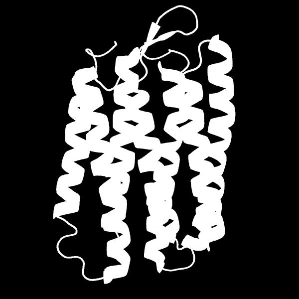
80 Bacteriorhodopsin 80

81 81
82 In addition to the protein itself, bacteriorhodopsin contains one retinal molecule which is a chromophore Retinal has a role in the vision of animals 82

83 Retinal 83

84 84
85 Operation of bacteriorhodopsin The retinal changes its conformation upon absorption of light This conformational change of retinal causes a change in the conformation of the protein This conformational change allows the protein to function as a proton pump and release a proton to the extracellular site 85

86 The photoisomerization cycle of retinal The cis-retinal binds to the protein as a Schiff-base This complex can absorb a photon and the cisretinal isomerizes to the all-trans form 86
87 Following the isomerization of the chromophore, a proton transfer occurs from the Schiff base to the Asp-85 residue of the protein Another aspartate residue of the protein, Asp-96, provides a proton to reprotonate the Schiff base A reprotonation of Asp-96 occurs from the cytoplasm of the cell 87

88 A reverse isomerization of retinal from the cis to the all-trans form while both Asp-85 and Asp-96 are protonated 88
89 Finally, Asp-85 releases the proton outside the cell Repeating this cycle many times, a proton gradient is established which can be used for ATP synthesis 89

90 Absorption spectrum of bacteriorhodopsin 90
91 Bacterial flagellum Some bacteria have a special organ called flagellum which takes part in the motion of the organism The flagellum consists of a basal body located in the cell wall of the bacterium and a filament attached to it The filament is built of a protein called flagellin The filament if attached to the basal body through a protein called hook which ensures a quasi rectangular junction 91
92 Gram-negative bacteria such as Escherichia coli have a basal body consisting of four protein rings L ring is located in the outer membrane P ring is located in the peptidoglycan layer M ring is located in the plasma membrane and S ring is attached to the plasma membrane 92

93 Flagellum of a Gram-negative bacterium 93

94 Flagellin 94
95 Operation of bacterial flagellum Several models have been proposed to explain the flagellar rotation These models agree that it is not the ATP but an electrochemical potential gradient through the plasma membrane which drives the rotation In E. coli a flow of protons into the cell generates a rotational motion 95
96 Proteins functioning in rotation Several proteins have been shown to participate in the generation of rotary motion MotA and MotB are membrane proteins being localized in the plasma membrane that form the stator and serve as channels to conduct ions across the membrane FligF, FliG, FliM and FliN proteins form the rotor itself 96


97 Schematic structure of the rotary complex 97


98 98

99 Proton acceptor groups are on the MS-ring equally spaced along the ring 99
100 A general scheme for energy conversion A general model is shown based on models describing the rotary motion generation process There are several disagreements between models with respect to the details of the process 100
101 General scheme of rotary motion generation Around the MS-ring, proton acceptor groups are located equally spaced From outside, a proton arrives to the nearest acceptor group through a channel formed by the MotA-MotB complex The proton binds to the acceptor group, lending it a positive charge There is another channel with a negatively charged group at its entrance 101
102 This negatively charged group attracts the positively charged group, causing the rotation of the whole ring Due to this rotation, the next acceptor group turns to the channel through which another proton arrives This cycle repeats several times, generating the rotation of the ring and thus the rotation of the flagellar filament as well 102
103 A proton arrives from outside through a channel formed by the MotA-MotB complex 103

104 The proton binds to the proton acceptor group 104
105 MS-ring rotation driven by the attraction of electric charges occurs 105
Membrane Structure and Membrane Transport of Small Molecules. Assist. Prof. Pinar Tulay Faculty of Medicine
 Membrane Structure and Membrane Transport of Small Molecules Assist. Prof. Pinar Tulay Faculty of Medicine Introduction Cell membranes define compartments of different compositions. Membranes are composed
Membranes. Chapter 5
 Membranes Chapter 5 Membrane Structure The fluid mosaic model of membrane structure contends that membranes consist of: -phospholipids arranged in a bilayer -globular proteins inserted in the lipid bilayer
Membrane Structure. Membrane Structure. Membrane Structure. Membranes
 Membrane Structure Membranes Chapter 5 The fluid mosaic model of membrane structure contends that membranes consist of: -phospholipids arranged in a bilayer -globular proteins inserted in the lipid bilayer
Membranes. Chapter 5. Membrane Structure
 Membranes Chapter 5 Membrane Structure Lipid Bilayer model: - double phospholipid layer - Gorter & Grendel: 1925 Fluid Mosaic model: consist of -phospholipids arranged in a bilayer -globular proteins inserted
Lecture Series 5 Cellular Membranes
 Lecture Series 5 Cellular Membranes Cellular Membranes A. Membrane Composition and Structure B. Animal Cell Adhesion C. Passive Processes of Membrane Transport D. Active Transport E. Endocytosis and Exocytosis
A. Membrane Composition and Structure. B. Animal Cell Adhesion. C. Passive Processes of Membrane Transport. D. Active Transport
 Cellular Membranes A. Membrane Composition and Structure Lecture Series 5 Cellular Membranes B. Animal Cell Adhesion E. Endocytosis and Exocytosis A. Membrane Composition and Structure The Fluid Mosaic
Biomembranes structure and function. B. Balen
 Biomembranes structure and function B. Balen All cells are surrounded by membranes Selective barrier But also important for: 1. Compartmentalization 2. Biochemical activities 3. Transport of dissolved
PETER PAZMANY CATHOLIC UNIVERSITY Consortium members SEMMELWEIS UNIVERSITY, DIALOG CAMPUS PUBLISHER
 PETER PAZMANY CATHOLIC UNIVERSITY SEMMELWEIS UNIVERSITY Development of Complex Curricula for Molecular Bionics and Infobionics Programs within a consortial* framework** Consortium leader PETER PAZMANY
Cell Membranes and Signaling
 5 Cell Membranes and Signaling Concept 5.1 Biological Membranes Have a Common Structure and Are Fluid A membrane s structure and functions are determined by its constituents: lipids, proteins, and carbohydrates.
PETER PAZMANY CATHOLIC UNIVERSITY Consortium members SEMMELWEIS UNIVERSITY, DIALOG CAMPUS PUBLISHER
 PETER PAZMANY CATHOLIC UNIVERSITY SEMMELWEIS UNIVERSITY Development of Complex Curricula for Molecular Bionics and Infobionics Programs within a consortial* framework** Consortium leader PETER PAZMANY
membranes cellular membranes basic structure basic structure chapter ECM CYTOPLASM
 membranes chapter 11-1 1 cellular membranes 3 compartmentalization intracellular compartments 1. receiving info membrane receptors recognition and interaction with other cells. import and export of molecules
Cell Membranes Valencia college
 6 Cell Membranes Valencia college 6 Cell Membranes Chapter objectives: The Structure of a Biological Membrane The Plasma Membrane Involved in Cell Adhesion and Recognition Passive Processes of Membrane
Membrane Structure. Membrane Structure. Membranes. Chapter 5
 Membranes Chapter 5 Membrane Structure The fluid mosaic model of membrane structure contends that membranes consist of: -phospholipids arranged in a bilayer -globular proteins inserted in the lipid bilayer
Membrane transport. Pharmacy Dr. Szilvia Barkó
 Membrane transport Pharmacy 04.10.2017 Dr. Szilvia Barkó Cell Membranes Cell Membrane Functions Protection Communication Import and and export of molecules Movement of the cell General Structure A lipid
Lecture Series 4 Cellular Membranes
 Lecture Series 4 Cellular Membranes Reading Assignments Read Chapter 11 Membrane Structure Review Chapter 21 pages 709-717 717 (Animal( Cell Adhesion) Review Chapter 12 Membrane Transport Review Chapter
Lecture Series 4 Cellular Membranes. Reading Assignments. Selective and Semi-permeable Barriers
 Lecture Series 4 Cellular Membranes Reading Assignments Read Chapter 11 Membrane Structure Review Chapter 12 Membrane Transport Review Chapter 15 regarding Endocytosis and Exocytosis Read Chapter 20 (Cell
Lipids and Membranes
 Lipids and Membranes Presented by Dr. Mohammad Saadeh The requirements for the Pharmaceutical Biochemistry I Philadelphia University Faculty of pharmacy Biological membranes are composed of lipid bilayers
3.2.3 Transport across cell membranes
 alevelbiology.co.uk 3.2.3 Transport across cell membranes SPECIFICATION The basic structure of all cell membranes, including cell-surface membranes and the membranes around the cell organelles of eukaryotes,
Membrane Structure, Resting membrane potential, Action potential. Biophysics seminar
 Membrane Structure, Resting membrane potential, Action potential Biophysics seminar 09.09.2013. Membrane structure Biological membranes consists of lipids and proteins to bind with non-covalent bond. Phospholipids
Chapter 2 Transport Systems
 Chapter 2 Transport Systems The plasma membrane is a selectively permeable barrier between the cell and the extracellular environment. It permeability properties ensure that essential molecules such as
1. Which of the following statements about passive and primary active transport proteins is FALSE?
 Biological Membranes 1. Which of the following statements about passive and primary active transport proteins is FALSE? A. They are both integral membrane proteins. B. They both show a high degree of selectivity.
Cellular Neurophysiology I Membranes and Ion Channels
 Cellular Neurophysiology I Membranes and Ion Channels Reading: BCP Chapter 3 www.bioelectriclab All living cells maintain an electrical potential (voltage) across their membranes (V m ). Resting Potential
Lecture Series 4 Cellular Membranes
 Lecture Series 4 Cellular Membranes Reading Assignments Read Chapter 11 Membrane Structure Review Chapter 12 Membrane Transport Review Chapter 15 regarding Endocytosis and Exocytosis Read Chapter 20 (Cell
membranes membrane functions basic structure membrane functions chapter 11-12
 membranes chapter - membrane functions Ca + hormone IP H + HO compartmentalization intracellular compartments scaffold for biochemical activities organize enzymes selectively permeable membrane allows
The Cell Membrane & Movement of Materials In & Out of Cells PACKET #11
 1 February 26, The Cell Membrane & Movement of Materials In & Out of Cells PACKET #11 Introduction I 2 Biological membranes are phospholipid bilayers with associated proteins. Current data support a fluid
Membrane Structure and Function
 Membrane Structure and Function Chapter 7 Objectives Define the following terms: amphipathic molecules, aquaporins, diffusion Distinguish between the following pairs or sets of terms: peripheral and integral
Bear: Neuroscience: Exploring the Brain 3e
 Bear: Neuroscience: Exploring the Brain 3e Chapter 03: The Neuronal Membrane at Rest Introduction Action potential in the nervous system Action potential vs. resting potential Slide 1 Slide 2 Cytosolic
Membrane Transport. Anatomy 36 Unit 1
 Membrane Transport Anatomy 36 Unit 1 Membrane Transport Cell membranes are selectively permeable Some solutes can freely diffuse across the membrane Some solutes have to be selectively moved across the
Membrane Structure and Function
 BIOL1040 Page 1 Membrane Structure and Function Friday, 6 March 2015 2:58 PM Cellular Membranes Fluid mosaics of lipids and proteins Phospholipids - abundant Phospholipids are amphipathic molecules (has
Lecture 36: Review of membrane function
 Chem*3560 Lecture 36: Review of membrane function Membrane: Lipid bilayer with embedded or associated proteins. Bilayers: 40-70% neutral phospholipid 10-20% negative phospholipid 10-30% cholesterol 10-30%
The Cell Membrane & Movement of Materials In & Out of Cells PACKET #11
 1 The Cell Membrane & Movement of Materials In & Out of Cells PACKET #11 Introduction I 2 Biological membranes are phospholipid bilayers with associated proteins. Current data support a fluid mosaic model
Chapter 4 Cell Membrane Transport
 Chapter 4 Cell Membrane Transport Plasma Membrane Review o Functions Separate ICF / ECF Allow exchange of materials between ICF / ECF such as obtaining O2 and nutrients and getting rid of waste products
10/28/2013. Double bilayer of lipids with imbedded, dispersed proteins Bilayer consists of phospholipids, cholesterol, and glycolipids
 Structure of a Generalized Cell MEMBRANES Figure 3.1 Plasma Membrane Fluid Mosaic Model Separates intracellular fluids from extracellular fluids Plays a dynamic role in cellular activity Glycocalyx is
The Cell Membrane. Usman Sumo Friend Tambunan Arli Aditya Parikesit. Bioinformatics Group Faculty of Mathematics and Science University of Indonesia
 The Cell Membrane Usman Sumo Friend Tambunan Arli Aditya Parikesit Bioinformatics Group Faculty of Mathematics and Science University of Indonesia Overview Cell membrane separates living cell from nonliving
BIOLOGY 103 Spring 2001 MIDTERM LAB SECTION
 BIOLOGY 103 Spring 2001 MIDTERM NAME KEY LAB SECTION ID# (last four digits of SS#) STUDENT PLEASE READ. Do not put yourself at a disadvantage by revealing the content of this exam to your classmates. Your
Cell Membrane and Transport
 Cell Membrane and Transport 29/06/2015 11:08 AM Describe the Characteristics of the phospholipid Bilayer. The Phospholipid bilayer is made up of a double layer of membrane lipids that have a hydrophobic
The Plasma Membrane - Gateway to the Cell
 The Plasma Membrane - Gateway to the Cell 1 Photograph of a Cell Membrane 2 Cell Membrane The cell membrane is flexible and allows a unicellular organism to move 3 Homeostasis Balanced internal condition
Diffusion across cell membrane
 The Cell Membrane and Cellular Transport Diffusion across cell membrane Cell membrane is the boundary between inside & outside separates cell from its environment Can it be an impenetrable boundary? NO!
The Plasma Membrane - Gateway to the Cell
 The Plasma Membrane - Gateway to the Cell 1 Photograph of a Cell Membrane 2 Cell Membrane The cell membrane is flexible and allows a unicellular organism to move 3 Homeostasis Balanced internal condition
Biology 4410 First Examination Version B
 Biology 4410 Spring 2006 Name First Examination Version B This examination consists of two parts, a multiple-choice section and an essay section. Be sure to put your name on both the mark-sense sheet and
Ch 3 Membrane Transports
 Ch 3 Membrane Transports what's so dynamic about cell membranes? living things get nutrients and energy from the envrionment this is true of the entire organism and each cell this requires transport in/out
In the Name of God, the Most Merciful, the Most Compassionate. Movement of substances across the plasma membrane
 *Quick Revision: In the Name of God, the Most Merciful, the Most Compassionate Movement of substances across the plasma membrane Passive transport (doesn't require metabolic energy) Active transport (requires
Chapters 9 and 10 Lipids and Membranes
 Chapters 9 and 10 Lipids and Membranes Lipids- a class of biological molecules defined by low solubility in water and high solubility in nonpolar solvents. Lipids contain or are derived from fatty acids.
CH 7.2 & 7.4 Biology
 CH 7.2 & 7.4 Biology LABEL THE MEMBRANE Phospholipids Cholesterol Peripheral proteins Integral proteins Cytoskeleton Cytoplasm Extracellular fluid Most of the membrane A phospholipid bi-layer makes up
Ch7: Membrane Structure & Function
 Ch7: Membrane Structure & Function History 1915 RBC membranes studied found proteins and lipids 1935 membrane mostly phospholipids 2 layers 1950 electron microscopes supported bilayer idea (Sandwich model)
Fall Name Student ID
 Name Student ID PART 1: Matching. Match the organelle to its function (11 points) 1.Proton motive force 2. Fluid Mosiac 3. Oxidative Phosphorylation 4. Pyruvate dehydrogenase 5. Electrochemical Force 6.
Draw and label a diagram to show the structure of membranes
 2.4 Membranes 2.4.1 - Draw and label a diagram to show the structure of membranes Phospholipid Bilayer - This is arranged with the hydrophilic phosphate heads facing outwards, and the hydrophobic fatty
The Cell Membrane. Cell membrane separates living cell from nonliving surroundings. Controls traffic in & out of the cell
 The Cell Membrane 1 Overview Cell membrane separates living cell from nonliving surroundings thin barrier = 8nm thick Controls traffic in & out of the cell selectively permeable allows some substances
Lipids and Membranes
 Lipids Lipids are hydrophobic or amphiphilic insoluble in water soluble in organic solvents soluble in lipids Lipids are used as energy storage molecules structural components of membranes protective molecules
PETER PAZMANY CATHOLIC UNIVERSITY Consortium members SEMMELWEIS UNIVERSITY, DIALOG CAMPUS PUBLISHER
 PETER PAZMANY CATHOLIC UNIVERSITY SEMMELWEIS UNIVERSITY Development of Complex Curricula for Molecular Bionics and Infobionics Programs within a consortial* framework** Consortium leader PETER PAZMANY
PETER PAZMANY CATHOLIC UNIVERSITY Consortium members SEMMELWEIS UNIVERSITY, DIALOG CAMPUS PUBLISHER
 SEMMELWEIS UNIVERSITY PETER PAZMANY CATHOLIC UNIVERSITY Development of Complex Curricula for Molecular Bionics and Infobionics Programs within a consortial* framework** Consortium leader PETER PAZMANY
The Cell Membrane AP Biology
 The Cell Membrane AP Biology! 2007-2008 Overview! Cell membrane separates living cell from nonliving surroundings " thin barrier = 8nm thick! Controls traffic in & out of the cell " selectively permeable
Plasma Membrane Function
 Plasma Membrane Function Cells have to maintain homeostasis, they do this by controlling what moves across their membranes Structure Double Layer of phospholipids Head (polar) hydrophiliclikes water -
Chapter 8 Cells and Their Environment
 Chapter Outline Chapter 8 Cells and Their Environment Section 1: Cell Membrane KEY IDEAS > How does the cell membrane help a cell maintain homeostasis? > How does the cell membrane restrict the exchange
The Cell Membrane. Lecture 3a. Overview: Membranes. What is a membrane? Structure of the cell membrane. Fluid Mosaic Model. Membranes and Transport
 Lecture 3a. The Cell Membrane Membranes and Transport Overview: Membranes Structure of cell membranes Functions of cell membranes How things get in and out of cells What is a membrane? Basically, a covering
Transport through membranes
 Transport through membranes Membrane transport refers to solute and solvent transfer across both cell membranes, epithelial and capillary membranes. Biological membranes are composed of phospholipids stabilised
Cell Membranes. Q: What components of the cell membrane are in a mosaic pattern?
 Cell Membranes The cell / plasma membrane is. Selective in that it allows things in and some things out of the cell. Recall that phospholipids have hydrophobic and hydrophilic. The term to describe this
1.14. Passive Transport
 Passive Transport 1.14 Simple Diffusion Cell s are selectively permeable only certain substances are able to pass through them. As mentioned in section 1.2, cell s are largely composed of a phospholipid
Phospholipids. Extracellular fluid. Polar hydrophilic heads. Nonpolar hydrophobic tails. Polar hydrophilic heads. Intracellular fluid (cytosol)
") Module 2C Membranes and Cell Transport All cells are surrounded by a plasma membrane. Eukaryotic cells also contain internal membranes and membrane- bound organelles. In this module, we will examine the
Module 2C Membranes and Cell Transport All cells are surrounded by a plasma membrane. Eukaryotic cells also contain internal membranes and membrane- bound organelles. In this module, we will examine the
Membrane Structure and Function. Selectively permeable membranes are key to the cell's ability to function
 Membrane Structure and Function Selectively permeable membranes are key to the cell's ability to function Amphipathic Molecules Have both hydrophilic and hydrophobic regions Phospholipids have hydrophilic
Membrane structure & function
 Membrane structure & function Multiple Choice Identify the choice that best completes the statement or answers the question. 1. The phospholipid bilayer describes a structure with. a. polar layers on the
AP Biology. Overview. The Cell Membrane. Phospholipids. Phospholipid bilayer. More than lipids. Fatty acid tails. Phosphate group head
 Overview The Cell Membrane Cell separates living cell from nonliving surroundings thin barrier = 8nm thick Controls traffic in & out of the cell selectively permeable allows some substances to cross more
BIOL 158: BIOLOGICAL CHEMISTRY II
 BIOL 158: BIOLOGICAL CHEMISTRY II Lecture 1: Membranes Lecturer: Christopher Larbie, PhD Introduction Introduction Cells and Organelles have membranes Membranes contain lipids, proteins and polysaccharides
Cell Transport & the Cell Membrane
 Cell Transport & the Cell Membrane I. Cell Membrane A. Structure Structure of the cell membrane is referred to as the Fluid Mosaic Model. It is made up of lipids, proteins and carbohydrates. The membrane
Division Ave High School Ms. Foglia AP Biology
 The Cell Membrane Phospholipids Phosphate head hydrophilic Fatty acid tails hydrophobic Arranged as a bilayer Phosphate attracted to water Fatty acid repelled by water 2007-2008 Aaaah, one of those structure
The main biological functions of the many varied types of lipids include: energy storage protection insulation regulation of physiological processes
 Big Idea In the biological sciences, a dehydration synthesis (condensation reaction) is typically defined as a chemical reaction that involves the loss of water from the reacting molecules. This reaction
1. Describe the difference between covalent and ionic bonds. What are the electrons doing?
 Exam 1 Review Bio 212: 1. Describe the difference between covalent and ionic bonds. What are the electrons doing? 2. Label each picture either a Carbohydrate, Protein, Nucleic Acid, or Fats(Lipid). a.
Chapter 7: Membrane Structure and Function
 Chapter 7: Membrane Structure and Function Concept 7.1 Cellular membranes are fluid mosaics of lipids and proteins 1. Phospholipids are amphipathic. Explain what this means. Name Period Amphipathic means
Chem Lecture 11 Molecular Motors Part 2
 Chem 452 - Lecture 11 Molecular Motors Part 2 Question of the Day. How is the movement of bacteria like a bumper car ride? Bacterial flagella are remarkably similar to an electrical motor. 2 Bacterial
Chapter 7: Membrane Structure & Function
 Chapter 7: Membrane Structure & Function 1. Membrane Structure 2. Transport Across Membranes 1. Membrane Structure Chapter Reading pp. 125-129 What are Biological Membranes? Hydrophilic head WATER They
Chapter 7: Membrane Structure & Function. 1. Membrane Structure. What are Biological Membranes? 10/21/2015. Why phospholipids? 1. Membrane Structure
 Chapter 7: Membrane Structure & Function 1. Membrane Structure 2. Transport Across Membranes 1. Membrane Structure Chapter Reading pp. 125-129 What are Biological Membranes? Hydrophilic head WATER They
PETER PAZMANY CATHOLIC UNIVERSITY Consortium members SEMMELWEIS UNIVERSITY, DIALOG CAMPUS PUBLISHER
 PETER PAZMANY CATHOLIC UNIVERSITY SEMMELWEIS UNIVERSITY Development of Complex Curricula for Molecular Bionics and Infobionics Programs within a consortial* framework** Consortium leader PETER PAZMANY
Inorganic compounds: Usually do not contain carbon H 2 O Ca 3 (PO 4 ) 2 NaCl Carbon containing molecules not considered organic: CO 2
 2 NaCl Carbon containing molecules not considered organic: CO 2") Organic Chemistry The study of carbon-containing compounds and their properties. Biochemistry: Made by living things All contain the elements carbon and hydrogen Inorganic: Inorganic compounds: All other
Organic Chemistry The study of carbon-containing compounds and their properties. Biochemistry: Made by living things All contain the elements carbon and hydrogen Inorganic: Inorganic compounds: All other
The Cell Membrane (Ch. 7)
") The Cell Membrane (Ch. 7) Phospholipids Phosphate head hydrophilic Fatty acid tails hydrophobic Arranged as a bilayer Phosphate attracted to water Fatty acid repelled by water Aaaah, one of those structure
The Cell Membrane (Ch. 7) Phospholipids Phosphate head hydrophilic Fatty acid tails hydrophobic Arranged as a bilayer Phosphate attracted to water Fatty acid repelled by water Aaaah, one of those structure
Lipids and Biological Membranes
 Lipids and Biological Membranes Lipids: Found in all living organisms Especially important as components of biological membranes Defined functionally, not structurally, as compounds that are totally or
Gateway to the Cell 11/1/2012. The cell membrane is flexible and allows a unicellular organism to move FLUID MOSAIC MODEL
 Gateway to the Cell The cell membrane is flexible and allows a unicellular organism to move Isolates the cell, yet allows communication with its surroundings fluid mosaics = proteins (and everything else)
Membranes 9/15/2016. Phospholipids. Phospholipid bilayer
 Membranes Phospholipids Type of complex lipid that forms biological membranes. Have a polar hydrophilic head and two nonpolar hydrophobic tails. Amphipathic. This causes the tails to cluster together in
CHAPTER 8 MEMBRANE STUCTURE AND FUNCTION
 CHAPTER 8 MEMBRANE STUCTURE AND FUNCTION Plasma Membrane Plasma membrane is selectively permeable, (allowing some substances to cross more easily than others) PM is flexible bends and changes shape
UNIVERSITY OF MEDICAL SCIENCES, ONDO DEPARTMENT OF PHYSIOLOGY PHS 211 TRANSPORT MECHANISM LECTURER: MR A.O. AKINOLA
 UNIVERSITY OF MEDICAL SCIENCES, ONDO DEPARTMENT OF PHYSIOLOGY PHS 211 TRANSPORT MECHANISM LECTURER: MR A.O. AKINOLA OUTLINE Introduction Basic mechanisms Passive transport Active transport INTRODUCTION
Main Functions maintain homeostasis
 The Cell Membrane Main Functions The main goal is to maintain homeostasis. Regulates materials moving in and out of the cell. Provides a large surface area on which specific chemical reactions can occur.
Chapter 7: Membrane Structure and Function. Key Terms:
 Key Terms: Selectively permeable Fluid mosaic model Amphipathic Phospholipid Bilayer Hydrophilic Hydrophobic Phosphate head Fatty acid tail Davson-Danielli Singer-Nicolson Freeze-Fracture EM Unsaturated
Interactions Between Cells and the Extracellular Environment
 Chapter 6 Interactions Between Cells and the Extracellular Environment Et Extracellular lll environment Includes all parts of the body outside of cells Cells receive nourishment Cells release waste Cells
Membrane Structure and Function
 Chapter 7 Membrane Structure and Function PowerPoint Lecture Presentations for Biology Eighth Edition Neil Campbell and Jane Reece Lectures by Chris Romero, updated by Erin Barley with contributions from
/ The following functional group is a. Aldehyde c. Carboxyl b. Ketone d. Amino
 Section A: Multiple Choice Select the answer that best answers the following questions. Please write your selected choice on the line provided, in addition to circling the answer. /25 1. The following
PETER PAZMANY CATHOLIC UNIVERSITY Consortium members SEMMELWEIS UNIVERSITY, DIALOG CAMPUS PUBLISHER
 PETER PAZMANY CATHOLIC UNIVERSITY SEMMELWEIS UNIVERSITY Development of Complex Curricula for Molecular Bionics and Infobionics Programs within a consortial* framework** Consortium leader PETER PAZMANY
Chapter 5: Cell Membranes and Signaling
 Chapter Review 1. For the diagram below, explain what information you would use to determine which side of the membrane faces the inside of the cell and which side faces the extracellular environment.
Concept 7.1: Cellular membranes are fluid mosaics of lipids and proteins
 Concept 7.1: Cellular membranes are fluid mosaics of lipids and proteins Lipids: Non-polar substances such as fat that contain C, H, O. Phospholipids: Lipid with phosphate group, very abundant in plasma
Consider the structure of the plasma membrane (fig. 8.6)- phospholipid bilayer with peripheral and integral proteins.
- phospholipid bilayer with peripheral and integral proteins.") Topic 8: MEMBRANE TRANSPORT (lectures 11-12) OBJECTIVES: 1. Have a basic appreciation of the chemical characteristics of substances that impact their ability to travel across plasma membranes. 2. Know
Topic 8: MEMBRANE TRANSPORT (lectures 11-12) OBJECTIVES: 1. Have a basic appreciation of the chemical characteristics of substances that impact their ability to travel across plasma membranes. 2. Know
Chapter 4: Cell Membrane Structure and Function
 Chapter 4: Cell Membrane Structure and Function Plasma Membrane: Thin barrier separating inside of cell (cytoplasm) from outside environment Function: 1) Isolate cell s contents from outside environment
Phospholipids. Phosphate head. Fatty acid tails. Arranged as a bilayer. hydrophilic. hydrophobic. Phosphate. Fatty acid. attracted to water
 The Cell Membrane Phospholipids Phosphate head hydrophilic Fatty acid tails hydrophobic Arranged as a bilayer Phosphate attracted to water Fatty acid repelled by water I want you to remember: Structure
Biology 4410 First Examination Version B
 Biology 4410 Spring 2006 Name First Examination Version B This examination consists of two parts, a multiple-choice section and an essay section. Be sure to put your name on both the mark-sense sheet and
Plasma Membranes. Plasma Membranes WJEC GCE BIOLOGY 4.6
 4.6 Repeat Fig 3.20A here Fluid Mosaic Model of the Plasma Membrane Carbohydrate chain Glycoprotein Intrinsic Protein Non-polar hydrophobic fatty acid Phospholipids Appearance of the Cell Membrane Seen
TRANSPORT ACROSS MEMBRANES
 Unit 2: Cells, Membranes and Signaling TRANSPORT ACROSS MEMBRANES Chapter 5 Hillis Textbook TYPES OF TRANSPORT ACROSS THE CELL (PLASMA) MEMBRANE: What do you remember? Complete the chart with what you
Will s Pre-Test. (4) A collection of cells that work together to perform a function is termed a(n): a) Organelle b) Organ c) Cell d) Tissue e) Prison
 A collection of cells that work together to perform a function is termed a(n): a) Organelle b) Organ c) Cell d) Tissue e) Prison") Will s Pre-Test This is a representative of Exam I that you will take Tuesday September 18, 2007. The actual exam will be 50 multiple choice questions. (1) The basic structural and functional unit of the
Will s Pre-Test This is a representative of Exam I that you will take Tuesday September 18, 2007. The actual exam will be 50 multiple choice questions. (1) The basic structural and functional unit of the
Comprehensive and Easy Course Notes for BIOL1040 Exams and Assessment
 Comprehensive and Easy Course Notes for BIOL1040 Exams and Assessment MODULE 1: PRINCIPLES OF CELL FUNCTION Membrane Structure & Function Cellular membranes are fluid mosaics of lipids and proteins Phospholipids
Paper 12: Membrane Biophysics Module 15: Principles of membrane transport, Passive Transport, Diffusion, Fick s law
 Paper 12: Membrane Biophysics Module 15: Principles of membrane transport, Passive Transport, Diffusion, Fick s law LEARNING OBJECTIVES OF MODULE: We would begin this module by considering some general
CWDHS Mr. Winch Grade 12 Biology
 The Cell Membrane Overview Cell separates living cell from nonliving surroundings thin barrier = 8nm thick Controls traffic in & out of the cell selectively permeable allows some substances to cross more
Plasma Membrane Structure and Function
 Plasma Membrane Structure and Function The plasma membrane separates the internal environment of the cell from its surroundings. The plasma membrane is a phospholipid bilayer with embedded proteins. The
NANO 243/CENG 207 Course Use Only
 L9. Drug Permeation Through Biological Barriers May 3, 2018 Lipids Lipid Self-Assemblies 1. Lipid and Lipid Membrane Phospholipid: an amphiphilic molecule with a hydrophilic head and 1~2 hydrophobic tails.
Cell Biology. The Plasma Membrane
 Cell Biology The Plasma Membrane recall Fluid Mosiac Model S.J. Singer Semipermeable membrane fluid portion is double layer of phospholipids (=phospholipid bilayer) mosaic portion is the proteins and carbohydrates