Hearing Inner ear: Cochlea:
|
|
|
- Aubrey Conley
- 5 years ago
- Views:
Transcription
 is the (membrane labyrinth). This membrane structure more or less duplicates the shape of the bony channels.")


1 Hearing Inner ear: The inner ear (labyrinth) is made up of two parts; one within the other, the (bony labyrinth) is a series of channels in the petrous portion of the temporal bone. Inside these channels, surrounded by a fluid called (perilymph) is the (membrane labyrinth). This membrane structure more or less duplicates the shape of the bony channels. It is filled with a fluid called (endolymph), and there is no communication between the space filled with endolymph and those filled with perilymph. Cochlea: The cochlear portion of the labyrinth is a coiled tube which in human is 3.5 mm long and makes 2¼ turns. Throughout its length, the basilar membrane and Reissner`s membrane divided it into three chamber s (scalae). The upper scala vestibuli and the lower scala tympani contain perilymph and communicate with each other at the apex of the cochlea through a small opening called the helicortrema. At the base of the cochlea, the cochlea, the scala vestibuli ends at the oval window, which is closed by the footplate of the stapes. The scala tympani end at the round window, a foramen on the medial wall of the middle ear that is closed by the flexible secondary tympanic membrane. The scala media, the middle cochlear chamber, is continuous with the membranous labyrinth and does not communicate with the other two scala. It contains endolymph. The scala vestibuli and scala media are separated from each other by Reissner`s membrane (also called the vestibular membrane); the scala tympani and scala media are separated from each other by the basilar membrane. On the surface of the basilar membrane lies the organ of Corti, which contains a series of electromechanically sensitive cells, the hair cells. The Reissner`s membrane is so thin and so easily 1


2 moved that it does not obstruct the passage of sound vibrations through fluid from the scala vestibuli into scala media. The basilar membrane contains 20,000 to 30,000 basilar fibers that project from the bony center of the cochlea, the modiolus, toward the outer wall. These fibers are stiff, elastic, reed like structure that are fixed at their basal ends in the central bony structure of the (the modiolus) but not fixed at their distal ends, except that the distal ends are embedded in the loose basilar membrane. Because the fibers are stiff and.الر شة الت تتحرك عند نفخ الهواء ف الهارمونكةharmonica free at one end, they can vibrate like the reeds of a The basilar membrane characterized by: 1. The lengths of the basilar fibers increase progressively beginning at the oval window and going from the base of the cochlea to the apex, increasing from a length of about 0.04 millimeter near the oval and round windows to 0.5 millimeter at the tip of the cochlea (the helicotrema ), a 12-fold increase in length 2. The diameters of the fibers, however, decrease from the oval window to the helicotrema, so their overall stiffness decreases more than 100-fold. As a result, the stiff, short fibers near the oval window of the cochlea vibrate best at a very high frequency, whereas the long, limber fibers near the tip of the cochlea vibrate best at a low frequency. 3. Another feature of the traveling wave is that it travels fast along the initial portion of the basilar membrane but becomes progressively slower as it goes farther into the cochlea. The cause of this difference is the high coefficient of elasticity of the basilar fibers near the oval window and a progressively decreasing coefficient farther along the membrane. Low-frequency resonance occurs near the helicotrema, mainly because of the less stiff fibers and also because of increased loading with extra masses of fluid that must vibrate along the cochlear tubules. The high-frequency resonance of the basilar membrane occurs near the base, where the sound waves enter the cochlea through the oval window. Transmission of sound waves in the cochlea (the traveling wave): When the foot of the stapes moves inward against the oval window and round window must bulge outward because the cochlea is bounded on all sides by bony walls. Therefore, the initial effect of a sound wave entering at the oval window is to cause the basilar membrane at the base of the cochlea to bend in the direction of the round window. However, the elastic tension that is built up in the basilar fibers as they bend toward the round window initiates a fluid wave that travels along the basilar membrane toward the helicortrema. 2


3 Each wave is relatively weak at the outset but becomes strong when it reaches that portion of the basilar membrane that has a natural resonant frequency equal to the respective sound frequency. At this point, the basilar membrane can vibrate back and forth with such ease that energy in the wave is dissipated. Consequently, the wave dies out at this point and fails to travel the remaining distance along the basilar membrane. Thus, a high-frequency sound wave travels only a short distance along the basilar membrane before it reaches its resonant point and dies out; a medium-frequency sound wave travels about halfway and then dies out; and finally, a very low frequency sound wave travels the entire distance along the membrane. This rapid initial transmission of the wave allows the high-frequency sounds to travel far enough into the cochlea to spread out and separate from one another on the basilar membrane. Without this rapid initial transmission, all the high-frequency waves would be bunched together within the first millimeter or so of the basilar membrane, and their frequencies could not be discriminated. Organ of Corti: Located on the basilar membrane is the organ of Corti, the structure that contains the hair cells which the auditory receptors. This organ extended from the apex to the base of the cochlea and consequently has a spiral shape. The processes of the hair cells pierce the tough, membrane-like reticular lamina that is supported by the rods of Corti. Outer hair cells 1. Three rows. 2. lateral to the tunnel formed by the rods of Corti 3. 20,000 outer hair cells. 4. Measuring 8 µm. 5. stereocilia, are embedded in the tectorial membrane to 10 % synapse with a network of cochlear nerve ending Inner hair cells 1. One row 2. medial to the tunnel formed by the rods of Corti inner hair cells. 4. measuring about 12 µm 5. stereocilia, are not embedded in the tectorial membrane ( just touch) to 95 % synapse with a network of cochlear nerve ending emphasizing their special importance for the detection of sound. 3
 of the cochlea.")


4 The nerve fibers stimulated by the hair cells lead to the spiral ganglion of Corti, which lies in the modiolus (center) of the cochlea. The spiral ganglion neuronal cells send axons (a total of about 30,000) into the cochlear nerve and then into the central nervous system at the level of the upper medulla. The outer ends of the hair cells are fixed tightly in a rigid structure composed of a flat plate, called the reticular lamina, supported by triangular rods of Corti, which are attached tightly to the basilar fibers. The basilar fibers, the rods of Corti, and the reticular lamina move as a rigid unit. The tips of the stereocilia on the hair cells are embedded in the tectorial membrane, and the bodies of hair cells rest on the basilar membrane 4
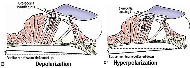
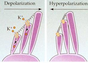
5 Depolarization of hair cell: Each hair cell has about 100 stereocilia on its apical border. 5


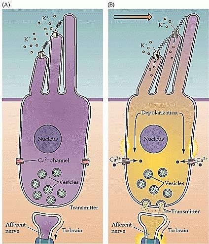
6 These stereocilia become progressively longer on the side of the hair cell away from the modiolus When the hair bundle is displaced in the direction of the tallest stereocilium depolarization occurs When the hair bundle is displaced in direction away from the tallest stereocilium hyperpolarization occurs When the hair bundle is displaced in direction perpendicular to stereocilium provides no change in membrane potential When the hair bundle is displaced in direction intermediate between these two directions produces depolarization or hyper-polarization that is proportionate to the degree to which the direction is toward or away from the Kino-cilium. The function of inner hair cells The function of inner hair cells is transmission of hearing signals The function of outer hair cells Outer hair cells contact by relatively few sensory axons and therefore accounted for only a small fraction of the auditory information that is sent onto the brain If the outer cells are damaged while the inner cells remain fully functional, a large amount of hearing loss occurs. The principle function of outer hair cells is to amplify the sound-induce vibration and thus increase our ability to hear very quiet sounds Unlike the inner hair cells, the outer hair cells do not signal the brain about incoming sounds. They instead actively and rapidly shortening occurs during depolarization and elongation occurs during hyperpolarization. They do this due to: 1. a very flexible basal membrane, 2. prestin a transmembrane protein that changes the length of outer hair cell. This behavior is known as electromotility. When the outer hair cells elongate, the motion of the basilar membrane is amplified. This modification of the basilar membrane is believed to improve and tune the stimulation of the inner hair cells. The outer hair cells therefore enhance the receptors of the inner hair cells, increasing their sensitivity to sound intensity and rendering them highly discriminatory between various pitches of sound. In support of this concept, a large number of retrograde nerve fibers pass from the brain stem to the vicinity of the outer hair cells (i.e. this will explain why there is afferent and efferent nerve fiber to both outer and inner hair cells) The differences between endolymph and perilymph Perilymph Endo-lymph Found in bony labyrinth Found in membranous labyrinth Like extracellular fluid Like intracellular fluid 6

 Primary auditory cortex (AI; area 41) The primary auditory cortex is directly excited by auditory nerve The primary auditory cortex has tonotopic maps")
7 Ultra-filtrated of plasma Low K and Protein, High Na and Ca Produced by stria vascularis High K and Protein, low Na, and Ca An electrical potential of about +80 millivolts exists all the time between endolymph and perilymph, with positivity inside the scala media and negativity outside. This is called the endocochlear potential, and it is generated by continual secretion of positive potassium ions into the scala media by the stria vascularis. Function of cerebral cortex in hearing 1) Primary auditory cortex (AI; area 41) The primary auditory cortex is directly excited by auditory nerve The primary auditory cortex has tonotopic maps "frequency map" for different tones. The primary auditory cortex unilateral lesions do not effect hearing because of completely bilateral sound representation. Sound frequency perception in the primary auditory cortex. Neurons in the auditory cortex are organized according to the frequency of sound to which they respond best. Neurons at one end of the auditory cortex respond best to low frequencies; neurons at the other respond best to high frequencies. The purpose of this frequency map (known as a tonotopic map) is unknown The frequency range to which each individual neuron in the auditory cortex responds is much narrower than that in the cochlear (i.e. sharpen the frequency). It is believed that this sharpening effect is caused mainly by the phenomenon of lateral inhibition. The same effect has been demonstrated to be important in sharpening patterns of somesthetic images, visual images, and other types of sensations. 2) Auditory association cortex or the secondary auditory cortex (AII; area 42) The auditory association areas surrounding the primary auditory cortex The auditory association areas are excited secondarily by impulses from the primary auditory cortex, as well as by some projections from thalamic association areas The auditory association areas are involved in the interpretation of sound. Many of the neurons in the auditory cortex, especially in the auditory association cortex, do not respond only to specific sound frequencies in the ear. It is believed that these neurons associate different sound frequencies with one another or associate sound information with information from other sensory areas of the cortex. Indeed, the parietal portion of the auditory association cortex partly overlaps somatosensory area II, which could provide an easy opportunity for the association of auditory information with somatosensory information. 3) Wernicke's area (Brodmann s area 22) Wernicke's area is concerned with the processing of auditory signals related to speech. Wernicke's area in the dominant hemisphere surrounding the auditory cortex Wernicke's area in the non-dominant hemisphere may be involved in understanding the tone, pitch, and لحن sound intensity and melody Wernicke's area is much more active on the left side than on the right side during language processing 7


8 The auditory pathways are also very plastic, and, like the visual and somasthetic pathways, they are modified by experience. Discrimination of sound Patterns by the auditory Cortex. Destruction of one side primary auditory cortex a. only slightly reduces hearing in the opposite ear b. it does not cause deafness in the same ear because of many crossover connections from side to side in the auditory neural pathway c. it does affect one s ability to localize the source of a sound, because comparative signals in both cortices are required for the localization function. Destruction of both primary auditory cortices in the human being greatly reduces one s sensitivity for hearing. Lesions that affect the auditory association areas but not the primary auditory cortex do not decrease a person s ability to hear and differentiate sound tones, or even to interpret at least simple patterns of sound. However, the person is often unable to فسرinterpret the meaning of the sound heard even though he or she hears them perfectly well and can even repeat them ال نفقد القدرة على السماع اوتفر ق االصوات ولكن عدم القدرة على تفسر معان االصوات وبالتال عدم القدرة على تكرارها Therefore, the auditory cortex is especially important in the discrimination of tonal andنغمة sequential patterns. soundتسلسل Determination of the direction from which sound come A person determines the horizontal direction from which sound comes by two principal means: (1) The intensity mechanism The intensity mechanism determine the distance depending on the difference between the intensities of the sounds reaching the two ears. The intensity mechanism operates best at higher frequencies because the head is a greater sound barrier at these frequencies. If a person is looking straight toward the source of the sound, the sound reaches both ears at exactly the same instant 8

9 If the right ear is closer to the sound than the left ear First, the paths are of different length because sound has to travel past the head to get to the left ear and sound intensity decreases with distance. Second, the head interferes with the sound-wave, casting the auditory equivalent of a shadow on the far ear. And, this sound shadow is more effective for sounds of higher frequency. (2) The time lag تأخر mechanism: The time lag between the entry of sound into one ear and its entry into the opposite ear (as little as 20 μs). This mechanism functions best at frequencies below 3000 cycles/sec The time lag mechanism discriminates direction much more exactly than the intensity mechanism because it does not depend on extraneous factors but only on the exact interval of time between two acoustical signals. The two aforementioned انفا mechanismsمذكور cannot tell whether the sound is emanating from in front of or behind the person or from above or below. This discrimination is achieved mainly by the pinnae of the تأك دempha sizing two ears. The shape of the pinna changes the quality of the sound entering the ear by specific sound frequencies depending on the direction from which the sound comes قوم ص وان االذن بتغ ر طب عة الصوت من خالل تغ ر تردد الصوت الواصل لالذن بناء على اتجاه الذي قدم منه الصوت 9
 senses head position, head movement, and whether our bodies are in motion.")
10 Equilibrium The vestibular system has two parts, the otolith organs (macula) and the semicircular canals. A mechanoreceptor (a hair cell with stereocilia) senses head position, head movement, and whether our bodies are in motion. The neural signals generated in the vestibular ganglion are transmitted through the vestibulecochlear nerve to the brain stem and cerebellum. 1. Sensory organ of utricle and saccule; They are2 millimeters in diameter They give information about static head position moving linearly the in 3 axis of the head The macula in the utricle and saccule contains an array of supporting cells and hair cells. The hair cells have stereocilia each and one large kinocilium. Hair cells stereocilia project into and embedded in the Otolithic membrane Otolithic membrane, contains a gelatinous mass otoliths (literally, ear stones ) or otoconia (literally, ear dust ). Otoconia are crystals of calcium carbonate range from 3 to 19 μm in length in humans Otoconia make the otolithic membrane heavier than the structures and fluids surrounding it. Gravity pulls on the dense otoliths, which deform the gelatinous mass and subsequently press on the stereocilia and influence the firing rate of the hair cells. Since gravity pulls constantly, the hair cells in the utricle and saccule provide tonic information about the orientation of the head. The nerve 10


 الراس مع الجذع")

 كما ف حالة صعود المصعد( downward a.")
11 fibers from the hair cells join those from the cristae in the vestibular division of the eighth cranial nerve A. Macula of utricle: 1. Utricle senses the Horizontal plane: موازي سطح االرض and Head orientation when person is upright position a. horizontal linear acceleration (motion in a straight line) They sense how quickly you are accelerating forward or ف الس ارةbackward,كما left or right (side by side) الراس مع الجذع الى االمام او الخلف b. up-down and forward-backward movements in the sagittal plane كما ف رفع ش ء من االرض 2. Macula of Saccule: The saccule senses the Vertical plane: مع مستوى االرض andعمودي Head orientation when person is lying down) كما ف حالة صعود المصعد( downward a. vertical linear acceleration in the sagittal plane (upward or b. head-tilts in the horizontal plane such as sideways head tilts and rapid lateral displacements, الرأس لحاله الى االمام او الخلف او على الجانب ن 11

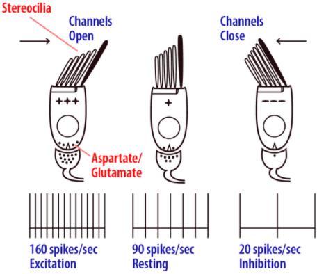
12 Note that the saccular and utricular maculae on one side of the head are mirror images of those on the other side. Thus, a tilt of the head to one side has opposite effects on corresponding hair cells of the two utricular maculae. This concept is important in understanding how the central connections of the vestibular periphery mediate the interaction of inputs from the two sides of the head When the stereocilia and kinocilium bend in the direction of the kinocilium causing receptor membrane depolarization this will increase firing rated of nerve above 100 per second. The reverse will causes receptor hyperpolarization this will increase firing rated of nerve below 100 per second in same manner as explained above. It is especially important that the hair cells are all oriented in different directions in the maculae of the utricles and saccules so that with different positions of the head, different hair cells become stimulated. The patterns of stimulation of the different hair cells apprise the brain of the position of the head with respect to the pull of gravity. In turn, the vestibular, cerebellar, and reticular motor nerve systems of the brain excite appropriate postural muscles to maintain proper equilibrium. This utricle and saccule system functions extremely effectively for maintaining equilibrium when the head is in the near-vertical position. Indeed, a person can determine as little as half a degree of disequilibrium when the body leans from the precise upright position. The impulses generated from these receptors are partly responsible for labyrinth righting reflexes. These reflexes are a series of responses integrated for the most part in the nuclei of the midbrain. The stimulus for the reflex is tilting of the head, which stimulates the otolithic organs; the response is compensatory contraction of the neck muscles to keep the head level. In cats, dogs, and primates, visual cues شاارات البصار ة canأأل initiate optical righting reflexes that right the animal in the absence of labyrinthine or body stimulation. In humans, the operation of these reflexes maintains the head in a stable position and the eyes fixed on visual targets despite movements of the body and the jerks and jolts of everyday life. The responses are initiated by vestibular stimulation, stretching of neck muscles, and movement of visual images on the retina, and the responses are the vestibulo-ocular reflex and other remarkably precise reflex contractions of the neck and extra-ocular muscles. Spatial orientation Orientation in space depends in part on input from the vestibular receptors visual cues proprioceptors cutaneous extero-ceptors, especially touch and pressure receptors. These four inputs are synthesized at a cortical level into a continuous picture of the individual orientation in space. 12

 semicircular ducts, are arranged at")
13 Although most of the responses to stimulation of the maculae are reflex in nature, vestibular impulses also reach the cerebral cortex. These impulses are presumably responsible for conscious perception of motion and supply part of the information necessary for orientation in space. Vertigo is the sensation of rotation in the absence of actual rotation and is a prominent symptom when one labyrinth is inflamed. Rotational movement; Semicircular canal: The three semicircular ducts in each vestibular apparatus, known as the anterior, posterior, and lateral (horizontal) semicircular ducts, are arranged at right angles to one another so that they represent all three planes in space. Each of the three semicircular canals senses just a onedimensional component of rotational acceleration. The three semicircular canals give information about dynamic head position Each of the three semicircular canals is responsible for a specific direction of head movement: One of the canals responds to the head tilting upwards or downwards, one responds to it tilting to the right or to the left, and one responds to it turning sideways. 13
 of")
 cells surmounted by a")

14 Inside the bony canals, the membranous canals are suspended in perilymph. A receptor structure, the crista ampullaris, is located in the expanded end (ampulla) of each of the membranous canals. Each crista consists of hair cells and supporting (sustentacular) cells surmounted by a gelatinous partition (cupula) that closes off the ampulla. The processes of the hair cells are embedded in the cupula, and the bases of the hair cells are in close contact with the afferent fibers of the vestibular division of the eighth cranial nerve. 14


15 Responses to rotational acceleration Rotational acceleration in the plane of a given semicircular canal stimulates its crista. The response passes into the following stages: First: The endolymph, because of its inertia, is displaced in a direction opposite to the direction of rotation. The fluid pushes on the cupula, deforming it. This bends the processes of the hair cells. This will increase the number of nerve impulses (depolarization) 15

16 Second: When a constant speed of rotation is reached, the fluid spins at the same rate as the body and the cupula swings back into the upright position in the middle of the ampulla because of its own elastic recoil This will returns the number of nerve impulses back to basilar level gradually Third: When the rotation suddenly stops, exactly opposite effects take place: The endolymph continues to rotate while the semicircular duct stops. This time, the cupula bends in the opposite direction, causing the hair cell to stop discharging entirely (hyperpolarization) Fourth: the endolymph stops moving and the cupula gradually returns to its resting position in 25 to 30 s, thus allowing hair cell discharge to return to its normal tonic level This will returns the number of nerve impulses back to basilar level gradually Movement of the cupula in one direction commonly causes an increase in the firing rate of single nerve fibers from the crista, whereas movement in the opposite direction commonly inhibits neural activity. Rotation causes maximal stimulation of the semicircular canals most nearly in the plane of rotation. Because the canals on one side of the head are a mirror image of those on the other side, the endolymph is displaced toward the ampulla on one side and away from it on the other. The pattern of stimulation reaching the brain therefore varies with the direction as well as the plane of rotation. Linear acceleration probably fails to displace the cupula and therefore does not stimulate the cristae. However, there is considerable evidence that when one part of the labyrinth is destroyed, other parts take over its functions. 16
Ear. Utricle & saccule in the vestibule Connected to each other and to the endolymphatic sac by a utriculosaccular duct
 Rahaf Jreisat *You don t have to go back to the slides. Ear Inner Ear Membranous Labyrinth It is a reflection of bony labyrinth but inside. Membranous labyrinth = set of membranous tubes containing sensory
Chapter 17, Part 2! The Special Senses! Hearing and Equilibrium!
 Chapter 17, Part 2! The Special Senses! Hearing and Equilibrium! SECTION 17-5! Equilibrium sensations originate within the inner ear, while hearing involves the detection and interpretation of sound waves!
Chapter 17, Part 2! Chapter 17 Part 2 Special Senses! The Special Senses! Hearing and Equilibrium!
 Chapter 17, Part 2! The Special Senses! Hearing and Equilibrium! SECTION 17-5! Equilibrium sensations originate within the inner ear, while hearing involves the detection and interpretation of sound waves!
What is the effect on the hair cell if the stereocilia are bent away from the kinocilium?
 CASE 44 A 53-year-old man presents to his primary care physician with complaints of feeling like the room is spinning, dizziness, decreased hearing, ringing in the ears, and fullness in both ears. He states
to vibrate the fluid. The ossicles amplify the pressure. The surface area of the oval window is
 Page 1 of 6 Question 1: How is the conduction of sound to the cochlea facilitated by the ossicles of the middle ear? Answer: Sound waves traveling through air move the tympanic membrane, which, in turn,
Chapter 15 Hearing & Equilibrium
 Chapter 15 Hearing & Equilibrium ANATOMY OF THE OUTER EAR EAR PINNA is the outer ear it is thin skin covering elastic cartilage. It directs incoming sound waves to the EXTERNAL AUDITORY CANAL, which is
Νευροφυσιολογία και Αισθήσεις
 Biomedical Imaging & Applied Optics University of Cyprus Νευροφυσιολογία και Αισθήσεις Διάλεξη 11 Ακουστικό και Αιθουσιαίο Σύστημα (Auditory and Vestibular Systems) Introduction Sensory Systems Sense of
Anatomy of the Ear Region. External ear Middle ear Internal ear
 Ear Lecture Objectives Make a list of structures making the external, middle, and internal ear. Discuss the features of the external auditory meatus and tympanic membrane. Describe the shape, position,
Vestibular physiology
 Vestibular physiology 2017 Utricle A flat epithelium: horizontal in the upright head Utricle Hair cells: no axons hair cells Utricle Hair cells synapse onto 8th nerve afferents. 8th nerve afferents Hair
A&P 1. Ear, Hearing & Equilibrium Lab. Basic Concepts. These notes follow Carl s Talk at the beginning of lab
 A&P 1 Ear, Hearing & Equilibrium Lab Basic Concepts These notes follow Carl s Talk at the beginning of lab In this "Lab Exercise Guide", we will be looking at the basics of hearing and equilibrium. NOTE:
The Physiology of the Senses Lecture 10 - Balance
 The Physiology of the Senses Lecture 10 - Balance www.tutis.ca/senses/ Contents Objectives... 1 The sense of balance originates from the labyrinth... 2 The auditory and vestibular systems have a common
A&P 1. Ear, Hearing & Equilibrium Lab. Basic Concepts. Pre-lab Exercises
 A&P 1 Ear, Hearing & Equilibrium Lab Basic Concepts Pre-lab Exercises In this "Lab Exercise Guide", we will be looking at the basics of hearing and equilibrium. NOTE: these notes do not follow the order
Auditory System. Barb Rohrer (SEI )
") Auditory System Barb Rohrer (SEI614 2-5086) Sounds arise from mechanical vibration (creating zones of compression and rarefaction; which ripple outwards) Transmitted through gaseous, aqueous or solid medium
Auditory System Barb Rohrer (SEI614 2-5086) Sounds arise from mechanical vibration (creating zones of compression and rarefaction; which ripple outwards) Transmitted through gaseous, aqueous or solid medium
Structure, Energy Transmission and Function. Gross Anatomy. Structure, Function & Process. External Auditory Meatus or Canal (EAM, EAC) Outer Ear
 Outer Ear") Gross Anatomy Structure, Energy Transmission and Function IE N O ME 1 Structure, Function & Process 4 External Auditory Meatus or Canal (EAM, EAC) Outer third is cartilaginous Inner 2/3 is osseous Junction
Gross Anatomy Structure, Energy Transmission and Function IE N O ME 1 Structure, Function & Process 4 External Auditory Meatus or Canal (EAM, EAC) Outer third is cartilaginous Inner 2/3 is osseous Junction
THE COCHLEA AND AUDITORY PATHWAY
 Dental Neuroanatomy Suzanne S. Stensaas, PhD February 23, 2012 Reading: Waxman, Chapter 16, Review pictures in a Histology book Computer Resources: http://www.cochlea.org/ - Promenade around the Cochlea
Otoconia: Calcium carbonate crystals Gelatinous mass. Cilia. Hair cells. Vestibular nerve. Vestibular ganglion
 VESTIBULAR SYSTEM (Balance/Equilibrium) The vestibular stimulus is provided by Earth s, and. Located in the of the inner ear, in two components: 1. Vestibular sacs - gravity & head direction 2. Semicircular
Course: PG- Pathshala Paper number: 13 Physiological Biophysics Module number M23: Posture and Movement Regulation by Ear.
 Course: PG- Pathshala Paper number: 13 Physiological Biophysics Module number M23: Posture and Movement Regulation by Ear Principal Investigator: Co-Principal Investigator: Paper Coordinator: Content Writer:
The cochlea: auditory sense. The cochlea: auditory sense
 Inner ear apparatus 1- Vestibule macula and sacculus sensing acceleration of the head and direction of gravity 2- Semicircular canals mainly for sensing direction of rotation of the head 1 3- cochlea in
Systems Neuroscience Oct. 16, Auditory system. http:
 Systems Neuroscience Oct. 16, 2018 Auditory system http: www.ini.unizh.ch/~kiper/system_neurosci.html The physics of sound Measuring sound intensity We are sensitive to an enormous range of intensities,
VESTIBULAR SYSTEM ANATOMY AND PHYSIOLOGY. Professor.Dr. M.K.Rajasekar MS., DLO.,
 VESTIBULAR SYSTEM ANATOMY AND PHYSIOLOGY Professor.Dr. M.K.Rajasekar MS., DLO., Life is hard for those who don t have a VOR During a walk I found too much motion in my visual picture of the surroundings
Hearing. By: Jimmy, Dana, and Karissa
 Hearing By: Jimmy, Dana, and Karissa Anatomy - The ear is divided up into three parts - Sound enters in through the outer ear and passes into the middle where the vibrations are received and sent to the
Hearing and Balance 1
 Hearing and Balance 1 Slide 3 Sound is produced by vibration of an object which produces alternating waves of pressure and rarefaction, for example this tuning fork. Slide 4 Two characteristics of sound
Deafness and hearing impairment
 Auditory Physiology Deafness and hearing impairment About one in every 10 Americans has some degree of hearing loss. The great majority develop hearing loss as they age. Hearing impairment in very early
Auditory System Feedback
 Feedback Auditory System Feedback Using all or a portion of the information from the output of a system to regulate or control the processes or inputs in order to modify the output. Central control of
AUDITORY APPARATUS. Mr. P Mazengenya. Tel 72204
 AUDITORY APPARATUS Mr. P Mazengenya Tel 72204 Describe the anatomical features of the external ear Describe the tympanic membrane (ear drum) Describe the walls of the middle ear Outline the structures
ENT 318 Artificial Organs Physiology of Ear
 ENT 318 Artificial Organs Physiology of Ear Lecturer: Ahmad Nasrul Norali The Ear The Ear Components of hearing mechanism - Outer Ear - Middle Ear - Inner Ear - Central Auditory Nervous System Major Divisions
MECHANISM OF HEARING
 MECHANISM OF HEARING Sound: Sound is a vibration that propagates as an audible wave of pressure, through a transmission medium such as gas, liquid or solid. Sound is produced from alternate compression
Gathering information the sensory systems; Vision
 Visual System Gathering information the sensory systems; Vision The retina is the light-sensitive receptor layer at the back of the eye. - Light passes through the cornea, the aqueous chamber, the lens,
Chapter 3: Anatomy and physiology of the sensory auditory mechanism
 Chapter 3: Anatomy and physiology of the sensory auditory mechanism Objectives (1) Anatomy of the inner ear Functions of the cochlear and vestibular systems Three compartments within the cochlea and membranes
Required Slide. Session Objectives
 Auditory Physiology Required Slide Session Objectives Auditory System: At the end of this session, students will be able to: 1. Characterize the range of normal human hearing. 2. Understand the components
9.01 Introduction to Neuroscience Fall 2007
 MIT OpenCourseWare http://ocw.mit.edu 9.01 Introduction to Neuroscience Fall 2007 For information about citing these materials or our Terms of Use, visit: http://ocw.mit.edu/terms. 9.01 Recitation (R02)
THE COCHLEA AND AUDITORY PATHWAY
 Dental Neuroanatomy Suzanne S. Stensaas, PhD April 14, 2010 Reading: Waxman, Chapter 16, Review pictures in a Histology book Computer Resources: http://www.cochlea.org/ - Promenade around the Cochlea HyperBrain
Vestibular System Dr. Bill Yates Depts. Otolaryngology and Neuroscience 110 Eye and Ear Institute
 Vestibular System Dr. Bill Yates Depts. Otolaryngology and Neuroscience 110 Eye and Ear Institute 412-647-9614 byates@pitt.edu What is the Vestibular System? The vestibular system is the sensory system,
cortical and brain stem control of motor function
 cortical and brain stem control of motor function cortical and brain stem control of motor function most voluntary movements initiated by the cerebral cortex are achieved when the cortex activates patterns
The Physiology of the Senses Lecture 10 - Balance
 The Physiology of the Senses Lecture 10 - Balance www.tutis.ca/senses/ Contents Objectives... 1 The sense of balance originates in the labyrinth.... 2 The vestibular system has two parts.... 3 The Anatomy
THE EAR AND HEARING Be sure you have read and understand Chapter 16 before beginning this lab. INTRODUCTION: hair cells outer ear tympanic membrane
 BIOLOGY 211: HUMAN ANATOMY & PHYSIOLOGY ****************************************************************************************************** THE EAR AND HEARING ******************************************************************************************************
Vestibular Physiology Richard M. Costanzo, Ph.D.
 Vestibular Physiology Richard M. Costanzo, Ph.D. OBJECTIVES After studying the material of this lecture, the student should be able to: 1. Describe the structure and function of the vestibular organs.
Auditory Physiology Richard M. Costanzo, Ph.D.
 Auditory Physiology Richard M. Costanzo, Ph.D. OBJECTIVES After studying the material of this lecture, the student should be able to: 1. Describe the morphology and function of the following structures:
The Vestibular System
 The Vestibular System Vestibular and Auditory Sensory Organs Bill Yates, Ph.D. Depts. Otolaryngology & Neuroscience University of Pittsburgh Organization of Sensory Epithelium Displacement of Stereocilia
Unit VIII Problem 9 Physiology: Hearing
 Unit VIII Problem 9 Physiology: Hearing - We can hear a limited range of frequency between 20 Hz 20,000 Hz (human hearing acuity is between 1000 Hz 4000 Hz). - The ear is divided into 3 parts. Those are:
Taste buds Gustatory cells extend taste hairs through a narrow taste pore
 The Special Senses Objectives Describe the sensory organs of smell, and olfaction. Identify the accessory and internal structures of the eye, and explain their function. Explain how light stimulates the
Auditory and vestibular system
 Auditory and vestibular system Sensory organs on the inner ear inner ear: audition (exteroceptor) and vestibular apparatus (proprioceptor) bony and membranous labyrinths within the temporal bone (os temporale)
Before we talk about the auditory system we will talk about the sound and waves
 The Auditory System PHYSIO: #3 DR.LOAI ZAGOUL 24/3/2014 Refer to the slides for some photos. Before we talk about the auditory system we will talk about the sound and waves All waves have basic characteristics:
4. Which letter in figure 9.1 points to the fovea centralis? Ans: b
 Chapter 9: The Sensory System 1. Proprioceptors are involved in the sense of A) pain. B) temperature. C) pressure. D) movement of limbs. 2. Which are chemoreceptors? A) taste B) olfactory C) proprioceptors
COGS 107B Week 2. Hyun Ji Friday 4:00-4:50pm
 COGS 107B Week 2 Hyun Ji Friday 4:00-4:50pm Lecture 3: Proprioception Principles: The Neuron Doctrine and The Law of Dynamic Polarization Proprioception Joint-protecting reflexes (ex. Knee jerk reflex)
Printable version - Hearing - OpenLearn - The Open University
 Skip to content Accessibility Sign in Contact Search the OU The Open University Study at the OU Research at the OU OU Community About the OU Hearing Printable page generated Saturday, 12 November 2011,
Anatomy and Physiology of Hearing
 Anatomy and Physiology of Hearing The Human Ear Temporal Bone Found on each side of the skull and contains the organs for hearing and balance Divided into four major portions: - squamous - mastoid - tympanic
Special Senses. Mechanoreception Electroreception Chemoreception Others
 Special Senses Mechanoreception Electroreception Chemoreception Others Recall our receptor types Chemically regulated: Respond to particular chemicals Voltage regulated: respond to changing membrane potential
For this lab you will use parts of Exercise #18 in your Wise lab manual. Please be sure to read those sections before coming to lab
 Bio 322 Human Anatomy Objectives for the laboratory exercise The Eye and Ear Required reading before beginning this lab: Saladin, KS: Human Anatomy 5 th ed (2017) Chapter 17 For this lab you will use parts
Chapter 18 Senses SENSORY RECEPTION 10/21/2011. Sensory Receptors and Sensations. Sensory Receptors and Sensations. Sensory Receptors and Sensations
 SENSORY RECEPTION Chapter 18 Senses s convert stimulus energy to action potentials s 1. Are specialized cells, or 2. Specialized endings that detect stimuli All stimuli are forms of energy s in eyes detect
Chapter 11: Sound, The Auditory System, and Pitch Perception
 Chapter 11: Sound, The Auditory System, and Pitch Perception Overview of Questions What is it that makes sounds high pitched or low pitched? How do sound vibrations inside the ear lead to the perception
Bony and membranous labyrinth. Vestibular system. János Hanics M.D.
 Bony and membranous labyrinth. Vestibular system. János Hanics M.D. The position of the inner ear The labyrinthes of the inner ear - Continuous cavity system in the petrous part of temporal bone - Cavity
SPECIAL SENSES: THE AUDITORY SYSTEM
 SPECIAL SENSES: THE AUDITORY SYSTEM REVISION OF PHYSICS: WAVES A wave is an oscillation of power, sound waves have two main characteristics: amplitude, which is the maximum displacement or the power of
SPECIAL SENSES PART I: OLFACTION & GUSTATION
 SPECIAL SENSES PART I: OLFACTION & GUSTATION 5 Special Senses Olfaction Gustation Vision Equilibrium Hearing Olfactory Nerves Extend through cribriform plate into nasal cavity on both sides of nasal septum
-Detect heat or cold and help maintain body temperature
 Sensory Receptors -Transduce stimulus energy and transmit signals to the central nervous system -Reception occurs when a receptor detectd a stimulus -Perception occurs in the brain as this information
SOLUTIONS Homework #3. Introduction to Engineering in Medicine and Biology ECEN 1001 Due Tues. 9/30/03
 SOLUTIONS Homework #3 Introduction to Engineering in Medicine and Biology ECEN 1001 Due Tues. 9/30/03 Problem 1: a) Where in the cochlea would you say the process of "fourier decomposition" of the incoming
Mechanical Properties of the Cochlea. Reading: Yost Ch. 7
 Mechanical Properties of the Cochlea CF Reading: Yost Ch. 7 The Cochlea Inner ear contains auditory and vestibular sensory organs. Cochlea is a coiled tri-partite tube about 35 mm long. Basilar membrane,
VESTIBULAR SYSTEM. Deficits cause: Vertigo. Falling Tilting Nystagmus Nausea, vomiting
 VESTIBULAR SYSTEM Objectives: Understand the functions of the vestibular system: What is it? How do you stimulate it? What are the consequences of stimulation? Describe the vestibular apparatus, the 2
University of Connecticut Schools of Medicine and Dental Medicine Systems Neuroscience Meds Vestibular System
 University of Connecticut Schools of Medicine and Dental Medicine Systems Neuroscience Meds 371 2007-08 Vestibular System S. Kuwada Reading: Purves et al. (2008, 4 th edition), Neuroscience, Chapter 14.
Chapter 7. Audition, the Body Senses, and the Chemical Senses. Copyright Allyn & Bacon 2004
 Chapter 7 Audition, the Body Senses, and the Chemical Senses This multimedia product and its contents are protected under copyright law. The following are prohibited by law: any public performance or display,
Presentation On SENSATION. Prof- Mrs.Kuldeep Kaur
 Presentation On SENSATION Prof- Mrs.Kuldeep Kaur INTRODUCTION:- Sensation is a specialty area within Psychology that works at understanding how are senses work and how we perceive stimuli in the environment.
Intro to Audition & Hearing
 Intro to Audition & Hearing Lecture 16 Chapter 9, part II Jonathan Pillow Sensation & Perception (PSY 345 / NEU 325) Fall 2017 1 Sine wave: one of the simplest kinds of sounds: sound for which pressure
CNS MCQ 2 nd term. Select the best answer:
 Select the best answer: CNS MCQ 2 nd term 1) Vestibular apparatus: a) Represent the auditory part of the labyrinth. b) May help in initiating the voluntary movements. c) Contains receptors concerned with
Hearing. By Jack & Tori
 Hearing By Jack & Tori 3 Main Components of the Human Ear. Outer Ear. Middle Ear. Inner Ear Outer Ear Pinna: >Visible part of ear and ear canal -Acts as a funnel to direct sound Eardrum: >Airtight membrane
Copyright 2009 Pearson Education, Inc.
 Outline Nervous System Sensory Systems I. II. III. IV. V. VI. Biol 105 Lecture 11 Chapter 9 Senses Sensory receptors Touch Vision Hearing and balance Smell Senses Sensory receptor cells Sensory receptors
Carlson (7e) PowerPoint Lecture Outline Chapter 7: Audition, the Body Senses, and the Chemical Senses
 PowerPoint Lecture Outline Chapter 7: Audition, the Body Senses, and the Chemical Senses") Carlson (7e) PowerPoint Lecture Outline Chapter 7: Audition, the Body Senses, and the Chemical Senses This multimedia product and its contents are protected under copyright law. The following are prohibited
Carlson (7e) PowerPoint Lecture Outline Chapter 7: Audition, the Body Senses, and the Chemical Senses This multimedia product and its contents are protected under copyright law. The following are prohibited
Vestibular Function and Anatomy. UTMB Grand Rounds April 14, 2004 Gordon Shields, MD Arun Gadre, MD
 Vestibular Function and Anatomy UTMB Grand Rounds April 14, 2004 Gordon Shields, MD Arun Gadre, MD System of balance Membranous and bony labyrinth embedded in petrous bone 5 distinct end organs 3 semicircular
Introduction. Senses our perception of what is out there 2 groups. General senses Special senses
 Introduction Senses our perception of what is out there 2 groups General senses Special senses Central Processing and Adaptation Adaptation the loss of sensitivity after continuous stimulation Tonic receptors
Senses and Sense Organs
 Senses and Sense Organs SENSORY SYSTEMS Human experience is effected by both internal and external stimuli. Humans are able to distinguish among many different types of stimuli by means of a highly developed
Perception of Sound. To hear sound, your ear has to do three basic things:
 Perception of Sound Your ears are extraordinary organs. They pick up all the sounds around you and then translate this information into a form your brain can understand. One of the most remarkable things
Overview of Sensory Receptors
 Sensory Systems Chapter 45 Overview of Sensory Receptors Sensory receptors provide information from our internal and external environments that is crucial for survival and success -Exteroceptors sense
SPHSC 462 HEARING DEVELOPMENT. Overview Review of Hearing Science Introduction
 SPHSC 462 HEARING DEVELOPMENT Overview Review of Hearing Science Introduction 1 Overview of course and requirements Lecture/discussion; lecture notes on website http://faculty.washington.edu/lawerner/sphsc462/
For more information about how to cite these materials visit
 Author(s): Matthew Velkey, 2009 License: Unless otherwise noted, this material is made available under the terms of the Creative Commons Attribution Non-commercial Share Alike 3.0 License: http://creativecommons.org/licenses/by-nc-sa/3.0/
Vestibular/Auditory Systems
 Vestibular/Auditory Systems Jay Zenner on February 3, 2012 Dental Neuroanatomy Scott Rogers Office: SOM 2C132 Boney Labyrinth Vestibular Apparatus Two Major Divisions Cochlea (anterior) VIII VII Semicircular
Lecture 6 Hearing 1. Raghav Rajan Bio 354 Neurobiology 2 January 28th All lecture material from the following links unless otherwise mentioned:
 Lecture 6 Hearing 1 All lecture material from the following links unless otherwise mentioned: 1. http://wws.weizmann.ac.il/neurobiology/labs/ulanovsky/sites/neurobiology.labs.ulanovsky/files/uploads/purves_ch12_ch13_hearing
Chapter 15 Lecture Outline
 Chapter 15 Lecture Outline See separate PowerPoint slides for all figures and tables preinserted into PowerPoint without notes. Copyright 2016 McGraw-Hill Education. Permission required for reproduction
Vestibular System. Dian Yu, class of 2016
 Vestibular System Dian Yu, class of 2016 Objectives 1. Describe the functions of the vestibular system: What is it? How do you stimulate it? What are the consequences of stimulation? 2. Describe the vestibular
The Ear. Dr. Heba Kalbouneh Assistant Professor of Anatomy and Histology
 The Ear Dr. Heba Kalbouneh Assistant Professor of Anatomy and Histology The Ear The ear consists of the external ear; the middle ear (tympanic cavity); and the internal ear (labyrinth), which contains
Hearing. istockphoto/thinkstock
 Hearing istockphoto/thinkstock Audition The sense or act of hearing The Stimulus Input: Sound Waves Sound waves are composed of changes in air pressure unfolding over time. Acoustical transduction: Conversion
THE VESTIBULAR APPRATUS AND PATHWAY
 Dental Neuroanatomy February 23, 2012 Suzanne Stensaas, Ph.D. Reading: Waxman Chapter 17 Also pp 105-108 on control of eye movments Computer Resources: HyperBrain Ch. 8 Vestibulospinal Pathway Quiz http://library.med.utah.edu/kw/animations/hyperbrain/pathways/
PSY 215 Lecture 10 Topic: Hearing Chapter 7, pages
 PSY 215 Lecture 10 Topic: Hearing Chapter 7, pages 189-197 Corrections: NTC 09-1, page 3, the Superior Colliculus is in the midbrain (Mesencephalon). Announcements: Movie next Monday: Case of the frozen
Auditory and Vestibular Systems
 Auditory and Vestibular Systems Objective To learn the functional organization of the auditory and vestibular systems To understand how one can use changes in auditory function following injury to localize
Physiology of human perception
 Physiology of human perception Vision Hearing Thermal and tactile sensations Basic introduction and the list and description of the tasks to be carried out Visible light: 400-700 nm. Vision or sight Anatomy
The Senses. Chapter 10 7/8/11. Introduction
 Chapter 10 The Senses Introduction A. Sensory receptors detect changes in the environment and stimulate neurons to send nerve impulses to the brain. B. A sensation is formed based on the sensory input.
What does it mean to analyze the frequency components of a sound? A spectrogram such as that shown here is the usual display of frequency components
 1 2 3 4 What does it mean to analyze the frequency components of a sound? A spectrogram such as that shown here is the usual display of frequency components as a function of time here during the production
P215 Basic Human Physiology Summer 2003 Lecture Exam #2
 PLEASE BE AWARE CONTENT COVERED ON EXAMS VARIES FROM ONE SEMESTER TO ANOTHER. THIS EXAM MAY NOT CONTAIN MATERIAL THAT WILL BE ON YOUR EXAM THIS SEMESTER, AND/OR MAY CONTAIN MATERIAL THAT WILL NOT BE COVERED
Cochlear anatomy, function and pathology I. Professor Dave Furness Keele University
 Cochlear anatomy, function and pathology I Professor Dave Furness Keele University d.n.furness@keele.ac.uk Aims and objectives of these lectures Introduction to gross anatomy of the cochlea Focus (1) on
Chapter Fourteen. The Hearing Mechanism. 1. Introduction.
 Chapter Fourteen The Hearing Mechanism 1. Introduction. 2. Hearing. 3. The Ear. 4. The External Ear. 5. The Inner Ear. 6. Frequency Discrimination. 7. The Organ of Corti. 8. Tests and Exrecises. 9. References.
Hearing: Physiology and Psychoacoustics
 9 Hearing: Physiology and Psychoacoustics Click Chapter to edit 9 Hearing: Master title Physiology style and Psychoacoustics The Function of Hearing What Is Sound? Basic Structure of the Mammalian Auditory
Sensory Physiology. Sensory Range Varies. Introduction to the Special Senses. How do we sense the world around us?
 Sensory Physiology How do we sense the world around us? We do not see things as they are; we see things as we are. --Anais Nin Anais Nin, French author 1903-1977 Sensory Range Varies Introduction to the
Sensory Processes Sensory Systems
 9 th Lecture (9b) Wed 04 Feb 2009 Vertebrate Physiology ECOL 437 (MCB/VetSci 437) Univ. of Arizona, spring 2009 Kevin Bonine & Kevin Oh Sensory Processing Chapter 13 1 Sensory Processes Sensory Systems
Sensory Processes Sensory Systems
 9 th Lecture (9b) Wed 04 Feb 2009 Vertebrate Physiology ECOL 437 (MCB/VetSci 437) Univ. of Arizona, spring 2009 Kevin Bonine & Kevin Oh Sensory Processes Sensory Systems Ch13 in your text Sensory Processing
Hearing Sound. The Human Auditory System. The Outer Ear. Music 170: The Ear
 Hearing Sound Music 170: The Ear Tamara Smyth, trsmyth@ucsd.edu Department of Music, University of California, San Diego (UCSD) November 17, 2016 Sound interpretation in the auditory system is done by
Music 170: The Ear. Tamara Smyth, Department of Music, University of California, San Diego (UCSD) November 17, 2016
 November 17, 2016") Music 170: The Ear Tamara Smyth, trsmyth@ucsd.edu Department of Music, University of California, San Diego (UCSD) November 17, 2016 1 Hearing Sound Sound interpretation in the auditory system is done by
Music 170: The Ear Tamara Smyth, trsmyth@ucsd.edu Department of Music, University of California, San Diego (UCSD) November 17, 2016 1 Hearing Sound Sound interpretation in the auditory system is done by
Special Senses. Accessory Structures of the Eye. The Eye and Vision. Accessory Structures of the Eye. Accessory Structures of the Eye
 8 PART A Special Senses PowerPoint Lecture Slide Presentation by Jerry L. Cook, Sam Houston University ESSENTIALS OF HUMAN ANATOMY & PHYSIOLOGY EIGHTH EDITION ELAINE N. MARIEB The Senses General senses
SENSORY SYSTEM VII THE EAR PART 1
 SENSORY SYSTEM VII THE EAR PART 1 Waves Sound is a compression wave The Ear Ear Outer Ear Pinna Outer ear: - Made up of the pinna and the auditory canal Auditory Canal Outer Ear Pinna (also called the
College of Medicine Dept. of Medical physics Physics of ear and hearing /CH
 College of Medicine Dept. of Medical physics Physics of ear and hearing /CH 13 2017-2018 ***************************************************************** o Introduction : The ear is the organ that detects
The speed at which it travels is a function of the density of the conducting medium.
 Sound is a compression wave which (unlike light) must have a medium to conduct it. If the medium is uniform in density, the sound will spread at as a uniform ring of circles (actually spheres). The speed
The olfactory epithelium is located at the roof of the nasal cavity. Nasal conchae cause turbulance of incoming air
 Special Senses I. Olfaction II. Gustation A. Anatomy and general info The olfactory epithelium is located at the roof of the nasal cavity Nasal conchae cause turbulance of incoming air Olfactory glands
Receptors / physiology
 Hearing: physiology Receptors / physiology Energy transduction First goal of a sensory/perceptual system? Transduce environmental energy into neural energy (or energy that can be interpreted by perceptual