Title macula by swept-source optical cohe.
|
|
|
- Hilda Carter
- 5 years ago
- Views:
Transcription
 Akagi-Kurashige, Yumiko; Miyake, Ma")

1 Title Three-dimensional tomographic featu macula by swept-source optical cohe Ellabban, Abdallah A; Tsujikawa, Ak Yamashiro, Kenji; Oishi, Akio; Ooto Author(s) Akagi-Kurashige, Yumiko; Miyake, Ma Hussein S; Radwan, Tarek M; Zaky, K Nagahisa Citation American journal of ophthalmology ( Issue Date -0 URL Right Elsevier Inc. Type Journal Article Textversion author Kyoto University
2 Abstract (MUST be submitted as a separate file) D FEATURES OF DOME SHAPED MACULA. Ellabban AA, et al. Abstract PURPOSE: To study the tomographic features and pathomorphology of dome-shaped maculas with swept-source optical coherence tomography (OCT). DESIGN: Prospective cross sectional study. METHODS: The macular area of highly myopic eyes ( patients) with dome-shaped macula was studied with swept-source OCT at 1,0 nm. Three-dimensional (D) datasets were obtained with raster scanning covering a 1 x mm area; D images of the posterior pole were constructed by auto-segmentation of the retinal pigment epithelium (RPE). RESULTS: In all reconstructed D images of the RPE, two outward concavities were seen within the posterior staphyloma and a horizontal ridge was formed between these two concavities. In of these eyes, this horizontal ridge was band-shaped. The vertical OCT section through the fovea showed a convex configuration of RPE, but the horizontal section showed an almost flat RPE line. In nine eyes, D images showed a typical dome-shaped convexity within the staphyloma. OCT scans showed no outward protrusions in the external scleral surface, but marked scleral thinning was seen consistent with the two outward concavities of the RPE. The sclera of the fovea (. ±. µm) was significantly thicker than that in all four quadrants of the parafoveal area ( µm, P <.001). CONCLUSIONS: In highly myopic eyes with a dome-shaped macula, a horizontal ridge is formed within the posterior staphyloma by uneven thinning of the sclera.
3 *Manuscript (incl. title, auth, corr.auth., demographics, references, legend) 1 1 D FEATURES OF DOME-SHAPED MACULA. Ellabban AA, et al. Page 0 THREE-DIMENSIONAL TOMOGRAPHIC FEATURES OF DOME-SHAPED MACULA BY SWEPT-SOURCE OPTICAL COHERENCE TOMOGRAPHY Short title: THREE-DIMENSIONAL FEATURES OF DOME-SHAPED MACULA ABDALLAH A. ELLABBAN, 1, AKITAKA TSUJIKAWA, 1 AKIKO MATSUMOTO, 1, KENJI YAMASHIRO, 1 AKIO OISHI, 1 SOTARO OOTO, 1 ISAO NAKATA, 1 YUMIKO AKAGI-KURASHIGE, 1 MASAHIRO MIYAKE, 1 HUSSEIN S. ELNAHAS, TAREK M. RADWAN, KHALED A. ZAKY, AND NAGAHISA YOSHIMURA 1 From 1 the Department of Ophthalmology and Visual Sciences, Kyoto University Graduate School of Medicine, Kyoto, Japan; the Department of Ophthalmology, Suez Canal University, Faculty of Medicine, Ismailia, Egypt; Topcon Corporation, Tokyo, Japan. Supplemental Material available at AJO.com Correspondence to Akitaka Tsujikawa, MD, Department of Ophthalmology and Visual Sciences, Kyoto University Graduate School of Medicine, Sakyo-ku, Kyoto -, Japan; phone: +1---; fax: ; tujikawa@kuhp.kyoto-u.ac.jp
4 1 D FEATURES OF DOME-SHAPED MACULA. Ellabban AA, et al. Page 1 INTRODUCTION High myopia is one of the leading causes of visual disturbance worldwide, and is particularly prevalent in Asians. 1- Axial elongation of the globe leads to complex changes in the topography of the posterior pole, with concomitant thinning of the retina, choroid, and sclera, and subsequent development of macular pathologies. - Posterior staphyloma, which is one of the characteristic features of high myopia, has been reported to be associated with various vision-threatening complications, including choroidal neovascularization, macular holes, schisis, chorioretinal atrophy, and visual field defects. - Using fundus examination, Curtin classified posterior staphyloma in eyes with high myopia into types. Based on optical coherence tomography (OCT), dome-shaped macula was first described by Gaucher and associates as an unexpected finding in myopic staphyloma, and was characterized as an inward convexity of the macula that occurred in highly myopic eyes within the concavity of a posterior staphyloma. They suggested that dome-shaped macula might be due to changes in choroidal thickness or to changes in scleral shape in highly myopic eyes. Thereafter, Imamura and associates, by using enhanced depth imaging OCT, reported that dome-shaped macula is due to a localized variation in thickness of the sclera in the macular area. However, the mechanisms which underlie this unusual tomographic feature remain unknown. In addition, most cases of dome-shaped macula in these previous reports were examined by only unidirectional OCT scans,,,, So far, little information is available on the topography of the posterior pole in eyes with dome-shaped macula. With the advances in OCT in the last decade, several retinal pathologies have been identified in highly myopic eyes. 1- However, imaging of highly myopic eyes with conventional OCT systems has several difficulties. Recent OCT generation has utilized swept-source laser as a light source. - The swept-source OCT at a longer wavelength allows deeper tissue penetration into the choroid and even sclera, especially in highly myopic eyes. Additionally, the higher imaging speed, larger scan window, and lower sensitivity roll-off with depth allow for larger scans and the production of three-dimensional (D) images of the posterior pole. In the study described herein, we examined eyes with dome-shaped macula using swept-source OCT at a longer wavelength in order to study the pathomorphology of dome-shaped macula by D imaging and to elucidate its association with macular complications.
5 1 D FEATURES OF DOME-SHAPED MACULA. Ellabban AA, et al. Page PATIENTS AND METHODS In the current prospective cross-sectional study, we examined eyes ( patients) with high myopia that showed a dome-shaped macular configuration within the posterior staphyloma. The macular area of the study subjects was examined using a prototype swept-source OCT at Kyoto University Hospital between the beginning of September and the end of February 1. The diagnosis of dome-shaped macula was based on the OCT findings of an inward bulge inside the chorioretinal posterior concavity of the macular area, per Gaucher and associates. This unusual feature of the macula in eyes with dome-shaped macula is especially clear on the vertical scans, and performing long vertical scans has been recommended in order to discern specific features. In the current study, we recruited patients with highly myopic eyes with dome-shaped macula on the basis of the presence of an inward bulge of the retinal pigment epithelium (RPE) of more than µm on vertical sections of OCT (Figure 1) above a presumed line tangent to the outer surface of RPE at the bottom of the posterior staphyloma. High myopia was defined as a refractive error of -.0 diopters or more and/or an axial length of. mm or greater. Eyes were excluded if they had a preexisting ocular disease or if they had a history of ocular surgery, other than cataract surgery. The original refractive errors were used for eyes that underwent cataract surgery. The type of posterior staphyloma was determined by location and size according to the classification of Curtin. In the current study, eyes with an inferior posterior staphyloma (type V) were excluded. All eyes of our patients had a posterior staphyloma of type I, II, III, or IX. The Institutional Review Board and Ethics Committee of Kyoto University approved this prospective study, which adhered to the tenets of the Declaration of Helsinki. Written informed consent for research participation was obtained from each subject before examination. All subjects underwent a comprehensive ocular examination, including autorefractometry (ARK1, Nidek, Gamagori, Japan), best-corrected visual acuity measurement with a -m Landolt chart, axial length measurement using ocular biometry (IOLMaster, Carl Zeiss Meditec, Jena, Germany), slit-lamp examination, intraocular pressure measurement, dilated color fundus photography (TRCLX, Topcon Corp., Tokyo, Japan), and swept-source OCT examination. All eyes with macular complications underwent simultaneous fluorescein angiography and indocyanine green angiography using Spectralis HRA+OCT (Heidelberg Engineering, Heidelberg, Germany). Eyes with poor images due to opaque media (e.g., cataracts or corneal opacity) were excluded from the study. SWEPT-SOURCE OCT AND SCAN PROTOCOLS
6 1 D FEATURES OF DOME-SHAPED MACULA. Ellabban AA, et al. Page The prototype swept-source OCT (Topcon Corp.) used in the current study has been previously described., In brief, this swept-source OCT uses a light source of a wavelength-sweeping laser centered at nm, with a repetition rate of 0,000 Hz. This OCT system has a depth of. mm in the scan window. Swept-source OCT examinations of the eligible subjects were performed by trained examiners after pupil dilation. In each subject, multi-averaged horizontal and vertical raster scans of 1 mm were obtained. Fifty single images, where each image consisted of 1,0 A-scans, were registered and averaged by software to create a multi-averaged single image. The vertical scan was centered on the fovea while the horizontal scan was centered on the midpoint between the fovea and optic disc. D imaging datasets were acquired by using a raster scan protocol of (horizontal) 1 (vertical) A-scans per dataset in 0. seconds. Each raster scan consisted of 1 B-scans and covered an area of 1 x mm centered on the fovea. Centration was confirmed by internal fixation and by a built-in camera within the swept-source OCT system. To improve signal to noise ratio, each image was de-speckled by the weighted moving averaging of three consecutive original B-scans. In each image, the RPE line was determined using the built-in automated segmentation-modifying tool, with manual corrections if necessary. D images were constructed to represent the curvature of the posterior pole. CHOROIDAL AND SCLERAL THICKNESS MEASUREMENT PROTOCOL Using the multi-averaged OCT images, we measured the choroidal and scleral thicknesses at the center of the fovea and at surrounding parafoveal regions at 00 µm superiorly, inferiorly, temporally and nasally, respectively. Retinal thickness was defined as the distance between the vitreoretinal interface and the outer border of the RPE; choroidal thickness was defined as the distance between the outer border of the RPE line and the chorioscleral interface; scleral thickness was defined as the distance between the chorioscleral interface and outer scleral border. Retinal, choroidal, and scleral thicknesses at the fovea were calculated as the average of the measurements in the vertical and horizontal OCT images at the fovea. We also measured the height of the inward bulge of the RPE above a presumed line tangent to the RPE at the bottom of the posterior staphyloma (Figure 1). The outer surface of the sclera was carefully identified in the OCT scans from retroocular structures such as episcleral tissue, Tenon s capsule, inferior oblique aponeurosis or orbital fat. Scleral tissues were identified by their lamellar morphology, continuity of the outer surface of the sclera within the raster lines in the vertical and
7 1 D FEATURES OF DOME-SHAPED MACULA. Ellabban AA, et al. Page horizontal scans and high reflectivity values. The outer border of the sclera at the fovea exceeded the scan window in two eyes and the outer scleral border was not clearly visible in three eyes. In these five eyes, we used the greatest measurable scleral thickness. In the parafoveal region, the scleral thickness could be identified in all eyes. STATISTICAL ANALYSIS Statistical analysis was computed using SPSS statistical software (version ; SPSS Inc., Chicago, IL, USA). All values are expressed as mean ± standard deviation. The measured visual acuity was converted to the logarithm of the minimum angle of resolution (logmar) for statistical analysis. The data were analyzed using the unpaired t-test. The Fischer exact test was used to analyze categorical variables. Choroidal and scleral thicknesses within different regions were compared using one-way analysis of variance with Tukey post-hoc analysis. A P value of less than.0 was considered to be statistically significant.
8 1 D FEATURES OF DOME-SHAPED MACULA. Ellabban AA, et al. Page RESULTS In the current study, eyes of patients ( men and women) with dome-shaped macula were studied by swept-source OCT at 1 µm wavelength. The characteristics of the subjects are summarized in Table 1. Their mean age was. ±. years (range: to years). Mean refractive error was. ±. diopters (range: -. to -.00 diopters) and mean axial length was. ±. mm (range:. to. mm). Mean visual acuity in logmar was 0. ± 0. (range: -0. to 1.). The dome-shaped macula was bilateral in (.%) patients and all patients were Japanese. SHAPE OF INWARD PROTRUSION IN EYES WITH DOME-SHAPED MACULA Multi-averaged sections with the swept-source OCT enabled clear visualization of the structure of the retina, choroid, and sclera. In all eyes, an inward bulge within the posterior staphyloma was seen clearly in the vertical sections. The height of the inward bulge above the tangent plane was. ±. µm (range:.0 to.0 µm). In the horizontal scan, however, the inward convexity was often detected less clearly. In some eyes, the horizontal scan showed a flat RPE line without any inward bulge within the staphyloma (Figure ). Reconstructed D images revealed curvature of the RPE in the posterior pole. In all eyes, two outward concavities of RPE were seen within the posterior staphyloma and a horizontal ridge was present between these two concavities, which was located at the fovea (Figure, supplemental movie 1, supplemental Figures 1 and ). In eyes (. %), the horizontal ridge was present as a band-shape and the vertical scan through the fovea showed a convex configuration, but the horizontal scan showed an almost flat RPE line. In the other nine eyes (. %), D imaging of the RPE showed a typical dome-shaped convexity within the staphyloma (Figure ) and both vertical and horizontal OCT scans showed a convex configuration. Of these nine eyes, the top of the convexity was located beneath the fovea in seven eyes and was most prominent at the nasal side of the fovea in two eyes. In two patients, one eye showed the typical dome-shaped convexity and the other eye showed a band-shaped ridge. There were no significant differences in axial length, refractive error, or choroidal thickness at the fovea, or the height of the inward bulge between these two types. However, the foveal scleral thickness was significantly thicker in eyes with a dome-shaped convexity (. ±. µm) than in eyes with a band-shaped ridge (1. ±. µm, P =.00). THICKNESS OF THE SCLERA IN DOME-SHAPED MACULA
9 1 D FEATURES OF DOME-SHAPED MACULA. Ellabban AA, et al. Page Table shows thickness of the choroid and of the sclera in the posterior pole. The choroid showed generalized thinning of the entire posterior pole (. ±.0 µm in the fovea). In OCT scans, no outward protrusions were seen in the external surface of the sclera. However, the sclera in the posterior pole showed an uneven thickness consistent with the two outward concavities of RPE within the staphyloma. The OCT sections showed marked scleral thinning. In the fovea, the sclera was relatively thick (. ±. µm), compared with parafoveal areas in all four quadrants ( µm, P <.001, respectively). In addition, the scleral thickness was more markedly reduced in the superior and inferior parafoveal areas than in the nasal or temporal parafoveal areas. The scleral thickness at 00 µm superiorly and inferiorly was significantly less than that nasally (P <.001 and =.001, respectively). MACULAR COMPLICATIONS Table shows the macular complications seen in our patients. Of the eyes with dome-shaped macula, a full-thickness macular hole was seen in only one (1.%). Extrafoveal retinal schisis was seen in nine eyes (. %), but foveal schisis was seen in only one (1.%). Twenty-one eyes (.%) showed choroidal neovascularization (CNV). Of these eyes, 1 had been treated with anti-vascular endothelial growth factor (VEGF) agents, had received photodynamic therapy and had received photodynamic therapy combined with anti-vegf agents. Table shows the comparison between eyes with CNV and eyes without CNV in the current study subjects. Visual acuity in logmar was significantly poorer in eyes with CNV (0. ± 0.) than in eyes without CNV (0. ± 0., P =.00), but there were no differences in axial length, refractive error, or retinal, choroidal, or scleral thickness at the fovea between these two groups. However, the mean height of the inward bulge was significantly less in eyes with CNV (1.0 ±. µm) than in eyes without CNV (. ±. µm, P =.0).
10 1 D FEATURES OF DOME-SHAPED MACULA. Ellabban AA, et al. Page DISCUSSION With the use of a light source at this longer wavelength, the current swept-source OCT allows high-contrast, long-penetration imaging of the entire choroid and sclera. When eyes with high myopia are scanned with a conventional OCT, the obtained images are often partially inverted or folded due to the limit of the scan window. The tunable laser source of the swept-source OCT has a lower signal decay versus depth compared to the existing spectral-domain OCT systems. Having a long scan window depth, the swept-source OCT used in the current study allows entire line scans of 1 mm, even in highly myopic eyes with posterior staphyloma. In addition, high-speed scanning coupled with the high sensitivity allowed high density scanning of the macular area (1 x mm ). By segmentation of the RPE line by means of the automated built-in software, we constructed D images of the curvature of the posterior pole in eyes with dome-shaped macula. Dome-shaped macula was first described by Gaucher and associates in 0 as an inward bulge of the macula in highly myopic eyes within the concavity of posterior staphyloma. However, there is no precise definition of dome-shaped macula. Originally, the authors reported that this unusual feature of the macular profile is especially clear on vertical scans, and Coco and associates recommended performing long vertical scans to identify this feature. Based on these previous reports, we used numerical data for the presence of an inward bulge of the RPE of more than µm on the vertical OCT section above a presumed line tangent to the RPE at the bottom of the posterior staphyloma. On vertical scans, the height of the inward bulge ranged from.0 µm to.0 µm. On horizontal scans, however, the inward convexity was often detected less clearly and only nine eyes showed an inward bulge of more than µm. A recent report by Coco and associates supports our current findings; when eyes with dome-shaped macula were examined with OCT, an inward bulge of the macula was detected on both vertical and horizontal scans in nine eyes, but only on the vertical scan in eyes. In reconstructed D images of the RPE, two outward concavities were seen within the posterior staphyloma and a horizontal ridge was formed between these two concavities in all eyes. Unexpectedly, this horizontal ridge was present in the typical dome-shaped convexity in only nine of our eyes; the remaining eyes showed a band-shaped ridge that extended horizontally from the optic disc through the fovea. However, statistical analysis showed no major difference between the two types of protrusion. In addition, two of our patients showed a band-shaped ridge in one eye and a dome shaped convexity in the other eye. A dome-shape convexity that is generally recognized as dome-shaped macula may be a variant of the more common band-shaped
11 1 D FEATURES OF DOME-SHAPED MACULA. Ellabban AA, et al. Page ridge between a pair of outward concavities within the posterior staphyloma. Recently, Moriyama and associates studied the topography of the highly myopic eyes with high resolution MRI. Although D MRI reconstructs the shape of the vitreous cavity, it does contribute to our understanding of the entire shape of eyeballs with high myopia. Those authors reported that.% of eyes with high myopia had more than one protrusion on the vitreous cavity and that eyes with three protrusions showed dome-shaped macula appearance on OCT sections. These eyes had one nasal protrusion and another temporal protrusion that was divided into two parts, superior and inferior, separated by the macula. Their observation supports our current finding of two outward concavities of the RPE within the posterior staphyloma. However, we could not confirm the presence of the nasal concavity in our patients because it is sometimes beyond the limit of our scan. In nine eyes with dome-shaped convexity, the top of the convexity was located under the fovea in seven eyes and was most prominent at the nasal side of the fovea in two eyes. The third outward concavity on the nasal side might contribute to formation of the dome-shaped convexity on the horizontal ridge. In our patients, no outward protrusions were seen in the external surface of the sclera, although two outward concavities of RPE were seen within the posterior staphyloma. Imamura and associates suggested that dome-shaped macula is the result of a relative localized variations in thickness of the sclera beneath the macula. Consistent with their report, the foveal scleral thickness in our patients was significantly greater than that of the parafoveal regions in all four quadrants. In addition, OCT sections showed marked scleral thinning, consistent with two outward concavities within the staphyloma. In our patients, scleral thickness at the fovea (. ±. µm) was in between data obtained from enucleated normal eyes (0. ± 0. mm) and OCT measurements of highly myopic eyes (1 ± µm). However, the scleral thickness within the two outer concavities within the staphyloma (. -. µm) was similar to that of highly myopic eyes. Curtin classified posterior staphylomas in eyes with high myopia into types. Types I and II staphylomas are reported to be most common in the Japanese population, with type I being seen in.% and type II in.% of individuals with high myopia. In younger patients with high myopia, posterior staphyloma is rarely detected. The development of posterior staphyloma at an older age is often accompanied by scleral thinning. In eyes with high myopia, the location of the ectatic change can vary between individuals, and the posterior staphyloma has been shown to not only deepen with age but also to change its shape over time. So we postulate that such asymmetric expansion of the eyeball could lead to formation of the dome-shaped macular configuration within the staphyloma. This asymmetric expansion may be secondary to a
12 1 D FEATURES OF DOME-SHAPED MACULA. Ellabban AA, et al. Page difference in structural strength of the sclera at the fovea due to either a regional difference in the biochemical structure of scleral lamellae or to organization of the collagen bundles that make the central part of the sclera in eyes with dome-shaped macula mechanically stronger than are the upper or lower regions. Although the mechanism of myopic CNV is still controversial, several factors have been reported to pose a risk for development of CNV, such as lacquer cracks,, patchy atrophy, choroidal thinning, dysfunction of choroidal circulation,, and posterior staphyloma protrusion. In the current study, CNV was present in.% of eyes, and the height of the inward bulge in eyes without CNV was significantly greater than in eyes with CNV, although the reason for this finding is uncertain. In eyes with greater dome-shaped macula, however, the change in the foveal contour from concave to convex within the incurvation of the posterior staphyloma may ameliorate the overall protrusion of the staphyloma or may alter the mechanical damage induced by lacquer cracks, resulting in a decreased risk of CNV. It is interesting to note that, although extrafoveal retinal schisis was present in nine eyes, only one eye had foveal schisis without either foveal detachment or macular hole formation. Although, in the population of the current study the mean spherical equivalent and axial length were relatively high, the incidence of foveoschisis was lower than in previous reports., The bulge in eyes with dome-shaped macula may act as a macular buckle-like mechanism, indenting the fovea similar to a macular exoplant or Ando plomb device, - and thus prevent or alleviate tractional forces over the fovea, thereby preventing schisis or detachment. The current study had some limitations. All measurements of choroidal and scleral thicknesses were done manually using a built-in caliper. In addition, all study subjects were Japanese. As the distribution of type of posterior staphyloma in Japanese is different from that in Caucasians, further studies in other ethnic groups are necessary. Furthermore, the current study is cross-sectional, so additional longitudinal studies are needed to elucidate clearly the scleral changes in the formation of dome-shaped macular configuration in eyes with high myopia. Very recently, Coco and associates postulated the concept of macular bending defined as a smooth macular elevation found in OCT in patients with high myopia related to either dome-shaped macula or the border of inferior staphyloma. In the current study, eyes with inferior staphyloma (type V) were excluded. Although we believe that these eyes are different from those with typical dome-shaped, macula, other authors have suggested that these may be variants. Gaucher and associates reported that all eyes with dome-shaped macula have a posterior staphyloma of type I or II, and Imamura and associates suggested that dome-shaped macula is a novel posterior anatomic characteristic of some highly myopic
13 1 D FEATURES OF DOME-SHAPED MACULA. Ellabban AA, et al. Page eyes, and is not a specific subtype of posterior staphyloma. In the current study, eyes with dome-shaped macula had either posterior staphyloma type I, II, or, less frequently, III or IX, while type V was excluded. We believe that dome-shaped macula is not related to the type of staphyloma but, rather, is related to an anatomic or structural change within the sclera, and can occur in eyes with any type of staphyloma.
14 1 D FEATURES OF DOME-SHAPED MACULA. Ellabban AA, et al. Page ACKNOWLEDGMENTS A. Funding/Support: This study was supported in part by the Japan Society for the Promotion of Science (JSPS), Tokyo, Japan (Grant-in-Aid for Scientific Research, no. ), and by the Japan National Society for the Prevention of Blindness, Tokyo, Japan. B. Financial Disclosure: A. Matsumoto, Topcon Corporation (Employment); N. Yoshimura, Topcon Corporation (Financial Support), Nidek (Financial Support, Consultant), Canon (Financial Support). C. Contributions of Authors: Conception and design of the study (AAE, AT, AM, HSE, TMR, KAZ, NY); analysis and interpretation (AAE, AT, AM, KY, AO, SO, IN, YA-K, MM, HSE, TMR, KAZ); writing of the article (AAE, AT); critical revision of the article (AAE, AT, AM, KY, AO, SO, IN, YA-K, MM, HSE, TMR, KAZ, NY); final approval of the article (AAE, AT, AM, KY, AO, SO, IN, YA-K, MM, HSE TMR, KAZ, NY); data collection (AAE, AM, KY, AO, SO, IN, YA-K, MM). E. Other Acknowledgments: none.
15 1 D FEATURES OF DOME-SHAPED MACULA. Ellabban AA, et al. Page 1 REFERENCES 1. Iwase A, Araie M, Tomidokoro A, Yamamoto T, Shimizu H, Kitazawa Y. Prevalence and causes of low vision and blindness in a Japanese adult population: the Tajimi Study. Ophthalmology 0;1():-1.. Xu L, Wang Y, Li Y, Cui T, Li J, Jonas JB. Causes of blindness and visual impairment in urban and rural areas in Beijing: the Beijing Eye Study. Ophthalmology 0;1():-.. Sawada A, Tomidokoro A, Araie M, Iwase A, Yamamoto T. Refractive errors in an elderly Japanese population: the Tajimi study. Ophthalmology 0;1():-0.. Curtin BJ. The posterior staphyloma of pathologic myopia. Trans Am Ophthalmol Soc ;:-.. Hayashi K, Ohno-Matsui K, Shimada N, et al. Long-term pattern of progression of myopic maculopathy: a natural history study. Ophthalmology ;1():-.. McBrien NA, Gentle A. Role of the sclera in the development and pathological complications of myopia. Prog Retin Eye Res 0;():-.. Gaucher D, Erginay A, Lecleire-Collet A, et al. Dome-shaped macula in eyes with myopic posterior staphyloma. Am J Ophthalmol 0;():0-.. Imamura Y, Iida T, Maruko I, Zweifel SA, Spaide RF. Enhanced depth imaging optical coherence tomography of the sclera in dome-shaped macula. Am J Ophthalmol ;1():-.. Spaide RF, Koizumi H, Pozzoni MC. Enhanced depth imaging spectral-domain optical coherence tomography. Am J Ophthalmol 0;():-0.. Byeon SH, Chu YK. Dome-shaped macula. Am J Ophthalmol ;1():01; author reply Mehdizadeh M, Nowroozzadeh MH. Dome-shaped macula in eyes with myopic posterior staphyloma. Am J Ophthalmol 0;():; author reply Sayanagi K, Morimoto Y, Ikuno Y, Tano Y. Spectral-domain optical coherence tomographic findings in myopic foveoschisis. Retina ;():-.. Costa RA, Skaf M, Melo LA Jr, et al. Retinal assessment using optical coherence tomography. Prog Retin Eye Res 0;():-.. Sayanagi K, Ikuno Y, Gomi F, Tano Y. Retinal vascular microfolds in highly myopic eyes. Am J Ophthalmol 0;1():-.. Takano M, Kishi S. Foveal retinoschisis and retinal detachment in severely myopic eyes with posterior staphyloma. Am J Ophthalmol ;1():-.. Sayanagi K, Ikuno Y, Tano Y. Tractional internal limiting membrane detachment in highly myopic eyes. Am J Ophthalmol 0;():-.
16 1 D FEATURES OF DOME-SHAPED MACULA. Ellabban AA, et al. Page. Shimada N, Ohno-Matsui K, Nishimuta A, et al. Detection of paravascular lamellar holes and other paravascular abnormalities by optical coherence tomography in eyes with high myopia. Ophthalmology 0;1():0-.. Fujiwara T, Imamura Y, Margolis R, Slakter JS, Spaide RF. Enhanced depth imaging optical coherence tomography of the choroid in highly myopic eyes. Am J Ophthalmol 0;():-0.. Forte R, Cennamo G, Pascotto F, de Crecchio G. En face optical coherence tomography of the posterior pole in high myopia. Am J Ophthalmol 0;():1-.. Ellabban AA, Tsujikawa A, Matsumoto A, et al. Macular choroidal thickness and volume in eyes with angioid streaks measured by swept source optical coherence tomography. Am J Ophthalmol 1;():-.. Hirata M, Tsujikawa A, Matsumoto A, et al. Macular choroidal thickness and volume in normal subjects measured by swept-source optical coherence tomography. Invest Ophthalmol Vis Sci ;():1-.. Ikuno Y, Maruko I, Yasuno Y, et al. Reproducibility of retinal and choroidal thickness measurements in enhanced depth imaging and high-penetration optical coherence tomography. Invest Ophthalmol Vis Sci ;():-.. Ohno-Matsui K, Akiba M, Moriyama M, Ishibashi T, Tokoro T, Spaide RF. Imaging retrobulbar subarachnoid space around optic nerve by swept-source optical coherence tomography in eyes with pathologic myopia. Invest Ophthalmol Vis Sci ;():-0.. Ohno-Matsui K, Akiba M, Moriyama M, et al. Acquired optic nerve and peripapillary pits in pathologic myopia. Ophthalmology 1;1():-.. Jirarattanasopa P, Ooto S, Tsujikawa A, et al. Assessment of macular choroidal thickness by optical coherence tomography and angiographic changes in central serous chorioretinopathy. Ophthalmology 1;1():-.. Motaghiannezam R, Schwartz DM, Fraser SE. In vivo human choroidal vascular pattern visualization using high-speed swept-source optical coherence tomography at nm. Invest Ophthalmol Vis Sci 1;():-.. Maruko I, Iida T, Sugano Y, Oyamada H, Sekiryu T. Morphologic choroidal and scleral changes at the macula in tilted disc syndrome with staphyloma using optical coherence tomography. Invest Ophthalmol Vis Sci ;(1):-.. Coco RM, Sanabria MR, Alegria J. Pathology associated with optical coherence tomography macular bending due to either dome-shaped macula or inferior staphyloma in myopic patients. Ophthalmologica 1;:-1.. Moriyama M, Ohno-Matsui K, Hayashi K, et al. Topographic analyses of shape of
17 1 D FEATURES OF DOME-SHAPED MACULA. Ellabban AA, et al. Page eyes with pathologic myopia by high-resolution three-dimensional magnetic resonance imaging. Ophthalmology ;1():-.. Vurgese S, Panda-Jonas S, Jonas JB. Scleral thickness in human eyes. PLoS One 1;(1):e.. Hsiang HW, Ohno-Matsui K, Shimada N, et al. Clinical characteristics of posterior staphyloma in eyes with pathologic myopia. Am J Ophthalmol 0;(1):-0.. Avila MP, Weiter JJ, Jalkh AE, Trempe CL, Pruett RC, Schepens CL. Natural history of choroidal neovascularization in degenerative myopia. Ophthalmology ;1(1):-1.. Ikuno Y, Sayanagi K, Soga K, et al. Lacquer crack formation and choroidal neovascularization in pathologic myopia. Retina 0;():-.. Ohno-Matsui K, Yoshida T, Futagami S, et al. Patchy atrophy and lacquer cracks predispose to the development of choroidal neovascularisation in pathological myopia. Br J Ophthalmol 0;():0-.. Ikuno Y, Jo Y, Hamasaki T, Tano Y. Ocular risk factors for choroidal neovascularization in pathologic myopia. Invest Ophthalmol Vis Sci ;():-.. Dimitrova G, Tamaki Y, Kato S, Nagahara M. Retrobulbar circulation in myopic patients with or without myopic choroidal neovascularisation. Br J Ophthalmol 0;():1-.. Wakabayashi T, Ikuno Y. Choroidal filling delay in choroidal neovascularisation due to pathological myopia. Br J Ophthalmol ;():1-.. Baba T, Ohno-Matsui K, Futagami S, et al. Prevalence and characteristics of foveal retinal detachment without macular hole in high myopia. Am J Ophthalmol 0;():-.. Theodossiadis GP, Theodossiadis PG. The macular buckling procedure in the treatment of retinal detachment in highly myopic eyes with macular hole and posterior staphyloma: mean follow-up of years. Retina 0;():-.. Mateo C, Burés-Jelstrup A, Navarro R, Corcóstegui B. Macular buckling for eyes with myopic foveoschisi secondary to posterior staphyloma. Retina 1;():-.. Tanaka T, Ando F, Usui M. Episcleral macular buckling by semirigid shaped-rod exoplant for recurrent retinal detachment with macular hole in highly myopic eyes. Retina 0;():-1.
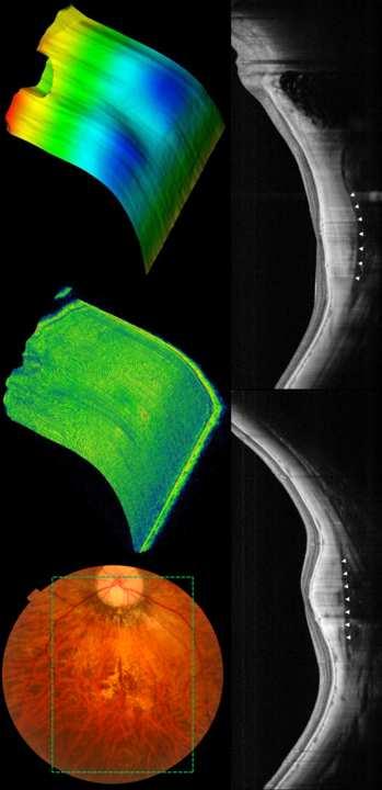
18 1 D FEATURES OF DOME-SHAPED MACULA. Ellabban AA, et al. Page FIGURE CAPTIONS Figure 1. Parameters associated with the dome-shaped macular configuration as measured from a multi-averaged vertical section obtained with swept-source optical coherence tomography. At the fovea, three parameters were measured: retinal thickness (blue line), choroidal thickness (yellow line), and scleral thickness (dashed white double arrow). The height of the inward bulge of the retinal pigment epithelium (red double arrow) was measured above the tangent plane (red dashed line) at the bottom of the posterior pole. At points 00 µm superior and 00 µm inferior to the fovea, the choroidal (yellow line) and scleral (dashed white double arrow) thicknesses were measured. In the horizontal scan, choroidal and scleral thicknesses were also measured at 00 µm temporal and nasal to the fovea. White arrowheads indicate the outer scleral border. Figure. Eyes with high myopia that show dome-shaped macular configuration within the posterior staphyloma. (Top left) Fundus photograph. (Top middle and right) Multi-averaged vertical (middle) and horizontal (right) scans through the fovea obtained by swept-source optical coherence tomography (OCT). (Top row) Both vertical and horizontal OCT scans show an inward convexity of the retinal pigment epithelium (RPE) within the staphyloma. The inward bulge is seen more clearly in the vertical scan. The top of the bulge (arrow) is located beneath the fovea. (nd row) Both vertical and horizontal OCT scans show an inward convexity of the RPE. The top of the bulge (arrow) is located at the nasal side of the fovea. (rd-th rows) Vertical OCT scans show clearly an inward bulge of RPE within the posterior staphyloma. The top of the bulge (arrows) is located beneath the fovea. The horizontal scan shows an almost flat contour of the RPE. Figure. An eye with high myopia that shows a band-shaped ridge within the posterior staphyloma. (Top left) Fundus photograph of the right eye of a -year-old woman (/ OD). Axial length was. mm and refractive error was -1. diopters. The dashed green rectangle outlines the area (1 x mm ) scanned by swept-source optical coherence tomography (OCT). (Top middle) Three-dimensional reconstructed image obtained by the swept-source OCT shows an inward convexity of the retina in the posterior pole. (Top right) Three-dimensional image reconstructed by segmentation of the retinal pigment epithelium (RPE) shows two outward concavities within the posterior staphyloma. A horizontal ridge is formed between these two outward concavities. (Bottom left) A vertical 1-mm line scan shows an inward bulge of the RPE beneath the fovea. The height of the RPE line above the tangent plane was µm. The external
19 1 D FEATURES OF DOME-SHAPED MACULA. Ellabban AA, et al. Page surface of the sclera shows a rather smooth curvature (white arrowheads). The sclera shows marked thinning at the areas of the two concavities and seems to be relatively thick beneath the fovea. (Bottom right) A horizontal 1-mm line scan shows an almost flat contour of the RPE. The scleral thinning at the parafoveal area is less evident, compared with the areas of the two outward concavities within the staphyloma. Figure. An eye with high myopia that shows a dome-shaped convexity within the posterior staphyloma. (Top left) Fundus photograph of the right eye of a -year-old woman (/ OD). Refractive error was -.0 diopters and the axial length was. mm. The dashed green rectangle outlines the area (1 x mm ) scanned by swept-source optical coherence tomography (OCT). Vertical and horizontal multi-averaged scans of 1 mm were obtained along the green arrows. (Top middle) Three-dimensional reconstructed image obtained by the swept-source optical coherence tomography shows an inward convexity of the retina in the posterior pole. (Top right) Three-dimensional image reconstructed by segmentation of the retinal pigment epithelium (RPE) shows the typical dome-shaped convexity within the staphyloma. (Bottom left) A vertical 1-mm line scan clearly shows an inward bulge of the RPE beneath the fovea. The height of the bulge of the RPE line above the tangent plane is µm. The external surface of the sclera is seen clearly (white arrowheads) and the sclera shows uneven thinning in the posterior pole, being relatively thick beneath the fovea. (Bottom right) A horizontal 1-mm line scan also shows an inward convexity in the posterior pole.
20 Tables D FEATURES OF DOME-SHAPED MACULA. Ellabban AA, et al. Table 1. Characteristics of Eyes with a Dome-Shaped Macula Number of eyes/patients / Sex (male/female) / Age (years). ±. ( to ) Axial length (mm). ±. (. to.) Refractive error (diopters). ±. (. to.0) Visual acuity (logmar) 0. ± 0. ( 0. to 1.) Intraocular pressure (mmhg). ±. ( to ) Height of inward bulge a (μm). ±. (.0 to.0) Foveal retinal thickness (μm) 0. ±. (0.0 to.) Foveal choroidal thickness (μm). ±.0 (.0 to 1.) Foveal scleral thickness (μm). ±. (.0 to.0) Type of posterior staphyloma I II III IX LogMAR, logarithm of minimal angle of resolution. a The height of the inward bulge was measured from a presumed line tangent to the outer surface of retinal pigment epithelium at the bottom of the posterior staphyloma in the vertical section of optical coherence tomography.
21 D FEATURES OF DOME-SHAPED MACULA. Ellabban AA, et al. Table. Choroidal and Scleral Thickness within the Macular Area in Eyes with a Dome-Shaped Macula Choroidal Thickness (µm) (P value a ) Scleral Thickness (µm) (P value a, P value b ) Subfovea. ±.0. ±. (NA, <.001) Parafovea at 00 µm superiorly. ±. (.0). ±.1 (<.001, <.001) Parafovea at 00 µm inferiorly.1 ±. (.). ± 1.1 (<.001,.001) Parafovea at 00 µm temporally.0 ±. (.).0 ±. (<.001,.0) Parafovea at 00 µm nasally. ±.0 (.0) 0. ±. (<.001, NA) a P values, compared with the values in the fovea; b P values, compared with the values in the nasal area (one way analysis of variance with Tukey post hoc analysis).
22 D FEATURES OF DOME-SHAPED MACULA. Ellabban AA, et al. Table. Complications in the Eyes with a Dome-Shaped Macula Choroidal neovascularization (.%) Serous retinal detachment (.%) Diffuse chorioretinal atrophy (.%) Patchy chorioretinal atrophy (.%) Lamellar macular hole (.%) Full-thickness macular hole 1 (1.%) Foveal schisis 1 (1.%) Extrafoveal retinal schisis (.%)
23 D FEATURES OF DOME-SHAPED MACULA. Ellabban AA, et al. Table. Characteristics of the Eyes with Dome-Shaped Macula with or without Choroidal Neovascularization Eyes without Choroidal Neovascularization Eyes with Choroidal Neovascularization P value Number of eyes (%) (.%) (.%) Sex (male/female) / /.0 a Age (years). ± 1.. ±.0. b Axial length (mm). ±.. ±.. b Refractive error (diopters). ±..0 ±..0 b Visual acuity (logmar) 0. ± ± b Foveal retinal thickness (μm) 0.1 ±.0 1. ± 1.. b Foveal choroidal thickness (μm). ±.. ±.. b Foveal scleral thickness (μm).0 ±.. ±.. b Height of the inward bulge c (μm). ±. 1.0 ±..0 b a Fischer exact test. b Unpaired t-test. c Height of the inward bulge was measured from the presumed line tangent to the outer surface of retinal pigment epithelium at the bottom of the posterior staphyloma on vertical optical coherence tomography scans. LogMAR, logarithm of minimal angle of resolution.

24 Figure 1 Click here to download high resolution image

25 Figure Click here to download high resolution image

26 Figure Click here to download high resolution image

27 Figure Click here to download high resolution image
28 Supplementary Figure 1

29 Supplementary Figure
30 Supplementary Material Legend D FEATURES OF DOME-SHAPED MACULA. Ellabban AA, et al. Page 0 Legend of Supplemental Material Supplemental figure 1. An eye with high myopia that shows a band-shaped ridge within the posterior staphyloma. (Top left) Fundus photograph of the right eye of a -year-old man (/ OD). Axial length was. mm and refractive error was -. diopters. The dashed green rectangle outlines the area (1 x mm ) scanned by swept-source optical coherence tomography (OCT). (Top middle) Three-dimensional reconstructed image obtained by the swept-source OCT shows an inward convexity of the retina at the posterior pole. (Top right) Three-dimensional image of retinal pigment epithelium (RPE) shows two outward concavities within the posterior staphyloma. A horizontal ridge is formed between these two concavities. (Bottom left) A vertical 1-mm line scan shows an inward bulge of the RPE beneath the fovea. The height of elevation of the RPE line above the tangent plane is µm. The external surface of the sclera shows a rather smooth curvature (white arrowheads). The sclera shows marked thinning in the areas of the two concavities and seems to be relatively thick beneath the fovea. (Bottom right) A horizontal 1-mm line scan shows that the contour of the RPE is almost flat. The scleral thinning at the parafoveal area is less evident, compared with the areas of the two outer concavities within the staphyloma. Supplemental figure. An eye with high myopia that shows a band-shaped ridge within the posterior staphyloma. (Top left) Fundus photograph of the right eye of a -year-old woman (/ OD). Refractive error was -1. diopters. The dashed green rectangle outlines the area scanned by swept-source optical coherence tomography (OCT). (Top middle) Three-dimensional reconstructed image obtained by the swept-source OCT shows an inward convexity of the retina at the posterior pole. (Top right) Three-dimensional image of retinal pigment epithelium (RPE) shows two outward concavities within the staphyloma, with a horizontal ridge between these two concavities. (Bottom left) A vertical 1-mm line scan shows an inward bulge of the RPE beneath the fovea. The height of elevation of the RPE line above the tangent plane is µm. The external surface of the sclera shows a rather smooth curvature (white arrowheads). The sclera shows marked thinning at the areas of the two concavities and seems to be relatively thick beneath the fovea. (Bottom right) A horizontal 1-mm line scan shows an almost flat contour of the RPE. The scleral thinning at the parafoveal area is less evident than within areas of the two outward concavities. Supplemental Video 1. A three-dimensional movie of an eye with dome-shaped macular configuration (shown in Figure ). Three-dimensional data sets covering an area of 1 x
31 D FEATURES OF DOME-SHAPED MACULA. Ellabban AA, et al. Page 1 mm were obtained with swept-source optical coherence tomography using a raster scan protocol of (horizontal) x 1 (vertical) A-scans. The three-dimensional constructed image shows an inward convexity of the retina at the posterior pole. By segmentation of the retinal pigment epithelium line, a reconstructed image of the posterior pole clearly shows two outward concavities in the macular area with a horizontal ridge between these two concavities.
DOME SHAPED MACULOPATHY. Ιωάννης Ν. Βαγγελόπουλος Χειρ. Οφθαλμίατρος - Βόλος
 DOME SHAPED MACULOPATHY Ιωάννης Ν. Βαγγελόπουλος Χειρ. Οφθαλμίατρος - Βόλος DOME SHAPED MACULOPATHY-DEFINITIONS The entity Dome Shaped Macula ( DSM ) was first described by Gaucher and associates in 2008
Abstracts DRI OCT-1. DRI OCT-1 See, Discover, Explore. Invest Ophthalmol Vis Sci Jul 1;52(8): Print 2011 Jul.
: Print 2011 Jul.") Abstracts Invest Ophthalmol Vis Sci. 2011 Jul 1;52(8):4971-8. Print 2011 Jul. Macular choroidal thickness and volume in normal subjects measured by swept-source optical coherence tomography. Hirata M,
Abstracts Invest Ophthalmol Vis Sci. 2011 Jul 1;52(8):4971-8. Print 2011 Jul. Macular choroidal thickness and volume in normal subjects measured by swept-source optical coherence tomography. Hirata M,
Optical Coherence Tomography Findings in Highly Myopic Eyes following Cataract Surgery
 Optical Coherence Tomography Findings in Highly Myopic Eyes following Cataract Surgery Fedra Hajizadeh, MD 1 Mohammad Riazi Esfahani, MD 1,2 Hooshang Faghihi, MD 3 Mehdi Khanlari, MD 4 Abstract Purpose:
Measurement of Choroidal Thickness in Normal Eyes Using 3D OCT-1000 Spectral Domain Optical Coherence Tomography
 pissn: 111-8942 eissn: 292-9382 Korean J Ophthalmol 212;26(4):255-259 http://dx.doi.org/1.3341/kjo.212.26.4.255 Original Article Measurement of Choroidal Thickness in Normal Eyes Using 3D OCT-1 Spectral
Dehiscence of detached internal limiting membrane in eyes with myopic traction maculopathy with spontaneous resolution
 Hirota et al. BMC Ophthalmology 2014, 14:39 RESEARCH ARTICLE Open Access Dehiscence of detached internal limiting membrane in eyes with myopic traction maculopathy with spontaneous resolution Kazunari
Kenji; Akiba, Masahiro; Yoshimura,
 Macular choroidal thickness and vol Titlestreaks measured by swept source op tomography. Ellabban, Abdallah A; Tsujikawa, Ak Author(s) Ogino, Ken; Hangai, Masanori; Ooto, Kenji; Akiba, Masahiro; Yoshimura,
International Journal of Ophthalmic Research
 International Journal of Ophthalmic Research Online Submissions: http://www.ghrnet.org/index./ijor/ doi:10.17554/j.issn.2409-5680.2017.03.55 Int. J. Ophthalmic Res 2017 June; 3(2): 226-230 ISSN 2409-5680
R&M Solutions
 Mohamed Hosny El-Bradey, MD., Assistant Professor of Ophthalmology, Tanta University. Wael El Haig, MD., Professor of Ophthalmology. Zagazeeg University. 1 Myopic CNV is considered the most common vision
Clinical Study Choroidal Thickness in Eyes with Unilateral Ocular Ischemic Syndrome
 Hindawi Publishing Corporation Journal of Ophthalmology Volume 215, Article ID 62372, 5 pages http://dx.doi.org/1.1155/215/62372 Clinical Study Choroidal Thickness in Eyes with Unilateral Ocular Ischemic
Myopia is a major cause of visual impairment in many
 Retina Association between Choroidal Morphology and Anti- Vascular Endothelial Growth Factor Treatment Outcome in Myopic Choroidal Neovascularization Seong Joon Ahn, 1 Se Joon Woo, 1 Ko Eun Kim, 2 and
Reproducibility of Choroidal Thickness Measurements Across Three Spectral Domain Optical Coherence Tomography Systems
 Reproducibility of Choroidal Thickness Measurements Across Three Spectral Domain Optical Coherence Tomography Systems The MIT Faculty has made this article openly available. Please share how this access
Observation of Posterior Precortical Vitreous Pocket Using Swept-Source Optical Coherence Tomography
 Anatomy and Pathology Observation of Posterior Precortical Vitreous Pocket Using Swept-Source Optical Coherence Tomography Hirotaka Itakura, Shoji Kishi, Danjie Li, and Hideo Akiyama Department of Ophthalmology,
RETINAL PIGMENT EPITHELIUM UNDULATIONS IN ACUTE STAGE OF VOGT-KOYANAGI-HARADA DISEASE
 RETINAL PIGMENT EPITHELIUM UNDULATIONS IN ACUTE STAGE OF VOGT-KOYANAGI-HARADA DISEASE Biomarker for Functional Outcomes After High-Dose Steroid Therapy KOUHEI HASHIZUME, MD,* YUTAKA IMAMURA, MD, TAKAMITSU
Focal choroidal excavation (FCE) is a newly recognized. Association of Focal Choroidal Excavation With Age-Related Macular Degeneration.
 is a newly recognized. Association of Focal Choroidal Excavation With Age-Related Macular Degeneration.") Retina Association of Focal Choroidal Excavation With Age-Related Macular Degeneration Yoshimasa Kuroda, 1 Akitaka Tsujikawa, 1 Sotaro Ooto, 1 Kenji Yamashiro, 1 Akio Oishi, 1 Hideo Nakanishi, 1 Kyoko
Retina Association of Focal Choroidal Excavation With Age-Related Macular Degeneration Yoshimasa Kuroda, 1 Akitaka Tsujikawa, 1 Sotaro Ooto, 1 Kenji Yamashiro, 1 Akio Oishi, 1 Hideo Nakanishi, 1 Kyoko
Choroidal Mapping; a Novel Approach for Evaluating Choroidal Thickness and Volume
 Imaging Technique Choroidal Mapping; a Novel Approach for Evaluating Choroidal Thickness and Volume Jila Noori 1, MD; Mohammad Riazi Esfahani 1,2, MD Fedra Hajizadeh 2, MD; Mohammad-Mehdi Zaferani 1, MD
Cirrus TM HD-OCT. Details define your decisions
 Cirrus TM HD-OCT Details define your decisions 2 With high-definition OCT Carl Zeiss Meditec takes you beyond standard spectral domain Built on 10 years experience at the vanguard of innovation, Carl Zeiss
NIH Public Access Author Manuscript JAMA Ophthalmol. Author manuscript; available in PMC 2013 September 10.
 NIH Public Access Author Manuscript Published in final edited form as: JAMA Ophthalmol. 2013 May ; 131(5): 693 694. doi:10.1001/jamaophthalmol.2013.692. Effect of Intravitreous Anti Vascular Endothelial
Fellow Eye Findings of Highly Myopic Subjects Operated for Retinal Detachment Associated with a Macular Hole
 Fellow Eye Findings of Highly Myopic Subjects Operated for Retinal Detachment Associated with a Macular Hole Guido Ripandelli, MD, Andrea Maria Coppé, MD, Vincenzo Parisi, MD, Mario Stirpe, MD Purpose:
Citation. As Published Publisher. Version
 Effect of Intravitreous Anti Vascular Endothelial Growth Factor Therapy on Choroidal Thickness in Neovascular Age-Related Macular Degeneration Using Spectral-Domain The MIT Faculty has made this article
Enhanced depth imaging (EDI) optical coherence tomography
 optical coherence tomography") Retina Macular Choroidal Thickness and Volume in Healthy Pediatric Individuals Measured by Swept-Source Optical Coherence Tomography Toshihiko Nagasawa, 1,2 Yoshinori Mitamura, 3 Takashi Katome, 3 Kayo
Retina Macular Choroidal Thickness and Volume in Healthy Pediatric Individuals Measured by Swept-Source Optical Coherence Tomography Toshihiko Nagasawa, 1,2 Yoshinori Mitamura, 3 Takashi Katome, 3 Kayo
Advances in OCT Murray Fingeret, OD
 Disclosures Advances in OCT Murray Fingeret, OD Consultant Alcon, Allergan, Bausch & Lomb, Carl Zeiss Meditec, Diopsys, Heidelberg Engineering, Reichert, Topcon Currently Approved OCT Devices OCT Devices
JMSCR Vol 07 Issue 01 Page January 2019
 www.jmscr.igmpublication.org Impact Factor (SJIF): 6.379 Index Copernicus Value: 79.54 ISSN (e)-2347-176x ISSN (p) 2455-0450 DOI: https://dx.doi.org/10.18535/jmscr/v7i1.64 Enhanced Depth imaging optical
Diagnosis and Treatment of Myopic Maculopathy. Tae Yokoi, MD, and Kyoko Ohno-Matsui, MD, PhD
 REVIEW ARTICLE Diagnosis and Treatment of Myopic Maculopathy Tae Yokoi, MD, and Kyoko Ohno-Matsui, MD, PhD Abstract: Visual impairment resulting from pathologic myopia is a serious issue worldwide. This
Choroidal Thickness and Biometric Markers for the Screening of Lacquer Cracks in Patients with High Myopia
 Choroidal Thickness and Biometric Markers for the Screening of Lacquer Cracks in Patients with High Myopia Nan-Kai Wang 1,2 *, Chi-Chun Lai 1,2, Chai Lin Chou 3, Yen-Po Chen 1,2, Lan-Hsin Chuang 1,4, An-Ning
Case Report Peripapillary Intrachoroidal Cavitation in Myopia Evaluated with Multimodal Imaging Comprising (En-Face) Technique
 Technique") Case Reports in Ophthalmological Medicine Volume 2015, Article ID 890876, 5 pages http://dx.doi.org/10.1155/2015/890876 Case Report Peripapillary Intrachoroidal Cavitation in Myopia Evaluated with Multimodal
Case Reports in Ophthalmological Medicine Volume 2015, Article ID 890876, 5 pages http://dx.doi.org/10.1155/2015/890876 Case Report Peripapillary Intrachoroidal Cavitation in Myopia Evaluated with Multimodal
IN MANY DEVELOPED COUNTRIES, A
 LINIAL SIENES haracteristics of Peripapillary etachment in Pathologic Myopia Noriaki Shimada, M; Kyoko Ohno-Matsui, M; Takeshi Yoshida, M; Kenjiro Yasuzumi, M; Ariko Kojima, M; Kanako Kobayashi, M; Soh
Assessment of Effects of Different Mydriatics on Choroidal Thickness by Examining Anterior Chamber Parameters
 Open Access World Journal of Ophthalmology & Vision Research Research Article Copyright All rights are reserved by Fahmy RM Assessment of Effects of Different Mydriatics on Choroidal Thickness by Examining
CLINICAL SCIENCES. Optical Coherence Tomography Findings in Myopic Traction Maculopathy
 Optical Coherence Tomography Findings in Myopic Traction Maculopathy Giacomo Panozzo, MD; Andrea Mercanti, MD CLINICAL SCIENCES Objective: To describe the features and incidence of epiretinal traction
Myopic Choroidal Neovascularization
 Myopic Choroidal Neovascularization A 10-year Follow-up Takeshi Yoshida, MD, Kyoko Ohno-Matsui, MD, Kenjiro Yasuzumi, MD, Ariko Kojima, MD, Noriaki Shimada, MD, Soh Futagami, MD, Takashi Tokoro, MD, Manabu
Optical Coherence Tomography (OCT) in Uveitis Piergiorgio Neri, BMedSc, MD, PhD Head Ocular Immunology Unit
 in Uveitis Piergiorgio Neri, BMedSc, MD, PhD Head Ocular Immunology Unit") The Eye Clinic Polytechnic University of Marche Head: Prof Alfonso Giovannini November, 1991 Optical Coherence Tomography (OCT) in Uveitis Piergiorgio Neri, BMedSc, MD, PhD Head Ocular Immunology Unit
The Eye Clinic Polytechnic University of Marche Head: Prof Alfonso Giovannini November, 1991 Optical Coherence Tomography (OCT) in Uveitis Piergiorgio Neri, BMedSc, MD, PhD Head Ocular Immunology Unit
Title: OCT Analysis Workshop: Interpretation of OCT printouts
 Title: OCT Analysis Workshop: Interpretation of OCT printouts Authors: David Yang, OD, FAAO Staff Optometrist, VA Palo Alto Health Care System Associate Clinical Professor, UC Berkeley School of Optometry
Cirrus TM HD-OCT. Details defi ne your decisions
 Cirrus TM HD-OCT Details defi ne your decisions 2 With high-defi nition OCT Carl Zeiss Meditec takes you beyond standard spectral domain Built on 10 years experience at the vanguard of innovation, Carl
Parapapillary Gamma Zone and Axial Elongation Associated Optic Disc Rotation: The Beijing Eye Study
 Anatomy and Pathology/Oncology Parapapillary Gamma Zone and Axial Elongation Associated Optic Disc Rotation: The Beijing Eye Study Jost B. Jonas, 1,2 Ya Xing Wang, 1 Qi Zhang, 1 Yuan Yuan Fan, 3 Liang
Optical Coherence Tomograpic Features in Idiopathic Retinitis, Vasculitis, Aneurysms and Neuroretinitis (IRVAN)
") Columbia International Publishing Journal of Ophthalmic Research (2014) Research Article Optical Coherence Tomograpic Features in Idiopathic Retinitis, Vasculitis, Aneurysms and Neuroretinitis (IRVAN)
Columbia International Publishing Journal of Ophthalmic Research (2014) Research Article Optical Coherence Tomograpic Features in Idiopathic Retinitis, Vasculitis, Aneurysms and Neuroretinitis (IRVAN)
Unlike other cranial nerves, the optic nerve (ON) is surrounded
 is surrounded") Clinical and Epidemiologic Research Imaging Retrobulbar Subarachnoid Space around Optic Nerve by Swept-Source Optical Coherence Tomography in Eyes with Pathologic Myopia Kyoko Ohno-Matsui, 1 Masahiro Akiba,
Clinical and Epidemiologic Research Imaging Retrobulbar Subarachnoid Space around Optic Nerve by Swept-Source Optical Coherence Tomography in Eyes with Pathologic Myopia Kyoko Ohno-Matsui, 1 Masahiro Akiba,
Yasser R. Serag, MD Tamer Wasfi, MD El- Saied El-Dessoukey, MD Magdi S. Moussa, MD Anselm Kampik, MD
 Microperimetric Evaluation of Brilliant Blue G- assisted Internal Limiting Membrane Peeling By Yasser R. Serag, MD Tamer Wasfi, MD El- Saied El-Dessoukey, MD Magdi S. Moussa, MD Anselm Kampik, MD The internal
OPTIC DISC PIT Pathogenesis and Management OPTIC DISC PIT
 OPTIC DISC PIT Pathogenesis and Management Abdel-Latif Siam Ain Shams University Cairo Egypt OPTIC DISC PIT Congenital pit is an atypical coloboma usually located on the temporal edge of the disc, associated
Oishi A, Miyamoto K, Yoshimura N. Etiology of carotid cavernous fistula in Japanese. Jpn J Ophthalmol. 2009;53:40-43.
 Kimura T, Takagi H, Miyamoto K, Kita M, Watanabe D, Yoshimura N. Macular hole with epiretinal membrane after triamcinolone-assisted vitrectomy for proliferative diabetic retinopathy. Retinal Cases Brief
Myopia is attributable to maculopathy, such as a retinal
 Retina Macular Microstructures and Prognostic Factors in Myopic Subretinal Hemorrhages Tomoko Asai, Yasushi Ikuno, and Kohji Nishida Department of Ophthalmology, Osaka University Graduate School of Medicine,
Acquired vitelliform detachment in patients with subretinal drusenoid deposits (reticular pseudodrusen)
") Zurich Open Repository and Archive University of Zurich Main Library Strickhofstrasse 39 CH-8057 Zurich www.zora.uzh.ch Year: 2011 Acquired vitelliform detachment in patients with subretinal drusenoid
Zurich Open Repository and Archive University of Zurich Main Library Strickhofstrasse 39 CH-8057 Zurich www.zora.uzh.ch Year: 2011 Acquired vitelliform detachment in patients with subretinal drusenoid
Structural examina.on: Imaging
 ManaMa: Glaucoma Structural examina.on: Imaging Luís Abegão Pinto, MD, PhD Department of Ophthalmology CHLC Lisbon Faculty of Medicine, Lisbon University 1 11-10- 2013 Structural changes Qualitative changes
ZEISS AngioPlex OCT Angiography. Clinical Case Reports
 Clinical Case Reports Proliferative Diabetic Retinopathy (PDR) Case Report 969 PROLIFERATIVE DIABETIC RETINOPATHY 1 1-year-old diabetic female presents for follow-up of proliferative diabetic retinopathy
OCT Angiography in Primary Eye Care
 OCT Angiography in Primary Eye Care An Image Interpretation Primer Julie Rodman, OD, MS, FAAO and Nadia Waheed, MD, MPH Table of Contents Diabetic Retinopathy 3-6 Choroidal Neovascularization 7-9 Central
doi: /j.ajo
 doi: 10.1016/j.ajo.2011.05.037 1 Evaluation of the Choroidal Thickness Using High Penetration Optical Coherence Tomography with Long Wavelength in Highly Myopic Normal Tension Glaucoma SHINICHI USUI, MD,
The Egyptian Journal of Hospital Medicine (January 2018) Vol. 70 (4), Page
 Vol. 70 (4), Page") The Egyptian Journal of Hospital Medicine (January 2018) Vol. 70 (4), Page 670-680 Enhanced Depth Imaging Optical Coherence Tomography: A Study of the Choroid in High Axial Myopia Samar O Zayed, Sherif
The Egyptian Journal of Hospital Medicine (January 2018) Vol. 70 (4), Page 670-680 Enhanced Depth Imaging Optical Coherence Tomography: A Study of the Choroid in High Axial Myopia Samar O Zayed, Sherif
HHS Public Access Author manuscript Ophthalmic Surg Lasers Imaging Retina. Author manuscript; available in PMC 2016 January 14.
 High-Speed Ultrahigh-Resolution OCT of Bruch s Membrane in Membranoproliferative Glomerulonephritis Type 2 Mehreen Adhi, MD, Sarah P. Read, MD, PhD, Jonathan J. Liu, PhD, James G. Fujimoto, PhD, and Jay
The choroid, a vascular meshwork between the retina and
 Retina Spatial Distribution of Posterior Pole Choroidal Thickness by Spectral Domain Optical Coherence Tomography Yanling Ouyang, 1 Florian M. Heussen, 1 Nils Mokwa, 1 Alexander C. Walsh, 1 Mary K. Durbin,
Mark Dunbar: Disclosure
 Important Things to Understand About OCT Mark T. Dunbar, O.D., F.A.A.O. Bascom Palmer Eye Institute University of Miami, School of Medicine Mark Dunbar: Disclosure Optometry Advisory Board for: Allergan
Case Report: Indocyanine Green Dye Leakage from Retinal Artery in Branch Retinal Vein Occlusion
 Case Report: Indocyanine Green Dye Leakage from Retinal Artery in Branch Retinal Vein Occlusion Hiroki Fujita, Kyoko Ohno-Matsui, Soh Futagami and Takashi Tokoro Department of Visual Science, Tokyo Medical
In 1990 our group first described reticular pseudodrusen as a. Choroidal Changes Associated with Reticular Pseudodrusen. Retina
 Retina Choroidal Changes Associated with Reticular Pseudodrusen Giuseppe Querques, 1,2 Lea Querques, 1,2 Raimondo Forte, 1 Nathalie Massamba, 1 Florence Coscas, 1 and Eric H. Souied 1 PURPOSE. To analyze
When optical coherence tomography (OCT)
") Macular Imaging: SD-OCT in nterior Segment Surgical Practice Many pathologic processes of the macula can be visualized or quantified only with this modality. y Steven G. Safran, MD When optical coherence
Macular Imaging: SD-OCT in nterior Segment Surgical Practice Many pathologic processes of the macula can be visualized or quantified only with this modality. y Steven G. Safran, MD When optical coherence
Retinal Nerve Fiber Layer Measurements in Myopia Using Optical Coherence Tomography
 Original Article Philippine Journal of OPHTHALMOLOGY Retinal Nerve Fiber Layer Measurements in Myopia Using Optical Coherence Tomography Dennis L. del Rosario, MD and Mario M. Yatco, MD University of Santo
Seiji T. Takagi, Yoshiyuki Kita, Asuka Takeyama, and Goji Tomita. 1. Introduction. 2. Subjects and Methods
 Ophthalmology Volume 2011, Article ID 914250, 5 pages doi:10.1155/2011/914250 Clinical Study Macular Retinal Ganglion Cell Complex Thickness and Its Relationship to the Optic Nerve Head Topography in Glaucomatous
Distribution and Severity of Myopic Maculopathy Among Highly Myopic Eyes
 Clinical and Epidemiologic Research Distribution and Severity of Myopic Maculopathy Among Highly Myopic Eyes Ou Xiao, 1 Xinxing Guo, 1,2 Decai Wang, 1 Monica Jong, 3,4 Pei Ying Lee, 5 Linxing Chen, 1 Ian
Published on Points de Vue International Review of Ophthalmic Optics (http://www.pointsdevue.com)
") Published on Points de Vue International Review of Ophthalmic Optics (http://www.pointsdevue.com) Home > OCT and retinal pathologies OCT and retinal pathologies Sylvain AURIOL, Véronique PAGOT-MATHIS e-mail
Published on Points de Vue International Review of Ophthalmic Optics (http://www.pointsdevue.com) Home > OCT and retinal pathologies OCT and retinal pathologies Sylvain AURIOL, Véronique PAGOT-MATHIS e-mail
Fundus Autofluorescence. Jonathan A. Micieli, MD Valérie Biousse, MD
 Fundus Autofluorescence Jonathan A. Micieli, MD Valérie Biousse, MD The retinal pigment epithelium (RPE) has many important functions including phagocytosis of the photoreceptor outer segments Cone Rod
Swept-Source OCT Angiography: SS OCT Angio TM
 Swept-Source OCT Angiography: SS OCT Angio TM Not available in all countries, please check with your distributor. 2015.09 Swept-Source OCT Angiography: SS OCT Angio TM Introduction Optical coherence tomography
Macular retinal and choroidal thickness in unilateral relentless placoid chorioretinitis analyzed by swept-source optical coherence tomography
 Dolz-Marco et al. Journal of Ophthalmic Inflammation and Infection 2014, 4:24 ORIGINAL RESEARCH Open Access Macular retinal and choroidal thickness in unilateral relentless placoid chorioretinitis analyzed
Measurement of Subfoveal Choroidal Thickness Using Spectral Domain Optical Coherence Tomography
 c l i n i c a l s c i e n c e Measurement of Subfoveal Choroidal Thickness Using Spectral Domain Optical Coherence Tomography Emily A. McCourt, MD; Brian C. Cadena, PhD; Cullen J. Barnett, CRA; Antonio
Indocyanine Green Angiographic Findings of Chorioretinal Folds
 Indocyanine Green Angiographic Findings of Chorioretinal Folds Miho Haruyama, Mitsuko Yuzawa, Akiyuki Kawamura, Chikayo Yamazaki and Youko Matsumoto Department of Ophthalmology, Nihon University School
OCT Interpretation in Retinal Disease
 OCT Interpretation in Retinal Disease Jay M. Haynie, OD, FAAO Financial Disclosure I have received honoraria or am on the advisory board for the following companies: Carl Zeiss Meditec Advanced Ocular
Method for comparing visual field defects to local RNFL and RGC damage seen on frequency domain OCT in patients with glaucoma.
 Method for comparing visual field defects to local RNFL and RGC damage seen on frequency domain OCT in patients with glaucoma. Donald C. Hood 1,2,* and Ali S. Raza 1 1 Department of Psychology, Columbia
Scleral buckling is a common surgical procedure for
 Suprachoroidal Buckling Technique A novel, less-invasive treatment option for rhegmatogenous retinal detachment and vitreoretinal interface pathologies. By Yusuke Oshima, MD; Ehab N. El Rayes, MD; Nagakazu
Clinical and optic coherence tomography findings of focal choroidal excavation in Chinese patients
 Guo et al. BMC Ophthalmology 2014, 14:63 RESEARCH ARTICLE Open Access Clinical and optic coherence tomography findings of focal choroidal excavation in Chinese patients Jie Guo 1, Lu Zhong 2, Chunhui Jiang
The Effect of Posterior Scleral Reinforcement for High Myopia Macular Splitting
 The Journal of International Medical Research 2011; 39: 662 666 The Effect of Posterior Scleral Reinforcement for High Myopia Macular Splitting XUEYING JI 1, *, JINGUO WANG 2, *, JINSONG ZHANG 1, HONGLIANG
OCT Assessment of the Vitreoretinal Relationship in CSME
 December 2007 Sonia Rani John et al. - IFIS 375 ORIGINAL ARTICLE OCT Assessment of the Vitreoretinal Relationship in CSME Dr. Manoj S. DNB FRCS, Dr. Unnikrishnan Nair MS DO FRCS, Dr. Gargi Sathish MS Introduction
Analysis of Peripapillary Atrophy Using Spectral Domain Optical Coherence Tomography
 Analysis of Peripapillary Atrophy Using Spectral Domain Optical Coherence Tomography The MIT Faculty has made this article openly available. Please share how this access benefits you. Your story matters.
Optical coherence tomography (OCT) is a new noninvasive
 is a new noninvasive") Use of Optical Coherence Tomography to Assess Variations in Macular Retinal Thickness in Myopia Marcus C. C. Lim, 1 Sek-Tien Hoh, 1 Paul J. Foster, 1,2 Tock-Han Lim, 3,4 Sek-Jin Chew, 1 Steve K. L. Seah,
Use of Optical Coherence Tomography to Assess Variations in Macular Retinal Thickness in Myopia Marcus C. C. Lim, 1 Sek-Tien Hoh, 1 Paul J. Foster, 1,2 Tock-Han Lim, 3,4 Sek-Jin Chew, 1 Steve K. L. Seah,
Spontaneous Large Serous Retinal Pigment Epithelial Tear
 This is an Open Access article licensed under the terms of the Creative Commons Attribution-NonCommercial-NoDerivs 3.0 License (www.karger.com/oa-license), applicable to the online version of the article
Il contributo dell'angio-oct: valutazione integrata della componente nervosa e vascolare della malattia glaucomatosa
 SIMPOSIO G.O.A.L. - LE NUOVE FRONTIERE DIAGNOSTICHE E LE LINEE DI INDIRIZZO AMBULATORIALI DEL GLAUCOMA Coordinatore e moderatore: D. Mazzacane Presidente: L. Rossetti Il contributo dell'angio-oct: valutazione
VISUAL ACUITY OUTCOMES OF RANIBIZUMAB TREATMENT IN PATHOLOGIC MYOPIC EYES WITH MACULAR RETINOSCHISIS AND CHOROIDAL NEOVASCULARIZATION
 VISUAL ACUITY OUTCOMES OF RANIBIZUMAB TREATMENT IN PATHOLOGIC MYOPIC EYES WITH MACULAR RETINOSCHISIS AND CHOROIDAL NEOVASCULARIZATION LALA CEKLIC, MD,* MARION R. MUNK, MD, PHD,* UTE WOLF-SCHNURRBUSCH,
A New Manual Retinal Thickness Measurement Protocol to Evaluate High Myopia Patients
 EURETINA Original Paper Received: November 20, 2012 Accepted after revision: May 19, 2013 Published online: August 28, 2013 A New Manual Retinal Thickness Measurement Protocol to Evaluate High Myopia Patients
3/23/2016. Diagnostic Services Taylor Pannell CRA, OCT-C. Services Available. Important info for the Tech to know. Visual Fields
 Services Available Diagnostic Services Taylor Pannell CRA, OCT-C Static and Kinetic Visual Fields Pachymetry Anterior and Posterior Segment OCT Fundus Photos FAF,FA,ICG Slit Lamp Photography Confocal HRT
PRIMUS 200 from ZEISS The essential OCT
 EN 00_00I The contents of the brochure may differ from the current status of approval of the product in your country. Please contact your regional representative for more information. Subject to change
Structural changes of the anterior chamber following cataract surgery during infancy
 Structural changes of the anterior chamber following cataract surgery during infancy Matthew Nguyen, Emory University Marla Shainberg, Emory University Allen Beck, Emory University Scott Lambert, Emory
Authors. Introduction. Introduction. Materials and Methods. Objective 10/27/2015
 Idiopathic Polypoidal Choroidal Vasculopathy (IPCV) in Thai Population Presenting with Choroidal Neovascularization (CNV) A multicenter study Authors Yonrawee Piyacomn 1, Chavakij Bhoomibunchoo 1, Yosanan
Moving forward with a different perspective
 Moving forward with a different perspective The Leader In Vision Diagnostics Offers A New Perspective Marco has served the eyecare community by offering exceptional lane products and automated high tech
PRIMUS 200 from ZEISS The essential OCT
 PRIMUS 200 from ZEISS The essential OCT Seeing beyond the surface. ZEISS PRIMUS 200 // INNOVATION MADE BY ZEISS Clear Visualization. Advanced Technology. Reliability. Essential elements of your first OCT.
EPIDEMIOLOGY. SECTION EDITOR: LESLIE HYMAN, PhD. Prevalence and Characteristics of Myopic Retinopathy in a Rural Chinese Adult Population
 EPIDEMIOLOGY SECTION EDITOR: LESLIE HYMAN, PhD Prevalence and Characteristics of Myopic Retinopathy in a Rural Chinese Adult Population The Handan Eye Study Li Qin Gao, MD; Wu Liu, MD; Yuan Bo Liang, MD,
Indocyanine Green Angiography Findings in Central Serous Chorioretinopathy
 IOSR Journal of Dental and Medical Sciences (IOSR-JDMS) e-issn: 2279-0853, p-issn: 2279-0861.Volume 15, Issue 5 Ver. III (May. 2016), PP 11-21 www.iosrjournals.org Indocyanine Green Angiography Findings
Effect of Internal Limiting Membrane Removal in Treatment of Retinal Detachment Caused by Myopic Macular Hole
 Korean J Ophthalmol Vol. 18:141-147, 2004 Effect of Internal Limiting Membrane Removal in Treatment of Retinal Detachment Caused by Myopic Macular Hole Heeyoon Cho, MD, Anho Choi, MD, Se Woong Kang, MD
Study of clinical significance of optical coherence tomography in diagnosis & management of diabetic macular edema
 Original Research Article Study of clinical significance of optical coherence tomography in diagnosis & management of diabetic macular edema Neha Kantilal Desai 1,*, Somesh Vedprakash Aggarwal 2, Sonali
Analysis of Changes in Corneal Shape and Refraction Following Scleral Buckling Surgery
 CLINICAL INVESTIGATIONS Analysis of Changes in Corneal Shape and Refraction Following Scleral Buckling Surgery Yuichi Okada, Seigo Nakamura, Eri Kubo, Namiki Oishi, Yukio Takahashi and Yoshio Akagi Department
DRI OCT Triton Series A Multimodal Swept Source OCT
 DRI OCT Triton Series A Multimodal Swept Source OCT Color Red-Free FA FAF Posterior Anterior See what others can t see. A Multimodal Swept Source OCT DEEP RANGE IMAGING Swept Source OCT imaging massively
Macular Hole Associated with Vogt-Koyanagi-Harada Disease at the Acute Uveitic Stage
 Published online: September 15, 2015 2015 The Author(s) Published by S. Karger AG, Basel 1663 2699/15/0063 0328$39.50/0 This article is licensed under the Creative Commons Attribution-NonCommercial 4.0
NIH Public Access Author Manuscript Ophthalmic Surg Lasers Imaging Retina. Author manuscript; available in PMC 2014 June 24.
 NIH Public Access Author Manuscript Published in final edited form as: Ophthalmic Surg Lasers Imaging Retina. 2014 ; 45(1): 32 37. doi:10.3928/23258160-20131220-04. Analysis of the Short Term Change in
Reports. Macular Thickness as a Potential Biomarker of Mild Alzheimer s Disease
 Reports Macular Thickness as a Potential Biomarker of Mild Alzheimer s Disease Although several postmortem findings in the retina of patients with Alzheimer s disease (AD) are available, 1 new biomarkers
Delayed Absorption of Subretinal Fluid after Retinal Reattachment Surgery and Associated Choroidal Features
 pissn: 1011-8942 eissn: 2092-9382 Korean J Ophthalmol 2017;31(5):402-411 https://doi.org/10.3341/kjo.2016.0033 Original Article Delayed Absorption of Subretinal Fluid after Retinal Reattachment Surgery
Diagnosis in AMD. Managing your AMD Patients
 Managing your AMD Patients Robert W. Dunphy, O.D., F.A.A.O. Diagnosis in AMD Have suspicion Identify relative risk Conduct surveillance Biometry Utilize technology to facilitate detection of change / stability
Optical Coherence Tomography in Diabetic Retinopathy. Mrs Samantha Mann Consultant Ophthalmologist Clinical Lead of SEL-DESP
 Optical Coherence Tomography in Diabetic Retinopathy Mrs Samantha Mann Consultant Ophthalmologist Clinical Lead of SEL-DESP Content OCT imaging Retinal layers OCT features in Diabetes Some NON DR features
Vitrectomy for Diabetic Cystoid Macular Edema
 Vitrectomy for Diabetic Cystoid Macular Edema Yukihiro Sato, Zeon Lee and Hiroyuki Shimada Department of Ophthalmology, Nihon University School of Medicine, Tokyo, Japan Purpose: We evaluated visual outcomes
Intravitreal Injection of Ranibizumab for Recovery of Macular Function in Eyes With Subfoveal Polypoidal Choroidal Vasculopathy
 Retina Intravitreal Injection of Ranibizumab for Recovery of Macular Function in Eyes With Subfoveal Polypoidal Choroidal Vasculopathy Ken Ogino, Akitaka Tsujikawa, Kenji Yamashiro, Sotaro Ooto, Akio Oishi,
S uperior segmental optic hypoplasia (SSOH), also termed
, also termed") 910 CLINICAL SCIENCE Optical coherence tomography of superior segmental optic hypoplasia K Unoki, N Ohba, W F Hoyt... See end of article for authors affiliations... Correspondence to: Kazuhiko Unoki, MD,
910 CLINICAL SCIENCE Optical coherence tomography of superior segmental optic hypoplasia K Unoki, N Ohba, W F Hoyt... See end of article for authors affiliations... Correspondence to: Kazuhiko Unoki, MD,
evaluation of vitreoretinal adhesions in exudative AMD using optical coherence tomography
 evaluation of vitreoretinal adhesions in exudative AMD using optical coherence tomography Dr. Mahmoud Alaa Abouhusssein, FRCO Lecturer of ophthalmology, Alexandria university Dr. Amir Ramadan Gomaa, MD
Efficacy of Anti-VEGF Agents in the Treatment of Age-Related Macular Degeneration
 Efficacy of Anti-VEGF Agents in the Treatment of Age-Related Macular Degeneration Marilita M. Moschos Abstract- Purpose: To evaluate by OCT and mf-erg the macular function in eyes with CNV due to ARMD
What Is O.C.T. and Why Should I Give A Rip? OCT & Me How Optical Coherence Tomography Changed the Life of a Small Town Optometrist 5/19/2014
 OCT & Me How Optical Coherence Tomography Changed the Life of a Small Town Optometrist Email: myoder@wcoil.com Mark A. Yoder, O.D. 107 N. Main Street PO Box 123 Bluffton, OH 45817 @yoderod 115.02 Histoplasma
Choroidal thickness and high myopia: a case control study of young Chinese men in Singapore
 Choroidal thickness and high myopia: a case control study of young Chinese men in Singapore Preeti Gupta, 1,2 Seang-Mei Saw, 1,2,3 Carol Y. Cheung, 1,2,4 Michael J. A. Girard, 1,5 Jean Martial Mari, 6
SOCT Copernicus REVO. * - Currently import and overlay are avaibale in manual mode only
 SOCT Copernicus REVO Easy Operation (Full auto & Auto mode) Auto alignment (Z-position, C-gate, Focus, Tomogram) Voice guide (support patient through examination) Powerful analysis tools Enhanced tomograms
RETINAL CONDITIONS RETINAL CONDITIONS
 GENERAL INFORMATION RETINAL CONDITIONS RETINAL CONDITIONS WHAT ARE RETINAL CONDITIONS? Retinal conditions affect the light-sensitive tissue at the back of eye known as the retina. They include diseases
Peripapillary circle of Zinn Haller revealed by fundus fluorescein angiography
 British Journal of Ophthalmology 1997;81:663 667 663 Department of Ophthalmology, School of Medicine, Hanyang University, Seoul, Korea M-K Ko D-S Kim Y-K Ahn Correspondence to: Myung-Kyoo Ko, MD, Department