Cellular Metabolism Modulates Ion Channels That Regulate Neuronal Excitability
|
|
|
- Tiffany Cannon
- 5 years ago
- Views:
Transcription
1 Cellular Metabolism Modulates Ion Channels That Regulate Neuronal Excitability The Harvard community has made this article openly available. Please share how this access benefits you. Your story matters Citation Lutas, Andrew Cellular Metabolism Modulates Ion Channels That Regulate Neuronal Excitability. Doctoral dissertation, Harvard University, Graduate School of Arts & Sciences. Citable link Terms of Use This article was downloaded from Harvard University s DASH repository, and is made available under the terms and conditions applicable to Other Posted Material, as set forth at nrs.harvard.edu/urn-3:hul.instrepos:dash.current.terms-ofuse#laa
2 Cellular Metabolism Modulates Ion Channels That Regulate Neuronal Excitability A dissertation presented by Andrew Lutas to The Division of Medical Sciences in partial fulfillment of the requirements for the degree of Doctor of Philosophy in the subject of Neurobiology Harvard University Cambridge, Massachusetts April, 2015
3 2015 Andrew Lutas All rights reserved.
4 Dissertation Advisor: Dr. Gary Yellen Andrew Lutas Cellular Metabolism Modulates Ion Channels That Regulate Neuronal Excitability Abstract Epilepsy is a common neurological disorder, affecting around 1% of the world s population. For many, drugs are available that can prevent their seizures. However, for close to one third of those who suffer from epilepsy, current medicines simply do not work. Surprisingly, a change in diet can dramatically stop seizures when medications cannot. This diet, known as the ketogenic diet, involves switching from a typical western diet of high carbohydrate content to one of almost entirely fats, which induces a state of ketosis or elevated circulating ketone bodies. The liver generates these ketone bodies from fat to be used by other tissues in the body as a fuel. In particular, during ketosis, the brain begins to utilize ketone bodies in addition to the usual fuel, glucose. The ketogenic diet is very effective at preventing seizures, but remains poorly understood. How might a change in fuel utilization in the brain have such a profound impact on epilepsy? One of the best known links between cellular metabolism and excitability is the ATP-sensitive potassium (K ATP ) channel. When the intracellular ratio of [ATP]:[ADP] decreases sufficiently, these channels open to generate a hyperpolarizing effect on cells. In the brain, this activity of the channel can limit the spiking of neurons. Remarkably, we have found that the presence of ketone bodies can also favor the openings of these channels providing a hypothesis for how the ketogenic diet might act to prevent seizures. iii
5 Yet, the mechanism for how changes in fuel metabolism in brain cells leads to increased K ATP channel opening is not known. This thesis presents work aimed at understanding whether decreases in glucose metabolism in neurons is capable of activating K ATP channels to affect neuronal firing. We find that, while disruption of glucose metabolism can activate K ATP channels, it requires that mitochondrial ATP production is lowered. In addition, disrupting glucose metabolism can also affect a nonselective cation current in spontaneously active neurons, leading to a slowing of firing. Together, these findings provide new understanding of metabolic conditions in neurons that modulate ion channel activity and ultimately neuronal excitability. iv
6 Table of Contents Abstract... iii Acknowledgements:... x Chapter I: Introduction... 1 Fueling the brain... 2 Neurons are high consumers of energy... 3 Ion channels modify neuronal firing in response to changes in metabolism... 4 The ATP-sensitive potassium channel (K ATP )... 5 Epilepsy, the Ketogenic Diet and K ATP Channels... 7 What changes in brain metabolism during ketosis lead to seizure protection?... 9 References: Chapter II: The ketogenic diet: metabolic influences on brain excitability and epilepsy 16 Abstract: The ketogenic diet treatment for epilepsy Metabolic changes associated with the ketogenic diet Do ketone bodies reduce neuronal excitability? Direct inhibition of vesicular glutamate loading by ketone bodies Figure 2.1: Ketone body inhibition of vesicular glutamate transport Ketone bodies can increase mitochondrial metabolism and decrease glycolysis22 Ketone bodies reduce neuronal excitability via K ATP channels Brain adenosine levels affect seizure susceptibility Reducing glucose usage might be more important Reduced glucose utilization confers seizure resistance Glycolytic inhibition is anticonvulsant Figure 2.2: K ATP channels mediate the seizure resistance of BAD mutant mice Reduced oxidative stress may be involved in the seizure protection of the ketogenic diet Neuroprotection and anti-epileptogenic effects of dietary treatment Conclusions Figure 2.3: Potential mechanisms of the ketogenic diet Figure 2.3: (Continued) Figure 2.3: (Continued) Acknowledgements v
7 References: Chapter III: Metabolism regulates the spontaneous firing of SNr neurons via K ATP and nonselective cation channels Abstract: Introduction: Methods: Results: βhb sustains SNr firing in the absence of glucose In the presence of mitochondrial fuels, inhibition of glycolysis with 2-DG or IAA lowers SNr firing Figure 3.1: The ketone body βhb sustains spontaneous SNr firing in the absence of glucose, but glycolytic inhibition slows the firing rate Figure 3.1: (Continued) The observed decrease in SNr firing is not mediated by K ATP channel activation56 Glycolytic inhibition can activate K ATP channels under slower flow rate conditions or in the absence of alternative fuels Figure 3.2: K ATP channel activation after glycolytic inhibition is conditional on the perfusion flow rate and the presence of mitochondrial fuels Figure 3.2: (Continued) Inhibition of glycolysis with IAA in the presence of βhb decreases a nonselective cation conductance Figure 3.3: Glycolytic inhibition hyperpolarizes the membrane potential by decreasing a constitutively active conductance Figure 3.3: (Continued) Figure 3.4: Glycolytic inhibition decreases a nonselective cationic current Figure 3.4: (Continued) The nonselective cation conductance reduced by inhibition of glycolysis is not carried by TRPC channels Figure 3.5: The reduction in firing rate produced by glycolytic inhibition does not require TRPC channels Discussion: In the absence of glycolysis, K ATP channel activation is conditional on perfusion flow rate and mitochondrial fuels The K ATP -independent reduction in SNr firing likely involves a decrease in a nonselective cation conductance vi
8 TRPC3 is not required for spontaneous firing of SNr neurons and is not necessary for the reduction in firing produced by inhibiting glycolysis Conclusion: Acknowledgements: References: Chapter IV: Metabolic and K ATP channel regulation of the cortical slow oscillation Abstract: Introduction: Slow Wave Sleep and the Cortical Slow Oscillation The role of K ATP channels in regulating cortical oscillations Methods: Results: Figure 4.1: Slow wave oscillations change in response to tolbutamide Tolbutamide effects the duration of UDS Tolbutamide effect on UDS does not require Kir Figure 4.2: Tolbutamide effect on Kir6.2 knockout brain slices Figure 4.3: Tolbutamide effect on SWO in Kir6.1-AAA brain slices Activity-dependent potassium channels may regulate UDS durations Figure 4.4: Blocking inhibitory synaptic transmission changes UDS Figure 4.4: (Continued) Metabolism regulates the in vitro cortical slow oscillation Figure 4.5: Low glucose alters UDS possibly by activating K ATP channels Conclusions: Shifting excitatory-inhibitory balance decreases up-state duration, but does not require K ATP channels Decreases in glucose metabolism can alter UDS and may involve K ATP channels The ketogenic diet may control seizures during slow wave sleep References: Chapter V: Next-Generation Sequencing of Individual Substantia Nigra Neurons to Elucidate the Machinery for Spontaneous Firing and Modulation Abstract: Introduction: Next-Gen sequencing techniques Obtaining whole transcriptome sequences from individual cells vii
9 Gene expression analysis of substantia nigra neurons Methods: Figure 5.1: Manually collecting GAD-eGFP positive SNr neurons Figure 5.1: (Continued) Results: Candidate nonselective cation channels Figure 5.2: Nonselective cation channel expression analysis Figure 5.2: (Continued) Figure 5.2: (Continued) Figure 5.2: (Continued) Figure 5.3: Distribution of log2 FPKM values for all genes in GAD positive cells Conclusions: Acknowledgements: References: Chapter VI: In vivo and in vitro brain oxygen tension measurements Acknowledgement: Abstract: Introduction: Oxygenation: Differences between in vitro and in vivo conditions In vitro experiments lack dynamic oxygenation and appropriate oxygenation is often not even considered Methods: Results: Figure 6.1: Oxygen tension measurements at different flow rates and in different solution bubbling conditions Neuronal firing in the SNr is sensitive to oxygen tension Oxygen tension in brain slices Cortical oxygen tension measurements in anesthetized mice Figure 6.2: Oxygen tension measurements (mmhg) in vivo from three anesthetized adult mice Conclusions: References: Chapter VII: Conclusions The role of K ATP channels in cortical slow oscillations viii
10 In the SNr, glycolysis may modulate the activity of a nonselective cation channel Future directions: Monitoring intracellular metabolites in SNr neurons Identifying the nonselective cation channel that sustains spontaneous SNr firing Conclusions: References: ix
11 Acknowledgements: This thesis would not be possible without the guidance and help of many people: My parents for their enduring support, understanding and love. My family aunts, uncles, and cousins both here and across the sea. Victoria Eells for our adventures in the world outside of science and her continuous support. My advisor Gary Yellen, for always being supportive and from whom I have learned a tremendous amount. My fellow labmates with whom I have shared many wonderful and fun hours in and outside of lab. Juan Martínez-François, Ricard Masia, Rex Hung, Mathew Tantama, Becky Mongeon, Caro Lahmann, Manlio Diaz-García, Carlos Alonso-Ron, Jon Dingus, Peter Chou, Binsen Li, and Veronica Burnham. My classmates for many memories and for all being amazing scientists. My dissertation advisors Chinfei Chen, Mike Do, and Bruce Bean, for their guidance and thoughtful comments. The Program in Neuroscience for making my graduate school experience productive and enjoyable. In particular, Karen Harmin, Rachel Wilson, Rick Born, David Cardozo, and all of the graduate students. x
12 I would also like to thank Alfredo Giménez-Cassina and Nika Danial for their comments and collaborations. This work was also made possible by the technical and scientific advice of many others over the years and for that I am very grateful. xi
13 Chapter I: Introduction 1
14 Fueling the brain While the brain takes up a small volume of an organism, it consumes a significant amount of the organism s energy up to 25% in primates (Mink et al., 1981). Therefore, it is not difficult to imagine that changes in an organism s metabolism may play a role in the function of the nervous system. Consider the exquisite sensitivity of the brain to metabolic insults such as stroke or diabetic hypoglycemia, and the changes to cerebral blood flow that can be used to assess brain activity (Norris, 2006). Typically, the brain, like most tissues, uses glucose as a primary energy source. Through a sequence of cytosolic reactions called glycolysis, glucose is broken down to generate the end product pyruvate. In addition to pyruvate, glycolysis generates 2 molecules of ATP and NADH per glucose molecule, which serve as major energy carriers in cells. Pyruvate can be used by mitochondria to fuel the citric acid cycle to generate even more NADH, which, in turn, is needed to establish a proton gradient across the inner mitochondrial membrane. This gradient drives the generation of more ATP molecules, the major energy currency of cells, through a process that consumes oxygen and is termed oxidative phosphorylation. Therefore, glucose and oxygen are key ingredients for fueling cellular metabolism. However, energy can also be generated in the absence of glucose. Some cells such as muscle or liver cells store energy by converting excess glucose into the polysaccharide glycogen. During periods when energy demands exceed energy supply, glycogen can be broken down to fuel metabolism. In the brain, astrocytes can store significant levels of glycogen (Brown and Ransom, 2007), and it is hypothesized that this 2
15 glycogen is used to generate lactate, which astrocytes feed to neurons (Pellerin et al., 2007). Another approach to make energy when glucose is low involves bypassing glycolysis altogether and fueling mitochondrial metabolism directly. During starvation states or if a diet low in carbohydrates is consumed, the liver generates ketone bodies from fatty acids (Rui, 2014). Two of the ketone bodies made, acetoacetate and β-hydroxybutyrate, can then be converted in energy-consuming organs to acetyl-coa, which can then be used to fuel the citric acid cycle in the brain. This state of increased circulation and usage of ketone bodies is known as ketosis and can be intentionally induced by consuming a ketogenic diet low in carbohydrates, but high in fats. The ketogenic diet has been shown to be an effective therapy for refractory epilepsy (Neal et al., 2009) and is discussed in detail in chapter 2 of this thesis. Neurons are high consumers of energy Neurons are excitable cells, capable of integrating electrical inputs in their dendrites, which can trigger the firing of action potentials. Some neurons express the machinery to be intrinsically active they fire spontaneously without the need of synaptic input. The ability of neurons to behave in these intricate ways is endowed by the chemical and electrical gradient they maintain across their plasma membrane. These gradients are actively generated by ion pumps that consume cellular energy, in the form of ATP, to move ions against their concentration gradient. When neurons receive synaptic input or fire trains of action potentials, ion pumps must work to restore and maintain the appropriate electrical gradient. As a consequence, neurons have high 3
16 energy demands which scale with the level of synaptic and spiking activity (Julia et al., 2012). To meet these energetic demands, organisms respond to changes in neuronal activity in ways that funnel more energy to those neurons. For example, blood flow increases in areas of elevated brain activity to provide more oxygen and glucose (Woolsey et al., 1991). As a result, the maintenance of adequate energy supply to neurons is a crucial control system of the body. When this system is impaired, as might occur during a stroke when cerebral blood flow is occluded (Donnan et al., 2008), very rapid loss of neuronal function occurs, indicating the high dependence of brain activity on constant energy supply. Ion channels modify neuronal firing in response to changes in metabolism Neurons themselves can sense and respond to changes in metabolism. Periods of pathological energy deprivation such as ischemia or anoxia can lead to the loss of neuronal function and even to cell death. However, neurons also express the machinery that allows them to respond to physiological changes in metabolism. In particular, they express receptors and ion channels that can respond to metabolically relevant signals. In vertebrates, an area of the brain called the hypothalamus is important for the control of feeding (Smeets et al., 2012). Neurons in the hypothalamus express receptors that respond to hormone signals from the digestive system and adipose tissue. Activation of these receptors leads to changes in the activity of ion channels that control the firing properties of hypothalamic neurons. In addition, certain hypothalamic neurons can directly sense changes in glucose levels that alter the concentration of ATP inside these 4
17 cells (Routh et al., 2014). These changes in ATP are sensed by cytosolic proteins as well as by membrane proteins like ATP-sensitive potassium (K ATP ) channels, which can lead to changes in neuronal firing. These metabolically sensitive channels are expressed not only in neurons specialized for regulating metabolism, but also in many other neurons of the brain. We have found that changes in metabolism in healthy mouse neurons are capable of activating K ATP channels. In the dentate gyrus of the hippocampus, we have shown that increased openings of K ATP channels are observed during trains of action potentials (Tanner et al., 2011). Given that K ATP channels are inhibited by ATP binding, during action potentials, energy consumption by the sodium/potassium pump leads to the disinhibition of K ATP channels. These openings are even more abundant when the ketone body β-hydroxybutyrate is provided to the brain slices. β-hydroxybutyrate does not directly affect K ATP channel activity (Ma et al., 2007), thus a shift in fuel utilization from glucose to ketone bodies might favor the activity of K ATP channels. Our work demonstrates that similar to other tissues of the body, K ATP channels in neurons are great candidates for the link between cellular metabolism and excitability. The ATP-sensitive potassium channel (K ATP ) K ATP channels were first described in cardiac myocytes (Noma, 1983). These channels are inhibited by elevated intracellular [ATP] as well as by sulfonylureas, an important class of drugs used to treat diabetes (Ashcroft and Gribble, 1998). In the pancreas, K ATP channels play a critical role in regulating the release of insulin from pancreatic β-cells. After meals, an elevated blood glucose level produces an increase in 5
18 the cytoplasmic ATP concentration, which inhibits pancreatic K ATP channels. The reduced K ATP channel activity depolarizes pancreatic β-cells, resulting in calcium influx through voltage-gated calcium channels. The calcium influx triggers the secretion of insulin, which results in a reduction in the blood glucose levels (Ashcroft et al., 1994; Aguilar-Bryan and Bryan, 1999; Nichols, 2006). Not surprisingly, gain-of-function mutations in K ATP channel subunits lead to neonatal diabetes (Koster et al., 2005), while loss-of-function mutations result in hyperinsulinism (Sharma et al., 2000). The K ATP channel is formed by four pore-forming subunits and four regulatory subunits (Ashcroft and Gribble, 1998; Aguilar-Bryan and Bryan, 1999). The pore-forming region is composed of four subunits that in vertebrates are encoded by the genes KIR6.1 (KCNJ8) or KIR6.2 (KCNJ11) (Inagaki et al., 1995; Sakura et al., 1995). The Kir6.x proteins can combine either as homomultimers or heteromultimers to form the channel pore. The structure of Kir6.x consists of two transmembrane segments referred to as M1 and M2. Binding of ATP to Kir6.x inhibits the channel activity, while binding of phosphatidylinositol 4,5-bisphosphate (PIP 2 ) has a stimulatory effect (Nichols, 2006). In addition to the four pore-forming subunits, K ATP channels are composed of four regulatory sulfonylurea receptors (SUR), which are encoded by one of two genes, SUR1 (ABCC-8) or SUR2 (ABCC-9) (Inagaki et al., 1996). Functional K ATP channels require the combination of Kir6.x and SUR because the C-terminus of Kir6.x contains a retention sequence that must be masked by SUR for proper trafficking to the plasma membrane (Tucker et al., 1997; Sharma et al., 1999; Zerangue et al., 1999). SURs are members of the ATP-binding cassette (ABC) superfamily, though they are named after their ability to bind sulfonylureas like glibenclamide and tolbutamide. Additionally, each SUR subunit 6
19 contains two intracellular nucleotide-binding domains that regulate the activity of the channel, allowing ADP (and also ATP) to stimulate channel activity and oppose the inhibitory effect of ATP on the Kir6 subunits. Finally, SUR subunits are the site of interaction of K ATP channel openers such as diazoxide and pinacidil (Nichols, 2006). K ATP channels are widely expressed throughout the brain as demonstrated by mrna and protein expression analysis (Karschin et al., 1997; Dunn-Meynell et al., 1998; Zawar et al., 1999; Thomzig et al., 2001, 2003, 2005; Zhou et al., 2002). In the ventromedial hypothalamus, glucose-sensing neurons contain K ATP channels that couple the metabolic state to neuronal activity (Miki et al., 2001). In the substantia nigra, K ATP channels protect the brain against hypoxia-induced seizures (Yamada et al., 2001). They also can prevent ischemic and anoxic damage to cortical neurons (Sun et al., 2006). Finally, the K ATP subunit Kir6.1 is also expressed by astrocytes (Thomzig et al., 2001), though what role K ATP channels play in astrocytes is unclear. Epilepsy, the Ketogenic Diet and K ATP Channels Epilepsy is a common neurological disorder, affecting around 1% of the world s population. While significant progress has been made in controlling seizures in epileptic patients, close to a third of people with epilepsy are not well treated by pharmacological agents. Interestingly, the earliest known form of treatment for epilepsy was fasting, dating back to the ancient Greeks in 400 B.C. Essentially, the treatment allowed only water to be consumed. Work in the early 1920 s demonstrated the effectiveness of fasting to reduce or stop seizures (Lennox and Cobb, 1928). In the 1920 s, it was discovered that three compounds, β-hydroxybutyrate, acetoacetate, and acetone, were 7
20 elevated during fasting or when a low-carbohydrate, high-fat diet was consumed (Woodyatt, 1921). These compounds, known as ketone bodies, were predicted to be the active components of the fasting treatment for epilepsy (Wilder, 1921). A new treatment, called the ketogenic diet, was developed to promote conditions that elevate ketone bodies, while still allowing for caloric intake (Wilder, 1921; Conklin, 1922; Peterman, 1925). The diet was widely used until the discovery of diphenylhydantoin in 1938, after which interest in the ketogenic diet diminished (Bailey et al., 2005; Wheless, 2008). The ketogenic diet remains an important treatment for epilepsy and its use has seen resurgence in the clinic. In particular, the ketogenic diet has been found to be effective in treating patients who previously have not achieved seizure control using anticonvulsant medication (Vining, 1998). For this reason, research directed at identifying the unknown mechanism of action of the ketogenic diet has increased over the past decade (Lutas and Yellen, 2013). Understanding the mechanism of action may lead to the development of new medications for epilepsy that may become first-line therapies in the future. Work from our group in collaboration with Nika Danial s laboratory at the Dana Farber Cancer Institute, has found that a genetic mouse model of altered fuel metabolism is seizure resistant (Giménez-Cassina et al., 2012). This mouse lacks the protein BAD, which in addition to its well established role as a pro-apoptotic protein regulates glucose oxidation in many cell types (Danial et al., 2003, 2008; Danial, 2008). Phosphorylation of BAD at a specific serine residue switches BAD from its apoptotic role to its role in regulating glucose utilization. Together with the Danial lab, we found that neurons and astrocytes from BAD knockout or mutant animals had altered fuel metabolism. Cells from these animals used glucose less efficiently than ketone bodies, 8
21 which is a metabolic scenario similar to that observed during fasting or when consuming a ketogenic diet. Therefore, we asked if these animals also showed seizure resistance and found that this was the case. Furthermore, seizure resistance was due to BAD s metabolic role and not its effects on apoptosis. Finally, and most astonishingly, the seizure resistance could be abolished by genetically ablating K ATP channels, indicating a likely role for these channels in seizure resistance. What changes in brain metabolism during ketosis lead to seizure protection? A major motivation for the work in this thesis is to understand how cellular metabolism, during states of ketosis, can affect brain activity to prevent seizures. We hypothesize that K ATP channels serve an important role in the ketogenic diet s mechanism of seizure resistance (Yellen, 2008). In particular, ketone bodies may increase K ATP channel activity in neurons and make them less excitable (Ma et al., 2007; Tanner et al., 2011). Ketone bodies bypass glycolysis and are metabolized in mitochondria via oxidative phosphorylation (Lopes-Cardozo and Klein, 1982; Haymond et al., 1983). While this increases total brain [ATP]/[ADP], it is hypothesized that the reduced glycolytic flux (Appleton and De Vivo, 1973; DeVivo et al., 1978) may actually lower [ATP] in a submembrane compartment. Reduced [ATP] at the plasma membrane would lead to increased K ATP channel activity (Ma et al., 2007). Submembrane ATP compartments have been demonstrated in some cells including red blood cells (Proverbio and Hoffman, 1977; Hoffman et al., 2009). However, the existence of a submembrane ATP compartment in cells in the brain has not been demonstrated and 9
22 compartmentalization of ATP has been questioned (Gribble et al., 2000; Knopp et al., 2001). To further understand how K ATP channels might regulate neuronal excitability and what metabolic conditions would support this regulation, we studied the firing of spontaneously active neurons of the substantia nigra pars reticulata (SNr). Chapter 3 presents the findings of this study, which show that K ATP channels are not simply linked to glycolytic ATP; rather, K ATP channels are activated most efficiently when mitochondrial ATP production is impaired, as occurs during hypoxic conditions or when glucose metabolism is reduced in conjunction with decrease oxygen availability. This K ATP channel activation is capable of decreasing neuronal excitability and therefore we suspect that a decrease in mitochondrial ATP production must occur to induce K ATP channel involvement. In addition, we found that a yet-unidentified nonselective cation channel in these neurons is sensitive to glycolysis inhibition. Therefore, in SNr neurons, changes in glucose metabolism may affect firing through a K ATP -independent mechanism. In chapter 5, I describe follow-up work using whole transcriptome sequencing to determine the identity of the nonselective cation channel that was found to be modulated in SNr neurons by changes in glycolysis. We also investigated the role of K ATP channels and glucose metabolism in a large neural network context. In chapter 4, I discuss the findings using an in vitro model of the cortical slow wave oscillation and show that K ATP channels are not involved in the transitions between oscillation states under basal conditions, but may be under low glucose conditions. Together, this thesis presents several avenues 10
23 that I have used to achieve an answer to a complex and yet-unresolved question: how can a change in diet make a brain less likely to have seizures? References: Aguilar-Bryan L, Bryan J (1999) Molecular biology of adenosine triphosphate-sensitive potassium channels. Endocr Rev 20: Appleton DB, De Vivo DC (1973) An experimental animal model for the effect of ketogenic diet on epilepsy. Proc Aust Assoc Neurol 10: Ashcroft F, Gribble F (1998) Correlating structure and function in ATP-sensitive K+ channels. Trends Neurosci 21: Ashcroft FM, Proks P, Smith PA, Ammälä C, Bokvist K, Rorsman P (1994) Stimulussecretion coupling in pancreatic beta cells. J Cell Biochem 55 Suppl: Bailey EE, Pfeifer HH, Thiele EA (2005) The use of diet in the treatment of epilepsy. Epilepsy Behav 6:4 8. Brown AM, Ransom BR (2007) Astrocyte glycogen and brain energy metabolism. Glia 55: Conklin HW (1922) Cause and treatment of epilepsy. J Am Osteopath Assoc 26: Danial N (2008) BAD: undertaker by night, candyman by day. Oncogene 27 Suppl 1:70. Danial NN et al. (2008) Dual role of proapoptotic BAD in insulin secretion and beta cell survival. Nat Med 14: Danial NN, Gramm CF, Scorrano L, Zhang C-Y, Krauss S, Ranger AM, Datta SR, Greenberg ME, Licklider LJ, Lowell BB, Gygi SP, Korsmeyer SJ (2003) BAD and glucokinase reside in a mitochondrial complex that integrates glycolysis and apoptosis. Nature 424: DeVivo D, Leckie M, Ferrendelli J, McDougal D (1978) Chronic ketosis and cerebral metabolism. Ann Neurol 3: Donnan GA, Fisher M, Macleod M, Davis SM (2008) Stroke. Lancet 371: Dunn-Meynell A, Rawson N, Levin B (1998) Distribution and phenotype of neurons containing the ATP-sensitive K+ channel in rat brain. Brain Res 814: Giménez-Cassina A, Martínez-François J, Fisher J, Szlyk B, Polak K, Wiwczar J, Tanner G, Lutas A, Yellen G, Danial N (2012) BAD-dependent regulation of fuel 11
24 metabolism and K(ATP) channel activity confers resistance to epileptic seizures. Neuron 74: Gribble FM, Loussouarn G, Tucker SJ, Zhao C, Nichols CG, Ashcroft FM (2000) A novel method for measurement of submembrane ATP concentration. J Biol Chem 275: Haymond M, Howard C, Ben-Galim E, DeVivo D (1983) Effects of ketosis on glucose flux in children and adults. Am J Physiol 245:8. Hoffman JF, Dodson A, Proverbio F (2009) On the functional use of the membrane compartmentalized pool of ATP by the Na+ and Ca++ pumps in human red blood cell ghosts. J Gen Physiol 134. Inagaki N, Gonoi T, Clement JP, Namba N, Inazawa J, Gonzalez G, Aguilar-Bryan L, Seino S, Bryan J (1995) Reconstitution of IKATP: an inward rectifier subunit plus the sulfonylurea receptor. Science 270: Inagaki N, Gonoi T, Clement JP, Wang CZ, Aguilar-Bryan L, Bryan J, Seino S (1996) A family of sulfonylurea receptors determines the pharmacological properties of ATPsensitive K+ channels. Neuron 16: Julia JH, Renaud J, David A (2012) Synaptic Energy Use and Supply. Neuron 75. Karschin C, Ecke C, Ashcroft F, Karschin A (1997) Overlapping distribution of K(ATP) channel-forming Kir6.2 subunit and the sulfonylurea receptor SUR1 in rodent brain. FEBS Lett 401: Knopp A, Thierfelder S, Doepner B, Benndorf K (2001) Mitochondria are the main ATP source for a cytosolic pool controlling the activity of ATP-sensitive K(+) channels in mouse cardiac myocytes. Cardiovasc Res 52: Koster JC, Permutt MA, Nichols CG (2005) Diabetes and insulin secretion: the ATPsensitive K+ channel (K ATP) connection. Diabetes 54: Lennox WG, Cobb S (1928) Studies in Epilepsy. Arch Neurol Psychiatry 20:771. Lopes-Cardozo M, Klein W (1982) Ketone-body utilization by homogenates of adult rat brain. Neurochem Res 7: Lutas A, Yellen G (2013) The ketogenic diet: metabolic influences on brain excitability and epilepsy. Trends Neurosci 36: Ma W, Berg J, Yellen G (2007) Ketogenic diet metabolites reduce firing in central neurons by opening K(ATP) channels. J Neurosci 27: Miki T, Liss B, Minami K, Shiuchi T, Saraya A, Kashima Y, Horiuchi M, Ashcroft F, Minokoshi Y, Roeper J, Seino S (2001) ATP-sensitive K+ channels in the hypothalamus are essential for the maintenance of glucose homeostasis. Nat Neurosci 4:
25 Mink JW, Blumenschine RJ, Adams DB (1981) Ratio of central nervous system to body metabolism in vertebrates: its constancy and functional basis. Am J Physiol Regul Integr Comp Physiol 241:R203 R212. Neal E, Chaffe H, Schwartz R, Lawson M, Edwards N, Fitzsimmons G, Whitney A, Cross J (2009) A randomized trial of classical and medium-chain triglyceride ketogenic diets in the treatment of childhood epilepsy. Epilepsia 50: Nichols CG (2006) KATP channels as molecular sensors of cellular metabolism. Nature 440: Noma A ATP-regulated K+ channels in cardiac muscle. Nature 305: Norris DG (2006) Principles of magnetic resonance assessment of brain function. J Magn Reson Imaging 23: Pellerin L, Bouzier-Sore A-K, Aubert A, Serres S, Merle M, Costalat R, Magistretti P (2007) Activity-dependent regulation of energy metabolism by astrocytes: an update. Glia 55: Peterman M (1925) The ketogenic diet in epilepsy. J Am Med Assoc 84: Proverbio F, Hoffman J (1977) Membrane compartmentalized ATP and its preferential use by the Na,K-ATPase of human red cell ghosts. J Gen Physiol 69: Routh VH, Hao L, Santiago AM, Sheng Z, Zhou C (2014) Hypothalamic glucose sensing: making ends meet. Front Syst Neurosci 8:236. Rui L (2014) Energy metabolism in the liver. Compr Physiol 4: Sakura H, Ammälä C, Smith PA, Gribble FM, Ashcroft FM (1995) Cloning and functional expression of the cdna encoding a novel ATP-sensitive potassium channel subunit expressed in pancreatic beta-cells, brain, heart and skeletal muscle. FEBS Lett 377: Sharma N, Crane A, Clement JP, Gonzalez G, Babenko AP, Bryan J, Aguilar-Bryan L (1999) The C terminus of SUR1 is required for trafficking of KATP channels. J Biol Chem 274: Sharma N, Crane A, Gonzalez G, Bryan J, Aguilar-Bryan L (2000) Familial hyperinsulinism and pancreatic beta-cell ATP-sensitive potassium channels. Kidney Int 57: Smeets PAM, Charbonnier L, van Meer F, van der Laan LN, Spetter MS (2012) Foodinduced brain responses and eating behaviour. Proc Nutr Soc 71: Sun H-S, Feng Z-P, Miki T, Seino S, French R (2006) Enhanced neuronal damage after ischemic insults in mice lacking Kir6.2-containing ATP-sensitive K+ channels. J Neurophysiol 95:
26 Tanner G, Lutas A, Martínez-François J, Yellen G (2011) Single K ATP channel opening in response to action potential firing in mouse dentate granule neurons. J Neurosci 31: Thomzig A, Laube G, Prüss H, Veh R (2005) Pore-forming subunits of K-ATP channels, Kir6.1 and Kir6.2, display prominent differences in regional and cellular distribution in the rat brain. J Comp Neurol 484: Thomzig A, Prüss H, Veh RW (2003) The Kir6.1-protein, a pore-forming subunit of ATPsensitive potassium channels, is prominently expressed by giant cholinergic interneurons in the striatum of the rat brain. Brain Res 986: Thomzig A, Wenzel M, Karschin C, Eaton MJ, Skatchkov SN, Karschin A, Veh RW (2001) Kir6.1 is the principal pore-forming subunit of astrocyte but not neuronal plasma membrane K-ATP channels. Mol Cell Neurosci 18: Tucker SJ, Gribble FM, Zhao C, Trapp S, Ashcroft FM (1997) Truncation of Kir6.2 produces ATP-sensitive K+ channels in the absence of the sulphonylurea receptor. Nature 387: Vining EPG (1998) A Multicenter Study of the Efficacy of the Ketogenic Diet. Arch Neurol 55:1433. Wheless JW (2008) History of the ketogenic diet. Epilepsia 49 Suppl 8:3 5. Wilder RM (1921) The effects of ketonemia on the course of epilepsy. Mayo Clin Proc 2: Woodyatt RT (1921) Objects and methods of diet adjustment in diabetes. Arch Intern Med 28:125. Woolsey TA, Rovainen CM, Cox SB, Henegar MH, Liang GE, Liu D, Moskalenko YE, Sui J, Wei L Neuronal units linked to microvascular modules in cerebral cortex: response elements for imaging the brain. Cereb Cortex 6: Yamada K, Ji J, Yuan H, Miki T, Sato S, Horimoto N, Shimizu T, Seino S, Inagaki N (2001) Protective role of ATP-sensitive potassium channels in hypoxia-induced generalized seizure. Science 292: Yellen G (2008) Ketone bodies, glycolysis, and KATP channels in the mechanism of the ketogenic diet. Epilepsia 49 Suppl 8: Zawar C, Plant T, Schirra C, Konnerth A, Neumcke B (1999) Cell-type specific expression of ATP-sensitive potassium channels in the rat hippocampus. J Physiol 514 ( Pt 2: Zerangue N, Schwappach B, Jan YN, Jan LY (1999) A new ER trafficking signal regulates the subunit stoichiometry of plasma membrane K(ATP) channels. Neuron 22:
27 Zhou M, Tanaka O, Suzuki M, Sekiguchi M, Takata K, Kawahara K, Abe H (2002) Localization of pore-forming subunit of the ATP-sensitive K+-channel, Kir6.2, in rat brain neurons and glial cells. Mol Brain Res 101:
28 Chapter II: The ketogenic diet: metabolic influences on brain excitability and epilepsy 16
29 Abstract: A dietary therapy for pediatric epilepsy known as the ketogenic diet has seen a revival in its clinical use in the past decade. Though the diet s underlying mechanism remains unknown, modern scientific approaches like genetic disruption of glucose metabolism are allowing for more detailed questions to be addressed. Recent work indicates that several mechanisms may exist for the ketogenic diet including disruption of glutamatergic synaptic transmission, inhibition of glycolysis, and activation of ATPsensitive potassium channels. Here we describe on-going work in these areas that is providing a better understanding of metabolic influences on brain excitability and epilepsy. The ketogenic diet treatment for epilepsy Trends in neuroscience come and go. In the 1920 s world of clinical neurology, momentum gathered behind the idea of treating epileptic seizures not with the few and rather inadequate medications available at the time, but by a radical change in diet: elimination of all but a tiny amount of ingested carbohydrate and substitution mostly by dietary fat, a so-called ketogenic diet. Then, despite many reports of the effectiveness of this diet in reducing seizures in patients with epilepsy, the diet was quick to fade from the clinical arsenal once the first really effective modern anticonvulsant drug, diphenylhydantoin, was introduced in the late 1930 s. Decades later and with dozens of new anticonvulsant drugs, roughly a third of epilepsy patients still fail to achieve significant relief from seizures with drug treatment 17
30 (Kwan and Brodie, 2000). Clinical interest in the ketogenic diet was renewed in the 1990 s, now specifically as a treatment for children with drug-resistant seizures. Its efficacy has been supported in retrospective and prospective studies as well as in a recent randomized trial (Neal et al., 2009), and many major centers now offer dietary therapy for epilepsy. But its utilization in epilepsy patients remains low for a variety of reasons: tolerability, compliance with the strict diet, management, and training in diet administration as well as a general reluctance on the part of neurologists to employ dietary therapy. Still, the diet often succeeds in control of seizures when drugs fail, indicating that the metabolic changes produced by the diet tap into anticonvulsant mechanisms that are not targeted by existing medications. Neurobiologists interest in dissecting and ultimately, in reverse engineering the nature of these mechanisms is on the rise, and here we review the latest insights from this work. How can altered diet, and the ensuing changes in brain metabolism, affect brain excitability? Neuronal excitability is intertwined with energy metabolism in multiple ways. At the most basic level, maintenance of neuronal function incurs a substantial energy demand, and this demand must be met by active cellular metabolism. It is also known that certain specialized neurons and neuroendocrine cells are tuned to sense metabolic changes, in order to regulate hormonal secretion, energy management, and feeding behavior. But in addition to these obvious links between excitability and metabolism, many neurons whose primary function is not the sensing of metabolism can also alter their excitability in response to metabolic changes. 18
31 Metabolic changes associated with the ketogenic diet The classic ketogenic diet consists of a 4:1 ratio of fats to proteins and carbohydrates (Peterman, 1925). This drastic decrease in carbohydrates reduces the amount of glucose utilization. Instead, fatty acids are used by the liver to produce the ketone bodies, beta-hydroxybutyrate (BHB) and acetoacetate, which fuel cellular metabolism in lieu of glucose. Much of the energy production of the body goes into fueling neurons, which have a high rate of energy expenditure. On the ketogenic diet, ketone bodies replace glucose as the major fuel source for the brain (DeVivo et al., 1978). Do ketone bodies reduce neuronal excitability? The two major areas of focus in research on the ketogenic diet have been the ketone bodies themselves and the metabolic changes associated with decreased glucose oxidation. Early clinical studies of dietary treatment of epilepsy attributed ketosis with seizure protection and injection of ketone bodies has been described to be anticonvulsant (Keith, 1933; Rho et al., 2002; Likhodii et al., 2003). In animal models, the level of ketosis has not correlated well with the degree of efficacy of the ketogenic diet (Bough et al., 1999, 2000; Likhodii et al., 2000; Dell et al., 2001). However, in humans, there is still evidence for the importance of elevated blood ketone bodies (Gilbert et al., 2000; van Delft et al., 2010) and brain ketone body levels may end up correlating with seizure protection. Several recent studies have directly examined the role of ketone bodies on neuronal excitability. 19
32 Direct inhibition of vesicular glutamate loading by ketone bodies One long-standing hypothesis is that ketone bodies may act directly as pharmacological agents, but possible targets have been unclear. Recently, glutamate transport into synaptic vesicles by the vesicular glutamate transporter, VGLUT2, was found to be inhibited by the ketone body acetoacetate (Juge et al., 2010) at concentrations that are expected during the ketogenic diet. This effect can decrease glutamate release by cultured neurons exposed to acetoacetate. Furthermore, in these experiments (though not in an earlier study (Thio et al., 2000)), acetoacetate reduced excitatory glutamatergic synaptic transmission at hippocampal CA1 neurons (Figure 2.1). There was no effect on inhibitory synaptic input, consistent with the lack of inhibition of the vesicular GABA transporter by acetoacetate. Inhibition of glutamate signaling by acetoacetate would be expected to reduce neuronal excitability. Indeed, neuronal hyperexcitability induced in rats by infusion of 4- aminopyridine, a potassium channel blocker and proconvulsant, was reduced by direct infusion of acetoacetate into the brain (Juge et al., 2010). It should be noted that, in this case, the 10 mm acetoacetate directly dialyzed into the rat brain was considerably higher than the effective concentrations required for inhibition of VGLUT2 and than expected during the ketogenic diet. Additionally, the relative seizure severity score used does not indicate the level of seizures induced (i.e. hypoactivity versus full tonic-clonic seizures), so it is unclear whether acetoacetate is effective on mild or severe seizure levels. 20
 Transport of glutamate into synaptic vesicles occurs via vesicular glutamate transporters (VGLUT).")
33 2 pa A B MCT Glutamate Acetoacetate (ACA) Glutamate Receptors 10 ms Glutamate Glutamate Cl VGLUT Cl ACA VGLUT Figure 2.1: Ketone body inhibition of vesicular glutamate transport A) Transport of glutamate into synaptic vesicles occurs via vesicular glutamate transporters (VGLUT). Acetoacetate (ACA), a ketone body whose level is elevated in patients on the ketogenic diet, was shown to be an inhibitor of VGLUT, competing with chloride for the site of allosteric regulation (Juge et al., 2010). B) When ACA was applied to hippocampal brain slices, glutamatergic synaptic transmission onto CA1 pyramidal cells was significantly reduced (Juge et al., 2010). This reduced glutamatergic signaling may reduce brain excitability and potentially contribute to the mechanism of the ketogenic diet. Abbreviations: mepsc, miniature excitatory postsynaptic current, **, P< Reproduced, with permission, from (Juge et al., 2010). 21
34 The reduction in glutamate release by acetoacetate is one promising candidate for how the diet might reduce seizures. However, organotypic hippocampal slice cultures chronically exposed to BHB were not protected from pharmacologically induced epileptiform activity (Samoilova et al., 2010). Additionally, acetoacetate rapidly breaks down to acetone or is converted to BHB, so it remains to be shown whether acetoacetate levels in the brain during dietary therapy are actually sufficient to chronically inhibit VGLUT2. Earlier work suggested that increased production of the inhibitory neurotransmitter GABA might result from changes in brain metabolism produced by ketogenic diet (Yudkoff et al., 2007). It is hypothesized that glutamate recycling via glutamine becomes more efficient when ketone bodies are available, and that this may improve GABA resynthesis for inhibitory neurotransmission even more than it affects glutamate repackaging for excitatory neurotransmission (Yudkoff et al., 2007). The higher GABA production would be expected to increase inhibitory signaling in the brain, though, in rodents, elevations in total brain GABA levels have not been found (Yudkoff et al., 2001). Such changes in GABA signaling could complement the hypothesized alteration in glutamate signaling produced by acetoacetate. Ketone bodies can increase mitochondrial metabolism and decrease glycolysis Because ketone bodies are directly metabolized by mitochondria, glycolysis is bypassed and even inhibited by the increase in mitochondrial metabolism (DeVivo et al., 1978; Haymond et al., 1983). This metabolic shift is expected to increase mitochondrial ATP production and decrease glycolytic ATP production. Glycolytic enzymes are often 22
35 found associated with membrane proteins (Paul et al., 1979; Mercer and Dunham, 1981; Dubinsky et al., 1998) and may produce a compartmentation of ATP at the plasma membrane (Proverbio and Hoffman, 1977; Hoffman et al., 2009). Indeed, it is believed that the pumps maintaining the intracellular ionic concentrations utilize glycolytic ATP (Mercer and Dunham, 1981). The submembrane consumption of ATP by pumps may activate nearby ATP-sensitive potassium (K ATP ) channels (Haller et al., 2001; Tanner et al., 2011), candidates for the link between metabolism and neuronal excitability. Intracellular ATP inhibits this channel and activation of the channel upon metabolic inhibition or ATP consumption generates a hyperpolarizing current that reduces cellular excitability (Ashcroft and Gribble, 1998). Their role is best characterized in pancreatic beta cells where open K ATP channels maintain the membrane potential at a negative, hyperpolarized level (Bennett et al., 2010). Increases in blood glucose levels lead to inhibition of K ATP channels by ATP, which then permits membrane depolarization and triggers insulin release. Similarly, in the hypothalamus, K ATP channels control the activity of glucose-sensitive neurons, which are important for regulation of energy consumption and body weight (Parton et al., 2007; Kong et al., 2010). K ATP channels are widely expressed in the brain (Karschin et al., 1997; Dunn- Meynell et al., 1998; Zawar et al., 1999) and may play a role in the anticonvulsant action of the ketogenic diet, possibly via a use-dependent mechanism (Yellen, 2008). Increased activity of the channel has been observed with bursts of action potentials in respiratory neurons (Haller et al., 2001) and in hippocampal dentate granule neurons (Tanner et al., 2011). Inhibition of the Na + /K + pump prevented the activation of the channels, supporting the hypothesis that ATP consumption by the pump released K ATP channels from ATP inhibition. These negative feedback actions of K ATP channels may be 23
36 augmented by the ketogenic diet, as the presence of elevated BHB also increased the activity of K ATP channels in dentate granule neurons (Tanner et al., 2011), possibly via the decrease in glycolytic ATP production. Ketone bodies reduce neuronal excitability via K ATP channels Consistent with increased K ATP channel activity, ketone bodies were found to directly reduce neuronal firing rates of spontaneously active substantia nigra pars reticulata (SNr) GABAergic neurons from mouse brain slices (Ma et al., 2007). SNr neurons are important regulators of motor output and have been considered a seizure gate (Depaulis et al., 1994). Addition of acetoacetate or BHB produced a slowing of firing rate that was mediated by the activation of K ATP channels and depended on GABA B receptors. The mechanism of activation of K ATP channels may involve reductions in submembrane ATP levels or, alternatively, it may involve G-protein signaling. Consistent with this alternate method of K ATP channel activation, G-protein activation of K ATP channels has been linked with GABA B receptors (Mironov and Richter, 2000) or adenosine A 1 receptors (Kawamura et al., 2010; Li et al., 2010). Brain adenosine levels affect seizure susceptibility Adenosine signaling via A 1 receptors can reduce neuronal excitability (Phillis and Wu, 1981), and an increased level of adenosine in the brain has been hypothesized as a mechanism for the anticonvulsant effect of the ketogenic diet (Masino and Geiger, 24
37 2008). Recent evidence shows that the ketogenic diet can reverse seizures in mice that are produced by disruption of adenosine A 1 receptor signaling. Electrographic seizure-like activity was recorded in the hippocampus of transgenic mice overexpressing adenosine kinase (Fedele et al., 2005) or mice lacking the adenosine A 1 receptor (Masino et al., 2011). In the adenosine kinase transgenic mice, this hyperexcitability was reduced when they were fed a ketogenic diet (Masino et al., 2011). The effectiveness of the ketogenic diet in reducing the seizures was reversed by injection of glucose or of DPCPX, a blocker of the adenosine A 1 receptor. These findings suggest that the ketogenic diet may increase extracellular adenosine and therefore adenosine signaling, thereby reversing the impairments producing the seizures in this model. However, it is not known whether dysfunctional adenosine signaling underlies seizures in other rodent models or in human cases. Therefore, it will be intriguing to see whether adenosine is involved in ketogenic diet seizure protection in classical rodent seizure models that do not rely directly on disruption of adenosine receptor signaling for production of seizures. Reducing glucose usage might be more important So far, we have examined recent ideas into how ketone bodies themselves may be important for prevention of neuronal hyperexcitability. Ketone body levels, though, have not consistently correlated with the level of seizure control. Instead, a decrease in glucose metabolism has been hypothesized to be important for the seizure protection. The anticonvulsant properties of the ketogenic diet can be rapidly reversed upon infusion 25
38 of glucose (Huttenlocher, 1976), supporting the importance of decreased glucose usage. Additionally, new modifications to the ketogenic diet, which do not necessarily generate ketosis, are also effective in children with intractable epilepsy (Pfeifer and Thiele, 2005). Thus, a reduction in glucose usage might be more important than ketone bodies. Unfortunately, dietary treatments in rodents have led to conflicting results in seizure models possibly as a result of strain differences or differences in the composition of the diet. This has made it difficult to compare ketogenic diet results in rodents with seizure protection in humans. Recently, more direct metabolic manipulations in rodents have allowed for some new insights into how decreased glucose utilization reduces seizures. Reduced glucose utilization confers seizure resistance One promising approach to assess seizure susceptibility is the use of mutant mice exhibiting chronic altered metabolism. Mice lacking the protein BAD (BCL-2- associated Agonist of Cell Death) (Chipuk et al., 2010), have reduced cellular glucose metabolism (Danial et al., 2008). Real-time mitochondrial oxygen consumption rates measured in cultured hippocampal neurons and astrocytes from BAD knockout mice showed reduced glucose oxidation and elevated BHB metabolism (Giménez-Cassina et al., 2012). Consistent with a shift away from glucose utilization, BHB levels were elevated in brain extracts from BAD knockout animals, while total brain ATP levels were unaffected, a result that is reminiscent of the characteristics of the ketogenic diet. Moreover, the reduced glucose metabolism in BAD knockout mice conferred resistance to acute seizures induced by kainic acid or pentylenetetrazol injection. The seizure resistance was not a result of BAD s apoptotic role, but rather its role in glucose 26
39 metabolism, as shown by parallel effects on seizures by BAD mutations with opposite effects on apoptosis. To elucidate the link between metabolic changes and neuronal excitability, the activity of K ATP channels in hippocampal brain slices from BAD mutant mice was also examined in this study (Giménez-Cassina et al., 2012). K ATP channels, recorded from dentate granule neurons, were significantly more active in the BAD mutant slices (Figure 2.2). Furthermore, whole cell K ATP currents were elevated in BAD mutant neurons and increasing the intracellular ATP concentration could decrease these currents. Supporting these in vitro results, mice lacking both BAD and Kir6.2, the pore-forming subunit of the K ATP channel, reversed the seizure resistance, providing genetic evidence that K ATP channels were important for the seizure resistance in BAD mutant mice. The mechanism of elevated K ATP channel activity in BAD mutant mice is not known. It was speculated that a down-regulation of glycolysis by the shift to ketone body oxidation might increase K ATP channel activity (Figure 2.2), but this has not been demonstrated. In addition, it is unknown whether the elevated K ATP activity recorded in dentate granule neurons occurs in other brain regions in BAD mutant mice. Dentate granule neurons are important in gating hyperexcitability from spreading beyond the dentate gyrus into other areas of the hippocampus (Heinemann et al., 1992; Coulter, 1999; Brenner et al., 2005; Hsu, 2007), but it seems unlikely that changes in these cells alone would be sufficient to confer the substantial seizure resistance of BAD mutant mice. Though these questions await further investigation, BAD mutant mice provide a new tool to dissect the mechanism of metabolic reduction of seizures. 27
40 Glycolytic inhibition is anticonvulsant The reduction in glucose levels and increase in ketone body metabolism observed during the ketogenic diet are consistent with a decrease in glycolysis. This has led to studies examining the ability of glycolytic inhibition to reduce seizures. The glucose analog, 2-deoxyglucose (2DG), inhibits glycolysis by decreasing glucose uptake (Nakada and Wick, 1956) and competing for phosphoglucose isomerase (Wick et al., 1957). 2DG is able to slow seizure progression in the rodent kindling seizure model (Garriga-Canut et al., 2006). This antikindling effect was proposed to result from decreased expression of brain-derived neurotrophic factor (BDNF) and the BDNF receptor, TrkB. The mechanism of the decreased expression may involve the repression of BDNF by the NADH binding protein CtBP and neuron restrictive silencing factor (NRSF). BDNF is a candidate proconvulsant and reduction of BDNF signaling via its receptor TrkB is expected to increase seizure resistance (He et al., 2004; McNamara and Scharfman, 2010). A recent study supported NRSF s role in the anticonvulsant properties of 2DG, but demonstrated that the ketogenic diet could still increase seizure resistance in mice lacking NRSF (Hu et al., 2011). NRSF might not be required for the ketogenic diet, or multiple redundant mechanisms might exist for the diet. 28


41 Knockout or mutation BAD Ketone bodies Glucose K ATP channels Figure 2.2: K ATP channels mediate the seizure resistance of BAD mutant mice BAD regulates mitochondrial metabolism of glucose and ketone bodies. In BAD knockout or mutant animals, glucose utilization is reduced while ketone body metabolism is elevated (Danial, 2008). This metabolic switch results in increased K ATP channel activity, as demonstrated by cellattached recordings of K ATP channels in dentate granule neurons in brain slices from BAD knockout and mutant brains (Giménez-Cassina et al., 2012). BAD-deficient mice are more resistant to seizures induced by injection of kainic acid (bottom left panel) and show reduced cortical seizure activity as recorded by electroencephalogram (EEG) (bottom right panel) (Giménez-Cassina et al., 2012). Reproduced, with permission, from (Giménez-Cassina et al., 2012) (middle and bottom panels). 29
42 Reduced oxidative stress may be involved in the seizure protection of the ketogenic diet Metabolic changes could also improve seizure resistance by reducing reactive oxygen species (ROS). Rats injected with fructose 1,6-bisphosphate, which has been shown to shift glucose utilization to the pentose phosphate pathway (Kelleher et al., 1995), were more resistant to acute seizures (Lian et al., 2007). Because the pentose phosphate pathway produces NADPH, which is used to reduce intracellular ROS, it is hypothesized that the improved antioxidant function might be important for seizure protection (Stringer and Xu, 2008). Several studies have presented evidence that the ketogenic diet augments mechanisms that attenuate ROS (Kim et al., 2007, 2010; Maalouf et al., 2007; Jarrett et al., 2008). While reduction of ROS is known to improve cell health, it is not fully understood if this would serve only a neuroprotective role or also function to directly reduce neuronal excitability. Neuroprotection and anti-epileptogenic effects of dietary treatment While this review focuses on relatively direct mechanisms by which altered metabolism may reduce seizures through altered neuronal excitability, a ketogenic diet may also protect against or even reverse the chronic sequelae of seizures. Cellular metabolic stress during seizures can lead to neuronal death, and the ketogenic diet may serve a neuroprotective role, both by supplying additional cellular fuels and by reducing production of damaging reactive oxygen species (Sullivan et al., 2004). 30
43 The brain can also respond to seizures by learning to have seizures more easily, a process known as epileptogenesis that involves changes in intrinsic excitability, synaptic connectivity, and synaptic plasticity (Noebels et al., 2012). Dietary treatment for epilepsy may reverse these changes by blocking seizure activity itself and allowing a slow unlearning of seizures many patients whose epilepsy is wellcontrolled for several years, either by dietary therapy or by conventional anticonvulsant medications, are able to remain seizure-free after stopping treatment. Some hypothesize that dietary treatment may also promote the unlearning of seizures by its effects on gene regulation (Bough et al., 2003; Garriga-Canut et al., 2006; McNamara and Scharfman, 2010; McDaniel et al., 2011). Conclusions Interest in dietary treatments for pediatric epilepsy has grown in recent years, which has motivated new research studies into the mechanism of the ketogenic diet. It is evident from recent advances that multiple mechanisms are likely at play (Figure 2.3). Ketone bodies could be acting directly to inhibit vesicular glutamate transport, but they also produce important changes in cellular metabolism that reduce seizures. Ketone bodies alter metabolism by bypassing glycolysis and increasing mitochondrial oxidation. This metabolic change may lead to activation of K ATP channels, which can reduce neuronal excitability. Recent evidence suggests that elevated K ATP activity might occur with reduced glucose oxidation, as observed in BAD mutant animals, and the increased K ATP activity confers seizure resistance. Inhibiting glycolysis directly using 2DG was also found to prevent seizures in rodent models through avenues involving decreased BDNF 31
44 signaling. Alternatively, reducing glycolysis by shifting glucose metabolism to the pentose phosphate pathway was shown to reduce seizures, possibly through improved ROS handling. Unraveling the complete picture of the ketogenic diet s anticonvulsive properties will require further studies on the ways in which cellular metabolism can shift membrane excitability. Newly developed methods for monitoring chronic seizures, genetic tools for manipulation of metabolism, and improved in vitro techniques for assessing metabolism and excitability will enable future progress in answering the outstanding questions. Diet may act through multiple simultaneous mechanisms that differ depending on cell-type and brain-region. Future work may link the individual findings from many studies of metabolic modification of neuronal activity in a unified explanation of the potent antiseizure action of the ketogenic diet. 32
45 Figure 2.3: Potential mechanisms of the ketogenic diet A) Schematic diagram illustrating the key metabolic pathways and diet targets. 1) Ketone bodies enter neurons via the monocarboxylate transporter (MCT) and are directly metabolized by mitochondria. 2) Glucose levels are reduced during the ketogenic diet as ketone bodies become the major fuel in the brain. Reduction of glucose metabolism in BAD mutant mice, or inhibition of glycolysis using 2-deoxyglucose, results in seizure protection (Garriga-Canut et al., 2006; Giménez-Cassina et al., 2012). 3) Ion pumps maintain ion homeostasis and consume intracellular ATP. Neuronal function is energetically demanding (Howarth et al., 2012), which allows for changes in metabolism to modulate neuronal function. 4) K ATP channels hyperpolarize neurons and may link changes in metabolism to neuronal excitability (Ma et al., 2007; Yellen, 2008). Decreases in cytosolic ATP, possibly via increased pump consumption of ATP, lead to increased K ATP channel activity (Haller et al., 2001; Tanner et al., 2011). 5) Extracellular adenosine can signal through A 1 receptors and G-proteins to activate hyperpolarizing conductances like the K ATP channel (Kawamura et al., 2010; Li et al., 2010). Release of adenine nucleotides through pannexin hemichannels or by exocytosis can modulate extracellular concentrations of adenosine. 6) Antioxidant capacity can be increased by shifts of glucose metabolism into the pentose phosphate pathway (PPP). This improves cellular handling of ROS and can act to protect neurons; such neuroprotection may be important for antiepileptogenesis (Kim et al., 2007; Maalouf et al., 2007; Jarrett et al., 2008; Stringer and Xu, 2008). 7) Gene expression can respond to changes in cellular metabolism, with downstream effects on excitability. 2-deoxyglucose inhibits glycolysis, which results in decreased in BDNF expression (Garriga-Canut et al., 2006). Other changes in gene expression are likely to occur during the ketogenic diet. B-D) Hypotheses for how metabolic changes could result in reduced hyperexcitability. B. Reduced Glycolysis reduced BDNF expression. Reduced glycolysis (for instance, by application of 2-deoxyglucose) leads to a decrease in cytosolic NADH, which in turn represses BDNF expression (Garriga-Canut et al., 2006). BDNF and its receptor, TrkB, have been implicated in epileptogenesis (the development of spontaneous seizures after status epilepticus) (He et al., 2004; Garriga-Canut et al., 2006). C. Increased adenosine Increased K ATP. In conditions of low glucose, extracellular adenosine levels may be elevated via release of adenosine or ATP from neurons through pannexin hemichannels (Kawamura et al., 2010). Activation of adenosine A 1 receptors could then increase K ATP channel activity and reduce neuronal excitability. D. Metabolic switch Increased K ATP. A switch from glucose to ketone body metabolism reduces the activity of glycolysis (DeVivo et al., 1978). The reduced glycolytic 33
46 Figure 2.3: (Continued) production of ATP, combined with pump consumption of ATP during neuronal activity, could lead to reduced ATP levels near the plasma membrane. This in turn would increase K ATP channel activity and reduce the excitability of neurons. The metabolic switch can be induced either by increased availability of ketone bodies (supplied by the liver via blood vessels, when on the ketogenic diet) or by mutation of BAD (white outlines) (Giménez-Cassina et al., 2012). Ketone bodies may also be produced from fatty acids by neighboring astrocytes (Guzmán and Blázquez, 2001). 34

47 Figure 2.3: (Continued) 35
48 Acknowledgements We thank members of the Yellen lab for helpful discussions. This work was supported by grants from the National Institutes of Health [F31 NS (A.L.), R01 NS and R56 NS (G.Y.)]. References: Ashcroft FM, Gribble FM (1998) Correlating structure and function in ATP-sensitive K+ channels. Trends Neurosci 21: Bennett K, James C, Hussain K (2010) Pancreatic β-cell KATP channels: Hypoglycaemia and hyperglycaemia. Rev Endocr Metab Disord 11: Bough K, Chen R, Eagles D (1999) Path analysis shows that increasing ketogenic ratio, but not beta-hydroxybutyrate, elevates seizure threshold in the Rat. Dev Neurosci 21: Bough K, Schwartzkroin P, Rho J (2003) Calorie restriction and ketogenic diet diminish neuronal excitability in rat dentate gyrus in vivo. Epilepsia 44: Bough K, Yao S, Eagles D (2000) Higher ketogenic diet ratios confer protection from seizures without neurotoxicity. Epilepsy Res 38: Brenner R, Chen Q, Vilaythong A, Toney G, Noebels J, Aldrich R (2005) BK channel beta4 subunit reduces dentate gyrus excitability and protects against temporal lobe seizures. Nat Neurosci 8: Chipuk J, Moldoveanu T, Llambi F, Parsons M, Green D (2010) The BCL-2 family reunion. Mol Cell 37: Coulter D (1999) Chronic epileptogenic cellular alterations in the limbic system after status epilepticus. Epilepsia 40 Suppl 1. Danial N (2008) BAD: undertaker by night, candyman by day. Oncogene 27 Suppl 1:70. Danial NN et al. (2008) Dual role of proapoptotic BAD in insulin secretion and beta cell survival. Nat Med 14: Dell C, Likhodii S, Musa K, Ryan M, Burnham W, Cunnane S (2001) Lipid and fatty acid profiles in rats consuming different high-fat ketogenic diets. Lipids 36:
49 Depaulis A, Vergnes M, Marescaux C (1994) Endogenous control of epilepsy: the nigral inhibitory system. Prog Neurobiol 42: DeVivo D, Leckie M, Ferrendelli J, McDougal D (1978) Chronic ketosis and cerebral metabolism. Ann Neurol 3: Dubinsky W, Mayorga-Wark O, Schultz S (1998) Colocalization of glycolytic enzyme activity and KATP channels in basolateral membrane of Necturus enterocytes. Am J Physiol 275:9. Dunn-Meynell A, Rawson N, Levin B (1998) Distribution and phenotype of neurons containing the ATP-sensitive K+ channel in rat brain. Brain Res 814: Fedele D, Gouder N, Güttinger M, Gabernet L, Scheurer L, Rülicke T, Crestani F, Boison D (2005) Astrogliosis in epilepsy leads to overexpression of adenosine kinase, resulting in seizure aggravation. Brain 128: Garriga-Canut M, Schoenike B, Qazi R, Bergendahl K, Daley T, Pfender R, Morrison J, Ockuly J, Stafstrom C, Sutula T, Roopra A (2006) 2-Deoxy-D-glucose reduces epilepsy progression by NRSF-CtBP-dependent metabolic regulation of chromatin structure. Nat Neurosci 9: Gilbert D, Pyzik P, Freeman J (2000) The ketogenic diet: seizure control correlates better with serum beta-hydroxybutyrate than with urine ketones. J Child Neurol 15: Giménez-Cassina A, Martínez-François J, Fisher J, Szlyk B, Polak K, Wiwczar J, Tanner G, Lutas A, Yellen G, Danial N (2012) BAD-dependent regulation of fuel metabolism and K(ATP) channel activity confers resistance to epileptic seizures. Neuron 74: Guzmán M, Blázquez C (2001) Is there an astrocyte-neuron ketone body shuttle? Trends Endocrinol Metab 12: Haller M, Mironov S, Karschin A, Richter D (2001) Dynamic activation of K(ATP) channels in rhythmically active neurons. J Physiol 537: Haymond M, Howard C, Ben-Galim E, DeVivo D (1983) Effects of ketosis on glucose flux in children and adults. Am J Physiol 245:8. He X-P, Kotloski R, Nef S, Luikart B, Parada L, McNamara J (2004) Conditional deletion of TrkB but not BDNF prevents epileptogenesis in the kindling model. Neuron 43: Heinemann U, Beck H, Dreier J, Ficker E, Stabel J, Zhang C (1992) The dentate gyrus as a regulated gate for the propagation of epileptiform activity. Epilepsy Res Suppl 7: Hoffman J, Dodson A, Proverbio F (2009) On the functional use of the membrane compartmentalized pool of ATP by the Na+ and Ca++ pumps in human red blood cell ghosts. J Gen Physiol 134:
50 Howarth C, Gleeson P, Attwell D (2012) Updated energy budgets for neural computation in the neocortex and cerebellum. J Cereb Blood Flow Metab 32: Hsu D (2007) The dentate gyrus as a filter or gate: a look back and a look ahead. Prog Brain Res 163: Hu X-L, Cheng X, Fei J, Xiong Z-Q (2011) Neuron-restrictive silencer factor is not required for the antiepileptic effect of the ketogenic diet. Epilepsia 52: Huttenlocher P (1976) Ketonemia and seizures: metabolic and anticonvulsant effects of two ketogenic diets in childhood epilepsy. Pediatr Res 10: Jarrett S, Milder J, Liang L-P, Patel M (2008) The ketogenic diet increases mitochondrial glutathione levels. J Neurochem 106: Juge N, Gray J, Omote H, Miyaji T, Inoue T, Hara C, Uneyama H, Edwards R, Nicoll R, Moriyama Y (2010) Metabolic control of vesicular glutamate transport and release. Neuron 68: Karschin C, Ecke C, Ashcroft F, Karschin A (1997) Overlapping distribution of K(ATP) channel-forming Kir6.2 subunit and the sulfonylurea receptor SUR1 in rodent brain. FEBS Lett 401: Kawamura M, Ruskin D, Masino S (2010) Metabolic autocrine regulation of neurons involves cooperation among pannexin hemichannels, adenosine receptors, and KATP channels. J Neurosci 30: Keith H (1933) Factors influencing experimentally produced convulsions. Arch Neurol Psychiatry. Kelleher J, Chan P, Chan T, Gregory G (1995) Energy metabolism in hypoxic astrocytes: protective mechanism of fructose-1,6-bisphosphate. Neurochem Res 20: Kim DY, Davis L, Sullivan P, Maalouf M, Simeone T, van Brederode J, Rho J (2007) Ketone bodies are protective against oxidative stress in neocortical neurons. J Neurochem 101: Kim DY, Vallejo J, Rho J (2010) Ketones prevent synaptic dysfunction induced by mitochondrial respiratory complex inhibitors. J Neurochem 114: Kong D, Vong L, Parton L, Ye C, Tong Q, Hu X, Choi B, Brüning J, Lowell B (2010) Glucose stimulation of hypothalamic MCH neurons involves K(ATP) channels, is modulated by UCP2, and regulates peripheral glucose homeostasis. Cell Metab 12: Kwan P, Brodie M (2000) Early identification of refractory epilepsy. N Engl J Med 342: Li D-P, Chen S-R, Pan H-L (2010) Adenosine inhibits paraventricular pre-sympathetic neurons through ATP-dependent potassium channels. J Neurochem 113:
51 Lian X-Y, Khan F, Stringer J (2007) Fructose-1,6-bisphosphate has anticonvulsant activity in models of acute seizures in adult rats. J Neurosci 27: Likhodii S, Musa K, Mendonca A, Dell C, Burnham W, Cunnane S (2000) Dietary fat, ketosis, and seizure resistance in rats on the ketogenic diet. Epilepsia 41: Likhodii S, Serbanescu I, Cortez M, Murphy P, Snead O, Burnham W (2003) Anticonvulsant properties of acetone, a brain ketone elevated by the ketogenic diet. Ann Neurol 54: Ma W, Berg J, Yellen G (2007) Ketogenic diet metabolites reduce firing in central neurons by opening K(ATP) channels. J Neurosci 27: Maalouf M, Sullivan P, Davis L, Kim D, Rho J (2007) Ketones inhibit mitochondrial production of reactive oxygen species production following glutamate excitotoxicity by increasing NADH oxidation. Neuroscience 145: Masino S, Geiger J (2008) Are purines mediators of the anticonvulsant/neuroprotective effects of ketogenic diets? Trends Neurosci 31: Masino S, Li T, Theofilas P, Sandau U, Ruskin D, Fredholm B, Geiger J, Aronica E, Boison D (2011) A ketogenic diet suppresses seizures in mice through adenosine A₁ receptors. J Clin Invest 121: McDaniel S, Rensing N, Thio L, Yamada K, Wong M (2011) The ketogenic diet inhibits the mammalian target of rapamycin (mtor) pathway. Epilepsia 52:11. McNamara JO, Scharfman HE (2010) Temporal lobe epilepsy and the BDNF receptor, TrkB. Epilepsia 51:46. Mercer R, Dunham P (1981) Membrane-bound ATP fuels the Na/K pump. Studies on membrane-bound glycolytic enzymes on inside-out vesicles from human red cell membranes. J Gen Physiol 78: Mironov S, Richter D (2000) Intracellular signalling pathways modulate K(ATP) channels in inspiratory brainstem neurones and their hypoxic activation: involvement of metabotropic receptors, G-proteins and cytoskeleton. Brain Res 853: Nakada H, Wick A (1956) The effect of 2-deoxyglucose on the metabolism of glucose, fructose, and galactose by rat diaphragm. J Biol Chem 222: Neal E, Chaffe H, Schwartz R, Lawson M, Edwards N, Fitzsimmons G, Whitney A, Cross J (2009) A randomized trial of classical and medium-chain triglyceride ketogenic diets in the treatment of childhood epilepsy. Epilepsia 50: Noebels J, Avoli M, Rogawski M, Olsen R, Delgado-Escueta A (2012) Jasper s Basic Mechanisms of the Epilepsies. Natl Cent Biotechnol Inf. 39
52 Parton L, Ye C, Coppari R, Enriori P, Choi B, Zhang C-Y, Xu C, Vianna C, Balthasar N, Lee C, Elmquist J, Cowley M, Lowell B (2007) Glucose sensing by POMC neurons regulates glucose homeostasis and is impaired in obesity. Nature 449: Paul R, Bauer M, Pease W (1979) Vascular smooth muscle: aerobic glycolysis linked to sodium and potassium transport processes. Science 206: Peterman M (1925) The ketogenic diet in epilepsy. J Am Med Assoc 84: Pfeifer H, Thiele E (2005) Low-glycemic-index treatment: a liberalized ketogenic diet for treatment of intractable epilepsy. Neurology 65: Phillis J, Wu P (1981) The role of adenosine and its nucleotides in central synaptic transmission. Prog Neurobiol 16: Proverbio F, Hoffman J (1977) Membrane compartmentalized ATP and its preferential use by the Na,K-ATPase of human red cell ghosts. J Gen Physiol 69: Rho J, Anderson G, Donevan S, White H (2002) Acetoacetate, acetone, and dibenzylamine (a contaminant in l-(+)-beta-hydroxybutyrate) exhibit direct anticonvulsant actions in vivo. Epilepsia 43: Samoilova M, Weisspapir M, Abdelmalik P, Velumian A, Carlen P (2010) Chronic in vitro ketosis is neuroprotective but not anti-convulsant. J Neurochem 113: Stringer J, Xu K (2008) Possible mechanisms for the anticonvulsant activity of fructose- 1,6-diphosphate. Epilepsia 49 Suppl 8: Sullivan P, Rippy N, Dorenbos K, Concepcion R, Agarwal A, Rho J (2004) The ketogenic diet increases mitochondrial uncoupling protein levels and activity. Ann Neurol 55: Tanner G, Lutas A, Martínez-François J, Yellen G (2011) Single K ATP channel opening in response to action potential firing in mouse dentate granule neurons. J Neurosci 31: Thio L, Wong M, Yamada K (2000) Ketone bodies do not directly alter excitatory or inhibitory hippocampal synaptic transmission. Neurology 54: Van Delft R, Lambrechts D, Verschuure P, Hulsman J, Majoie M (2010) Blood betahydroxybutyrate correlates better with seizure reduction due to ketogenic diet than do ketones in the urine. Seizure 19: Wick A, Drury D, Nakada H, Wolfe J (1957) Localization of the primary metabolic block produced by 2-deoxyglucose. J Biol Chem 224: Yellen G (2008) Ketone bodies, glycolysis, and KATP channels in the mechanism of the ketogenic diet. Epilepsia 49 Suppl 8:
53 Yudkoff M, Daikhin Y, Melø T, Nissim I, Sonnewald U, Nissim I (2007) The ketogenic diet and brain metabolism of amino acids: relationship to the anticonvulsant effect. Annu Rev Nutr 27: Yudkoff M, Daikhin Y, Nissim I, Lazarow A (2001) Brain amino acid metabolism and ketosis. J Neurosci Res 66: Zawar C, Plant T, Schirra C, Konnerth A, Neumcke B (1999) Cell-type specific expression of ATP-sensitive potassium channels in the rat hippocampus. J Physiol 514 ( Pt 2:
54 Chapter III: Metabolism regulates the spontaneous firing of SNr neurons via K ATP and nonselective cation channels 42
55 Abstract: Neurons use glucose to fuel glycolysis and provide substrates for mitochondrial respiration, but neurons can also use alternative fuels that bypass glycolysis and feed directly into mitochondria. To determine if neuronal pacemaking depends on active glucose metabolism, we switched the metabolic fuel from glucose to alternative fuels, lactate or β-hydroxybutyrate, while monitoring the spontaneous firing of GABAergic neurons in mouse substantia nigra pars reticulata (SNr) brain slices. We found that alternative fuels, in the absence of glucose, sustained SNr spontaneous firing at basal rates. But glycolysis may still be supported by glycogen in the absence of glucose. To prevent any glycogen-fueled glycolysis, we directly inhibited glycolysis using either 2- deoxyglucose or iodoacetic acid. Inhibiting glycolysis in the presence of alternative fuels lowered SNr firing to a slower sustained firing rate. Surprisingly, we found that the decrease in SNr firing was not mediated by ATP-sensitive potassium (K ATP ) channel activity. Though if we lowered the perfusion flow rate or omitted the alternative fuel, K ATP channels were activated and could silence SNr firing. The K ATP -independent slowing of SNr firing that occurred with glycolytic inhibition in the presence of alternative fuels was consistent with a decrease in a nonselective cationic conductance. While mitochondrial metabolism alone can prevent severe energy deprivation and K ATP channel activation in SNr neurons, active glucose metabolism appears important for keeping open a class of ion channels that is crucial for the high spontaneous firing rate of SNr neurons. 43
56 Introduction: Neuronal firing is energetically expensive (Howarth et al., 2012), and therefore metabolism constrains firing and may influence neuronal processing (Kann et al., 2014). Neuronal metabolism is thought to be primarily fueled by glucose, but neurons can also use alternative fuels, such as β-hydroxybutyrate (βhb), a ketone body (DeVivo et al., 1978), during starvation states or when certain diets are consumed. These alternative fuels bypass glycolysis and directly fuel mitochondria. However, while neurons are capable of using alternative fuels, glycolysis is still crucial to drive certain cellular processes in neurons (Dhar-Chowdhury et al., 2007; Ivannikov et al., 2010; Zala et al., 2013). Whether neuronal firing is also coupled to local energy production from glycolysis is not known. In other cell types, glycolytic enzymes are associated with the plasma membrane where they can provide rapid, local production of ATP for ion pumps (Proverbio and Hoffman, 1977; Mercer and Dunham, 1981; Paul et al., 1989; Hoffman et al., 2009). We wanted to better understand whether and how changes in glycolysis can lead to changes in neuronal excitability. We used the spontaneous firing rate of substantia nigra pars reticulata (SNr) neurons (Zhou and Lee, 2011) as a read-out of metabolic influences on neuronal excitability because their high firing rates likely generate a heavy metabolic burden, and local production of ATP at sites of high energy consumption may be important to sustain their spontaneous firing. To test if SNr spontaneous firing requires active glycolysis, we switched between glucose metabolism and metabolism of alternative, mitochondrial fuels, which bypass glycolysis. We used several different interventions to eliminate the contribution of the 44
57 glucose-specific glycolytic pathway, while maintaining cell health by supplementing with mitochondrial fuels and sufficient oxygenation conditions. We found that mitochondrial fuels, in the absence of glucose-fueled metabolism, could sustain the spontaneous firing of SNr neurons and prevent energy deprivation. The spontaneous firing was sustained at a slower rate when we used glycolytic inhibitors iodoacetate (IAA) or 2-deoxyglucose (2- DG) to eliminate any contribution of glycogen-fueled glycolysis. We determined that the lower SNr firing rate was likely produced by a reduction in a nonselective cation conductance. Our findings suggest that mitochondrial metabolism contributes a major portion of energy in SNr neurons, while glucose metabolism may modulate SNr firing by augmenting the activity of a nonselective cation channel. Methods: Brain Slice Preparation: All procedures involving animals were approved by the Harvard Medical Area Standing Committee on Animals. Experiments were performed using brains of male and female 13 to 20 day old wild-type (WT) mice (C57/BL6; Charles River Laboratories), Kir6.2 knockout mice (Miki et al., 1998) that we have backcrossed into C57/BL6 background, TRPC3 knockout mice (Hartmann et al., 2008) in 129/Sv background, and knockout mice lacking all seven of the canonical TRP (TRPC) channels also in 129/Sv background. The sevenfold TRPC knockout line (-/- genotype for TRPC1 through TRPC7) was created by crossing the individual TRPC KO alleles (Freichel et al., 2001; Stowers et al., 2002; Dietrich et al., 2005; Dietrich et al., 2007; Hartmann et al., 2008; Perez-Leighton et al., 2011; Phelan et al., 2013) to obtain knockout animals lacking all 45
58 seven TRPC channels, and these mice are viable, fertile, and healthy in appearance (L. Birnbaumer, unpublished observation). Mice were first anesthetized via isoflurane inhalation and decapitated. Using a vibrating tissue slicer (Vibratome 3000 or Campden 7000smz-2), we made acute coronal midbrain slices (275 µm) containing the substantia nigra region. Three coronal slices per animal were generally obtained and were hemi-sectioned to obtain 6 total slices containing the SNr region. All slicing procedures were performed in ice-cold slicing solution. Slices were immediately incubated in ACSF at 37 C for 35 minutes and afterwards were kept at room temperature in ACSF for 25 minutes to 3 hours before being used for recording. Slicing solution and ACSF were continuously oxygenated with 95% O 2 and 5% CO 2. Solutions: Slicing solution consisting of (mm): 215 sucrose, 2.5 KCl, 24 NaHCO 3, 1.25 NaH 2 PO 4, 0.5 CaCl 2, 7 MgSO 4, and 10 D-glucose (~ 310 mosm, ph = 7.4). ACSF consisted of (mm): 125 NaCl, 2.5 or 4 KCl, 25 NaHCO 3, 1.25 NaH 2 PO 4, 1.5 CaCl 2, 1 MgSO 4, (MgCl 2 in barium experiments) and 10 D-glucose (~ 300 mosm, ph = 7.4). For low sodium ACSF, 125 mm NaCl was replaced by 125 mm N-Methyl-D-Glucamine (NMDG) chloride. Other fuel sources were provided in the ACSF solution in addition to glucose or replacing glucose as described. The ph of the ACSF was unchanged by the addition of alternate fuels. For βhb (2-3 mm), we used sodium (R)-3-hydroxybutyrate, which is the specific enantiomer of βhb that can be metabolized. This concentration of βhb is similar to circulating plasma levels observed in children consuming a ketogenic diet (Huttenlocher, 1976). For lactate addition, sodium L-lactate (5 mm) was used. 46
59 For loose patch cell-attached recordings, the pipette solution consisted of (mm): 125 NaCl, 2.5 KCl, 10 HEPES, 2 CaCl 2, 1 MgCl 2 (~ 300 mosm; ph 7.3). For whole-cell recordings, the pipette solution consisted of (mm): 140 K-gluconate, 10 NaCl, 10 HEPES, 1 MgCl 2, 0.1 EGTA, (~ mosm; ph 7.3). For perforated-patch recordings, amphotercin B (200 µg/ml) and Alexa 488 (10 µm; Invitrogen) were added to the whole-cell recording solution and this solution was vortex-mixed immediately before each neuron was patched. Electrophysiology: We recorded from GABAergic neurons of the SNr, which can be identified by several characteristics including anatomical location, high firing rates, narrow action potentials, and minimal contribution of I h current (Zhou and Lee, 2011). By recording at 34 C and using extracellular concentrations of calcium (1.5 mm), magnesium (1 mm), and potassium (4 mm) adjusted to near physiological values (Hansen, 1985), we found that SNr neurons fired approximately spikes per second, which is similar to firing rates observed in rodents in vivo (Sanderson et al., 1986; Gulley et al., 1999; Deransart et al., 2003; Maurice et al., 2003). Recordings were performed in a dual-perfusion chamber where the slice received continuous supply of oxygenated ACSF from above and below (Hájos et al., 2009). The slice was placed on a metal grid (Supertech Instruments) and held down with a slice anchor (Warner Instruments; slice anchor kit for RC-22C). The chamber was produced in-house using a 3D printer (Objet 30, Stratasys) and consisted of two pieces that when assembled sandwiched the metal grid in between. The chamber contained two solution inputs: one to flow solution above the slice and one below the slice. A total 47
60 flow rate of 5 ml/min (2.5 ml/min/line) was typically used. In some experiments, the total flow rate was reduced to 1 ml/min. Bath temperature was maintained at 34 C using inline heaters (Warner Instruments) for each perfusion line. During experiments, solutions were preheated (~ 36 C) by maintaining solution bottles in a water bath (VWR) to prevent out-gassing. Neurons were visualized using an upright microscope (BX51WI; Olympus) equipped with IR-DIC and controlled using TILL Vision (TILL Photonics). Spontaneous action potentials were recorded in a loose-patch cell-attached configuration with seal resistance of approximately MΩ. Borosilicate pipettes (Warner Instruments) were used with tip resistances of ~ 2 MΩ. Long term cell-attached recordings can alter neuronal properties (Alcami et al., 2012), which we found to occur with recordings greater than 20 minutes. We avoided performing very long cell-attached recordings, but if needed, cells were patched for only short durations to obtain baseline conditions and then repatched later to record the firing rate after drug application. Only one neuron per brain slice was recorded when application of any pharmacology was tested. Data were collected with an Axopatch 200B or Multiclamp 700B (Molecular Devices). Loose-patch recordings were low-pass filtered at either 1 or 4 khz and sampled at 5 or 10 khz respectively. Current-clamp recordings were low-pass filtered at 8 khz and sampled at 20 khz. Voltage-clamp experiments were low-pass filtered at 4 khz and sampled at 10 khz. Signals were digitized using a Digidata 1321A (Molecular Devices) and acquired using pclamp 10 (Molecular Devices). 48
61 Changes in steady-state current with application of IAA and βhb were tested in voltage-clamp, perforated-patch configuration at a holding potential of -70 mv. Wholecell current-clamp recordings of action potentials and membrane potential were performed with zero current injection. For the characterization of current-voltage relationships, recordings were performed in whole-cell voltage-clamp configuration immediately upon breaking into the neuron to avoid changes associated with dialysis of the intracellular conditions. Neurons were held at -30 mv to inactivate large voltagegated potassium currents. Voltage steps of 150 ms duration were made in 10 mv decrements from -0 mv to -110 mv, and steady-state current was calculated from the average of a 10 ms window at the end of each 150 ms voltage step. These experiments were completed within one minute of breaking into the neuron. All voltage-clamp and current-clamp experiments were corrected for liquid junction potentials, 15 mv for normal ACSF and 22 mv for NMDG-based low sodium ACSF. For whole-cell experiments, pipettes had tip resistances of MΩ and were not fire-polished. For perforated-patch recordings, pipettes were tip-filled with amphotericin-free whole-cell recording solution and then back-filled with perforated-patch solution. Neurons were sealed before the perforated-patch solution arrived at the tip of the pipette, which we monitored by imaging the Alexa 488 dye. Access resistance gradually decreased over 10 minutes after establishing a multi-gigaohm seal and recordings were initiated when access resistance was <100 MΩ. The integrity of the perforated-patch recording was monitored by imaging the Alexa 488 dye, and recordings were stopped if dye entered the neuron, which usually coincided with a sharp decrease in the access resistance indicating a rupture in the membrane at the pipette tip. All perforated-patch 49
62 recordings were performed at room temperature (~ 23 C) to reduce the occurrence of spontaneous membrane rupture during recordings. Pharmacology: All chemicals used were obtained from Sigma-Aldrich. Most cell-attached recordings and all whole-cell experiments were performed in the presence of synaptic blockers of ionotropic glutamate and GABA receptors to eliminate spontaneous synaptic events. Kynurenic acid (1 mm) was used to block glutamate receptors and picrotoxin (100 µm) was used to block GABA A receptors. Synaptic blockers were dissolved directly into the bath ACSF solution. No difference was observed in basal firing rates between experiments performed in the presence or absence of synaptic blockers, consistent with previous findings (Atherton and Bevan, 2005). Additional experiments performed in the presence of blockers of the metabotropic glutamate receptor mglur1a (LY ; 50 µm; Tocris), GABA B receptors (CGP 55845; 2 µm; Tocris), or adenosine A1 receptors (DPCPX; 5 µm; Tocris) did not alter our findings and these experiments were not included in this paper s analysis. All hydrophobic drugs were dissolved in DMSO to obtain stock solutions. Final DMSO concentrations in ACSF were < 0.1% and this concentration of DMSO had no effect on SNr spontaneous firing. The concentration of IAA (1 mm) was chosen to maximally inhibit GAPDH activity in brain slices and was tenfold higher than the concentration shown to specifically block GAPDH in cultured astrocytes (Schmidt and Dringen, 2009). Slices were exposed to IAA only for short periods of time to prevent the reaction of IAA with other enzymes that can occur with long incubations. 50
63 Data Analysis: Analysis was performed using Clampfit 10 (Molecular Devices) and Origin 9.1 (Origin Lab). Recordings were digitally high-pass filtered at 1 Hz and a threshold was used to detect individual action potentials. The number of action potentials within 10 second bins was used to determine firing frequency. For population averages, 30 seconds in each condition (e.g. control or IAA) were used as a measure of firing frequency. For normalized firing rate plots, individual experiments were normalized to a 30 second baseline average. Descriptive statistics are reported as mean ± SEM. Sample size reported indicates number of neurons and typically only one neuron was used per slice. For comparisons between two populations, paired or unpaired two-tail Student s t-test was used and p-values are reported. For multiple comparisons, one-way ANOVA with Bonferroni post hoc test with alpha = 0.05 was used. An asterisk indicates significance at the p < 0.05 level. Results: To ask how spontaneous SNr firing is affected by a metabolic fuel switch from glucose to alternative fuels, we used three manipulations that disrupt glucose-fueled metabolism while leaving mitochondrial metabolism uninhibited. In the first manipulation, we removed glucose from the bath solution while simultaneously providing the ketone body βhb, which bypasses glycolysis and directly fuels mitochondrial metabolism. In the second approach, we again replaced glucose with βhb, but also added a glucose analog, 2-DG, which can inhibit glycolysis (Wick et al., 1957). In the third intervention, 51
64 instead of 2-DG, we used IAA, an inhibitor of the glycolytic enzyme GAPDH (Sabri and Ochs, 1971; Schmidt and Dringen, 2009). βhb sustains SNr firing in the absence of glucose We initially tried the most conservative manipulation by simply replacing extracellular glucose (10 mm) with βhb (2.5 mm). We had found that if we removed glucose without any addition of βhb, SNr firing was almost completely abolished by 20 minutes (Figure 3.1A; red symbols). In contrast, we found the spontaneous firing rate was fully sustained for at least 20 minutes in the absence of glucose if we supplied the alternative fuel βhb (Figure 3.1A; blue symbols). These data suggest that mitochondrial respiration of βhb can sustain, at least for tens of minutes, the high spontaneous firing rates of SNr neurons in the absence of glucose. However, neurons can contain glycogen stores that through glycogenolysis can provide substrate for glycolysis in the absence of glucose (Saez et al., 2014). While we removed the main substrate of glycolysis, glucose, we did not directly inhibit glycolysis and therefore, our intention to shift metabolism only to mitochondria may not have been fully achieved. To address this, we used two methods to directly block glycolysis and prevent the possibility for glycogen mobilization to preserve glycolysis. The first method used the glucose analog, 2-DG, which is taken up by cells and phosphorylated by hexokinase, but unable to proceed further through glycolysis. In the second method, we used IAA, an irreversible cysteine modifier that disrupts the function of the glycolytic enzyme GAPDH by alkylating its reactive cysteine residue. Each of these two methods may have non-specific effects in addition to their inhibition of glycolysis, but if we 52
65 observe similar results using both methods, we would expect these consistencies to be a result of their similar effect on glycolysis. In the presence of mitochondrial fuels, inhibition of glycolysis with 2-DG or IAA lowers SNr firing We repeated our experiments using glucose removal with addition of βhb, but now we also blocked glycolysis using 2-DG (5 mm or 10 mm). We found that SNr firing decreased to a lower firing rate within ten minutes of the switch to 2-DG and βhb (Figure 3.1B, blue line) and this lower firing rate was ~ 10 spikes/s (~ 32%) slower than the basal firing rate (Figure 3.1D; blue square). In contrast, if βhb was withheld, replacement of glucose with 2-DG (5 mm) almost completely silenced SNr firing (Figure 3.1B & D; black line) indicating that βhb was utilized in the absence of functional glycolysis to sustain SNr firing, but at a slower firing rate. We observed a similar outcome using IAA (1 mm) instead of 2-DG to block glycolysis (Figure 3.1C & D); however, the decrease in firing rate was significantly greater with IAA (Figure 3.1E). Because IAA may affect glutathione (Schmidt and Dringen, 2009) and possibly increase the amount of reactive oxygen species (ROS), we performed two manipulations that would reduce ROS. We inhibited glycolysis with IAA in the presence of βhb and glucose, which allows glucose to proceed through the pentose phosphate pathway (PPP) and increase NADPH production. In the presence of glucose, the effect of IAA 53
66 Figure 3.1: The ketone body βhb sustains spontaneous SNr firing in the absence of glucose, but glycolytic inhibition slows the firing rate A) SNr spontaneous firing rate in the presence of glucose (10 mm; 31.4 ± 3.2 spikes/s; black symbols, n = 10). Spontaneous firing rate (29.3 ± 4.5 spikes/s; blue symbols; n = 8) recorded from neurons in the absence of glucose, but presence of βhb (2.5 mm) for at least 20 minutes was not significantly different from the firing rate in glucose (p > 0.05; one-way ANOVA with Bonferroni). In the absence of external fuel (0 mm glucose; 0 mm βhb), SNr spontaneous firing was almost completely silent (0.9 ± 0.4 spikes/s; red symbols; n = 10). B) When glycolysis was inhibited using 2-DG (5 mm) in the absence of glucose and βhb (black line), the spontaneous firing of a SNr neuron decreased and then sharply dropped until firing was silenced. Return of glucose in the external solution rapidly restored the spontaneous firing. In the presence of βhb (2.5 mm; blue line), glycolytic inhibition with 2-DG (5 mm) in the absence of glucose decreased the spontaneous firing of a SNr neuron, but did not silence it. C) When glycolysis was inhibited with IAA (1 mm) in glucose solution (10 mm; black trace), the firing rate of a representative SNr neuron decreased and then sharply stopped firing action potentials. However, in the presence of the ketone body βhb (2.5 mm; blue trace) and absence of glucose, the firing of a representative SNr neuron was sustained after glycolytic inhibition with IAA at a lower firing rate. D) Average firing rate of SNr neurons in control and test conditions. The initial firing rate of SNr neurons in glucose (10 mm) was 32.7 ± 2.9 spikes/s (black squares & line; n = 12), but after removal of glucose and addition of 2-DG (5 mm) the firing rate was almost completely silenced within 15 minutes (0.8 ± 0.3 spikes/s). The firing rate of SNr neurons in the absence of glucose, but presence of βhb (2.5 mm) was 33.1 ± 3.7 spikes/s (blue square & line). After addition of 2-DG (5 mm or 10 mm), the firing rate of those SNr neurons was significantly reduced, but not silenced (22.2 ± 2.6 spikes/s; n = 13; p = ; Student s paired t-test). Addition of IAA (1 mm) to spontaneously firing SNr neurons (37.7 ± 9.7 spikes/s; n = 6; black circles & line) in glucose (10 mm) completely silenced SNr firing. When exogenous βhb (2.5 mm; blue circles & line) replaced glucose, the spontaneous firing of SNr neurons (37.2 ± 2.7 spikes/s) was significantly decreased after addition of IAA (1 mm), but was not silenced (17.8 ± 1.4 spikes/s; n = 20; p = ; Student s paired t-test). Similarly, when lactate (5 mm; n = 5; red circles & line) replaced glucose, SNr spontaneous firing (39.8 ± 4.5 spikes/s) was decreased after addition of IAA (21.6 ± 3.4 spikes/s; p = 0.002; Student s paired t-test). The mitochondrial poisons rotenone (Rot; 1 µm) and oligomycin (Oligo; 1 µm) silenced SNr firing in glucose solution (33.7 ± 3.0 vs 0.8 ± 0.5 spikes/s; n = 6; black diamonds & line). In the presence of rotenone (1 µm) and oligomycin (1 µm), βhb (2.5 mm) did not sustain SNr firing after treatment with IAA (40.2 ± 7.4 vs 0.2 ± 0.2 spikes/s; n = 4; blue diamonds & line). E) Replacement of glucose with 2-DG and βhb decreased SNr firing by 54
67 Firing rate (spike/s) Percent decrease in firing rate Firing rate (spikes/s) Normalized firing rate Normalized firing rate Figure 3.1: (Continued) A 60 n.s. B 1.5 Glucose βhb C 1.5 Glucose βhb DG 1 IAA D G 0 G 2.5 βhb * 0 G * * Time (min) Glucose βhb Lactate E Time (min) * * n.s Ctrl 2-DG Ctrl IAA Ctrl Rot & Oligo 0 2-DG + 0 G + βhb IAA + 0 G + βhb IAA +10 G + βhb 32.3 ± 5.0% (n = 13), which was significantly less than the decrease observed when using IAA to inhibit glycolysis (p < 0.05; one-way ANOVA with Bonferroni). Inhibition of glycolysis with IAA was performed with βhb either in the absence of glucose (blue symbols) or with 10 mm glucose (red symbols). The percent decrease in firing rate was not significantly different (p > 0.05; one-way ANOVA with Bonferroni) between experiments without glucose (49.5 ± 3.4%; n = 20) or while maintaining glucose (64.2 ± 5.8%; n = 12). In the presence of the antioxidant TEMPOL (2 mm), the percent decrease in firing rate (64.2 ± 5.2%; n = 8) after inhibition of glycolysis with IAA in the presence of βhb and glucose was not significantly different from control experiments without TEMPOL. All error bars indicate SEM and asterisks indicate significance at the p < 0.05 level. 55
68 was not significantly different from the effect observed in the absence of glucose (Figure 3.1E). In addition, we performed experiments in the presence of an antioxidant, 4- hydroxy-tempo (TEMPOL), which has been demonstrated to be a potent scavenger of ROS (Wilcox, 2010), and we found that the decrease in firing rate after addition of IAA was not attenuated by the ROS scavenger (Figure 3.1E). These results suggest that the effect of glycolytic inhibition in the presence of βhb is independent of a change in ROS or the PPP. To determine if the change in firing rate produced by glycolytic inhibition was specific to the mitochondrial fuel used, we inhibited glycolysis with IAA in the presence of lactate (5 mm). With lactate, inhibition of glycolysis with IAA produced a similar decrease in firing rate as with βhb, indicating that the reduction in SNr firing was not just specific to βhb (Figure 3.1D; red circle). To further confirm that mitochondrial respiration was crucial to sustain SNr firing in the absence of glucose, we poisoned mitochondria with rotenone (1 µm) and oligomycin (1 µm) and found that βhb was no longer able to sustain firing in the absence of glycolysis (Figure 3.1D; blue diamond). Interestingly, rotenone and oligomycin silenced SNr firing in the presence of glucose (10 mm) as well, indicating that glycolysis alone is incapable of sustaining SNr firing (Figure 3.1D; black diamond). Together, these data demonstrate that in the absence of glycolysis, mitochondrial respiration can utilize alternative fuels to sustain SNr firing, but the firing rate is slower. The observed decrease in SNr firing is not mediated by K ATP channel activation 56
69 The slower firing rate in the presence of mitochondrial fuels and absence of functional glycolysis may reflect the disruption of processes that are preferentially influenced by glycolysis. The ATP-sensitive potassium (K ATP ) channel can hyperpolarize cells when intracellular ATP levels are reduced (Ashcroft and Gribble, 1998; Nichols, 2006) and may be sensitive to changes in the ATP pool generated by glycolysis (Dhar- Chowdhury et al., 2005). SNr neurons express K ATP channels (Karschin et al., 1997; Richards et al., 1997) and have been previously shown to generate changes in SNr firing upon changes in metabolism (Yamada et al., 2001; Ma et al., 2007). We tested whether an increase in K ATP channel activity was responsible for the slower firing rate we observed when we inhibited glycolysis in the presence of mitochondrial fuels. When we blocked K ATP channels using glibenclamide (Glib, 200 nm) and we inhibited glycolysis with 2-DG (5 mm) in the presence of βhb (2.5 mm), SNr firing was still significantly reduced (Figure 3.2A), and the percent decrease in firing rate (31.5 ± 2.9 %, n = 8) was not different from the decrease observed in the absence of glibenclamide (32.3 ± 5.0 %, n = 13, p = 0.9, Student s unpaired t-test). In addition, glibenclamide (10 µm) was unable to reverse the decrease in firing rate produced by glycolytic inhibition with IAA in the presence of βhb (Figure 3.2A). We also used mice lacking Kir6.2, a pore-forming subunit of the K ATP channel, which eliminates functional K ATP channels (Miki et al., 1998). In these K ATP knockout animals, glycolytic inhibition with IAA in the presence of βhb still decreased SNr firing (Figure 3.2A) and the percent decrease in firing rate (51.6 ± 3.4 %, n = 8) was not significantly different from the decrease observed in WT SNr neurons (49.5 ± 3.4 %; n = 20; p = 0.7; Student s unpaired t-test). Together, these data indicate that activation of K ATP channels is not required for slower SNr firing upon inhibition of glycolysis in the presence of βhb. 57
70 Glycolytic inhibition can activate K ATP channels under slower flow rate conditions or in the absence of alternative fuels We wondered whether our elevated flow rate conditions might contribute to the lack of K ATP channel involvement by providing a highly oxygenated condition that favored robust mitochondrial metabolism. We tested this by decreasing the perfusion flow rate from 5 ml/min to 1 ml/min, a lower flow rate often used in brain slice studies. In contrast to the K ATP -independent slowing of firing observed at 5 ml/min (Figure 3.2B), with the slower flow rate of 1 ml/min (Figure 3.2C), we found that βhb (2.5 mm) was unable to prevent the loss of SNr firing with application of IAA in 6 out of 8 neurons tested. Addition of glibenclamide (200 nm) could partially restore firing in silenced neurons, indicating that K ATP channels contributed to the loss of SNr firing and that involvement of K ATP channels was dependent on the perfusion flow rate used. 58
71 Figure 3.2: K ATP channel activation after glycolytic inhibition is conditional on the perfusion flow rate and the presence of mitochondrial fuels A) The spontaneous firing rate of SNr neurons, recorded in the continuous presence of the K ATP channel blocker glibenclamide (Glib; 200 nm), was significantly decreased (circle symbols; 28.8 ± 4.2 vs 19.3 ± 2.5 spikes/s; n = 8; p = 0.001; Student s paired t-test) after inhibition of glycolysis with 2-DG (5 mm) in the absence of glucose but presence of βhb (2.5 mm). The decreased firing rate after inhibition of glycolysis with IAA (1 mm) in the presence of βhb (2.5 mm) was not reversed after addition of Glib (10 µm; square symbols; 18.2 ± 1.7 vs 17.0 ± 1.8 spikes/s; n = 6; p > 0.05; one-way ANOVA with Bonferroni). IAA (1 mm) significantly decreased the firing rate of Kir6.2 knockout SNr neurons (triangle symbols; 33.3 ± 3.6 vs 15.7 ± 1.6 spikes/s; n = 8; p = ; Student s paired t-test). B) With a flow rate of 5 ml/min, βhb (2.5 mm) sustained the spontaneous firing of a SNr neuron after inhibition of glycolysis with IAA (1 mm). Further addition of the K ATP channel blocker Glib (10 µm) did not reverse the decrease in firing rate. a c) Traces depict cell-attached recordings of spontaneous firing at the indicated times (Scale bar: 50 pa; 200 ms). C) With a lower flow rate of 1 ml/min, βhb (2.5 mm) was unable to sustain the firing rate of a SNr neuron. Addition of Glib (200 nm) could partially restore the firing rate. a c) Traces depict cell-attached recordings of spontaneous firing at the indicated times (Scale bar: 20 pa; 200 ms). D) Cell-attached recordings of spontaneous firing rates with inhibition of glycolysis by IAA (1 mm) in the presence of glucose (10 mm). IAA completely silenced SNr firing of control neurons (n = 6; black trace). When K ATP channels were inhibited using Glib (200 nm; 10 minute preincubation; n = 4; blue trace) or eliminated in Kir6.2 knockout mice (n = 6; red trace), SNr firing displayed a transient increase followed by a complete silencing. E) Representative whole-cell recordings showing the time course of the effect of IAA (1 mm) application on the normalized firing rate of SNr neurons in the presence of glucose. In a control neuron, application of IAA promptly decreased the spontaneous firing rate without any increase in firing rate (black line). In a neuron preincubated in Glib (200 nm; blue line) or in a neuron from a Kir6.2 knockout mouse (red line), the firing rate increased after application of IAA and then stopped firing. F) Summarized data from all whole-cell experiments with application of IAA in the presence of glucose. After application of IAA, control neurons had a hyperpolarized resting potential (-74.6 ± 3.5 mv; n = 9; black symbols). After IAA, neurons preincubated in Glib (200 nm; blue symbols) had more depolarized resting potentials (-60.5 ± 2.7 mv; n = 6; p < 0.05; one-way ANOVA with Bonferroni) and neurons from Kir6.2 knockout animals (red symbols) also rested more depolarized (-53.2 ± 3.8 mv; n = 6; p < 0.05; one-way ANOVA with Bonferroni). All error bars indicate SEM and asterisks indicate significance at the p < 0.05 level. 59
72 Figure 3.2: (Continued) A Wild-type n.s. * 60 * DG + βhb Glib B Ctrl IAA + βhb a) IAA +βhb 40 Glib b) a) 20 b) c) c) a) b) c) 20 b) Ctrl Glib KO Time (min) 15 c) Time (min) F Normalized firing rate Normalized firing rate IAA Glib 40 E 2.0 a) IAA +βhb Time (min) D IAA + βhb 1 ml/min flow rate Firing rate (spikes/s) 60 Ctrl + Glib C 5 ml/min flow rate Firing rate (spikes/s) Kir6.2 -/ IAA Ctrl Glib KO Time (min) Vm (mv) Firing rate (spikes/s) Wild-type * * Ctrl Glib KO
73 To further characterize the role of K ATP channel activation, we inhibited glycolysis with IAA in the presence of glibenclamide (200 nm) and only supplied glucose (10 mm) as a fuel source. We had previously observed that with functional K ATP channels, IAA could rapidly silence SNr firing when no mitochondrial fuels were provided (Figure 3.2D; black line). In contrast, if we blocked K ATP channels with glibenclamide (Figure 3.2D; blue line) or eliminated them genetically (Figure 3.2D; red line), we observed a transient decrease in firing rate followed by a large, transient increase in firing rate after inhibition of glycolysis with IAA. To further examine these changes in SNr firing rate, we performed whole-cell current-clamp recordings where we again inhibited glycolysis without providing a mitochondrial fuel, either in control (Figure 3.2E; black line), in the presence of glibenclamide (Figure 3.2E; blue line), or from Kir6.2 knockout neurons (Figure 3.2E; red line). We found that WT neurons rested at hyperpolarized potentials after inhibition of glycolysis with IAA (Figure 3.2F; black symbols). However, in the presence of glibenclamide (Figure 3.2F; blue symbols) or in brain slices from Kir6.2 knockout mice (Figure 3.2F; red symbols), SNr neurons had final resting potentials that were significantly more depolarized than in control experiments. Together, these data indicate that K ATP channels can be activated by inhibition of glycolysis, but only under slower flow rate conditions or when no mitochondrial fuels are provided. The activation of K ATP channels under these conditions silences SNr firing and maintains SNr neurons at a hyperpolarized resting potential. Inhibition of glycolysis with IAA in the presence of βhb decreases a nonselective cation conductance 61
74 To determine the mechanism for the K ATP -independent slowing of SNr firing that we had observed with βhb-fueled metabolism during well oxygenated conditions, we examined the changes in membrane voltage and currents during inhibition of glycolysis. We recorded action potentials immediately upon establishing whole-cell recordings to avoid washing out intracellular metabolites. Consistent with our cell-attached recordings, we found that basal firing rates were lower in neurons preincubated in IAA and βhb (Figure 3.3A; blue trace) compared to control neurons in glucose (Figure 3.3A; black trace). There was no noticeable difference in the action potential waveform between the control neurons (Figure 3.3B; black line) and neurons in the presence of IAA and βhb (Figure 3.3B; blue line); however, the afterhyperpolarization appeared more pronounced in the presence of IAA and βhb. To monitor changes in voltage and current over time with application of IAA, we turned to a perforated-patch technique to prevent disruption of intracellular metabolites during longitudinal recordings. Because we had not observed a change in the action potential shape, we performed current-clamp recordings in the presence of the sodium channel blocker lidocaine (1 mm) to eliminate action potentials and to better observe any changes in steady-state membrane potential. We found that inhibition of glycolysis with IAA in the presence of βhb hyperpolarized the resting membrane potential by 13.7 ± 1.3 mv (Figure 3.3C). These experiments were performed in the continuous presence of glibenclamide (200 nm) to confirm that the changes in membrane potential we observed with glycolytic inhibition were independent of K ATP channel activation. 62
75 Figure 3.3: Glycolytic inhibition hyperpolarizes the membrane potential by decreasing a constitutively active conductance A) Action potentials were recorded immediately upon establishing a whole-cell recording from control neurons or neurons preincubated in IAA (1 mm) and βhb (2.5 mm). Representative traces from two separate neurons, one in control condition (black line) and one preincubated in IAA and βhb (blue line), show that IAA increases interspike intervals. B) The action potential waveform of neurons incubated in IAA in the presence of βhb (n = 7; blue trace) was similar to action potentials from control neurons (n = 5; black trace), but did have a more prominent afterhyperpolarization. (Scale bar: 20 mv; 5 ms). C) With action potentials blocked using lidocaine (1 mm) and with the K ATP channel blocker glibenclamide (200 nm) present, application of IAA (1 mm) in the continued presence of βhb (2.5 mm) decreased the membrane potential of SNr neurons recorded in perforated-patch configuration (-53.8 ± 2.0 mv to ± 1.3 mv; n = 8; p = ; Student s paired t-test). D) Perforated-patch voltage-clamp recordings (holding potential of -70 mv) of SNr neurons in the presence of lidocaine (1 mm) exhibited a decrease in inward current after application of IAA (1 mm) with βhb (2.5 mm; ± 7.4 pa to -6.1 ± 3.7 pa; n = 4; p = ; Student s paired t-test). All error bars indicate SEM and asterisks indicate significance at the p < 0.05 level. 63
76 Current (pa) Vm (mv) Vm (mv) Vm (mv) Figure 3.3: (Continued) A 25 Ctrl IAA + βhb B 0-25 Ctrl IAA + βhb -50 C Time (ms) -40 * IAA + βhb D Current (pa) Time (min) IAA + βhb Time (min) Ctrl Ctrl * IAA + βhb IAA + βhb 64
77 Next, we used voltage-clamp to monitor changes in ionic current, again during inhibition of glycolysis with IAA in the presence of βhb. In perforated-patch recordings, IAA decreased the steady-state inward current at -70 mv by 35.8 ± 4.0 pa (Figure 3.3D). To better characterize the identity of this inward current, we performed a current-voltage analysis by stepping the voltage in 10 mv decrements. For these experiments, we used whole-cell recordings, which allowed for lower series resistance and therefore better voltage control, and measurements were made immediately upon breaking in to the neurons to avoid effects due to washout of intracellular metabolites. We also performed these recordings in the presence of lidocaine (1 mm) and tetraethylammonium (TEA; 1 mm) to eliminate large currents and improve the voltage control. When we compared neurons that had been exposed to IAA in the presence of βhb (Figure 3.4A; blue squares) to control neurons in glucose (Figure 3.4A; black squares), we observed a change in steady-state current over the voltage range between -60 mv and -30 mv. We analyzed this change at the single voltage of -50 mv which is approximately the resting potential we observed in these neurons in the absence of action potentials. We found that the current at -50 mv was significantly different between control neurons and neurons preincubated in IAA and βhb (Figure 3.4E). This change in steady-state current was also observed when experiments were performed in the continuous presence of barium (1 mm BaCl 2 ; Figure 3.4B) to block inward-rectifier potassium channels (Hibino et al., 2009) as well as members of the tandem-pore potassium channel family (Ma et al., 2011). In the presence of barium, the inward current at -50 mv was significantly reduced in the neurons preincubated in IAA and βhb (Figure 3.4F), and the magnitude of the change in current was similar to that observed in experiments without barium (Figure 3.4E). 65
78 Figure 3.4: Glycolytic inhibition decreases a nonselective cationic current A) Steady-state current-voltage (I-V) relationship of control neurons (n = 10; black solid squares) versus neurons preincubated (> 10 mins) in IAA (1 mm) and βhb (2.5 mm; n = 10; blue squares) over the voltage range from -100 mv to -10 mv. B) Steady-state currents were measured in the presence of barium chloride (1 mm) in control condition (10 mm glucose, black diamonds, n = 10) and in neurons preincubated with IAA and βhb (blue diamonds; n = 10). Neurons in IAA and βhb had decreased steady-state inward current. C) With lowered external sodium, the steady-state I-V relationship of control neurons (n = 10; black circles) was similar to that of neurons preincubated in IAA and βhb in lowered sodium (n = 9; blue circles). For comparison, the I-V plot of control neurons in standard sodium condition is shown (gray squares). D) Steady-state I-V relationship was similar for neurons in IAA and βhb (blue squares), and neurons preincubated (> 10 mins) in the nonspecific TRP channel blocker 2-aminoethoxydiphenyl borate (2-APB; 200 µm; n = 6; red squares). Both I-V plots from neurons in IAA and βhb, and neurons in 2-APB were different than the control neurons (gray squares). E) The steady-state current (pa) at -50 mv of control neurons (-66.0 ± 16.7; n = 10) was significantly different than neurons preincubated in IAA and βhb (54.9 ± 10.7; n = 10; p < 0.05). The steady-state current at -50 mv from neurons preincubated in 2-APB (200 µm) was also significantly different from control neurons (57.9 ± 22.3; n = 6; p < 0.05), but not significantly different from neurons in IAA and βhb. Neurons in low external sodium (27 mm NaCl) had an average steady-state current at -50 mv that was also significantly different from control neurons (95.9 ± 10.3; n = 10; p < 0.05). In the low sodium condition, IAA in the presence of βhb did not significantly alter steady-state current at -50 mv compared to control neurons in low sodium (112.3 ± 20.4; n = 9; p > 0.05). Significance of pair-wise comparisons at the p < 0.05 level was determined by one-way ANOVA with Bonferroni. F) The steady-state current (pa) at -50 mv in the presence of barium from control neurons ( ± 14.8; n = 10) was significantly different from neurons preincubated in IAA and βhb (-73.7 ± 17.4; n = 10; p = ; Student s unpaired t-test). The decrease in inward current at -50 mv generated by IAA and βhb in the presence of barium (132.0 ± 22.8 pa) was similar in magnitude as the decreased observed without barium (120.5 ± 19.8 pa). Asterisks indicate significance at the p < 0.05 level. Comparisons that are not significantly different (p > 0.05) are also indicated (n.s.). All error bars indicate SEM. All experiments were performed in the presence of lidocaine (1 mm) and TEA (1 mm). 66
79 Current (pa) at -50 mv Current (pa) at -50 mv Current (pa) Current (pa) Current (pa) Current (pa) Figure 3.4: (Continued) A Normal Sodium ACSF Ctrl IAA + βhb B ACSF with Barium Ctrl IAA + βhb C Vm (mv) Low Sodium ACSF D Vm (mv) Normal Sodium ACSF L.S. Ctrl L.S. IAA + βhb Ctrl IAA + βhb 2-APB Ctrl E Vm (mv) Ctrl * * IAA + βhb * 2-APB -250 n.s. Low Low Na + Na + + IAA + βhb Vm (mv) F Ctrl * -250 ACSF with Barium IAA + βhb 67
80 Based on the characteristics of the current-voltage plot, we hypothesized that the change in steady-state current could be produced by a decrease in a nonselective cation conductance. To determine if our metabolic fuel switch altered the conductance of nonselective cation channel, we reduced the external sodium concentration from ~152 mm to ~27 mm by replacing sodium chloride with NMDG chloride. Lowering external sodium has been previously shown to eliminate a large proportion of the tonic nonselective cation conductance in SNr neurons (Atherton and Bevan, 2005; Zhou et al., 2008). In the low sodium condition, inward current through a nonselective cation channel should be reduced. Indeed, the inward steady-state current at -50 mv was significantly reduced in the low sodium condition compared to control neurons in the normal sodium condition (Figure 3.4E). In addition, if inhibition of glycolysis with IAA is decreasing a nonselective cation conductance, we expected to see a smaller change in current upon application of IAA in the low sodium condition. We found that in the low external sodium condition, the steady-state current after inhibition of glycolysis with IAA in the presence of βhb (Figure 3.4C & E) was not significantly different from control neurons in low sodium, and also not different from neurons preincubated in IAA in the presence of βhb in the normal sodium condition (Figure 3.4E). These data show that the current modulated by glycolytic inhibition in the presence of βhb is sodium dependent and consistent with a nonselective cation current. We suspected that the nonselective cation current could be carried by a TRP channel with outward rectification (Clapham, 2003). In SNr neurons, a steady-state current with similar characteristics has been previously attributed to a TRP channel (Zhou et al., 2008). To test this, we blocked TRP channels using the nonspecific TRP channel inhibitor 2-aminoethoxydiphenyl borate (2-APB; 200 µm) and found that steady- 68
81 state current in the presence of 2-APB (Figure 3.4D; red squares) was altered from the control conditions (Figure 3.4D; gray squares), in a fashion similar to the effect of IAA with βhb (Figure 3.4D; blue squares). Comparing the steady-state current at the single voltage of -50 mv, we found that both IAA with βhb and 2-APB significantly reduced the steady-state inward current, compared to controls, and by similar magnitudes (Figure 3.4E). These data suggested that the change in steady-state inward current produced by IAA in the presence of βhb was consistent with a decrease in a TRP channel. The nonselective cation conductance reduced by inhibition of glycolysis is not carried by TRPC channels A previous study reported that murine SNr neurons express a single type of TRP channel, TRPC3, which is involved in maintaining the more depolarized potential of SNr neurons that allows them to fire spontaneously at high rates (Zhou et al., 2008). We reasoned that the constitutively active nonselective cationic current affected by IAA could be carried by TRPC3. We tested this by recording from TRPC3 knockout animals and found, surprisingly, that SNr neurons were spontaneously active with firing rates comparable to WT animals (28.6 ± 4.3 spikes/s; n = 10). When glycolysis was inhibited with IAA in the presence of βhb (Figure 3.5A), SNr firing was reduced in TRPC3 knockout neurons indicating that TRPC3 is not required for the decrease in firing rate produced by inhibition of glycolysis (Figure 3.5B). We considered the possibility that in the absence of TRPC3, other members of the TRPC family of TRP channels could compensate. To test this, we recorded from SNr neurons from mice lacking all seven members of the TRPC family. We found that SNr 69
82 Firing rate (spikes/s) Firing rate (spikes/s) Firing rate (spikes/s) A 60 TRPC3 -/- B 60 TRPC3 -/- * C 60 All TRPC -/- * 40 IAA + βhb a) a) b) b) Time (min) 0 Ctrl IAA +βhb 0 Ctrl IAA +βhb Figure 3.5: The reduction in firing rate produced by glycolytic inhibition does not require TRPC channels A) Loose-patch cell-attached recording of a TRPC3 knockout SNr neuron showed a reduction in firing rate after application of IAA (1 mm) in the presence of βhb (3 mm). a b) Traces depict cell-attached recordings of spontaneous firing before and after application of IAA in the presence of βhb (Scale bar: 50 pa; 200 ms). B) Glycolytic inhibition in the presence of βhb (2.5 or 3 mm) decreased the spontaneous firing rate of TRPC3 knockout SNr neurons (28.6 ± 4.3 to 11.2 ± 2.8 spikes/s; n = 10; p = ; Student s paired t-test). C) SNr neurons lacking all seven TRPC channels are spontaneously active. Inhibition of glycolysis with IAA in the presence of βhb (2.5 mm) significantly reduces the firing rate of these neurons (28.7 ± 3.4 to 12.6 ± 1.8 spikes/s; n = 7; p = 0.001; Student s paired t-test). 70
83 neurons lacking all TRPC channels were still spontaneously active (Figure 3.5C). Furthermore, the firing rate of these neurons was still decreased after inhibition of glycolysis with IAA in the presence of βhb (Figure 3.5C). These data indicate that none of the TRPC channels are required for the spontaneous firing of SNr neurons and that the decrease in firing rate produced by glycolytic inhibition in the presence of βhb is not mediated by a decrease in the activity of a TRPC channel. Discussion: To ask if neuronal excitability is linked to glucose-fueled metabolism, we examined how a fuel switch from glucose to an alternative fuel affected the spontaneous firing of SNr neurons. With glycolysis inhibited using 2-DG or IAA, the alternative fuels, βhb or lactate, sustain SNr firing but at a slower rate indicating that active glycolysis is important for a portion of the spontaneous firing. The effect likely does not involve changes in the PPP because we observe similar results when we block glycolysis with IAA in the presence or absence of glucose which should have opposite consequences on the PPP. The effect is also independent of changes in ROS because the antioxidant TEMPOL did not alter the response to IAA. Therefore, a change in glucose metabolism via glycolysis appears to modulate the firing rate of SNr neurons. In the hypothalamus, changes in glucose metabolism alter K ATP channel activity, which can regulate neuronal firing and control feeding behavior (Sohn, 2012), but the role of K ATP channels may be different in other brain regions. We considered whether the activation of K ATP channels produced the decrease in SNr firing rate. In the SNr, we find that K ATP channels are not required for the decrease in firing rate after inhibition of 71
84 glycolysis. Instead, inhibition of glycolysis in the presence of mitochondrial fuels appears to decrease a nonselective cation conductance and this slows SNr firing. To activate K ATP channels, we needed to inhibit glycolysis either without providing mitochondrial fuels or with a lower perfusion flow rate to decrease the oxygenation of the brain slice. Therefore, glycolysis influences SNr firing through a K ATP -independent mechanism when mitochondrial metabolism is robust and a K ATP -dependent mechanism during conditions of reduced mitochondrial metabolism. These data may provide relevant insight into how a metabolic fuel switch from glucose to ketone bodies can prevent seizures, a phenomenon that is used clinically to treat pediatric epilepsy (Bailey et al., 2005). Low carbohydrate dietary therapies for treating epilepsy have been around for centuries and have recently seen a renewed interest following clinical studies demonstrating their efficacy (Neal et al., 2009). During starvation or while consuming a ketogenic diet, patients experience an increase in circulating ketone body levels (DeVivo et al., 1978). The brain uses ketone bodies as a fuel source, which likely shifts cellular metabolism away from glucose. This metabolic change is thought to underlie the mechanism of seizure protection of the ketogenic diet (Lutas and Yellen, 2013); however, the exact mechanism remains unknown. Our finding that SNr neurons fire less in the absence of glucose-fueled metabolism suggests that changes in metabolic fuel can alter neuronal excitability, which may be important for seizure protection because the SNr is implicated in the gating of seizure progression and inhibiting SNr firing is anticonvulsant (Iadarola and Gale, 1982; McNamara et al., 1984; Depaulis et al., 1994). We also implicate the closure of a nonselective cation channel in the mechanism of the reduced excitability and we speculate that this channel may play a role in the mechanism for seizure resistance during a ketogenic diet. 72
85 In the absence of glycolysis, K ATP channel activation is conditional on perfusion flow rate and mitochondrial fuels When we set out to test the contribution of glycolysis to SNr firing, we wanted to prevent the impairment of mitochondrial metabolism. To accomplish this, we provided the mitochondrial fuel βhb or lactate, but we also wanted to ensure that mitochondria could sufficiently utilize the oxidative substrate. Therefore, we used a flow rate of 5 ml/min in a dual perfusion chamber, as this was previously shown to provide adequate oxygenation of brain slices (Hájos et al., 2009). Under this higher flow rate condition, we completely sustain spontaneous firing with βhb in the absence of glucose and we observe no activation of K ATP channels. However, we tested whether lower flow rate conditions that are often used in brain slice experiments might produce different results. When we lower the flow rate to 1 ml/min and block glycolysis in the presence of βhb, K ATP channels activate, which hyperpolarizes SNr neurons and results in the loss of firing. This resembles the activation of K ATP channels that occurs when we inhibit glycolysis without providing βhb, suggesting that at the lower flow rate of 1 ml/min, we have lowered the oxygenation to the brain slice and reduced the mitochondrial utilization of βhb. Therefore, the activation of K ATP channels is dependent on the oxygenation conditions of the brain slice, and flow rate should be taken into consideration when studying K ATP channel activity in brain slices. Our data supports a role of K ATP channels in SNr neurons during more oxygen starved conditions but minimal K ATP contribution during well oxygenated brain conditions. While we know that the higher flow rate supplies more freshly oxygenated solution to the brain slice, the large differences in oxygen delivery 73
86 between normal perfusion of brain tissue by capillary blood containing red blood cells (and thus hemoglobin) oxygenated by 20% oxygen partial pressure, and brain slice superfusion with 95% oxygen (but no oxygen carrier) makes it difficult to know which condition more accurately models the in vivo behavior. We also find that we can activate K ATP channels if we inhibit glycolysis without providing an alternative fuel source, which disrupts all neuronal energy metabolism by eliminating substrate needed for mitochondrial ATP production. This conclusion is supported by the finding that mitochondrial blockers, rotenone and oligomycin, produce a loss of SNr firing similar to the loss of firing produced by glycolytic inhibition when glucose is the only fuel source provided. Under this effectively complete metabolic inhibition, K ATP channels activate to hyperpolarize SNr neurons and prevent depolarization-induced hyperexcitability. In the absence of K ATP channels, complete metabolic inhibition depolarizes SNr neurons and transiently increases the firing rate. This increase in firing rate resembles changes in SNr firing rate observed during hypoxia in Kir6.2 knockout mice (Yamada et al., 2001). Typically, hypoxia decreases SNr firing by activating K ATP channels; however, in the absence of Kir6.2-containing K ATP channels, hypoxia induces an increase in SNr firing rate by a yet unknown mechanism. The findings of Yamada et al. together with our data, support the idea that K ATP channels prevent depolarization after metabolic inhibition (Ben-Ari et al., 1990) and therefore play an important neuroprotective role when mitochondrial oxidation is impaired (Yamada and Inagaki, 2005; Sun et al., 2006). 74
87 The K ATP -independent reduction in SNr firing likely involves a decrease in a nonselective cation conductance To determine the mechanism of the K ATP -independent decrease in firing rate, we characterized the change in conductance produced by the fuel switch to non-glucose fueled metabolism. Our data from these experiments suggest that a decrease in a constitutively active nonselective cationic current mediates the change in SNr firing. This conclusion is supported by our current-voltage characterization of the change produced by IAA, which indicates that a steady-state, sodium-dependent, inward current is reduced after glycolytic inhibition. The change in current produced by IAA is similar in magnitude and shape to the change in current produced by the nonspecific TRP channel blocker 2-APB and this current resembles a TRP-like nonselective cationic current previously described in these neurons (Zhou et al., 2008). In addition, the outward rectification observed at negative voltages resembles members of the TRP channel family (Clapham, 2003). TRPC3 is not required for spontaneous firing of SNr neurons and is not necessary for the reduction in firing produced by inhibiting glycolysis A previous study reported that murine SNr neurons express a single TRP channel, TRPC3, which is constitutively active in SNr neurons and required for the spontaneous firing of these neurons (Zhou et al., 2008). Therefore, we tested whether the current reduced by glycolytic inhibition is carried by TRPC3 channels. Surprisingly, we find that SNr neurons from TRPC3 knockout animals fire spontaneously at rates similar to WT neurons. Additionally, application of IAA decreases firing rates of TRPC3 75
88 knockout neurons to the same extent as WT neurons. These data indicate that TRPC3 channels are not required for the reduction in firing rate we observe with glycolytic inhibition in the presence of βhb and suggest that TRPC3 channels are not required for the baseline activity of SNr neurons. Other types of TRP channels have been described in SNr neurons including the TRPM subtype in guinea pig SNr neurons (Zhou et al., 2008; Lee et al., 2013) and multiple TRPC channel subtypes in rat midbrain GABAergic neurons (Michel et al., 2005). To test the possibility that other TRPC channel subtypes were compensating for the loss of TRPC3, we recorded from SNr neurons from mice lacking all seven TRPC channel members. Surprisingly, SNr neurons are spontaneously active in the absence of all TRPC channels and inhibition of glycolysis in the presence of βhb decreases their firing rate. Thus the identity of the nonselective cation channels that sustain SNr spontaneous firing remains an outstanding question. Conclusion: Our studies demonstrate that changes in cellular metabolism can influence spontaneous firing of SNr neurons. We find that SNr neurons are capable of utilizing mitochondrial fuels in the absence of glucose, but that oxygen levels play a critical role in this ability. Similarly, K ATP channels, which can have strong effects on SNr firing, are activated only under conditions that favor decreased mitochondrial respiration. Finally, we find that a nonselective cation channel, which is important for SNr spontaneous firing, closes when glycolysis is inhibited in the presence of mitochondrial fuels. These findings 76
89 provide insights into how changes in glucose metabolism, possibly during starvation states or while consuming a ketogenic diet, can alter neuronal excitability. Acknowledgements: We thank members of the Yellen lab for valuable discussions and comments. We are also grateful to Drs. Bruce Bean, Michael Do, and Chinfei Chen for helpful advice. Kir6.2 knock-out mice were generously provided by Drs. Susumu Seino and Colin Nichols. This work was supported by NIH/NINDS grants R01 NS to G.Y. and F31 NS to A.L., and by the Intramural Research Program of the NIH (Project Z01-ES ) to L.B. The authors declare no competing financial interests. References: Alcami P, Franconville R, Llano I, Marty A (2012) Measuring the firing rate of highresistance neurons with cell-attached recording. J Neurosci 32: Ashcroft F, Gribble F (1998) Correlating structure and function in ATP-sensitive K + channels. Trends Neurosci 21: Atherton JF, Bevan MD (2005) Ionic mechanisms underlying autonomous action potential generation in the somata and dendrites of GABAergic substantia nigra pars reticulata neurons in vitro. J Neurosci 25: Bailey E, Pfeifer H, Thiele E (2005) The use of diet in the treatment of epilepsy. Epilepsy Behav 6:4-8. Ben-Ari Y, Krnjević K, Crépel V (1990) Activators of ATP-sensitive K + channels reduce anoxic depolarization in CA3 hippocampal neurons. Neuroscience 37: Clapham D (2003) TRP channels as cellular sensors. Nature 426:
90 Depaulis A, Vergnes M, Marescaux C (1994) Endogenous control of epilepsy: the nigral inhibitory system. Prog Neurobiol 42: Deransart C, Hellwig B, Heupel-Reuter M, Léger JF, Heck D, Lücking C (2003) Singleunit analysis of substantia nigra pars reticulata neurons in freely behaving rats with genetic absence epilepsy. Epilepsia 44: DeVivo D, Leckie M, Ferrendelli J, McDougal D (1978) Chronic ketosis and cerebral metabolism. Ann Neurol 3: Dhar-Chowdhury P, Malester B, Rajacic P, Coetzee WA (2007) The regulation of ion channels and transporters by glycolytically derived ATP. Cell Mol Life Sci 64: Dhar-Chowdhury P, Harrell MD, Han SY, Jankowska D, Parachuru L, Morrissey A, Srivastava S, Liu W, Malester B, Yoshida H (2005) The glycolytic enzymes, glyceraldehyde-3-phosphate dehydrogenase, triose-phosphate isomerase, and pyruvate kinase are components of the K ATP channel macromolecular complex and regulate its function. J Biol Chem 280: Dietrich A, Kalwa H, Storch U, Mederos y Schnitzler M, Salanova B, Pinkenburg O, Dubrovska G, Essin K, Gollasch M, Birnbaumer L, Gudermann T (2007) Pressure-induced and store-operated cation influx in vascular smooth muscle cells is independent of TRPC1. Pflügers Arch 455: Dietrich A, Mederos Y Schnitzler M, Gollasch M, Gross V, Storch U, Dubrovska G, Obst M, Yildirim E, Salanova B, Kalwa H, Essin K, Pinkenburg O, Luft FC, Gudermann T, Birnbaumer L (2005) Increased vascular smooth muscle contractility in TRPC6-/- mice. Mol Cell Biol 25: Freichel M, Suh SH, Pfeifer A, Schweig U, Trost C, Weissgerber P, Biel M, Philipp S, Freise D, Droogmans G, Hofmann F, Flockerzi V, Nilius B (2001) Lack of an endothelial store-operated Ca 2+ current impairs agonist-dependent vasorelaxation in TRP4-/- mice. Nat Cell Biol 3: Gulley J, Kuwajima M, Mayhill E, Rebec G (1999) Behavior-related changes in the activity of substantia nigra pars reticulata neurons in freely moving rats. Brain Res 845: Hájos N, Ellender TJ, Zemankovics R, Mann EO, Exley R, Cragg SJ, Freund TF, Paulsen O (2009) Maintaining network activity in submerged hippocampal slices: importance of oxygen supply. Eur J Neurosci 29: Hansen A (1985) Effect of anoxia on ion distribution in the brain. Physiol Rev 65: Hartmann J, Dragicevic E, Adelsberger H, Henning HA, Sumser M, Abramowitz J, Blum R, Dietrich A, Freichel M, Flockerzi V, Birnbaumer L, Konnerth A (2008) TRPC3 78
91 channels are required for synaptic transmission and motor coordination. Neuron 59: Hibino H, Inanobe A, Furutani K, Murakami S, Findlay I, Kurachi Y (2009) Inwardly rectifying potassium channels: their structure, function, and physiological roles. Physiol Rev 90: Hoffman J, Dodson A, Proverbio F (2009) On the functional use of the membrane compartmentalized pool of ATP by the Na + and Ca ++ pumps in human red blood cell ghosts. J Gen Physiol 134: Howarth C, Gleeson P, Attwell D (2012) Updated energy budgets for neural computation in the neocortex and cerebellum. J Cerebr Blood F Met 32: Huttenlocher P (1976) Ketonemia and seizures: metabolic and anticonvulsant effects of two ketogenic diets in childhood epilepsy. Pediatr Res 10: Iadarola M, Gale K (1982) Substantia nigra: site of anticonvulsant activity mediated by gamma-aminobutyric acid. Science 218: Ivannikov MV, Sugimori M, Llinás RR (2010) Calcium clearance and its energy requirements in cerebellar neurons. Cell Calcium 47: Kann O, Papageorgiou IE, Draguhn A (2014) Highly energized inhibitory interneurons are a central element for information processing in cortical networks. J Cerebr Blood F Met 34: Karschin C, Ecke C, Ashcroft F, Karschin A (1997) Overlapping distribution of K ATP channel-forming Kir6.2 subunit and the sulfonylurea receptor SUR1 in rodent brain. FEBS Letters 401: Lee C, Machold R, Witkovsky P, Rice M (2013) TRPM2 channels are required for NMDA-induced burst firing and contribute to H 2 O 2 -dependent modulation in substantia nigra pars reticulata GABAergic neurons. J Neurosci 33: Lutas A, Yellen G (2013) The ketogenic diet: metabolic influences on brain excitability and epilepsy. Trends Neurosci 36: Ma W, Berg J, Yellen G (2007) Ketogenic diet metabolites reduce firing in central neurons by opening K ATP channels. J Neurosci 27: Ma XY, Yu JM, Zhang SZ, Liu XY, Wu BH, Wei XL, Yan JQ, Sun HL, Yan HT, Zheng JQ (2011) External Ba 2+ block of the two-pore domain potassium channel TREK-1 defines conformational transition in its selectivity filter. J Biol Chem 286:
92 Maurice N, Thierry AM, Glowinski J, Deniau JM (2003) Spontaneous and evoked activity of substantia nigra pars reticulata neurons during high-frequency stimulation of the subthalamic nucleus. J Neurosci 23: McNamara J, Galloway M, Rigsbee L, Shin C (1984) Evidence implicating substantia nigra in regulation of kindled seizure threshold. J Neurosci 4: Mercer R, Dunham P (1981) Membrane-bound ATP fuels the Na/K pump. Studies on membrane-bound glycolytic enzymes on inside-out vesicles from human red cell membranes. J Gen Physiol 78: Michel F, Fortin G, Martel P, Yeomans J, Trudeau L-E (2005) M3-like muscarinic receptors mediate Ca 2+ influx in rat mesencephalic GABAergic neurones through a protein kinase C-dependent mechanism. Neuropharmacology 48: Miki T, Nagashima K, Tashiro F, Kotake K, Yoshitomi H, Tamamoto A, Gonoi T, Iwanaga T, Miyazaki J, Seino S (1998) Defective insulin secretion and enhanced insulin action in K ATP channel-deficient mice. Proc Natl Acad Sci USA 95: Neal EG, Chaffe H, Schwartz RH, Lawson MS, Edwards N, Fitzsimmons G, Whitney A, Cross JH (2009) A randomized trial of classical and medium-chain triglyceride ketogenic diets in the treatment of childhood epilepsy. Epilepsia 50: Nichols CG (2006) K ATP channels as molecular sensors of cellular metabolism. Nature 440: Paul R, Hardin C, Raeymaekers L, Wuytack F, Casteels R (1989) Preferential support of Ca 2+ uptake in smooth muscle plasma membrane vesicles by an endogenous glycolytic cascade. FASEB 3: Perez-Leighton CE, Schmidt TM, Abramowitz J, Birnbaumer L, Kofuji P (2011) Intrinsic phototransduction persists in melanopsin-expressing ganglion cells lacking diacylglycerol-sensitive TRPC subunits. Eur J Neurosci 33: Phelan KD, Shwe UT, Abramowitz J, Wu H, Rhee SW, Howell MD, Gottschall PE, Freichel M, Flockerzi V, Birnbaumer L, Zheng F (2013) Canonical transient receptor channel 5 (TRPC5) and TRPC1/4 contribute to seizure and excitotoxicity by distinct cellular mechanisms. Mol Pharmacol 83: Proverbio F, Hoffman J (1977) Membrane compartmentalized ATP and its preferential use by the Na,K-ATPase of human red cell ghosts. J Gen Physiol 69: Richards C, Shiroyama T, Kitai S (1997) Electrophysiological and immunocytochemical characterization of GABA and dopamine neurons in the substantia nigra of the rat. Neuroscience 80:
93 Sabri MI, Ochs S (1971) Inhibition of glceraldehyde-3-phosphate dehydrogenase in mammalian nerve by iodoacetic acid. J Neurochem 18: Saez I, Duran J, Sinadinos C, Beltran A, Yanes O, Tevy MF, Martínez-Pons C, Milán M, Guinovart JJ (2014) Neurons have an active glycogen metabolism that contributes to tolerance to hypoxia. J Cerebr Blood F Met 34: Sanderson P, Mavoungou R, Albe-Fessard D (1986) Changes in substantia nigra pars reticulata activity following lesions of the substantia nigra pars compacta. Neurosci letters 67: Schmidt MM, Dringen R (2009) Differential effects of iodoacetamide and iodoacetate on glycolysis and glutathione metabolism of cultured astrocytes. Front Neuroenergetics. Sohn JW (2012) Ion channels in the central regulation of energy and glucose homeostasis. Front Neurosci 7:85. Stowers L, Holy TE, Meister M, Dulac C, Koentges G (2002) Loss of sex discrimination and male-male aggression in mice deficient for TRP2. Science 295: Sun HS, Feng ZP, Miki T, Seino S, French RJ (2006) Enhanced neuronal damage after ischemic insults in mice lacking Kir6.2-containing ATP-sensitive K + channels. J Neurophysiol 95: Wick A, Drury D, Nakada H, Wolfe J (1957) Localization of the primary metabolic block produced by 2-deoxyglucose. J Biol Chem 224: Wilcox CS (2010) Effects of tempol and redox-cycling nitroxides in models of oxidative stress. Pharmacol Therapeut 126: Yamada K, Inagaki N (2005) Neuroprotection by K ATP channels. J Mol Cell Cardiol 38: Yamada K, Ji J, Yuan H, Miki T, Sato S, Horimoto N, Shimizu T, Seino S, Inagaki N (2001) Protective role of ATP-sensitive potassium channels in hypoxia-induced generalized seizure. Science 292: Zala D, Hinckelmann MV, Yu H, Lyra da Cunha MM, Liot G, Cordelières FP, Marco S, Saudou F (2013) Vesicular glycolysis provides on-board energy for fast axonal transport. Cell 152: Zhou FM, Lee CR (2011) Intrinsic and integrative properties of substantia nigra pars reticulata neurons. Neuroscience 198: Zhou FW, Matta SG, Zhou FM (2008) Constitutively active TRPC3 channels regulate basal ganglia output neurons. J Neurosci 28:
94 Chapter IV: Metabolic and K ATP channel regulation of the cortical slow oscillation 82
95 Abstract: A characteristic feature of deep sleep stages is a cortical slow oscillation. Cortical neurons oscillate between two states: an up-state during which neurons are depolarized and a down-state during which neurons are hyperpolarized. While the oscillation is initiated by a synaptic mechanism, potassium channels are believed to be important for the transition to the down-state and regulating the duration of the two states. In particular, the ATP-sensitive potassium channel (K ATP ) has been shown to be important in regulating the slow oscillation. Changes in metabolism can alter K ATP activity, which is one mechanism for how cellular metabolism can alter the electrical state of the cell. We examined the role of K ATP channels in controlling the cortical slow oscillation using an in vitro mouse model. We find that while the effects of some pharmacological manipulations suggest involvement of K ATP channels in the down-state transition, these effects persist in slices from mice lacking functional K ATP channels. Furthermore, experiments using more specific pharmacological tools do not support a role for K ATP channels in slow oscillation state transitions. However, we do find that cellular metabolism can alter the slow oscillation and that K ATP channels may play a role in metabolic regulation of cortical slow oscillations. Introduction: Slow Wave Sleep and the Cortical Slow Oscillation Non-rapid eye movement sleep is characterized by sleep spindles and slow waves, which appear as large fluctuations in electroencephalogram (EEG) voltage 83
96 recordings (Blake and Gerard, 1937). Spindles occur at a frequency of 7-14 Hz and originate from thalamocortical neurons. Slow waves are composed of a delta rhythm (1-4 Hz) and a cortical slow oscillation (0.1-1 Hz) (Steriade et al., 1993b). The slow oscillation occurs during periods of deep sleep and under anesthesia. Like other sleep oscillations, it is regulated by the presence of the neuromodulators acetylcholine, norepinephrine (Steriade et al., 1993a), and adenosine (Fellin et al., 2009; Halassa et al., 2009). Intracellular recordings from cortical neurons of anesthetized animals reveal that the slow oscillation is generated by periods of synchronized activity and periods of quiescence (Steriade et al., 1993b; Amzica and Steriade, 1995a, 1995b; Volgushev et al., 2006). These periods are referred to as up-down-states (UDS). During up-states, all cortical neurons are depolarized and fire action potentials. On the other hand, downstates are characterized by hyperpolarized membrane potentials, which silence cortical neurons. The slow oscillation is a cortical phenomenon, as it does not require thalamic input (Timofeev et al., 2000) and can occur in cortical brain slices that are maintained under physiological conditions (Sanchez-Vives and McCormick, 2000). The brain slice slow oscillation has been helpful in addressing the cellular mechanism of UDS. The slow oscillation appears to be generated by recurrent synaptic activity in cortical networks and not by intrinsic mechanisms. This conclusion is supported by the observation that hyperpolarizing a cortical neuron does not eliminate the UDS and blocking fast excitatory synaptic transmission eliminates the slow oscillation in vitro (Sanchez-Vives and McCormick, 2000). Intracellular recordings of cortical neurons reveal that barrages of excitatory and inhibitory post-synaptic potentials (PSPs) occur during up-states (Shu et al., 2003; Haider et al., 2006). Furthermore, inhibitory input is 84
97 necessary for maintaining the durations of UDS. Completely blocking fast inhibitory activity results in a shift from the slow oscillation to paroxysmal seizure-like activity in vivo and in brain slices. Partially blocking inhibitory input increases the amplitude of the depolarization and shortens the duration of up-states (Sanchez-Vives et al., 2010). Consequently, it is thought that the up-state is maintained by a balance in cortical excitatory and inhibitory input (Haider and McCormick, 2009) and this is supported by the finding that excitatory and inhibitory conductances are correlated in vitro (Shu et al., 2003) and in vivo (Haider et al., 2006). The initiation of an up-state is believed to be triggered by a summation of spontaneous miniature excitatory post-synaptic potentials (mepsps) (Sanchez-Vives and McCormick, 2000; Shu et al., 2003; Haider et al., 2006; Chauvette et al., 2010). While any cortical area or layer has the ability to initiate an up-state, large layer V pyramidal neurons tend to trigger an up-state first (Sanchez-Vives and McCormick, 2000; Chauvette et al., 2010). Presumably, the extensive dendritic branching of these pyramidal neurons allows for greater convergence of mepsps that leads to action potential threshold faster. Once one or several neurons are active, the rest of the cortical neurons receive barrages of EPSPs from feedback excitation and also transition to the up-state. The slow oscillation then propagates horizontally as a traveling wave (Sanchez-Vives and McCormick, 2000) The mechanism of the transitions from the up-state to the down-state is not completely understood, though several hypotheses exist. Since the up-state is triggered by excitatory synaptic activity, it was proposed that inhibitory input triggers the transition to the down-state (Contreras et al., 1996). Several lines of evidence suggest that this 85
98 transition is more likely mediated by potassium channels. Firstly, intracellular dialysis with cesium based solutions, which block potassium channels, eliminated or reduced the down-state hyperpolarization (Metherate and Ashe, 1993; Steriade et al., 1993b). Secondly, if inhibitory input triggers the termination of an up-state, blocking inhibitory input should prolong the duration of the up-state. On the contrary, when GABA A receptors are partially blocked, the up-states duration becomes shorter (Mann et al., 2009; Sanchez-Vives et al., 2010). Additionally, the up-state depolarization is more pronounced and action potential firing is more intense. These observations are compatible with the hypothesis that an activity-dependent mechanism terminates the upstate, such as activity-dependent potassium channels. Furthermore, afterhyperpolarizations (AHPs) occur after up-states and produce a relative refractory period during the down-state, which affects the frequency of the slow oscillation (Sanchez-Vives and McCormick, 2000). This AHP is larger after blocking inhibitory synaptic input supporting a role of increased potassium channel activity (Sanchez-Vives et al., 2010). In particular, sodium-dependent potassium channels have previously been shown to produce a slow AHP (Contreras et al., 1996; Wallén et al., 2007). Modeling of the UDS demonstrated that a sodium-dependent potassium channel might be sufficient to terminate the up-state (Compte et al., 2003). Another possibility is that depletion of ATP during the up-state, primarily by the Na + /K + ATPase, leads to the activation of ATP-sensitive potassium channels (K ATP ), which then hyperpolarize the neurons (Cunningham et al., 2006). Even though these lines of evidence suggest a termination mechanism that involves activity dependent channels, it is still possible that termination of up-states is 86
99 simply synaptic. During partial inhibition of GABAA receptors, the greater intensity of firing during up-states might enhance inhibitory synaptic transmission, which can signal via postsynaptic GABA B receptors. GABA B receptor activation has been implicated in the termination of the up-state, though the channel affected by GABA B receptor activation is unknown in this case (Mann et al., 2009). A possibility that has been suggested in other situations is that GABA B receptor activity and K ATP channels are coupled (Roeper et al., 1990; Ma et al., 2007). The role of K ATP channels in regulating cortical oscillations K ATP channels have been implicated in regulating the termination of cortical upstates in vitro in the adult rat entorhinal cortex (Cunningham et al., 2006). The authors found that tolbutamide could increase the duration of the up-state, while the K ATP channel opener diazoxide almost entirely abolished the slow oscillation. These findings support the idea that activity-dependent potassium channels are involved in regulating the duration and termination of the up-state. It has been shown that increasing the neuronal firing rate during up-states by blocking GABAergic input using bicuculline and gabazine (Mann et al., 2009; Sanchez-Vives et al., 2010) or by increasing the temperature (Reig et al., 2010) shortens the up-state and enhances the AHP. While evidence in support of the role of activity-dependent potassium channels continues to increase, the key channels involved have not been fully characterized. It is known that during periods of intense neuronal firing, Na + /K + ATPase and Ca 2+ ATPase activity increase to restore the resting ionic conditions. The Na + /K + ATPase accounts for much of neuronal ATP consumption (Attwell and Laughlin, 2001) and may produce a 87
100 decrease in intracellular ATP during cortical up-states, thereby recruiting K ATP channels. Indeed, it has been observed that the Na + /K + ATPase inhibitor, ouabain, can abolish the cortical slow oscillation in vitro (Cunningham et al., 2006). Further characterization of the role of K ATP channels in regulating the slow oscillation will improve our understanding of neocortical rhythms, slow wave sleep, and K ATP channel function. In particular, the slow oscillation is interesting to us because during certain forms of epilepsy, for example Lennox-Gastaut syndrome, patients experience seizures during slow wave sleep. The slow oscillation progresses into a characteristic seizure pattern called spike-wave seizures (Amzica and Steriade, 1999). Understanding how slow oscillations are generated will provide significant insight into how the oscillation can develop into seizures. In addition, Lennox-Gastaut syndrome is particularly responsive to treatment using the ketogenic diet. It is possible that the role of K ATP channel regulation of the slow oscillation is also involved in the mechanism of preventing the transition of the slow oscillation into seizures. Methods: Both the interface chamber (Haas style; Harvard Apparatus) and submerged chamber (Dual perfusion; 3D printed; Stratasys) were used to record slow oscillations from horizontal slices of the mouse (P13-P18; C57bl6; Charles River) entorhinal cortex. However, all data present in this chapter was obtained using the interface chamber because the signal-to-noise was significantly better (Figure 4.1A). Slices were prepared on a Vibratome 3000 at a thickness of 350 μm in sucrose substituted ACSF solution (Lutas et al., 2014). Afterwards, slices were incubated at 37 C for 35 minutes in ACSF 88
101 under interface conditions. Slices were either immediately used for recordings or stored under interface conditions at room temperature for several hours. To observe spontaneous slow cortical activity, the ACSF solution must contain physiological concentrations of calcium (1-1.2 mm), potassium (3-3.5 mm) and magnesium (1 mm) (Sanchez-Vives and McCormick, 2000). Additionally, the temperature of the slice must be maintained at near physiological temperatures (34 C- 37 C) (Reig et al., 2010). For the submerged recording condition, the perfusion flow rate must be increased to >3 ml/min. Most of our recordings were performed using the interface chamber with a flow rate of <1 ml/min. For submerged conditions, the flow rate was 5 ml/min in a dual perfusion chamber. Slow oscillations in the local field potential can be recorded extracellularly using a borosilicate pipette (<1 MΩ; Warner Instruments) filled with ACSF solution. The signal (~ uv) was amplified fold before acquisition using a Multiclamp 700B (Molecular Devices). The up-state and down-state durations were calculated by thresholding the power in the 8-50 Hz band after Fourier transform. Drugs were added to the ACSF solution and washed on to slices via the perfusion system. Results: In most studies of the in vitro slow oscillation, an interface chamber is used to maintain the slices. This chamber exposes the upper surface of the slice to a continuous flow of humidified oxygen. 89
102 Figure 4.1: Slow wave oscillations change in response to tolbutamide A) Extracellular field potential recordings from layer III of the entorhinal cortex using an interface chamber or submerged chamber. Enlarged section of recording depicts difference in signal amplitude between the interface and submerged chamber conditions. B) Representative traces for before, during, and after application of tolbutamide (0.3 mm). C) Averaged timecourse of application of tolbutamide (0.3 mm) demonstrating increase in up-state duration and decrease in down-state duration (n = 8). 90
103 Using horizontal brain slices of the entorhinal cortex and hippocampal region from juvenile (P13-18) mice, I have recorded spontaneous slow oscillations in the extracellular local field potential (LFP) (Figure 4.1A). UDS are detected by setting a threshold after first taking the power of the frequencies between 8-50 Hz after Fourier transform. While these recordings are very robust using an interface chamber, I have found that they can also be recorded using a more conventional chamber where slices are submerged under a continuous perfusion of solution albeit using a higher flow rate (~3-5 ml/min vs 1 ml/min) (Figure 4.1A). The advantage of the submerged slice recording setup is the ability to use water-immersed objectives allowing for visualization of individual neurons. With the improved visualization, patch-clamp recordings can be made from selected neurons and fluorescent sensors can be utilized. However, the smaller amplitude of the events made it more difficult to analyze and therefore all experiments presented here were performed in an interface chamber. Tolbutamide effects the duration of UDS Initially, I set out to reproduce the finding that tolbutamide increases the duration of the up-state in vitro (Cunningham et al., 2006). I applied 0.3 mm tolbutamide to brain slices and observed an increase the duration of the up-state gradually over minutes (Figure 4.1B-C). Furthermore, I found that the down-state duration decreased rapidly with the application of tolbutamide (Figure 4.1C). In addition, by continuously 91
104 monitoring the slow oscillation throughout the entire experiment, I noticed that the effect on the up-state duration is not simply an increase in the duration. There is an initial decrease in the up-state duration after tolbutamide application that reverses with continued drug presence. The observed effects on the UDS were not a result of the vehicle (0.1% ethanol) used as this was controlled for by including the vehicle in the tolbutamide-free baseline solution or by performing experiments with tolbutamide dissolved directly into the ACSF. Tolbutamide effect on UDS does not require Kir6.2 To confirm the specificity of the effect of tolbutamide, the experiments were repeated using brain slices from mice lacking the gene encoding Kir6.2. We expected that, in these brain slices, tolbutamide would have no significant effect. Surprisingly, we found that application of tolbutamide had an almost identical effect as in the wild-type brain slices (Figure 4.2A). Furthermore, the baseline properties of the oscillation were no different than wild-type mice suggesting that Kir6.2 is not involved in regulating the UDS (Figure 4.2B). In addition, application of the high-affinity K ATP inhibitory glibenclamide (100 nm - 1 µm) also had no effect on the oscillation in wild-type brain slices (Figure 4.2C). Previous studies have reported glibenclamide-insensitive K ATP channels (Pu et al., 2008). However, together with the experiments using Kir6.2 knockout brain slices, these results suggest that the mechanism of action of tolbutamide is likely K ATP -independent. 92
105 Figure 4.2: Tolbutamide effect on Kir6.2 knockout brain slices A) Tolbutamide application to Kir6.2 KO brain slices demonstrates that Kir6.2 containing K ATP channels do not mediate the tolbutamide effect on SWO. B) Bar graphs of average up-state and down-state durations of WT (n = 19) and Kir6.2 KO (n = 14) SWO. C) Application of a higher affinity K ATP channel blocker, glibenclamide (1 μm), had no affect on SWO. 93
106 There are several possible interpretations for these findings. One scenario is that slow oscillations in the juvenile brain slice are different than those reported in the adult rat brain slice. This seems unlikely because the properties of the oscillations I record are identical to those presented by previous studies (Sanchez-Vives and McCormick, 2000; Dickson et al., 2003; Cunningham et al., 2006). Moreover, this oscillation has been found in brain slices from a wide variety of animals and is believed to be a basic property of cortical networks. In addition, I was able to record an effect of tolbutamide that was similar to the one previously reported (Cunningham et al., 2006), though the previous study did not use glibenclamide nor did they use any genetic manipulation to confirm the specificity of their result. The major concern, then, is that tolbutamide is acting independently of K ATP channels. Evidence for actions of tolbutamide outside of its ability to block K ATP channels has been shown before. For example, it has been demonstrated that sulfonylureas can bind to a Rap guanine nucleotide exchange factor called Epac2 (Hinke, 2009; Zhang et al., 2009), which can interact with K ATP channels (Kang et al., 2008) and enhance vesicle release (Hinke, 2009). While this is a possibility, the finding that glibenclamide had no effect does not support the involvement of Epac2, as it is activated by glibenclamide (Zhang et al., 2009) and we observed no effect of glibenclamide on UDS. Another possibility is that Kir6.2 knockout mice may still contain ATP-sensitive currents in cortical neurons. K ATP channels can also be formed by the Kir6.1 subunit, though expression of Kir6.1 is typically very low in the brain. However, it is possible that functional K ATP channels could be composed of Kir6.1 in the cortex or that Kir6.1 compensates for the loss of Kir6.2. This is an intriguing possibility because Kir6.1 has 94
107 been shown to be present at pre-synaptic active zones and may be involved in controlling vesicle release (Soundarapandian et al., 2007). Considering that recurrent synaptic activity plays a major role in the cortical slow oscillation, K ATP channels composed of Kir6.1 might regulate synaptic transmission and thereby affect UDS. To address this possibility, we obtained mice conditionally expressing the Kir6.1- AAA dominant-negative mutation (Malester et al., 2007). These mice express a pore mutant of Kir6.1 that has had the three amino acids (Gly-Phe-Gly) in the selectivity filter replaced by three alanines (Tong et al., 2006). Kir6.1-AAA has been shown to disrupt K ATP channels composed of both Kir6.1 and Kir6.2 suggesting that both Kir6 subunits can coassemble. I targeted the expression of functional dominant-negative Kir6.1-AAA to neurons using cre recombinase driven by the Nestin promoter. Nestin is an intermediate filament protein found in neural precursor cells (Lendahl et al., 1990). In the Nestin-cre; Kir6.1-AAA mice, expression of cre recombinase will remove a stop cassette containing GFP and allow for expression of dominant negative Kir6.1-AAA. Therefore, all cells with expression of the dominant negative Kir6.1 will not be fluorescent. I found that mice expressing dominant negative Kir6.1 under the control of the nestin promoter had normal SWO and the effect of tolbutamide persisted in these animals (Figure 4.3). Therefore, the most straightforward explanation is that the actions of tolbutamide are not mediated by K ATP channels. 95
108 Figure 4.3: Tolbutamide effect on SWO in Kir6.1-AAA brain slices The effect of tolbutamide (0.3 mm) application on slow oscillations in brain slices from Nestincre;Kir6.1-AAA mice. Up-state and down-state durations are still modulated by application of tolbutamide. 96
109 Activity-dependent potassium channels may regulate UDS durations The slow oscillation up-state is sustained by a balance of excitatory and inhibitory input. It was thus thought that if inhibitory input surpasses excitatory input, the up-state duration should decrease. Conversely, if inhibitory input should decrease, the up-state duration should increase. Several groups have now observed that this is not the case. Instead, reduction of inhibitory synaptic transmission increases the intensity of the firing during up-state and decreases the duration (Sanchez-Vives and McCormick, 2000; Mann et al., 2009; Sanchez-Vives et al., 2010). Furthermore, simply increasing the temperature of the brain slice also increases the intensity of the firing during the up-state and decreases the up-state duration (Reig et al., 2010). This decrease in the up-state duration is accompanied by an increase in the slow afterhyperpolarization (sahp) after up-state termination. These studies support a role of activity-dependent potassium channels in terminating the up-state. K ATP channels may be involved in this activity-dependent reduction of the upstate duration. It is expected that the increase in firing rate after blocking GABAergic input will increase the activity of the Na + /K + ATPase and potentially increase K ATP channel activity. Na + /K + ATPase has previously been linked to the regulation of K ATP channel activity (Haller et al., 2001; Tanner et al., 2011). If intracellular ATP consumption by the Na + /K + ATPase is indeed important for regulating the slow oscillation, inhibition of K ATP channels should eliminate the effect of increase pump activity. To test this, I partially blocked inhibitory transmission using low concentrations of picrotoxin (1.3 µm) and recorded the decrease in the up-state duration. As previously reported, I observed that the up-state duration decreased along with the decrease in 97
110 Figure 4.4: Blocking inhibitory synaptic transmission changes UDS A) Picrotoxin (1.3 µm) decreases both up-state and down-state duration. n= 4 slices. B) Multi-unit activity (MUA) was assessed by measuring power within the frequency range Hz. Picrotoxin produced an increase in MUA that accompanied the decrease in up-state duration. C) Preapplication of tolbutamide (0.3 mm) was unable to prevent the decrease in up-state and downstate duration produced by picrotoxin. 98
111 Figure 4.4: (Continued) 99
112 down-state (Figure 4.4A). In addition, by measuring the power in the frequency range above 200 Hz, I could assess the level of multi-unit activity (MUA) that reflects an increase in spiking during up-states. As expected, I observed an increase in the MUA, which supports previous working indicating that increased spiking intensity during upstates leads to decreases in up-state duration (Figure 4.4B). I then tried to block this effect by pre-application of tolbutamide. As previously observed, tolbutamide (0.3 mm) increases up-state duration and decreases the downstate duration. However, tolbutamide did not prevent the ability of picrotoxin to decrease up-state duration (Figure 4.4C); instead, the action of picrotoxin appeared larger in the presence of tolbutamide. Metabolism regulates the in vitro cortical slow oscillation. A previous study observed that lowering extracellular glucose concentrations decreases the duration of the up-state (Cunningham et al., 2006). They hypothesized that activation of K ATP channels underlies the decrease in the up-state duration. I have also observed an effect of lowering the glucose concentration, though the most prominent effect is an increase in the down-state duration (Figure 4.5A). The lengthening of the down-state can be reversed by application of tolbutamide suggesting that lowering the glucose concentration activates K ATP channels (Figure 4.5A); however, the nonspecific effects of tolbutamide I have previously observed make it impossible to definitely argue for a role of K ATP channels. Further experiments using glibenclamide and genetic ablation of K ATP channels are needed. If these results support a role of K ATP channels than a reasonable hypothesis would be that K ATP channels can provide a tonic 100
113 Figure 4.5: Low glucose alters UDS possibly by activating K ATP channels A) Reducing the extracellular glucose concentration from 10 mm to 5 mm produces a decrease in the up-state duration and an increase in the down-state duration. B) This increase in the downstate duration can be reversed by application of tolbutamide (0.3 mm; n = 2) 101
114 hyperpolarization during both up-states and down-states. For the down-state, this would simply regulate how easy it is for synaptic inputs to trigger an up-state transition. For the up-state, K ATP regulation of the membrane potential will determine how easy it is to reach threshold and fire an action potential. Conclusions: I set out to test whether tolbutamide could affect the UDS in juvenile mouse brain slices in hopes of developing an interesting model to test how changes in metabolism might affect K ATP channels. I found that like previously reported in adult rat slices (Cunningham et al., 2006), application of tolbutamide increases the duration of up-states and decreases the duration of down-states. However, attributing this change in UDS to K ATP channels has been complicated by the finding that glibenclamide, a higher affinity K ATP channel inhibitor, has only a minor effect on the durations of the UDS. Furthermore, tolbutamide prolongs the duration of the up-state in Kir6.2 knockout mice. Conditional expression of the Kir6.1-AAA dominant-negative transgene in neurons was also unable to prevent the effect of tolbutamide indicating that K ATP channels did not mediate the effect. Shifting excitatory-inhibitory balance decreases up-state duration, but does not require K ATP channels In experiments using picrotoxin, it is expected that the increase in firing rate after blocking GABAergic input will increase the activity of the Na + /K + ATPase and potentially 102
115 increase K ATP channel activity. I did observe that application of picrotoxin decreased the up-state duration, but preincubation with tolbutamide did not prevent this action of picrotoxin. These data suggest that the action of picrotoxin on UDS does not require K ATP channels. There are several other possible candidates for the activity dependent mechanism of up-state shortening in the presence of picrotoxin. The small-conductance calcium-activated potassium channel (SK channel) may play a role in up-state shortening; however, application of the SK channel specific inhibitor, apamin, reduces the duration of the up-state (data not shown), which is opposite to what you would expect if SK channel activity shortened up-state durations. Another candidate, the sodium-activated potassium channel, has previously been suggested to be involved in terminating up-states in a cortical network model (Compte et al., 2003). Finally, KCNQ2/3 channels are involved in the sahp in the hippocampus and may be involved in the AHP after up-states in the cortex. Future pharmacological and genetic manipulations of these candidate channels may explain how up-state durations shorten when inhibitory input is suppressed in the entorhinal cortex. Decreases in glucose metabolism can alter UDS and may involve K ATP channels While K ATP channels do not appear to function in establishing UDS under basal conditions, it is possible that changes in metabolism may modulate UDS by activating K ATP channels. I tested whether decreasing the glucose concentration could alter the duration of UDS. Indeed, lowering glucose decreases up-state duration and increases the down-state duration. Furthermore, application of tolbutamide could reverse the effect 103
116 of low glucose on the down-state duration. This suggests that at least for the duration of the down-state, K ATP channels may be opened by the low glucose state. However, tolbutamide is not a specific enough blocker of K ATP channels. Therefore, further experiments using high affinity antagonists such as glibenclamide or genetic manipulations of K ATP channels will need to be performed to determine if K ATP channels mediated the effects of low glucose on UDS. The ketogenic diet may control seizures during slow wave sleep Previous work from our lab has demonstrated that ketone bodies can increase K ATP channel openings in dentate granule neurons (Tanner et al., 2011). In the SNr, our lab showed that ketone bodies can decrease neuronal firing rate, an effect that is mediated by K ATP channel activity (Ma et al., 2007). Ketone bodies bypass glycolysis and are instead directly metabolized by mitochondria. It is believed that this decrease in glycolysis can lead to a reduction in the intracellular submembrane ATP concentration and subsequently an increase in K ATP channel open probability. I have shown that low glucose may activate KATP channels and alter UDS. Therefore, ketone body metabolism may alter the slow oscillation as well. In pediatric epilepsy patients consuming a ketogenic diet, it was found that sleep quality and quantity was improved (Hallböök et al., 2007). Additionally, the ketogenic diet is an efficient treatment of Lennox-Gastaut syndrome, a form of epilepsy that is characterized by seizures during slow wave sleep (Freeman, 2009; Freeman et al., 2009). It will be particularly interesting to address the role of ketone body metabolism in regulating slow oscillations in light of the potential benefits for preventing sleep seizures. 104
117 References: Amzica F, Steriade M (1995a) Disconnection of intracortical synaptic linkages disrupts synchronization of a slow oscillation. J Neurosci 15: Amzica F, Steriade M (1995b) Short- and long-range neuronal synchronization of the slow (< 1 Hz) cortical oscillation. J Neurophysiol 73: Amzica F, Steriade M (1999) Spontaneous and artificial activation of neocortical seizures. J Neurophysiol 82: Attwell D, Laughlin S (2001) An energy budget for signaling in the grey matter of the brain. J Cereb Blood Flow Metab 21: Blake H, Gerard RW (1937) Brain potentials during sleep. Am J Physiol 119: Chauvette S, Volgushev M, Timofeev I (2010) Origin of active states in local neocortical networks during slow sleep oscillation. Cereb Cortex 20: Compte A, Sanchez-Vives M V, McCormick DA, Wang X-J (2003) Cellular and network mechanisms of slow oscillatory activity (<1 Hz) and wave propagations in a cortical network model. J Neurophysiol 89: Contreras D, Timofeev I, Steriade M (1996) Mechanisms of long-lasting hyperpolarizations underlying slow sleep oscillations in cat corticothalamic networks. J Physiol 494 ( Pt 1: Cunningham MO, Pervouchine DD, Racca C, Kopell NJ, Davies CH, Jones RSG, Traub RD, Whittington MA (2006) Neuronal metabolism governs cortical network response state. Proc Natl Acad Sci U S A 103: Dickson CT, Biella G, de Curtis M (2003) Slow periodic events and their transition to gamma oscillations in the entorhinal cortex of the isolated Guinea pig brain. J Neurophysiol 90: Fellin T, Halassa MM, Terunuma M, Succol F, Takano H, Frank M, Moss SJ, Haydon PG (2009) Endogenous nonneuronal modulators of synaptic transmission control cortical slow oscillations in vivo. Proc Natl Acad Sci U S A 106: Freeman J, Vining E, Kossoff E, Pyzik P, Ye X, Goodman S (2009) A blinded, crossover study of the efficacy of the ketogenic diet. Epilepsia 50: Freeman JM (2009) The ketogenic diet: additional information from a crossover study. Haider B, Duque A, Hasenstaub AR, McCormick DA (2006) Neocortical network activity in vivo is generated through a dynamic balance of excitation and inhibition. J Neurosci 26: Haider B, McCormick DA (2009) Rapid neocortical dynamics: cellular and network mechanisms. Neuron 62:
118 Halassa MM, Florian C, Fellin T, Munoz JR, Lee SY, Abel T, Haydon PG, Frank MG (2009) Astrocytic Modulation of Sleep Homeostasis and Cognitive Consequences of Sleep Loss. Neuron 61: Hallböök T, Lundgren J, Rosén I (2007) Ketogenic diet improves sleep quality in children with therapy-resistant epilepsy. Epilepsia 48: Haller M, Mironov S, Karschin A, Richter D (2001) Dynamic activation of K(ATP) channels in rhythmically active neurons. J Physiol 537: Hinke SA (2009) Epac2: a molecular target for sulfonylurea-induced insulin release. Sci Signal 2:pe54. Kang G, Leech CA, Chepurny OG, Coetzee WA, Holz GG (2008) Role of the camp sensor Epac as a determinant of KATP channel ATP sensitivity in human pancreatic beta-cells and rat INS-1 cells. J Physiol 586: Lendahl U, Zimmerman LB, McKay RD (1990) CNS stem cells express a new class of intermediate filament protein. Cell 60: Lutas A, Birnbaumer L, Yellen G (2014) Metabolism regulates the spontaneous firing of substantia nigra pars reticulata neurons via KATP and nonselective cation channels. J Neurosci 34: Ma W, Berg J, Yellen G (2007) Ketogenic diet metabolites reduce firing in central neurons by opening K(ATP) channels. J Neurosci 27: Malester B, Tong X, Ghiu I, Kontogeorgis A, Gutstein DE, Xu J, Hendricks-Munoz KD, Coetzee WA (2007) Transgenic expression of a dominant negative K(ATP) channel subunit in the mouse endothelium: effects on coronary flow and endothelin-1 secretion. FASEB J 21: Mann EO, Kohl MM, Paulsen O (2009) Distinct roles of GABA(A) and GABA(B) receptors in balancing and terminating persistent cortical activity. J Neurosci 29: Metherate R, Ashe JH (1993) Ionic flux contributions to neocortical slow waves and nucleus basalis-mediated activation: whole-cell recordings in vivo. J Neurosci 13: Pu JL, Ye B, Kroboth SL, McNally EM, Makielski JC, Shi NQ (2008) Cardiac sulfonylurea receptor short form-based channels confer a glibenclamide-insensitive KATP activity. J Mol Cell Cardiol 44: Reig R, Mattia M, Compte A, Belmonte C, Sanchez-Vives M V (2010) Temperature modulation of slow and fast cortical rhythms. J Neurophysiol 103: Roeper J, Hainsworth AH, Ashcroft FM (1990) Tolbutamide reverses membrane hyperpolarisation induced by activation of D2 receptors and GABAB receptors in isolated substantia nigra neurones. Pfl??gers Arch Eur J Physiol 416:
119 Sanchez-Vives M V, Mattia M, Compte A, Perez-Zabalza M, Winograd M, Descalzo VF, Reig R (2010) Inhibitory modulation of cortical up states. J Neurophysiol 104: Sanchez-Vives M V, McCormick DA (2000) Cellular and network mechanisms of rhythmic recurrent activity in neocortex. Nat Neurosci 3: Shu Y, Hasenstaub A, McCormick DA (2003) Turning on and off recurrent balanced cortical activity. Nature 423: Soundarapandian MM, Wu D, Zhong X, Petralia RS, Peng L, Tu W, Lu Y (2007) Expression of functional Kir6.1 channels regulates glutamate release at CA3 synapses in generation of epileptic form of seizures. J Neurochem 103: Steriade M, Amzica F, Nuñez A (1993a) Cholinergic and noradrenergic modulation of the slow (approximately 0.3 Hz) oscillation in neocortical cells. J Neurophysiol 70: Steriade M, Nuñez A, Amzica F (1993b) A novel slow (< 1 Hz) oscillation of neocortical neurons in vivo: depolarizing and hyperpolarizing components. J Neurosci 13: Tanner G, Lutas A, Martínez-François J, Yellen G (2011) Single K ATP channel opening in response to action potential firing in mouse dentate granule neurons. J Neurosci 31: Timofeev I, Grenier F, Bazhenov M, Sejnowski TJ, Steriade M (2000) Origin of slow cortical oscillations in deafferented cortical slabs. Cereb Cortex 10: Tong X, Porter LM, Liu G, Dhar-Chowdhury P, Srivastava S, Pountney DJ, Yoshida H, Artman M, Fishman GI, Yu C, Iyer R, Morley GE, Gutstein DE, Coetzee WA (2006) Consequences of cardiac myocyte-specific ablation of KATP channels in transgenic mice expressing dominant negative Kir6 subunits. Am J Physiol Heart Circ Physiol 291:H543 H551. Volgushev M, Chauvette S, Mukovski M, Timofeev I (2006) Precise long-range synchronization of activity and silence in neocortical neurons during slow-wave oscillations [corrected]. J Neurosci 26: Wallén P, Robertson B, Cangiano L, Löw P, Bhattacharjee A, Kaczmarek LK, Grillner S (2007) Sodium-dependent potassium channels of a Slack-like subtype contribute to the slow afterhyperpolarization in lamprey spinal neurons. J Physiol 585: Zhang C-L, Katoh M, Shibasaki T, Minami K, Sunaga Y, Takahashi H, Yokoi N, Iwasaki M, Miki T, Seino S (2009) The camp sensor Epac2 is a direct target of antidiabetic sulfonylurea drugs. Science 325:
120 Chapter V: Next-Generation Sequencing of Individual Substantia Nigra Neurons to Elucidate the Machinery for Spontaneous Firing and Modulation 108
121 Abstract: GABAergic neurons in the substantia nigra pars reticulata fire spontaneously without the need of synaptic input to drive excitation. Therefore, the intrinsic properties of these neurons are crucial for their firing behavior. Ideally, one would like to know all of the proteins expressed that contribute to spontaneous firing ion channels, ion transporters, and intracellular signaling pathways. In addition, the magnitude of the contribution of the each expressed ion channel to the firing properties would need to be known. Based on our previous work showing that the nonselective cation conductance that provides an important driving force for spiking of SNr neurons is not mediated by the predicted TRPC class of TRP channels, we set out to characterize the ion channels expressed in SNr neurons. To do this, we dissociated fluorescently labeled GABAergic neurons from the SNr as well as neighboring dopaminergic neurons and we sequenced cdna libraries generated from individual cells. We succeeded in obtaining gene expression information for the entire transcriptome of SNr neurons, which to our knowledge has not been performed before. We use this information to determine the relative expression levels of ion channels in GABAergic and dopaminergic neurons of the SN. We report here the expression of potential nonselective cation channels in individual SNr GABAergic neurons. Introduction: The mammalian genome is composed of roughly 24,000 genes; however, gene expression is dynamic, especially during development, and typically individual cells will 109
122 express only a subset of those genes at any given time. The expression pattern of a cell is a major part of what defines a cell-type. Being able to know the genes expressed in a given cell is a powerful tool that allows for a better understanding of how that cell developed and how it functions. Recent advances in highly parallelized sequencing termed next-gen sequencing has allowed for faster and cheaper sequencing of whole genomes (Mardis, 2011). Next-Gen sequencing techniques High-throughput sequencing was developed to dramatically reduce the cost and time of sequencing whole genomes. The basic principle of next-gen sequencing is to massively parallelize the process, which typically means fragmenting the DNA into small pieces and then sequencing these fragments concurrently. While fragmenting DNA, sequencing fragments, and then reassembling the sequence was performed before next-gen sequencing using Sanger sequencing, the ability to concurrently sequence millions of fragments is what allowed for the tremendous leap in sequencing abilities of this decade (Mardis, 2011). Sequencing platforms can read base-pairs simultaneously from millions or billions of fragments, which has allowed for the sequencing of entire human genomes in mere hours. There are several methods available for massively parallelized sequence, but one major method that has been commercialized by the company Illumina is called sequencing by synthesis (Chen, 2014). In sequencing by synthesis, fragments of DNA are annealed to the bottom of a chamber called the flow cell. Each fragment is 110
123 duplicated to generate millions of clusters of identical fragments, which allows for higher signal-to-noise in the sequencing reading phase. Sequencing is then performed by the use of fluorescently tagged nucleotides that are added one-by-one to generate newly synthesized copies of the fragments (Ju et al., 2006). These nuceotides are called reversible terminator bases for their ability to be blocked off and prevent further addition of bases, which allows the synthesis process to halt after each addition of a base. During this halt, the unbound nuceotides are washed away and a camera then images the fluorescence of each cluster of fragments simultaneously to determine the identity of the base. The blocking agent with the fluorescent moiety is then removed, and the next nucleotide is allowed to incorporate. This process cycles through 50 to 300 times depending on the read length desired. These sequenced fragments can then be aligned to a reference genome to determine gene identity. Over the past ten years, many advances in computational approaches have been made to handle the large datasets generated by high-throughput sequencing. In particular, several pipelines have been developed to go from short sequence reads to statements about statistically significant differential expression. Recently, single-cell sequencing initiatives have motivated new approaches to normalize and determine gene expression (Garber et al., 2011). Obtaining whole transcriptome sequences from individual cells Large amounts of starting material (DNA or RNA) are often needed for next-gen sequencing. This is usually not a hindrance when whole organism or tissue sequencing 111
124 is performed, but when performing sequencing from individual cells, the issue of input material becomes a significant concern. Isolation of particular cells followed by pooling of these cells is one way to overcome this issue. However, heterogeneity in expression patterns even within the same population of cells can exist and may be masked by pooling of cells. In addition, it is often not possible to obtain large numbers of a particular cell type to pool. This is particularly true for neurons within the brain. Significant research has been devoted to improving the methods of RNA processing to allow for the generation of cdna libraries and sequencing of samples with very low starting material (Saliba et al., 2014). Ways to process samples with low input amounts 10 to 30 picograms of RNA involve efficient extraction of RNA, conversion to cdna, and typically PCR amplification to generate sufficient quantities of DNA for fragmentation (Saliba et al., 2014). One technique that has been developed to perform RNA-seq from single-cells is called Smart-seq (Picelli et al., 2014), and a protocol based on this technique was used in this chapter. Gene expression analysis of substantia nigra neurons This chapter describes efforts to obtain RNA from individual GABAergic and dopaminergic neurons of the substantia nigra, and to sequence cdna libraries generated from these single-cell lysates. We were motivated to do this by our previous findings described in chapter 3. Briefly, we found that the nonselective cation channel, TRPC3, which had been argued to be the sole member of the TRP channel family expressed in GABAergic neurons of the SNr (Zhou et al., 2008) is not required for the 112
125 tonic firing of SNr neurons. Furthermore, we found that elimination of all seven members of the TRPC class of TRP channels did not eliminate tonic firing in SNr neurons nor significantly alter the average firing rate of the population. These findings indicate that other types of nonselective cation channels must be constitutively active to keep SNr neurons within the voltage range for spontaneous firing. To identify these other channels, we might adopt a candidate approach and perform qrt-pcr to probe for the expression of these candidates. However, this approach would be biased by our selection of candidates and the quality of each probe. Instead, we decided that obtaining gene expression information for all genes using RNA-seq would allow us to most thoroughly determine the identities of ion channels in these neurons. In addition, this method would simultaneously provide a wealth of data that would further our goal to understand the physiology of these neurons. For example, we would also learn about receptor expression, about expression of intracellular signaling molecules, and could potentially identify cell-type specific markers that might be used in the future to target these cells. We have made significant progress in achieving our goal. To obtain single-cell cdna libraries, we have manually collected fluorescently labeled GABAergic neurons from dissociations of the SNr region. For comparison purposes, we also collected putative dopaminergic neurons from the substantia nigra. These single-cells were processed by the Broad Institute to isolate RNA and generate sufficient quantities of cdna for next-gen sequencing. We present two key findings about ion channel expression in SNr GABAergic neurons: TRPM channels are expressed in addition to 113
126 TRPC-type TRP channels, and other types of nonselective cation channels like NALCN are expressed. Methods: Manual single-cell collection for RNA-seq: Collection of single SNr neurons was performed using a protocol modified from a previously published manual cell sorting method (Hempel et al., 2007). To identify GABAergic SNr neurons, Gad67-eGFP mice (Jackson Laboratories) were used between the ages of P14 and P18. Coronal mouse brain slices (275 µm) were prepared with modifications to the protocol previously described (Lutas et al., 2014). Slices containing the substantia nigra region were incubated for 30 minutes at room temperature in slice solution bubbled with 95% O 2 / 5% CO 2, which also contained a proteolytic enzyme (Pronase E, 2 mg/ml, P5147 Sigma). To silence neuronal firing and prevent excitotoxicity, slices were then rinsed with HEPES-based ACSF that either had NaCl completed replaced by equiosmolar sucrose or contained 1 mm lidocaine if NaCl was used. HEPES-based ACSF also contained 1% FBS. The region containing the SNr was microdissected under a dissecting scope from two brain slices, which resulted in four SNr pieces being obtained per mouse. These SNr regions were placed into an Eppendorf tube containing 500 µl of HEPES-based ACSF with 1% FBS. The tissue was then gently triturated with fire-polished Pasteur pipettes of progressively smaller tip diameter (400, 250, 150 µm). Typically less than 10 passages were performed with each Pasteur pipette and these were performed slowly to avoid forming bubbles. The solution contained the dissociated tissue was then diluted in 20 ml of HEPES-based ACSF with 114
127 1% FBS and poured into a sterile 140 mm petri dish (VWR). The petri dish was placed in an inverted fluorescence microscope (Nikon Eclipse TE300) to detect GFP positive cells. Individual cells were collected by aspiration into unfilamented borosilicate pipettes (Warner Instruments, G150-4) pulled to tip diameter around the size of a cell soma (10-30 µm). Pipettes were positioned using a micromanipulator (Sutter, MPC100) while visually monitoring with 160x magnification. Pipettes were not pre-filled with solution and slight positive pressure using a syringe connected via tygon tubing to the pipette holder was maintained when entering the solution in the petri dish to prevent much solution from entering the pipette. Capillary action was also minimized by using unfilamented capillary tubes. Typically only a few millimeters of solution entered the pipette tip and the meniscus of the solution was clearly visible in the field of view. When a GFP positive cell was identified, the pipette was rapidly lowered into the solution and brought into close apposition to the cell. Slight suction via syringe was applied to gently capture the cell and the pipette was immediately withdrawn from the solution. The pipette tip was broken into the bottom of a PCR tube contained 5 µl of TCL-buffer (Qiagen) and pressure was applied via syringe to eject any solution in the pipette. The PCR tube was then flash frozen in ethanol with dry ice and stored at -80 C until being sent for library preparation and sequencing. Around 15 cells were collected from each dissociated cell preparation and this collection was completed in 1-2 hours. Solutions: HEPES-based ACSF (mm): 200 sucrose or 150 NaCl, 6 MgSO 4, 2 KCl, 0.5 CaCl 2,10 HEPES, 10 Glucose. When NaCl was used, 1 mm Na-lidocaine was included. ph 115
128 adjusted to 7.4 with NaOH. 1% FBS was added to the solution before being used. Solutions used for dissociation and collection of cells were filtered using 0.2 µm syringe filters (VWR). RNA processing and sequencing: Individual neurons were lysed in 5 μl of TCL buffer (Qiagen) and processed at the Broad Institute to generate cdna libraries using a protocol based on the Smart-seq2 method (Picelli et al., 2014). Paired-end reads were generated using an Illumina mi-seq for the initial 26 pilot samples and with an Illumina Next-seq500 for the 96 sample dataset. Analysis of sequencing results: Short sequence reads were preprocessed by the Broad Institute to remove adapter sequences. Sequences were mapped to mouse genome (Genome Reference Consortium GRCm38) using the Tuxedo pipeline that implements TopHat sequence alignment and CuffLinks to determine gene expression. Normalized data is reported as fragments per kilobase pair per million mapped reads (FPKM). Data has been log2 transformed for heatmap plots. 116
129 Figure 5.1: Manually collecting GAD-eGFP positive SNr neurons A) Image of coronal brain slice from a GAD67-eGFP mouse. One SNr region is partially microdissected. GFP fluorescence is visible in the SNr region. Schematic depicts the process of plating dissociated cells and manually placing individual GFP positive cells into PCR tubes. B) Schematic depicting mrna extraction from individual PCR tubes, preparation of cdna libraries, and sequencing to generate short reads that can be aligned to a reference mouse genome to obtain gene expression results. C) Heatmap depicting log2 transformed FPKM values for marker genes from 80 samples that passed a first set of filtering criteria. Colorbar indicates the FPKM values after log2 transformation. 117

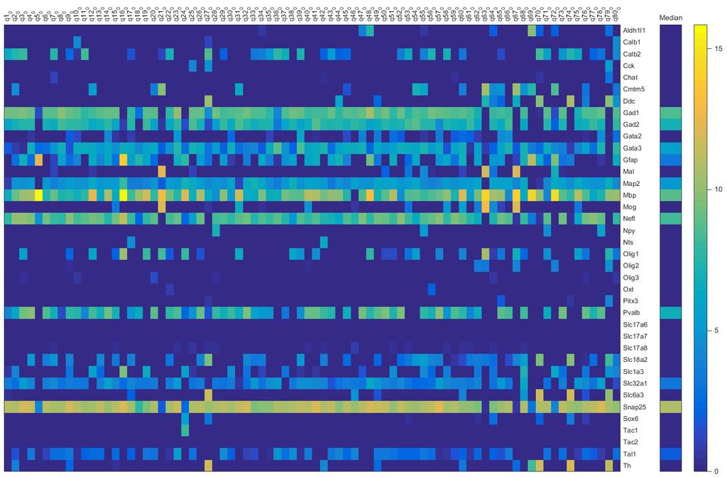
130 Figure 5.1: (Continued) A) B) C) 118
131 Results: We performed an initial pilot study using 26 manually collected SNr neurons (23 GFP positive neurons and 3 GFP negative neurons). These neurons were collected as described in the methods, but the sequencing platform used was an older generation Illumina mi-seq that only generated several thousand reads per cell. Nonetheless, this pilot study demonstrated that cdna libraries could be generated from individually collected SNr neurons and sequencing results could be obtained. The number of genes detected per cell was low, on the order of a few thousand genes, but this could be greatly improved by sequencing samples more deeply. Therefore, we collected another sample set consisting of 96 individually collected neurons from 6 mice (87 GFP-positive cells and 9 GFP-negative cells). Libraries from these individual samples were sequenced and we were able to generate several million reads per cell for 80 of the 96 samples (Figure 5.1C). Reads were then mapped to a mouse reference genome and normalized gene expression results were obtained (FPKM). We first assessed samples for contamination by other cell-types (Figure 5.1C). We found that almost all GFP-positive cells collected express marker genes consistent with GABAergic neurons and did not express marker genes for other neuronal types. However, some cells did express markers for glial cell types. In particular, several samples had significant expression of the oligodendrocyte marker gene olig1. We therefore filtered our samples to obtain 11 GAD positive neurons that expressed little to no marker genes for other neuronal and glial cell types. 119
132 Candidate nonselective cation channels To obtain a list of potential nonselective cation channels, we examined channels that have been described in the literature to be nonselective cation channels. We first examined expression of the 27 known TRP channel members in all the GAD positive cells (Figure 5.2A) as well as in a subset of GAD positive cells with the lowest contamination (Figure 5.2B). In addition, we examined expression for other known nonselective cation channels (Figure 5.2C-D). Because we were interested in genes consistently expressed in the population of GABAergic SNr neurons, and to avoid false positives, we determined the median expression level for each gene. We found consistent expression of several expected ion channel genes: TRPC3 (Zhou et al., 2008), TRPM2 (Lee et al., 2013), and HCN (Ibanez-Sandoval et al., 2007). In addition, we identify several nonselective cation channel genes not previously described in SNr neurons: TRPM7, TRPC4, PANX2, and NALCN. We also observed consistent expression of the lysosomal TRP channel MCOLN1 (Wang et al., 2014; Medina et al., 2015); however, to our knowledge, this channel does not localize to the plasma membrane and therefore is unlikely to be regulating SNr excitability. Using the distribution of the expression levels of all genes for GAD positive cells (Figure 5.3), we can assess the relative abundance of the nonselective cation channels in comparison to the rest of the genes. The distribution is centered near a log2 FPKM value of 4. Therefore, genes like NALCN, are expressed at levels near the average of all genes. 120
 Expression of TRP channel genes in low contamination GAD positive cells.")
133 Figure 5.2: Nonselective cation channel expression analysis A) A) Heatmap depicting the expression of TRP channels genes in all GAD positive cells. The median value for each gene is shown on the right. B) Expression of TRP channel genes in low contamination GAD positive cells. C) Expression of other nonselective cation channels in all GAD positive cells. D) Expression of other nonselective cation channels in low contamination GAD positive cells. Colorbars indicate the FPKM values after log2 transformation. 121
")
134 Figure 5.2: (Continued) B) 122
")
135 Figure 5.2: (Continued) C) 123
")
136 Figure 5.2: (Continued) D) 124
137 Figure 5.3: Distribution of log2 FPKM values for all genes in GAD positive cells Histogram of the distribution of log2 FPKM values for all GAD positive neurons and for GAD positive cells with low contamination. The distribution is centered near a value of
138 Conclusions: We were able to isolate and manually collect individual GABAergic neurons from the SNr region of the mouse brain using a GAD67-eGFP mouse line. From each of these cells, we obtained gene expression data for the entire transcriptome and found that the vast majority of samples generated sufficient reads to assess gene expression. Because we sequenced cells individually, we were able to assess the level of contamination by other cells types and focus our analysis on pure GABAergic neurons. While single-cell gene expression analysis is susceptible to genes being undetected because of the low starting material and the stochastic nature of sampling of the mrna population, our analysis of many cells allows us to determine which genes appear more consistently. However, trying to accurately cluster within this population of GABAergic SNr neurons would be more difficult and is beyond the initial scope of this study. We were mainly interested in determining which nonselective cation channels are expressed in SNr neurons. Therefore, we present gene expression results only for nonselective cation channels from these GABAergic neurons. We show that SNr neurons consistently express TRPC3 and TRPC4, but also other nonselective cation channels including: TRPM2, TRPM7, PANX2, and NALCN. We are particularly intrigued by NALCN, which appears to be highly expressed. These channels have not been thoroughly investigated in the brain, but previous evidence indicates a crucial role for these channels in neuronal function (Lu et al., 2007). PANX2 hemichannels are also interesting candidates that may be further tested using the drugs probenecid and carbenoxolone. We hope that this candidate list will allow for targeted analysis of the contribution of these channels in SNr firing and lead to a better understanding of how 126
139 these cells fire spontaneously. In addition, we have found that a nonselective cation channel is modulated by glycolysis and using this candidate list, we may uncover the identity of this metabolically sensitive channel. Acknowledgements: We thank Dr. Jesse Gray and Dr. Suzie Yorozu for helpful advice for manual cell collection. We are especially grateful to Dr. Magali Soumillon (Broad Institute) for valuable conversations about single-cell collection and sequencing. In addition, Dr. Soumillon performed all RNA processing and sequencing. We also thank Nikhil Sharma and Dr. Paul Greer for sharing their knowledge of single-cell RNA-seq. References: Chen C-Y (2014) DNA polymerases drive DNA sequencing-by-synthesis technologies: both past and present. Front Microbiol 5:305. Garber M, Grabherr MG, Guttman M, Trapnell C (2011) Computational methods for transcriptome annotation and quantification using RNA-seq. Nat Methods 8: Hempel CM, Sugino K, Nelson SB (2007) A manual method for the purification of fluorescently labeled neurons from the mammalian brain. Nat Protoc 2: Ibanez-Sandoval O, Carrillo-Reid L, Galarraga E, Tapia D, Mendoza E, Gomora JC, Aceves J, Bargas J (2007) Bursting in Substantia Nigra Pars Reticulata Neurons In Vitro: Possible Relevance for Parkinson Disease. J Neurophysiol 98: Ju J, Kim DH, Bi L, Meng Q, Bai X, Li Z, Li X, Marma MS, Shi S, Wu J, Edwards JR, Romu A, Turro NJ (2006) Four-color DNA sequencing by synthesis using cleavable fluorescent nucleotide reversible terminators. Proc Natl Acad Sci U S A 103:
140 Lee C, Machold R, Witkovsky P, Rice M (2013) TRPM2 channels are required for NMDA-induced burst firing and contribute to H(2)O(2)-dependent modulation in substantia nigra pars reticulata GABAergic neurons. J Neurosci 33: Lu B, Su Y, Das S, Liu J, Xia J, Ren D (2007) The Neuronal Channel NALCN Contributes Resting Sodium Permeability and Is Required for Normal Respiratory Rhythm. Cell 129: Lutas A, Birnbaumer L, Yellen G (2014) Metabolism regulates the spontaneous firing of substantia nigra pars reticulata neurons via KATP and nonselective cation channels. J Neurosci 34: Mardis ER (2011) A decade s perspective on DNA sequencing technology. Nature 470: Medina DL, Di Paola S, Peluso I, Armani A, De Stefani D, Venditti R, Montefusco S, Scotto-Rosato A, Prezioso C, Forrester A, Settembre C, Wang W, Gao Q, Xu H, Sandri M, Rizzuto R, De Matteis MA, Ballabio A (2015) Lysosomal calcium signalling regulates autophagy through calcineurin and TFEB. Nat Cell Biol 17: Picelli S, Faridani OR, Björklund AK, Winberg G, Sagasser S, Sandberg R (2014) Fulllength RNA-seq from single cells using Smart-seq2. Nat Protoc 9: Saliba A-E, Westermann AJ, Gorski SA, Vogel J (2014) Single-cell RNA-seq: advances and future challenges. Nucleic Acids Res 42: Wang W, Zhang X, Gao Q, Xu H (2014) Mammalian Transient Receptor Potential (TRP) Cation Channels (Nilius B, Flockerzi V, eds). Berlin, Heidelberg: Springer Berlin Heidelberg. Zhou F-W, Matta S, Zhou F-M (2008) Constitutively active TRPC3 channels regulate basal ganglia output neurons. J Neurosci 28:
141 Chapter VI: In vivo and in vitro brain oxygen tension measurements Acknowledgement: Andrew Lutas performed all in vitro oxygen measurements in brain slices. Carolina Lahmann performed the in vivo cranial window surgery and both Andrew Lutas and Carolina Lahmann conducted in vivo oxygen measurements. 129
142 Abstract: Oxidative phosphorylation is dependent on oxygen availability. Decreases in oxygen tension can reduce the amount of energy produced by mitochondria. We found that changes in flow rate onto SNr brain slices altered the ability of SNr neurons to utilize ketone bodies and prevent activation of K ATP channels. We speculated, based on work from other labs, that these changes in flow rate produced changes in oxygen tension. To better understand how oxygen availability regulates SNr activity, we manipulated oxygen tension in the bath solution. Lowering oxygen tension decreased SNr firing, which could be reversed by inhibitors of K ATP channels. To determine if the effects of flow rate on SNr activity were mediated by oxygen tension, we monitor oxygen tension using Clark-style electrodes. In addition, we perform in vivo measurements of oxygen at varying depths of anesthesia to determine how oxygen levels in the brain changed with brain activity. This work in progress will provide us with a stronger handle on oxygen levels, energy production, and neuronal activity. With this information in hand, we hope to gain a mechanistic understanding of brain energy status and neuronal excitability. Introduction: Oxygenation: Differences between in vitro and in vivo conditions The brain is extensively vascularized, with areas of the brain typically no farther than 60 μm from a blood vessel. During normal physiological brain activity, oxygen delivery is responsive to increases in brain activity indicating that adequate brain oxygenation is important and dynamic. This is further highlighted by the fact that 130
143 impaired oxygen delivery to the brain results in immediate loss of consciousness and eventually leads to significant cell death. Local impairment of oxygen delivery, as might occur during a stroke, also has profound consequences. Depending on the area experiencing ischemia, symptoms including loss of bodily function, numbness, and pain can occur. Oxygen enters the blood stream via the alveoli of the lungs. In the blood, hemoglobin acts as an oxygen carrier and is typically % saturated. In humans, there is normally around 15 grams of hemoglobin per liter of blood, which means that a very high concentration of oxygen is carried. The oxygen partial pressure in blood varies between 100 mmhg in arterial blood down to around 40 mmhg in venous blood. In addition, the saturation of hemoglobin is modulated by several key factors: ph, temperature, and 2,3-diphosphoglycerate (DPG). In the brain, oxygen partial pressure is reported to be between 10 and 80 mmhg (Ndubuizu and LaManna, 2007), with gradients possibly existing with distance away from blood vessels (Kasischke et al., 2011). Measurements of brain oxygen tension demonstrate that oxygen levels change with brain activity. This change in oxygen is mediated by oxygen consumption by neurons and glial cells, and also by changes in oxygen delivery. Blood flow increases and vessel dilation typically occur in areas of increased brain activity. These brain activity correlated vascular dynamics have been utilized to monitor brain activity non-invasively in fmri studies (Logothetis and Pfeuffer, 2004). 131
144 In vitro experiments lack dynamic oxygenation and appropriate oxygenation is often not even considered Most of our understanding of cellular metabolism comes from in vitro studies where the oxygenation is usually determined by the solution bathing the cells or tissue. For brain slice experiments, there are two types of recording conditions that are often used. One method involves maintaining slices at the interface between solution and humidified air using an interface recording chamber. In these conditions, solution flows very slowly, making it difficult to perform pharmacological manipulations, but oxygen tension is maintained high by blowing humidified oxygenated gas across the surface of the slice. This type of chamber has been used to study network dynamics in slices and provides much better results for long-term recordings of the sort of oscillations that are typically observed in vivo. For example, cortical slow wave oscillations that are observed during deep sleeping states of an animal can be readily reproduced in vitro using interface-like chambers. The other, more widely used, method of in vitro brain slice recordings uses a completely submerged condition where slices are immersed in flowing bath solution. Slices are held in the submerged condition either by affixing them to a glass coverslip or by using a harp to weigh down the slice. The submerged condition affords faster solution flow rate, which is ideal for fast wash in of pharmacological agents. Furthermore, the ability to use water immersion objectives allows for visualization of individual neurons in brain slices. A downside of the completely submerged condition appears to be the increased difficulty in preserving large network activity such as the cortical slow 132
145 oscillation. However, this obstacle can be surpassed by increasing the flow rate to the brain slice and by allowing solution to flow on both surfaces of the slice. What does increasing the flow rate achieve in the submerged condition? Measurements of the oxygen tension in brain slices using Clark-style polarographic oxygen electrodes have revealed that solution flow rate strongly affects the oxygen tension in brain slices (Hájos and Mody, 2009; Hájos et al., 2009; Ivanov et al., 2011). Higher flow rates allow for slices to be maintained at a higher oxygen tension and therefore provide more oxygen to neurons, especially in the deeper parts of the brain slice. The fact that network level activity can be only observed when flow rates are increased indicates that these phenomena must be highly dependent on the availability of oxygen and therefore on the level of mitochondrial energy production. These differences in in vitro oxygenation conditions are often not considered. This is particularly concerning when reports of interesting phenomena are described using brain slice experiments. For example, whether induction of long-term potentiation, an important mechanism of memory storage, can be observed in slice experiments may be dependent on the metabolic conditions of the network. However, while it is difficult to answer the question of what the best oxygenation situation of a slice is for reflecting the true in vivo scenario, the ability to readily control the oxygenation of a slice does afford some benefits. The ability to observe a set of neuronal behaviors in one metabolic condition versus another provides insight into when those behaviors might be expected to occur in vivo. For instance, if certain neuronal firing properties are only observed in lower oxygen conditions, it might be that in vivo those properties would only occur when the brain was oxygen deprived. 133
146 In this chapter, we describe studies aimed at measuring oxygen tension and the correlation between oxygen and neuronal firing properties. In particular, we characterize the effect of perfusion flow rate on oxygen tension levels in brain slice experiments. We find that these different oxygen conditions bias the cell towards more or less K ATP channel activity. Finally, we measure oxygen tension in vivo using Clark-style microelectrodes in anesthetized mice and find oxygen tension values dramatically higher than expected. Methods: In vitro experiments in mouse brain slices Brain slices were prepared from P13-P18 mice as previously described and recordings were performed using a dual-perfusion chamber (Lutas et al., 2014). Oxygen measurements were made using a Clark-style oxygen electrode with < 10 μm tip size (OX-10; Unisense). A two or three point calibration of the electrode was performed before and after the experiment as indicated in the manual. The electrode was lowered into the bath under the control of a micromanipulator (Sutter; MP-225). Depth measurements were read off the micromanipulator LCD screen. All experiments and oxygen electrode calibrations were performed at 34 C. In vivo mouse primary somatosensory cortex oxygen measurements 134
147 Adult mice were deeply anesthetized via inhalation of 3% isoflurane in 100% oxygen. Anesthesia was maintained at 1.5% isoflurane in oxygen. Local anesthetics (50 μl of a 1:1 mixture of 0.25% lidocaine and % bupivacaine) were injected at the incision site. The analgesic ketoprofen (10 mg/kg) was injected subcutaneously at the beginning of the surgery. Body temperature was maintained at 37 C by a heated pad below the mouse and regulated by rectal temperature measurements. A small craniotomy was performed on adult mice to make a cranial window over the primary somatosensory (barrel) cortex region (-1.5 AP, -3.0 ML from bregma over the right S1 barrel field). Following the craniotomy procedure, mice were maintained under anesthesia while the Clark-style oxygen electrode was inserted 200 μm into the barrel cortex using a micromanipulator (Narishige). Oxygen measurements were performed while changing the isoflurane concentration to alter the depth of anesthesia. Blood oxygen saturation and breathing rate were monitored using a pulse oximeter (Starr Lifesciences) placed on the shaved thigh of the mouse. Results: In chapter 2, we reported that the ability of the ketone body β-hydroxybutyrate to prevent the loss of SNr firing when glycolysis was inhibited was dependent on the flow rate used. When flow rate was lowered to 1 ml/min, activation of K ATP channels frequently occurred when glycolysis was inhibited in the presence of β-hydroxybutyrate. We interpreted this finding as a decreased ability of mitochondria to utilize the ketone body because of the decreased oxygen tension under slower flow rate conditions. This 135
148 interpretation is supported by several studies, which measured oxygen tension in brain slices being perfused at different flow rates (Hájos and Mody, 2009; Ivanov et al., 2011). We examined this directly in our recording conditions so that we could correlate the oxygen tension in our brain slices to the level of K ATP channel activity. We initially examined how changes in flow rate from 5 ml/min to 1 ml/min affected the oxygen tension measured in the recording chamber. We found that oxygen tension in the dual perfusion chamber during 5 ml/min flow rate was around 450 mmhg (Figure 6.1A), which is less than the ~700 mmhg measured from ACSF directly bubbled with 95% oxygen. This indicates that even at 5 ml/min, there is already a loss of oxygen tension from the bottle to the recording chamber. When we lowered the flow rate to 1 ml/min, we recorded a rapid decrease in oxygen tension that leveled out around 150 mmhg (Figure 6.1A), which is around the oxygen tension of air. Therefore, under the low flow rate condition, the oxygen tension at the slice is similar to an unbubbled solution. This finding was supported by the recordings made in ACSF bubbled with 95% air/ 5% CO 2. This solution is equilibrated with air, but is bubbled so as to provide the 5% CO 2 to obtain the correct ph of our bicarbonate bubbled solution. The oxygen tension in the solution bubbled with air was similar to the tension recorded in the 1 ml/min flow rate of 95% oxygen bubble solution (Figure 6.1A). In addition, changing the flow rate between 5 ml/min and 1 ml/min of the ACSF bubbled with air had minimal effect on the oxygen tension measured in the chamber (Figure 6.1A). Therefore, under sufficiently low flow rates, oxygen outgassing results in complete equilibration with air. 136
149 Figure 6.1: Oxygen tension measurements at different flow rates and in different solution bubbling conditions. A) With high flow rates (5 ml/min), oxygen tension in the slice chamber is around 450 mmhg. Low flow rates or solutions bubbled with 95% air give oxygen tension values near 150 mmhg. B) Changing bath solution to one bubbled with 95% air resulted in decreased SNr firing that was reversed by application of the K ATP channel blocker glibenclamide (Glib; 200 nm). C) Oxygen tension measurements made at various distances above and below the slice surface. The physiological range of oxygen tension values in rodent brains is indicated ( mmhg). 137
150 Neuronal firing in the SNr is sensitive to oxygen tension We had previously shown that lowering flow rate affects the ability of ketone bodies to prevent loss of SNr firing during inhibition of glycolysis. In addition, we have observed that lowering flow rate to 1 ml/min effectively decreases oxygen tension to that of air. Therefore, we performed experiments using ACSF bubbled with 95% air/ 5% CO 2 to see how changing oxygen tension without changing flow rate could affect the firing of SNr neurons. We found that when flow rate was maintained at 5 ml/min, but the solution was bubbled with 95% air, the firing rate of SNr neurons decreased (Figure 6.1B). This decrease varied from cell to cell, with some cells almost completely going silent, while others responded with a smaller decrease in firing rate. In addition, we found that application of the K ATP channel blocker glibenclamide (200 nm) could completely reverse the decrease in firing rate of SNr neurons (Figure 6.1B). These data indicate that SNr neurons are sensitive to the oxygen tension and that activation of K ATP channels mediates the change in firing rate. Oxygen tension in brain slices Oxygen must diffuse into brain slices to reach cells deeper in the slice. As cells in the slice consume oxygen, an oxygen gradient develops from the surface of the slice towards the center of the tissue. We measured oxygen tension at distances above the slice and with increasing depth into the slice (Figure 6.1C). We found that oxygen tension drops as we approached the slice and continued to decrease with depth in the slice. The values measured within the slice ranged from ~ 100 mmhg down to ~
151 mmhg, which is within the physiological range measured in vivo in healthy brains (Ndubuizu and LaManna, 2007). We expect that under low flow rate conditions, because the initial oxygen tension in the solution is 150 mmhg, the oxygen tension in brain slices would be much lower than those observed with high flow rates. However, we have not yet made these measurements. Cortical oxygen tension measurements in anesthetized mice To begin to correlate our findings in brain slices to those in vivo, we made oxygen tension measurements in the barrel cortex of anesthetized adult mice. Surprisingly, we found the oxygen tension to be significantly higher than those reported previously for mouse brain (Mathiesen et al., 2013; Thrane et al., 2013); however, these studies were performed under anesthesia using injectable anesthetics instead of isoflurane. Interestingly, we found that decreasing the depth of anesthesia could change the oxygen tension towards those previously reported (Figure 6.2). These high oxygen tension values under deep anesthesia are higher even than our brain slice measurements using 5 ml/min flow rate and solution bubbled with 95 % oxygen. Therefore, under these high oxygen tension conditions in vivo, we would predict that K ATP channels would be closed in neurons, assuming they behave in vivo as we have observed in brain slices. 139

152 Figure 6.2: Oxygen tension measurements (mmhg) in vivo from three anesthetized adult mice. Oxygen tension measurements were made in anesthetized adult mice through a cranial window centered over the barrel cortex. Measurements were made at 200 µm below the surface of the brain. White areas indicate normal anesthesia of 1.5% isoflurane. Colored regions indicate changes in isoflurane to the indicated value. Lower isoflurane levels resulted in lower oxygen tension values. 140
E. Haritov, E. Angeleska and N. Boyadjieva Department of pharmacology and toxicology, Medical Faculty, Medical University So a
 :.,..,, KETOGENIC DIET AND EPILEPSY: THE ROLE OF KETONE BODIES IN THE INFLUENCE N NEURONAL EXCITABILITY E. Haritov, E. Angeleska and N. Boyadjieva Department of pharmacology and toxicology, Medical Faculty,
Medical Biochemistry and Molecular Biology department
 Medical Biochemistry and Molecular Biology department Cardiac Fuels [Sources of energy for the Cardiac muscle] Intended learning outcomes of the lecture: By the end of this lecture you would be able to:-
Neurovascular Coupling
 Cerebral Blood Flow and Brain Activation UCLA NITP July 2012 Neurovascular Coupling Richard B. Buxton University of California, San Diego rbuxton@ucsd.edu... The subject to be observed lay on a delicately
Chemistry 1120 Exam 4 Study Guide
 Chemistry 1120 Exam 4 Study Guide Chapter 12 12.1 Identify and differentiate between macronutrients (lipids, amino acids and saccharides) and micronutrients (vitamins and minerals). Master Tutor Section
Karin Borges, PhD. October The urgent need for new metabolic (dietary) treatments of epilepsy
 treatments of epilepsy") Karin Borges, PhD October 2016 The urgent need for new metabolic (dietary) treatments of epilepsy Epilepsy is a common neurological disease that is characterized by recurrent seizures due to the spontaneous
Karin Borges, PhD October 2016 The urgent need for new metabolic (dietary) treatments of epilepsy Epilepsy is a common neurological disease that is characterized by recurrent seizures due to the spontaneous
GABA Induced Depolarization: A Tale of Opposing Forces
 GABA Induced Depolarization: A Tale of Opposing Forces December 2010 Jong M. Rho, MD Alberta Children s Hospital University of Calgary, Alberta, Canada American Epilepsy Society Annual Meeting Disclosures
5.0 HORMONAL CONTROL OF CARBOHYDRATE METABOLISM
 5.0 HORMONAL CONTROL OF CARBOHYDRATE METABOLISM Introduction: Variety of hormones and other molecules regulate the carbohydrates metabolism. Some of these have already been cited in previous sections.
Chapter 2. The Cellular and Molecular Basis of Cognition Cognitive Neuroscience: The Biology of the Mind, 2 nd Ed.,
 Chapter 2. The Cellular and Molecular Basis of Cognition Cognitive Neuroscience: The Biology of the Mind, 2 nd Ed., M. S. Gazzaniga, R. B. Ivry, and G. R. Mangun, Norton, 2002. Summarized by B.-W. Ku,
Chapter 2. The Cellular and Molecular Basis of Cognition
 Chapter 2. The Cellular and Molecular Basis of Cognition Cognitive Neuroscience: The Biology of the Mind, 2 nd Ed., M. S. Gazzaniga,, R. B. Ivry,, and G. R. Mangun,, Norton, 2002. Summarized by B.-W. Ku,
Integration Of Metabolism
 Integration Of Metabolism Metabolism Consist of Highly Interconnected Pathways The basic strategy of catabolic metabolism is to form ATP, NADPH, and building blocks for biosyntheses. 1. ATP is the universal
MOLECULAR AND CELLULAR NEUROSCIENCE
 MOLECULAR AND CELLULAR NEUROSCIENCE BMP-218 November 4, 2014 DIVISIONS OF THE NERVOUS SYSTEM The nervous system is composed of two primary divisions: 1. CNS - Central Nervous System (Brain + Spinal Cord)
! acts via the autonomic nervous system. ! maintaining body weight within tight limits. ! ventromedial (VMN) ! arcuate (ARC) ! neuropeptide Y (NPY)
 ! arcuate (ARC) ! neuropeptide Y (NPY)") Brain Regulates energy homeostatis Glucose Sensing in the Brain Seminar 2012 Gareth Price! acts in response to circulating signals of nutrient states! acts via the autonomic nervous system Ensures a balance
Brain Regulates energy homeostatis Glucose Sensing in the Brain Seminar 2012 Gareth Price! acts in response to circulating signals of nutrient states! acts via the autonomic nervous system Ensures a balance
Oxidation of Long Chain Fatty Acids
 Oxidation of Long Chain Fatty Acids Dr NC Bird Oxidation of long chain fatty acids is the primary source of energy supply in man and animals. Hibernating animals utilise fat stores to maintain body heat,
Cell Injury MECHANISMS OF CELL INJURY
 Cell Injury MECHANISMS OF CELL INJURY The cellular response to injurious stimuli depends on the following factors: Type of injury, Its duration, and Its severity. Thus, low doses of toxins or a brief duration
Name: KEY. Chem 3511 Exam 4
 Chem 3511 Exam 4 The exam starts on the next page. It has 12 questions, worth a total of 100 points. Please write legibly and don t assume that long answers are required if there is a lot of space left
Relating Neurophysiology and Imaging Signals
 UCLA NITP July 2010 Relating Neurophysiology and Imaging Signals Richard B. Buxton University of California, San Diego ASL-fMRI, LDF Blood Oxygenation BOLD-fMRI, NIRS, optical Blood Volume MION-fMRI, VASO,
3) Most of the organelles in a neuron are located in the A) dendritic region. B) axon hillock. C) axon. D) cell body. E) axon terminals.
 Most of the organelles in a neuron are located in the A) dendritic region. B) axon hillock. C) axon. D) cell body. E) axon terminals.") Chapter 48 Neurons, Synapses, and Signaling Multiple-Choice Questions 1) A simple nervous system A) must include chemical senses, mechanoreception, and vision. B) includes a minimum of 12 ganglia. C) has
Chapter 48 Neurons, Synapses, and Signaling Multiple-Choice Questions 1) A simple nervous system A) must include chemical senses, mechanoreception, and vision. B) includes a minimum of 12 ganglia. C) has
Principles of Anatomy and Physiology
 Principles of Anatomy and Physiology 14 th Edition CHAPTER 25 Metabolism and Nutrition Metabolic Reactions Metabolism refers to all of the chemical reactions taking place in the body. Reactions that break
Chapter 9 Notes. Cellular Respiration and Fermentation
 Chapter 9 Notes Cellular Respiration and Fermentation Objectives Distinguish between fermentation and anaerobic respiration. Name the three stages of cellular respiration and state the region of the cell
7 Pathways That Harvest Chemical Energy
 7 Pathways That Harvest Chemical Energy Pathways That Harvest Chemical Energy How Does Glucose Oxidation Release Chemical Energy? What Are the Aerobic Pathways of Glucose Metabolism? How Is Energy Harvested
Cellular Pathways That Harvest Chemical Energy. Cellular Pathways That Harvest Chemical Energy. Cellular Pathways In General
 Cellular Pathways That Harvest Chemical Energy A. Obtaining Energy and Electrons from Glucose Lecture Series 12 Cellular Pathways That Harvest Chemical Energy B. An Overview: Releasing Energy from Glucose
In glycolysis, glucose is converted to pyruvate. If the pyruvate is reduced to lactate, the pathway does not require O 2 and is called anaerobic
 Glycolysis 1 In glycolysis, glucose is converted to pyruvate. If the pyruvate is reduced to lactate, the pathway does not require O 2 and is called anaerobic glycolysis. If this pyruvate is converted instead
THE GLUCOSE-FATTY ACID-KETONE BODY CYCLE Role of ketone bodies as respiratory substrates and metabolic signals
 Br. J. Anaesth. (1981), 53, 131 THE GLUCOSE-FATTY ACID-KETONE BODY CYCLE Role of ketone bodies as respiratory substrates and metabolic signals J. C. STANLEY In this paper, the glucose-fatty acid cycle
Electrical Properties of Neurons. Steven McLoon Department of Neuroscience University of Minnesota
 Electrical Properties of Neurons Steven McLoon Department of Neuroscience University of Minnesota 1 Neuronal Communication Neurons communicate with other cells, often over long distances. The electrical
Synaptic Transmission: Ionic and Metabotropic
 Synaptic Transmission: Ionic and Metabotropic D. Purves et al. Neuroscience (Sinauer Assoc.) Chapters 5, 6, 7. C. Koch. Biophysics of Computation (Oxford) Chapter 4. J.G. Nicholls et al. From Neuron to
Summarized by B.-W. Ku, E. S. Lee, and B.-T. Zhang Biointelligence Laboratory, Seoul National University.
 Chapter 2. The Cellular l and Molecular Basis of Cognition Cognitive Neuroscience: The Biology of the Mind, 3 rd Ed., M. S. Gazzaniga, R. B. Ivry, and G. R. Mangun, Norton, 2008. Summarized by B.-W. Ku,
Lecture 5: Cell Metabolism. Biology 219 Dr. Adam Ross
 Lecture 5: Cell Metabolism Biology 219 Dr. Adam Ross Cellular Respiration Set of reactions that take place during the conversion of nutrients into ATP Intricate regulatory relationship between several
Cross-talk between Two Partners Regulates the Gating of the K ATP Channel Pore
 International Conference on Metabolic Syndromes Cross-talk between Two Partners Regulates the Gating of the K ATP Channel Pore Dr Hussein N Rubaiy h.rubaiy@leeds.ac.uk School of Medicine, University of
Chapter 7- Metabolism: Transformations and Interactions Thomson - Wadsworth
 Chapter 7- Metabolism: Transformations and Interactions 2008 Thomson - Wadsworth Simple Overview of Energy Metabolism The sum of all chemical reactions that go on in living cells Introduction Energy Heat-
Review of Neurochemistry
 Review of Neurochemistry UNIVERSITY OF PNG SCHOOL OF MEDICINE AND HEALTH SCIENCES DISCIPLINE OF BIOCHEMISTRY AND MOLECULAR BIOLOGY PBL MBBS YEAR III SEMINAR VJ Temple 1 CEREBRAL METABOLISM What are some
Lecture 3 (Oct 5 th ): NEURONS AND NERVE IMPULSES Lecture Outline
: NEURONS AND NERVE IMPULSES Lecture Outline") Lecture 3 (Oct 5 th ): NEURONS AND NERVE IMPULSES Lecture Outline 1) CNS vs. PNS 2) Structure of Neurons parts of a neuron: soma, dendrites, axons 3) Glial Cells 4) Mitosis and Regeneration in Neurons
Lecture 3 (Oct 5 th ): NEURONS AND NERVE IMPULSES Lecture Outline 1) CNS vs. PNS 2) Structure of Neurons parts of a neuron: soma, dendrites, axons 3) Glial Cells 4) Mitosis and Regeneration in Neurons
1- Which of the following statements is TRUE in regards to eukaryotic and prokaryotic cells?
 Name: NetID: Exam 3 - Version 1 October 23, 2017 Dr. A. Pimentel Each question has a value of 4 points and there are a total of 160 points in the exam. However, the maximum score of this exam will be capped
Integrative Metabolism: Significance
 Integrative Metabolism: Significance Energy Containing Nutrients Carbohydrates Fats Proteins Catabolism Energy Depleted End Products H 2 O NH 3 ADP + Pi NAD + NADP + FAD + Pi NADH+H + NADPH+H + FADH2 Cell
Sleep-Wake Cycle I Brain Rhythms. Reading: BCP Chapter 19
 Sleep-Wake Cycle I Brain Rhythms Reading: BCP Chapter 19 Brain Rhythms and Sleep Earth has a rhythmic environment. For example, day and night cycle back and forth, tides ebb and flow and temperature varies
Changes in Extracellular Ionic Composition q
 Changes in Extracellular Ionic Composition q JL Stringer, Baylor College of Medicine, Houston, TX, United States Ó 2017 Elsevier Inc. All rights reserved. Introduction 1 Background 1 Methods 2 Recent Results
Neuroscience: Exploring the Brain, 3e. Chapter 4: The action potential
 Neuroscience: Exploring the Brain, 3e Chapter 4: The action potential Introduction Action Potential in the Nervous System Conveys information over long distances Action potential Initiated in the axon
1) Drop off in the Bi 150 box outside Baxter 331 or to the head TA (jcolas).
 Drop off in the Bi 150 box outside Baxter 331 or to the head TA (jcolas).") Bi/CNS/NB 150 Problem Set 3 Due: Tuesday, Oct. 27, at 4:30 pm Instructions: 1) Drop off in the Bi 150 box outside Baxter 331 or e-mail to the head TA (jcolas). 2) Submit with this cover page. 3) Use a
Bi/CNS/NB 150 Problem Set 3 Due: Tuesday, Oct. 27, at 4:30 pm Instructions: 1) Drop off in the Bi 150 box outside Baxter 331 or e-mail to the head TA (jcolas). 2) Submit with this cover page. 3) Use a
Metabolism. Chapter 5. Catabolism Drives Anabolism 8/29/11. Complete Catabolism of Glucose
 8/29/11 Metabolism Chapter 5 All of the reactions in the body that require energy transfer. Can be divided into: Cell Respiration and Metabolism Anabolism: requires the input of energy to synthesize large
BIOLOGY 103 Spring 2001 MIDTERM LAB SECTION
 BIOLOGY 103 Spring 2001 MIDTERM NAME KEY LAB SECTION ID# (last four digits of SS#) STUDENT PLEASE READ. Do not put yourself at a disadvantage by revealing the content of this exam to your classmates. Your
How Nicotinic Signaling Shapes Neural Networks
 How Nicotinic Signaling Shapes Neural Networks Darwin K. Berg Division of Biological Sciences University of California, San Diego Nicotinic Cholinergic Signaling Uses the transmitter ACh to activate cation-selective
Synaptic Communication. Steven McLoon Department of Neuroscience University of Minnesota
 Synaptic Communication Steven McLoon Department of Neuroscience University of Minnesota 1 Course News The first exam is next week on Friday! Be sure to checkout the sample exam on the course website. 2
Chapter 7 Cellular Respiration and Fermentation*
 Chapter 7 Cellular Respiration and Fermentation* *Lecture notes are to be used as a study guide only and do not represent the comprehensive information you will need to know for the exams. Life Is Work
How Cells Release Chemical Energy. Chapter 8
 How Cells Release Chemical Energy Chapter 8 Impacts, Issues: When Mitochondria Spin Their Wheels More than forty disorders related to defective mitochondria are known (such as Friedreich s ataxia); many
Chemical Energy. Valencia College
 9 Pathways that Harvest Chemical Energy Valencia College 9 Pathways that Harvest Chemical Energy Chapter objectives: How Does Glucose Oxidation Release Chemical Energy? What Are the Aerobic Pathways of
When cells are already maximally potentiated LTP is occluded.
 When cells are already maximally potentiated LTP is occluded. Stein, V et al., (2003) J Neurosci, 23:5503-6606. Also found in Rat Barrel Cortex Ehrlich & Malinow (2004) J. Neurosci. 24:916-927 Over-expression
Hompes Method Lesson 29 Organic Acids Part One
 Hompes Method Lesson 29 Organic Acids Part One Health for the People Ltd not for reuse without expressed permission Organic Acids - Introduction The ultimate tool for laboratory evaluations in nutritional
Chapter 45: Synapses Transmission of Nerve Impulses Between Neurons. Chad Smurthwaite & Jordan Shellmire
 Chapter 45: Synapses Transmission of Nerve Impulses Between Neurons Chad Smurthwaite & Jordan Shellmire The Chemical Synapse The most common type of synapse used for signal transmission in the central
Reading Assignments. A. Energy and Energy Conversions. Lecture Series 9 Cellular Pathways That Harvest Chemical Energy. gasoline) or elevated mass.
 or elevated mass.") Lecture Series 9 Cellular Pathways That Harvest Chemical Energy Reading Assignments Review Chapter 3 Energy, Catalysis, & Biosynthesis Read Chapter 13 How Cells obtain Energy from Food Read Chapter 14
Lecture Series 9 Cellular Pathways That Harvest Chemical Energy Reading Assignments Review Chapter 3 Energy, Catalysis, & Biosynthesis Read Chapter 13 How Cells obtain Energy from Food Read Chapter 14
Chapter 7 Nerve Cells and Electrical Signaling
 Chapter 7 Nerve Cells and Electrical Signaling 7.1. Overview of the Nervous System (Figure 7.1) 7.2. Cells of the Nervous System o Neurons are excitable cells which can generate action potentials o 90%
ATP-sensitive potassium channelopathies: focus on insulin secretion
 Review series ATP-sensitive potassium channelopathies: focus on insulin secretion Frances M. Ashcroft University Laboratory of Physiology, Oxford University, Oxford, United Kingdom. ATP-sensitive potassium
Biol 219 Lec 7 Fall 2016
 Cellular Respiration: Harvesting Energy to form ATP Cellular Respiration and Metabolism Glucose ATP Pyruvate Lactate Acetyl CoA NAD + Introducing The Players primary substrate for cellular respiration
The Brain & Homeostasis. The Brain & Technology. CAT, PET, and MRI Scans
 The Brain & Homeostasis Today, scientists have a lot of information about what happens in the different parts of the brain; however they are still trying to understand how the brain functions. We know
Sample Lab Report 1 from 1. Measuring and Manipulating Passive Membrane Properties
 Sample Lab Report 1 from http://www.bio365l.net 1 Abstract Measuring and Manipulating Passive Membrane Properties Biological membranes exhibit the properties of capacitance and resistance, which allow
Chapter 2: Cellular Mechanisms and Cognition
 Chapter 2: Cellular Mechanisms and Cognition MULTIPLE CHOICE 1. Two principles about neurons were defined by Ramón y Cajal. The principle of connectional specificity states that, whereas the principle
Energy Production In A Cell (Chapter 25 Metabolism)
") Energy Production In A Cell (Chapter 25 Metabolism) Large food molecules contain a lot of potential energy in the form of chemical bonds but it requires a lot of work to liberate the energy. Cells need
Energy Production In A Cell (Chapter 25 Metabolism) Large food molecules contain a lot of potential energy in the form of chemical bonds but it requires a lot of work to liberate the energy. Cells need
Serotonergic Control of the Developing Cerebellum M. Oostland
 Serotonergic Control of the Developing Cerebellum M. Oostland Summary Brain development is a precise and crucial process, dependent on many factors. The neurotransmitter serotonin is one of the factors
Energy Metabolism. Chapter Cengage Learning. All Rights Reserved.
 Energy Metabolism Chapter 7 Introduction Energy Heat, mechanical, electrical, and chemical Metabolism How the body uses foods to meet its needs Chemical Reactions in the Body Energy metabolism How body
CHAPTER 7 10/16/2012. How cells release Chemical Energy
 CHAPTER 7 10/16/2012 How cells release Chemical Energy 1 7.1 OVERVIEW OF CARBOHYDRATE BREAKDOWN PATHWAYS Organisms stay alive by taking in energy. Plants and all other photosynthetic autotrophs get energy
4. Which step shows a split of one molecule into two smaller molecules? a. 2. d. 5
 1. Which of the following statements about NAD + is false? a. NAD + is reduced to NADH during both glycolysis and the citric acid cycle. b. NAD + has more chemical energy than NADH. c. NAD + is reduced
Part 11: Mechanisms of Learning
 Neurophysiology and Information: Theory of Brain Function Christopher Fiorillo BiS 527, Spring 2012 042 350 4326, fiorillo@kaist.ac.kr Part 11: Mechanisms of Learning Reading: Bear, Connors, and Paradiso,
Na + K + pump. The beauty of the Na + K + pump. Cotransport. The setup Cotransport the result. Found along the plasma membrane of all cells.
 The beauty of the Na + K + pump Na + K + pump Found along the plasma membrane of all cells. Establishes gradients, controls osmotic effects, allows for cotransport Nerve cells have a Na + K + pump and
The Nervous System. Nervous System Functions 1. gather sensory input 2. integration- process and interpret sensory input 3. cause motor output
 The Nervous System Nervous System Functions 1. gather sensory input 2. integration- process and interpret sensory input 3. cause motor output The Nervous System 2 Parts of the Nervous System 1. central
Abstracts and affiliations
 Dopamine Discovery Day August 30, 2012 Rikshospitalet Store auditorium, Oslo, Norway Organized by Linda H. Bergersen & Vidar Gundersen Institute of Basic Medical Sciences & Centre for Molecular Biology
1) Drop off in the Bi 150 box outside Baxter 331 or to the head TA (jcolas).
 Drop off in the Bi 150 box outside Baxter 331 or to the head TA (jcolas).") Bi/CNS/NB 150 Problem Set 3 Due: Tuesday, Oct. 27, at 4:30 pm Instructions: 1) Drop off in the Bi 150 box outside Baxter 331 or e-mail to the head TA (jcolas). 2) Submit with this cover page. 3) Use a
Bi/CNS/NB 150 Problem Set 3 Due: Tuesday, Oct. 27, at 4:30 pm Instructions: 1) Drop off in the Bi 150 box outside Baxter 331 or e-mail to the head TA (jcolas). 2) Submit with this cover page. 3) Use a
LESSON 2.2 WORKBOOK. Metabolism: Glucose is the middleman for ATP
 DEFINITIONS OF TERMS Homeostasis The tendency toward a relatively stable equilibrium that is maintained by physiological processes. For a complete list of defined terms, see the Glossary. LESSON 2.2 WORKBOOK
Ion Channels Graphics are used with permission of: Pearson Education Inc., publishing as Benjamin Cummings (http://www.aw-bc.com)
") Ion Channels Graphics are used with permission of: Pearson Education Inc., publishing as Benjamin Cummings (http://www.aw-bc.com) Page 1. Introduction At synapses, ions move across cell membranes through
Ion Channels Graphics are used with permission of: Pearson Education Inc., publishing as Benjamin Cummings (http://www.aw-bc.com) Page 1. Introduction At synapses, ions move across cell membranes through
CHY2026: General Biochemistry. Lipid Metabolism
 CHY2026: General Biochemistry Lipid Metabolism Lipid Digestion Lipid Metabolism Fats (triglycerides) are high metabolic energy molecules Fats yield 9.3 kcal of energy (carbohydrates and proteins 4.1 kcal)
Seizure: the clinical manifestation of an abnormal and excessive excitation and synchronization of a population of cortical
 Are There Sharing Mechanisms of Epilepsy, Migraine and Neuropathic Pain? Chin-Wei Huang, MD, PhD Department of Neurology, NCKUH Basic mechanisms underlying seizures and epilepsy Seizure: the clinical manifestation
Exam 3. Short Answer/Terminology (1 pt each except where noted, 35 pts total) Version B. Zoology 250, Fall 2003
 Version B. Zoology 250, Fall 2003") Exam 3 Version B Zoology 250, Fall 2003 5 pages, 50 points total please check to see that your copy is complete. Please SIGN your name here if you would like me to post your grade by the last five digits
Exam 3 Version B Zoology 250, Fall 2003 5 pages, 50 points total please check to see that your copy is complete. Please SIGN your name here if you would like me to post your grade by the last five digits
Fall Name Student ID
 Name Student ID PART 1: Matching. Match the organelle to its function (11 points) 1.Proton motive force 2. Fluid Mosiac 3. Oxidative Phosphorylation 4. Pyruvate dehydrogenase 5. Electrochemical Force 6.
By Silas Aho. also used for treatment of epilepsy, a debilitating neurological disease. The success of this diet as an
 1 Ketosis on the Epileptic Brain By Silas Aho I m sure some of you reading this paper may remember a diet in the early 2000 s called the Atkins diet that was based on eating a diet high in fats/proteins
THE NERVOUS SYSTEM. Homeostasis Strand
 THE NERVOUS SYSTEM Homeostasis Strand Introduction In general, a nervous system has three overlapping functions : 1. Sensory input conduction of signals from sensory receptors to integration centres 2.
Chapter 5 subtitles GABAergic synaptic transmission
 CELLULAR NEUROPHYSIOLOGY CONSTANCE HAMMOND Chapter 5 subtitles GABAergic synaptic transmission INTRODUCTION (2:57) In this fifth chapter, you will learn how the binding of the GABA neurotransmitter to
Organization of the nervous system. [See Fig. 48.1]
![Organization of the nervous system. [See Fig. 48.1]](/thumbs/90/103926552.jpg "Organization of the nervous system. [See Fig. 48.1]") Nervous System [Note: This is the text version of this lecture file. To make the lecture notes downloadable over a slow connection (e.g. modem) the figures have been replaced with figure numbers as found
Nervous System [Note: This is the text version of this lecture file. To make the lecture notes downloadable over a slow connection (e.g. modem) the figures have been replaced with figure numbers as found
Intracranial Studies Of Human Epilepsy In A Surgical Setting
 Intracranial Studies Of Human Epilepsy In A Surgical Setting Department of Neurology David Geffen School of Medicine at UCLA Presentation Goals Epilepsy and seizures Basics of the electroencephalogram
Basics of Computational Neuroscience: Neurons and Synapses to Networks
 Basics of Computational Neuroscience: Neurons and Synapses to Networks Bruce Graham Mathematics School of Natural Sciences University of Stirling Scotland, U.K. Useful Book Authors: David Sterratt, Bruce
ENERGY FROM INGESTED NUTREINTS MAY BE USED IMMEDIATELY OR STORED
 QUIZ/TEST REVIEW NOTES SECTION 1 SHORT TERM METABOLISM [METABOLISM] Learning Objectives: Identify primary energy stores of the body Differentiate the metabolic processes of the fed and fasted states Explain
Structure of the Mitochondrion. Cell Respiration. Cellular Respiration. Catabolic Pathways. Photosynthesis vs. Cell Respiration ATP 10/14/2014
 Structure of the Mitochondrion Cellular Respiration Chapter 9 Pgs. 163 183 Enclosed by a double membrane Outer membrane is smooth Inner, or cristae, membrane is folded - this divides the mitochondrion
Transfer of food energy to chemical energy. Includes anabolic and catabolic reactions. The cell is the metabolic processing center
 Metabolism There are a lot of diagrams here. DO NOT, I repeat, DO NOT get overly anxious or excited about them. We will go through them again slowly!! Read the slides, read the book, DO NOT TAKE NOTES.
TA Review. Neuronal Synapses. Steve-Felix Belinga Neuronal synapse & Muscle
 TA Review Steve-Felix Belinga sbelinga@wustl.edu Neuronal synapse & Muscle Neuronal Synapses 1 Things you should know beyond the obvious stuff 1. Differences between ionotropic and metabotropic receptors.
The Role of Mitral Cells in State Dependent Olfactory Responses. Trygve Bakken & Gunnar Poplawski
 The Role of Mitral Cells in State Dependent Olfactory Responses Trygve akken & Gunnar Poplawski GGN 260 Neurodynamics Winter 2008 bstract Many behavioral studies have shown a reduced responsiveness to
Correlated network activity in the developing hippocampus: role in synaptogenesis
 Enrico Cherubini Correlated network activity in the developing hippocampus: role in synaptogenesis SPACE PHYSICS and BIOLOGY Dubna, December 19-23, 2010 The construction of the brain relies on genetic
7 Cellular Respiration and Fermentation
 CAMPBELL BIOLOGY IN FOCUS URRY CAIN WASSERMAN MINORSKY REECE 7 Cellular Respiration and Fermentation Lecture Presentations by Kathleen Fitzpatrick and Nicole Tunbridge, Simon Fraser University SECOND EDITION
Cellular respiration and fermentation 04/18/2016 BI102
 Cellular respiration and fermentation 04/18/2016 BI102 Announcements Exam 1 after lecture Don t forget to do the online assignments every week! Quiz 2 and lab 2 review Cellular Respiration Cells require
Resp & Cell Comm Review
 Resp & Cell Comm Review Two main catabolic processes: fermentation: partial degradation of sugars in the absence of oxygen. cellular respiration: uses oxygen to complete the breakdown of many organic molecules.
Cellular Respiration
 Cellular Respiration 1. To perform cell work, cells require energy. a. A cell does three main kinds of work: i. Mechanical work, such as the beating of cilia, contraction of muscle cells, and movement
Chapter 4 Neuronal Physiology
 Chapter 4 Neuronal Physiology V edit. Pg. 99-131 VI edit. Pg. 85-113 VII edit. Pg. 87-113 Input Zone Dendrites and Cell body Nucleus Trigger Zone Axon hillock Conducting Zone Axon (may be from 1mm to more
Mitochondria and ATP Synthesis
 Mitochondria and ATP Synthesis Mitochondria and ATP Synthesis 1. Mitochondria are sites of ATP synthesis in cells. 2. ATP is used to do work; i.e. ATP is an energy source. 3. ATP hydrolysis releases energy
Energy stores in different organs for a 155 lb male, in Calories
 Energy stores in different organs for a 155 lb male, in Calories Organ Glucose/ Glycogen Triacyl Glycerols* Liver 400 450 400 Brain 8 0 0 Mobile Proteins Muscle 1,200 450 24,000 Adipose Tissue 80 135,000
Ch. 45 Continues (Have You Read Ch. 45 yet?) u Central Nervous System Synapses - Synaptic functions of neurons - Information transmission via nerve
 u Central Nervous System Synapses - Synaptic functions of neurons - Information transmission via nerve") Ch. 45 Continues (Have You Read Ch. 45 yet?) u Central Nervous System Synapses - Synaptic functions of neurons - Information transmission via nerve impulses - Impulse may be blocked in its transmission
Ch. 45 Continues (Have You Read Ch. 45 yet?) u Central Nervous System Synapses - Synaptic functions of neurons - Information transmission via nerve impulses - Impulse may be blocked in its transmission
CHY2026: General Biochemistry UNIT 7& 8: CARBOHYDRATE METABOLISM
 CHY2026: General Biochemistry UNIT 7& 8: CARBOHYDRATE METABOLISM Metabolism Bioenergetics is the transfer and utilization of energy in biological systems The direction and extent to which a chemical reaction
We are IntechOpen, the world s leading publisher of Open Access books Built by scientists, for scientists. International authors and editors
 We are IntechOpen, the world s leading publisher of Open Access books Built by scientists, for scientists 4,000 116,000 120M Open access books available International authors and editors Downloads Our
Skeletal Muscle Contraction 4/11/2018 Dr. Hiwa Shafiq
 Skeletal Muscle Contraction 4/11/2018 Dr. Hiwa Shafiq Skeletal Muscle Fiber About 40 per cent of the body is skeletal muscle, and 10 per cent is smooth and cardiac muscle. Skeletal muscles are composed
Skeletal Muscle and the Molecular Basis of Contraction. Lanny Shulman, O.D., Ph.D. University of Houston College of Optometry
 Skeletal Muscle and the Molecular Basis of Contraction Lanny Shulman, O.D., Ph.D. University of Houston College of Optometry Like neurons, all muscle cells can be excited chemically, electrically, and
Homeostasis. Endocrine System Nervous System
 Homeostasis Endocrine System Nervous System 2004-2005 Regulation Why are hormones needed? chemical messages from one body part to another communication needed to coordinate whole body homeostasis & regulation
9/28/2016. Neuron. Multipolar Neuron. Astrocytes Exchange Materials With Neurons. Glia or Glial Cells ( supporting cells of the nervous system)
") Neuron Multipolar Neuron https://www.youtube.com/watch?v=lw-psbnu5xago to :38 Glia or Glial Cells ( supporting cells of the nervous system) 10X more numerous than neurons but one-tenth the size make up
Neuron Multipolar Neuron https://www.youtube.com/watch?v=lw-psbnu5xago to :38 Glia or Glial Cells ( supporting cells of the nervous system) 10X more numerous than neurons but one-tenth the size make up
Table of Contents. Section 1 Glycolysis and Fermentation. Section 2 Aerobic Respiration
 Table of Contents Section 1 Glycolysis and Fermentation Section 2 Aerobic Respiration Objectives Identify the two major steps of cellular respiration. Describe the major events in glycolysis. Compare lactic
5-Nervous system II: Physiology of Neurons
 5-Nervous system II: Physiology of Neurons AXON ION GRADIENTS ACTION POTENTIAL (axon conduction) GRADED POTENTIAL (cell-cell communication at synapse) SYNAPSE STRUCTURE & FUNCTION NEURAL INTEGRATION CNS
Portions from Chapter 6 CHAPTER 7. The Nervous System: Neurons and Synapses. Chapter 7 Outline. and Supporting Cells
 CHAPTER 7 The Nervous System: Neurons and Synapses Chapter 7 Outline Neurons and Supporting Cells Activity in Axons The Synapse Acetylcholine as a Neurotransmitter Monoamines as Neurotransmitters Other
QUIZ/TEST REVIEW NOTES SECTION 7 NEUROPHYSIOLOGY [THE SYNAPSE AND PHARMACOLOGY]
![QUIZ/TEST REVIEW NOTES SECTION 7 NEUROPHYSIOLOGY [THE SYNAPSE AND PHARMACOLOGY]](/thumbs/86/94924826.jpg "QUIZ/TEST REVIEW NOTES SECTION 7 NEUROPHYSIOLOGY [THE SYNAPSE AND PHARMACOLOGY]") QUIZ/TEST REVIEW NOTES SECTION 7 NEUROPHYSIOLOGY [THE SYNAPSE AND PHARMACOLOGY] Learning Objectives: Explain how neurons communicate stimulus intensity Explain how action potentials are conducted along
QUIZ/TEST REVIEW NOTES SECTION 7 NEUROPHYSIOLOGY [THE SYNAPSE AND PHARMACOLOGY] Learning Objectives: Explain how neurons communicate stimulus intensity Explain how action potentials are conducted along