PETER PAZMANY CATHOLIC UNIVERSITY Consortium members SEMMELWEIS UNIVERSITY, DIALOG CAMPUS PUBLISHER
|
|
|
- Maurice Gilmore
- 6 years ago
- Views:
Transcription


1 PETER PAZMANY CATHOLIC UNIVERSITY SEMMELWEIS UNIVERSITY Development of Complex Curricula for Molecular Bionics and Infobionics Programs within a consortial* framework** Consortium leader PETER PAZMANY CATHOLIC UNIVERSITY Consortium members SEMMELWEIS UNIVERSITY, DIALOG CAMPUS PUBLISHER The Project has been realised with the support of the European Union and has been co-financed by the European Social Fund *** **Molekuláris bionika és Infobionika Szakok tananyagának komplex fejlesztése konzorciumi keretben ***A projekt az Európai Unió támogatásával, az Európai Szociális Alap társfinanszírozásával valósul meg TÁMOP /2/A/KMR

2 Peter Pazmany Catholic University Faculty of Information Technology BEVEZETÉS A FUNKCIONÁLIS NEUROBIOLÓGIÁBA INTRODUCTION TO FUNCTIONAL NEUROBIOLOGY By Imre Kalló Contributed by: Tamás Freund, Zsolt Liposits, Zoltán Nusser, László Acsády, Szabolcs Káli, József Haller, Zsófia Maglóczky, Nórbert Hájos, Emilia Madarász, György Karmos, Miklós Palkovits, Anita Kamondi, Lóránd Erőss, Róbert Gábriel, Zoltán Kisvárdai, Zoltán Vidnyánszky TÁMOP /2/A/KMR
3 Epilepsy and neurodegenerative disorders Imre Kalló & Zsófia Maglóczky Pázmány Péter Catholic University, Faculty of Information Technology I. Epilepsy as a disease. II. Functional morphological changes in the epileptic hippocampus. III. Experimental models of epilepsy TÁMOP /2/A/KMR

4 Functional morphological alterations in epileptic diseases: cell death and reorganisation EPILEPSY: It is a chronic functional disturbance characterized by spontaneously recurrent seizures and different etiology. FUNCTIONAL BACKGROUND: Large number of cells fire synchronously. EPIDEMIOLOGY: About 2% of the population is affected. MOST FREQUENT: Focal epilepsy with temporal lobe origin (TLE). Questions arise: What is the mechanism of the synchronous discharges? What is the structural basis of this functional disturbance? TÁMOP /2/A/KMR
5 Classification of epileptic fits 1. Partial (focal, local) fits - simple partial seizures (no disturbance of consciousness) with motor, somatosensory, autonomic, psychic symptoms - complex partial fits (there is disturbance of consciousness) it may start with a simple partial onset, which is followed by the disturbance of consciousness with automatisms or it is dominated by the disturbance of consciousness from the beginning THEY CAN GENERALISE SECONDARILY 2. Generalized fits - absence (with disturbance of consciousness) (PM) it may be accompined by automatism, clonus, atonia, tonus, autonomic components - tonic-clonic seizures, (GM) (only tonus, only clonus, only atonia) - myoclonus (involuntary muscle contractions, localised or generalised, upper limb is more frequently affected) 3. Non-classified seizures - e.g. febrile seizure S T A T U S E P I L E P T I C U S TÁMOP /2/A/KMR
6 Etiology of epileptic seizures - perinatal anomalies - brain injury (infarcts) - tumor, pressure injury of the brain, head trauma - unknown reason - neuronal infection - vascular malformation - developmental malformation of the nervous system (dysplasia, migrational disturbances, microgyria, heterotopia etc.) - genetic errors - intoxications (alcohol, medicines, drugs, herbicides etc.) TÁMOP /2/A/KMR
7 Cortical and temporal epilepsy is often accompanied by developmental malformations dual pathology Malformation of Cortical Development (MCD) Types of MCD - proliferation-related (reduced, increased, time-shifted) - migration-related (e.g. heterotopia) - organization-related (polimicrogyria, microdysgenesis, schizencephalia) - others Focal appearance: Focal Cortical Dysplasia (FCD) Ectopic neurons, immature neurons, giant cells, abnormal layer formation In general, there are fewer inhibitory cells in MCD, consequently its epileptogenic state is hipothetised! TÁMOP /2/A/KMR

8 Frontal focal dysplasia Cells in the white matter Hypertophic neurons SMI 32 staining TÁMOP /2/A/KMR

9 Epilepsy as a disease Epilepsy is frequently accompanied by other psychiatric diseases: depression, psychotic symptoms, personality changes, decay of cognitive capabilities, anxiety, increased rate of suicides etc TÁMOP /2/A/KMR

10 Treatment for epilepsy There is no causal therapy. Either patients get over the epilepsy spontaneously Or receive treatments, which aim to prevent seizures. - antiepileptic drug treatment - antiepileptic surgery TÁMOP /2/A/KMR

11 Antiepileptic surgery It is recommended only, if the source of the epileptic seizure (the focus) is known. Most frequently the temporal lobe is targeted, and portions are removed such as the hippocampus, subiculum, entorhinal cortex or temporal cortex TÁMOP /2/A/KMR

12 Antiepileptic surgery In case of focal epileptic seizures and drug therapy resistant epilepsy, the epileptic focus can be removed. Photo: István Ulbert TÁMOP /2/A/KMR
13 Localization of the epileptic focus - Focus in the cerebral cortex: Usually it is associated to developmental abnormalities e.g. dysgenezis, dysplasia, migrational disturbances, abnormal gyrification, etc. -Focus in the temporal pole: Affected areas are the hippocampus, amygdala, subiculum, entorhinal, perirhinal, piriform corticies, temporal cortex, insula. The focus can be one of these regions, or even more of them. Sometimes the focus migrates from one place to the other. Dual pathology is also possible, e.g. when the focus is in the entorhinal cortex, the subiculum or the amygdala, dysplasia or minor abnormalities might be also present in cortical areas. -Tumor may also cause seizures - Febrile seizure, head trauma may also cause recurrent seizures TÁMOP /2/A/KMR
14 Functional neuromorphological studies on the epileptic reorganisation of the hippocampus sampled from patients with temporal lobe epilepsy Most frequently affected brain region is the hippocampus, which is partially removed from the brain of drug therapy resistant patients. Molecular biological, cellular and/or neuronal network studies (licenced!) can be carried out on the tissue samples removed TÁMOP /2/A/KMR
15 Levels of epileptic reorganisation Intracellular changes (receptors, ion channels, gene transcription, second messenger systems, enzyme activity, cellular organelles etc.) Cellular events (cell death, cell division, cell migration, morphological deformations, gliosis, quantitative and qualitative alterations in neurochemical markers) Changes at neuronal network level (changes of intercellular connections, axonal decay/ sprouting) Changes in the activity of cells/cell groups Alterations in large pathways connecting brain regions (decay or sprouting in neuronal pathways) Changes affecting the whole CNS (hormonal or metabolic alterations, synthesis/degradation of neurotransmitters, changes in the EEG pattern etc.) TÁMOP /2/A/KMR


16 Structure of the human hippocampus (Nissl-staining) TÁMOP /2/A/KMR


17 Golgi-staining, human dentate gyrus Camillo Golgi ( ) 1873: discovery of staining 1906: Nobel prize, shared with Ramon y Cajal TÁMOP /2/A/KMR

18 Golgi-staining, human gyrus dentatus, granule cells TÁMOP /2/A/KMR


19 The hippocampal trisynaptic loop entorhinal input Schaffercollaterals Mossy fibres TÁMOP /2/A/KMR

20 3-step immunostaining applied in the studies TÁMOP /2/A/KMR
 Mossy cell CA3 pyramidal")

 2011")
21 Granule cell Principal cells (human control tissue) Mossy cell CA3 pyramidal cell CA1, CA2 pyramidal cell Perforant pathway Mossy fibres Schaffer collaterals Drawing was made by Lucia Wittner (PhD thesis, 2004) TÁMOP /2/A/KMR
22 Blue:RAT. RED: HUMAN. BLACK: BOTH Principal cells of the hippocampus AREA PRINCIPAL CELL TRANSMITTER NEUROCHEMICAL MARKER Cornu Ammonis-CA1 Pyramidal cell glutamate Calbindin, GluR2/3-R, NeuN CA2 Pyramidal cell glutamate Calbindin, GluR2/3-R, NeuN CA3 Pyramidal cell glutamate GluR2/3-R, NeuN CA3c Pyramidal cell glutamate GluR2/3-R, NeuN Hilus Mossy cell glutamate CART peptide, GluR2/3-R CGRP, (calretinin in mouse, partially in monkey) Gyrus dentatus Granule cell glutamate, GABA (No GABA transporter) Calbindin, GluR2/3-R, Dynorphin, CART peptide, NeuN TÁMOP /2/A/KMR


23 Common neurochemical features Granule cells, CA1 and CA2 pyramidal cells are CB-immunoreactive rat human Calbindin-immunostaining TÁMOP /2/A/KMR
24 Layer-specific input of principal cells pyramidal cells CA3 Schaffer collaterals CA3 Only CA3 mossy terminals Septal + comissural fibers Sulcus Str. pyramidale Str. lucidum Str. radiatum Str. lacunosummoleculare CA1 CA1 pyramidal cell axons towards subiculum Septal+comissural fibers Schaffer-collaterals Perforant pathway (ecx) TÁMOP /2/A/KMR
25 Layer-specific input of principal cells granule cells Sulcus GD Str. moleculare Str. garnulosum Perforant pathway (ecx) Comissural fibers SUM input Local interneurons HILUS Mossy fibers TÁMOP /2/A/KMR

26 rat Granule cells human Mossy terminals terminals active zones there are no recurrents Acsády et al., 1998, J. Neurosci Basal dendrites in the hilus 20% Seress & Ribak, 1992, Brain Res TÁMOP /2/A/KMR
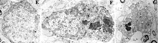
27 Mossy cells Complex spines developing (mossy fibers terminate on it) adult Seress L. Az emberi hippocampus születés utáni fejlődése. Lege Artis Medicine, (6):489, 5. ábra TÁMOP /2/A/KMR
 2.")
28 Functional types of inhibitory cells according to their targets 1. Perisomatic inhibitory cells: terminate on principal cells bodies, proximal dendrites and axon initial segments; regulate the output activity (basket and chandelier or axo-axonic cells) 2. Dendritic inhibitory cells: terminate on distal dendrites of principal cells; regulate the input activity 3. Interneuron selective cells: regulate the activity of interneurons Freund and Buzsaki, Hippocampus, 1996, 6, TÁMOP /2/A/KMR

29 Role of dendritic and perisomatic inhibition Dendritic inhibition Perisomatic inhibition STIMULI OF THE EXTERNAL WORLD DENDRITIC TREE: Input plasticity CELL BODY: Generation of output signal EFFECTS OF OUR INTERNAL WORLD AXON: Signal transmission TÁMOP /2/A/KMR
30 Function of neurochemically different inhibitory cells in the human hippocampus Red: calcium binding proteins; Blue: neuropeptides; Orange: receptor Parvalbumin-containing interneurons Calbindin-containing interneurons Calretinin-containing interneurons Cholecystokinin-containing interneurons Somatostatin-containing interneurons Neuropeptid Y-containing interneurons Substance P receptor expressing interneurons basket and axo-axonic cells, perisomatic inhibition (+ any species examined) dendritic inhibition, + axo-axonic cell, perisomatic inhibition (rat: only dendritic) Dendritic and interneuron specific inhibition (rat: different) Perisomatic and dendritic inhibition (+rat) Dendritic inhibition (+rat) Dendritic inhibition (+rat) Dendritic inhibition (rat: different) TÁMOP /2/A/KMR


31 Pathological types of TLE patients regarding the principal cell loss Control 1: mild group, similar to the control TÁMOP /2/A/KMR


32 Pathological types of TLE patients regarding the principal cell loss 2: patchy type patchy pyramidal cell loss 3: sclerotic type Profound CA1 pyramidal cell loss 4. gliotic type loss of all cell types including the resistant cells (granule cells, CBimmunostained interneurons) TÁMOP /2/A/KMR


33 Hippocampal sclerosis Control CA1 so: stratum oriens; sp: stratum pyramidale; sr: stratum radiatum; sl-m: stratum lacunosum-moleculare; DG: dentate gyrus s.p. Epileptic CA1 Calbindinimmunostaining s.r. s.l-m. s.o.,p.,r. s.l-m. G.D. G.D TÁMOP /2/A/KMR

34 Control Gliosis The amount of glial fibers increases significantly in the hippocampus of epileptic patients. Gliosis is very characteristic for the sclerotic CA1 region, and also present in the dentate gyrus. GFAP immunostaining. Epileptic, sclerotic Maglóczky Zs: A hippocampális neuronhálózatok átalakulása krónikus temporális lebeny epilepsziában. In: Halász P (ed.) Hippocampus, mint neuropszichiátriai betegségek közös nevezője. Budapest: Melinda Kiadó, pp TÁMOP /2/A/KMR


35 Gliosis The amount of glial fibers increases significantly in the hippocampus of epileptic patients. Large amount of glial fibres is very characteristic for the sclerotic CA1 region, and also present in the dentate gyrus. There is also increased amount of glial fibers visible in the hippocampus of non-sclerotic patients. Calbindin immunostaining Epileptic Dentate Gyrus Epileptic CA1 region Magloczky et al. Neuroscience 2000, Wittner et al. Neuroscience, TÁMOP /2/A/KMR


36 Mossy fiber sprouting enhanced internal excitatory pathway The number of granule cell axons terminals increases in the str. moleculare of dentate gyrus and CA3 region. These fibers terminate primarily on principal cells. Ann.Neurol., , Epilepsia, J Neurosci, , J Neurosci, Neuroscience, Neuroscience, Large fraction of the fibers terminates also on dendrites of local interneurons. If the inhibitory cells receive excess stimulation, many of those will dye. A subset of these neurons, however will survive and transmit a more effective inhibition. Maglóczky, Neuroscience : TÁMOP /2/A/KMR
.")

37 Sprouting of excitatory input pathways Control Epileptic The supramammillary pathway (SUM) innervates the granule cells of the DG with excitatory terminals, which are arranged in a thin layer in controls (arrows). In contrast, in epileptic patients this layer occupies the whole stratum moleculare, where the axons terminate mainly on granule cells. The SUM contains calretinin. The number of local calretinin-containing inhibitory cells is reduced. SUM fibers form asymmetric synapses (C,E), whereas the axon terminals of the local interneurons establish symmetric synapses (D,F). Calretinin immunostaining. Maglóczky et al Neuroscience : TÁMOP /2/A/KMR
38 Abnormal localization of interneurons Alterations of input characteristics of the interneurons. Receptor mis-match in the controls. Abnormal localization of interneurons (migration; arrows). Substance P receptor-immunoreactive inhibitory cells can be detected in the stratum moleculare (sm) of the epileptic hippocampus, in turn, such neurons are localised mainly in the hilus (H) of the control hippocampi. The number of cells are reduced in the hippocampus. Control Epileptic Maglóczky könyvfejezet Gabro kiadó TÁMOP /2/A/KMR
 Number of cells studied Stratum oriens Stratum pyramidale and radiatum Control (n=33) 4.25 1.04 10.52 3.28 Mild (n=30) 6.63 2.88 11.59 4.")
39 Dendritic morphology of Substance P receptor-expressing neurons Tóth K. et al Neuroscience Number of ramifications/cell (mean stdev) Number of cells studied Stratum oriens Stratum pyramidale and radiatum Control (n=33) Mild (n=30) Patchy, non sclerotic (n=28) Sclerotic (n=18) Stratum oriens, pyramidale and radiatum = TÁMOP /2/A/KMR

40 - Changes in the neurochemical markers (e.g. parvalbumin (PV) disappears from the inhibitory cells, number of immunoreactive (IR) cell bodies decreases, but the IR terminals remain visible) - Axonal sprouting of interneurons. PV-containing axo-axonic cells establish more synapses on the AIS of granule cells of epileptic patients than in controls. Parvalbumin immunostaining. Axonal sprouting of local interneurons Wittner et al. Neuroscience, 2001 Number of patients (Number of AIS) Control n=10 (n=95) Control n=5 (n=88) Patient n=21 (n=43) Patient n=9 (n=47) Patient n=22 (n=58) Non-sclerotic patient n=15; (n=74) Total length of the studied AISs Synaptic coverage (µm synapse/100 µm AIS) Mean of control 0.52 Mean of epileptic patients TÁMOP /2/A/KMR
 cells is")

41 Reduction of the number of local interneurons and their axonal sprouting Control Epileptic sm sg sg sm -The number of somatostatinimmunoreactive (SOM-IR) cells is reduced, and the SOM-IR axons show sprouting in the dentate gyrus. Somatostatin immunosatining. De Lanerolle et al., Brain Res. 495: TÁMOP /2/A/KMR

 - Deformation of interneurons (arrows, dendritic")

42 CA1: The morphology of inhibitory cells undergoes changes, the principal cells show functional alterations Control Epileptic - Changes of neurochemical markers (calbindin disappears from the pyramidal cells in the non sclerotic CA1 region) - Deformation of interneurons (arrows, dendritic growth, spine formation, hypertrophy) Control Non-sclerotic Sclerotic Calbindin immunostaining Wittner et al. Neuroscience, TÁMOP /2/A/KMR
 2011.10.15. TÁMOP 4.1.2-08/2/A/KMR-2009-0006 43")
43 DG: The morphology of inhibitory cells undergoes changes, the principal cells show functional alterations - Dispersion of granule cell layer (sg: stratum granulosum) - Changes of neurochemical markers (calbindin disappears from the granule cells - Deformation of interneurons (arrows; dendritic growth, spine formation, hypertrophy) TÁMOP /2/A/KMR


44 Axonal sprouting of local interneurons The majority of calbindin-containing inhibitory cells are preserved in the epileptic hippocampus. Contrasting the controls however, they do not project onto principal cells; they rather establish connections with each other resulting in disinhibition. Wittner et al. Neuroscience, TÁMOP /2/A/KMR


45 Loss of interneurons calretinin-containing cells, human TLE K. Tóth et. al. Brain TÁMOP /2/A/KMR


46 Calretinin-immunostained dendrites CONTROL EPILEPTIC Toth K. et al. Brain TÁMOP /2/A/KMR
47 Control Epileptic CR IS CR IS CR IS Toth K. Et al Brain CR IS CR IS CR IS Interneuron-specific inhibitory cell Synchronized dendritic inhibitory cells Pyramidal cells, no plasticity in dendrites Degenerating interneuron-specific inhibitory cell Asynchronous dendritic inhibitory cells Pyramidal dendrites with associative plasticity TÁMOP /2/A/KMR
 2011.10.15. TÁMOP 4.1.2-08/2/A/KMR-2009-0006 48")
48 DG: The morphology of inhibitory cells undergoes changes, the principal cells show functional alterations CONTROL sg EPILEPTIC sg - Dispersion of granule cell layer (sg: stratum granulosum) - Changes of neurochemical markers (calbindin partially disappears from the granule cells - Deformation of interneurons (arrows; dendritic growth, spine formation, hypertrophy) TÁMOP /2/A/KMR
49 Functional morphological changes at neuronal network level the degree of it is in correlation with the loss of principal cells 1. Cell death: Pyramidal cells of CA1 and CA3c regions, sensitive inhibitory cells (calretinin-, somatostatin-, neuropeptid Y-containing cells supplemented with parvalbumin- and Substance P receptor- containing cells of the CA1 region) and reduction of the number of mossy cells. 2. Migration of cells: Dispersion of granule cells, Substance P receptor-expressing inhibitory cells 3. Deformation of cellular morphology: Extra dendrites, formation of dendritic- and somatic spines, hypertrophy of cell body (calbindin- and Substance P receptor-containing inhibitory cells) 4. Neurochemical changes: Reduction of calbindin-level in the granule cells, and its increase in the interneurons, reduction of parvalbumin-level in the perisomatic inhibitory cells 5. Axonal sprouting changes of external and internal neuronal connections - local principal cells: sprouting of mossy fibers and the axons of CA1 pyramidal cells - external input pathways: axons within the supramammillary pathway (and the subicular input) - local inhibitory interneurons a) enhancement of the perisomatic inhibition in the dentate gyrus b) axonal sprouting of calbindin-containing interneurons in the CA1 region, change in the target cells - dendritic inhibition of CA1 pyramidal cells is replaced with the inhibition of interneurons 6. Glial fibers - increased deposition TÁMOP /2/A/KMR
50 Changes of neurochemical marker-content Calbindin: may disappear from principal cells, but not from interneurons Parvalbumin: may disappear from cells, dendrites, sometimes from terminals Calretinin: seems to be stably present SP: may APPEAR in principal cells NPY: stably present in interneurons, mrna may appear in granule cells. NPY appears in mossy fibres. CCK: seems to be stably present TÁMOP /2/A/KMR
51 Fate of inhibitory neurons in the epileptic hippocampus Interneuron types Black: looser Red: winner Green: looser&winner Parvalbumin/ perisomatic Calbindin/ dendritic (CCK) SPR/ dendritic Calretinin/ dendritic + interneuron specific Nonsclerotic CA1 Sclerotic CA1 Nonsclerotic DG Survive Vulnerable Survive/ sprouting Survive/ sprouting Survive/ dendritic growing Survive/ dendritic degeneration Subset of them survive/dendritic growth, spine formation, sprouting Survive Sclerotic DG Survive/ PV disappear, sprouting Survive/ growth Vulnerable Survive Survive/ migration, dendritic growth Vulnerable Survive/ dendritic degeneration Subset of them vulnerable TÁMOP /2/A/KMR
52 Epilepsy and the inhibitory neuronal network EPILEPTIC REORGANISATION = Cell death + sprouting: changes of the cellular connections and excitability Loss of interneuron specific inhibitory cells results in a reduction of the effectiveness of dendritic inhibition. Reduction of the dendritic inhibition and the sprouting of excitatory pathways result in an abnormal potentiation of the excitatory input. The increased perisomatic inhibition may increase the probability of synchronised cellular activity. The neuronal network becomes destabilised and consequently seizures develop more easily. Cellular death is induced by the increase of calcium levels deriving from the extracellular space, threshold phenomena. Loss of interneurons in the CA1 may depend on the survival of target cells TÁMOP /2/A/KMR
53 Epileptic reorganisation Cell death + alterations in the neuronal connectivity and excitability 1. Reduction of dendritic inhibition. 2. Reduction of interneuron-specific inhibition 3. Increase of excitatory input onto dendrites 4. Increase of perisomatic inhibition The neuronal network becomes destabilised, synchronisation is increased within. Seizures develop more easily. 1. Loss of inhibitory neurons develops in all types of epilepsy, independent of the sclerosis. 2. Axon sprouting (excitatory and inhibitory) develops in all types of epilepsy, independent of the sclerosis. 3. Loss of principal cells is likely to depend on the extent of excitation, threshold phenomena. EPILEPSY = SCLEROSIS and DUAL PATHOLOGY - other regions are also reorganised! TÁMOP /2/A/KMR
54 Function of hippocampus (+ limbic system) memory (transition of short-term and long-term) learning spatial orientation emotional background of events, behavioral regulation Types of memory: explicit (declarative) - hippocampus dependent epizodic, semantic, visual implicit (procedural) hippocampus independent Szirmai: Neurológia TÁMOP /2/A/KMR
55 Participation of the left and right hippocampus in memory processing DOMINANT (left, by right-handed people) speach recognition word recognition memorising words echoing words object of tales SUBDOMINANT (right) vizual capabilities face recognition spatial rotation of images details of tales TÁMOP /2/A/KMR
 -Seizure induced by electric")

56 Experimental epilepsy models (according to the triggering methods) - Genetic modifications - Kindling (repeated small electric or chemical stimulation, till the level of spontaneously recurrent seizures) -Seizure induced by electric stimulation -Application of excitatory amino acid analogues -Alteration of the levels of inhibitory-excitatory amino acids - Alteration of the operation of ion channels -Alteration of the kationic concentrations etc TÁMOP /2/A/KMR
 Tissue is sampled from control animal, and the seizure is triggered with a chemical agent. Epileptogenesis is studed, i.e. behavior of single cells in response to a seizure.")
.")
57 Experimental epilepsy models (according to the phenotype) 1. Acute seizure model (slice, cell culture) Tissue is sampled from control animal, and the seizure is triggered with a chemical agent. Epileptogenesis is studed, i.e. behavior of single cells in response to a seizure. There is no network effect and reorganisation. This is the model of synchronous activity. 2. Chronic epilepsy model It is studied in animals producing spontaneous seizures (such animals are produced by application of pilocarpine, kindling, or kainic acid). There is reorganization. The effect of long-term rearrangement and the network changes can be studied in this model TÁMOP /2/A/KMR
58 Epilepsy models EVOKED GENETIC CULTURED GENETICALLY MODIFIED IN VIVO IN VITRO TISSUE CULTURE, SLICE (control, chronic epilepsy) CHRONIC ACUTE (In animals producing (Seizure are induced by the manipulation acutely spontaneous seizures) there is no recurrent seizure) - kindling -4-amino piridin -pilocarpin -febrile seizure-model -kainic acid TÁMOP /2/A/KMR
59 Experimental models of Temporal Lobe Epilepsy Kindling model - It provides a model only for seizure, there is no/a few/ cell death - There is sprouting of mossy fiers, level of calbindin is reduced - It is a partial seizure model Pilocarpine (non-specific muscarin-receptor agonist) model - Administration of mg/kg pilocarpine i.p. (+scopolamine to reduce peripheral cholinergic effects) -Acute effect is status epilepticus (24 h) then a latent period (days-week) - Chronically recurrent seizures - Cell death characteristic for TLE is in the hippocampus TÁMOP /2/A/KMR
60 Experimental models of Temporal Lobe Epilepsy Kainic acid (glutamate analogue, effects through kainate receptors) model -Highest density of receptor of the drug on the pyramidal cells of CA3 region and the mossy fibers - direct effect trough its specific receptors -it spares the axons, indirect effects through axonal pathways - can be administered: intraperitonially, subcutaneously, intracerebroventricularly, intracerebrally -resultant cell death varies, seizures are always similar to the one characterises the TLE, status epilepticus appeares if it applied in large dose. It is suitable also for chemical kindling. - ipsilateral kainate injection in the hippocampus/entorhinal cortex results in cell death in the contralateral hippocampus, the appearance of which is very similar to the one observed in the hippocampus of TLE patients TÁMOP /2/A/KMR


61 Kainate model, ipsilateral kainate injection into the CA3 region Magloczky and Freund, Neuroscience TÁMOP /2/A/KMR
62 Cell death in the contralateral hippocampus after ipsilateral kainate injection: Loss of CA3 and CA1 pyramidal cells shows similar histology to the one observed in human hippocampal sclerosis. Gallyas silver impregnation. Magloczky et al. Neuroscience TÁMOP /2/A/KMR


63 Calbindin-immunoreactive cells in two models of epilepsy Kainate model (rat) PILO model (mouse) CA1 Gyrus dentatus TÁMOP /2/A/KMR
 2011.10.15. TÁMOP 4.1.2-08/2/A/KMR-2009-0006 64")
64 Two types of cell loss in pilocarpine-induced epilepsy (weak-strong SE) TÁMOP /2/A/KMR


65 Two types of cell loss in pilocarpineinduced epilepsy (weak-strong SE) CA1 CA TÁMOP /2/A/KMR

66 AP model, calbindin-containing cells in the CA1 region Control 3-AP-treated Slezia et al., Neurobiol. Des TÁMOP /2/A/KMR


67 Kainate model, calretinin-containing cells, CA1, rat Control Epileptic TÁMOP /2/A/KMR

68 4-AP model, calretinin-containing cells in the rat CA1 region Control 4-AP-treated Slezia et al., Neurobiol. Des TÁMOP /2/A/KMR
 Control 1. non-sclerotic 2.")


69 Classification of TLE patients according to the extent of cell death (n=50 ) Control 1. non-sclerotic 2. non-sclerotic 3. sclerotic 4. gliotic 1: Kindling, low dose kainate 4-AP injection 3: Unilateral, medium dose kainate, Contralateral hc., pilocarpine 4: Icv, intrahippocampal or systemic injection of large dose kainate TÁMOP /2/A/KMR

70 Drawing of Escher may also demonstrate the relationship between the epileptic reorganization and the epileptic seizures TÁMOP /2/A/KMR
PETER PAZMANY CATHOLIC UNIVERSITY Consortium members SEMMELWEIS UNIVERSITY, DIALOG CAMPUS PUBLISHER
 PETER PAZMANY CATHOLIC UNIVERSITY SEMMELWEIS UNIVERSITY Development of Complex Curricula for Molecular Bionics and Infobionics Programs within a consortial* framework** Consortium leader PETER PAZMANY
epilepticus (SE) or trauma. Between this injury and the emergence of recurrent
 or trauma. Between this injury and the emergence of recurrent") Introduction Epilepsy is one of the oldest medical disorders known. The word epilepsy derived from the Greek word epilamhanein, meaning to be seized or to be overwhelmed by surprise. Epilepsy is one of
Introduction Epilepsy is one of the oldest medical disorders known. The word epilepsy derived from the Greek word epilamhanein, meaning to be seized or to be overwhelmed by surprise. Epilepsy is one of
CYTOARCHITECTURE OF CEREBRAL CORTEX
 BASICS OF NEUROBIOLOGY CYTOARCHITECTURE OF CEREBRAL CORTEX ZSOLT LIPOSITS 1 CELLULAR COMPOSITION OF THE CEREBRAL CORTEX THE CEREBRAL CORTEX CONSISTS OF THE ARCHICORTEX (HIPPOCAMPAL FORMA- TION), PALEOCORTEX
TNS Journal Club: Interneurons of the Hippocampus, Freund and Buzsaki
 TNS Journal Club: Interneurons of the Hippocampus, Freund and Buzsaki Rich Turner (turner@gatsby.ucl.ac.uk) Gatsby Unit, 22/04/2005 Rich T. Introduction Interneuron def = GABAergic non-principal cell Usually
NIH Public Access Author Manuscript Epilepsia. Author manuscript; available in PMC 2011 July 1.
 NIH Public Access Author Manuscript Published in final edited form as: Epilepsia. 2010 July ; 51(Suppl 3): 115 120. doi:10.1111/j.1528-1167.2010.02624.x. Dynamic changes of CB1 receptor expression in hippocampi
Anatomy of the Hippocampus
 Anatomy of the Hippocampus Lecture 3.2 David S. Touretzky September, 2015 Human Hippocampus 2 Human Hippocampus 3 Hippocampus Means Seahorse Dissected human hippocampus next to a specimen of hippocampus
SYNAPTIC COMMUNICATION
 BASICS OF NEUROBIOLOGY SYNAPTIC COMMUNICATION ZSOLT LIPOSITS 1 NERVE ENDINGS II. Interneuronal communication 2 INTERNEURONAL COMMUNICATION I. ELECTRONIC SYNAPSE GAP JUNCTION II. CHEMICAL SYNAPSE SYNAPSES
PETER PAZMANY CATHOLIC UNIVERSITY Consortium members SEMMELWEIS UNIVERSITY, DIALOG CAMPUS PUBLISHER
 PETER PAZMANY CATHOLIC UNIVERSITY SEMMELWEIS UNIVERSITY Development of Complex Curricula for Molecular Bionics and Infobionics Programs within a consortial* framework** Consortium leader PETER PAZMANY
Mossy cells in epilepsy: rigor mortis or vigor mortis?
 140 Opinion Mossy cells in epilepsy: rigor mortis or vigor mortis? Anna d. H. Ratzliff, Vijayalakshmi Santhakumar, Allyson Howard and Ivan Soltesz Mossy cells are bi-directionally connected through a positive
Supplementary Figure 1. SDS-FRL localization of CB 1 in the distal CA3 area of the rat hippocampus. (a-d) Axon terminals (t) in stratum pyramidale
 Axon terminals (t) in stratum pyramidale") Supplementary Figure 1. SDS-FRL localization of CB 1 in the distal CA3 area of the rat hippocampus. (a-d) Axon terminals (t) in stratum pyramidale (b) show stronger immunolabeling for CB 1 than those in
Supplementary Figure 1. SDS-FRL localization of CB 1 in the distal CA3 area of the rat hippocampus. (a-d) Axon terminals (t) in stratum pyramidale (b) show stronger immunolabeling for CB 1 than those in
Changes of CB1 cannabinoid receptor distribution in temporal lobe epilepsy
 Changes of CB1 cannabinoid receptor distribution in temporal lobe epilepsy PhD Dissertation Mária Rita Karlócai János Szentágothai School of Neurosciences Semmelweis University Supervisor: Dr. Zsófia Maglóczky,
Neurogenesis and its Association to Epileptogenesis in Temporal Lobe Epilepsy
 Neurogenesis and its Association to Epileptogenesis in Temporal Lobe Epilepsy Vanessa Marques Donegá Cover page: Figure adapted from Siebzehnrubl FA. and Blumcke I., 2008 Supervisor: Dr. P.N.E. de Graan
Antiepileptic agents
 Antiepileptic agents Excessive excitability of neurons in the CNS Abnormal function of ion channels Spread through neural networks Abnormal neural activity leads to abnormal motor activity Suppression
Memory Systems II How Stored: Engram and LTP. Reading: BCP Chapter 25
 Memory Systems II How Stored: Engram and LTP Reading: BCP Chapter 25 Memory Systems Learning is the acquisition of new knowledge or skills. Memory is the retention of learned information. Many different
GABA A Receptors and GABAergic Interneurons in Chronic Temporal Lobe Epilepsy
 GABA A Receptors and GABAergic Interneurons in Chronic Temporal Lobe Epilepsy December 4, 2010 Carolyn R. Houser Professor, David Geffen School of Medicine at UCLA and VA Greater Los Angeles Healthcare
Free recall and recognition in a network model of the hippocampus: simulating effects of scopolamine on human memory function
 Behavioural Brain Research 89 (1997) 1 34 Review article Free recall and recognition in a network model of the hippocampus: simulating effects of scopolamine on human memory function Michael E. Hasselmo
Correlated network activity in the developing hippocampus: role in synaptogenesis
 Enrico Cherubini Correlated network activity in the developing hippocampus: role in synaptogenesis SPACE PHYSICS and BIOLOGY Dubna, December 19-23, 2010 The construction of the brain relies on genetic
CASE 48. What part of the cerebellum is responsible for planning and initiation of movement?
 CASE 48 A 34-year-old woman with a long-standing history of seizure disorder presents to her neurologist with difficulty walking and coordination. She has been on phenytoin for several days after having
BIPN 140 Problem Set 6
 BIPN 140 Problem Set 6 1) The hippocampus is a cortical structure in the medial portion of the temporal lobe (medial temporal lobe in primates. a) What is the main function of the hippocampus? The hippocampus
PETER PAZMANY CATHOLIC UNIVERSITY Consortium members SEMMELWEIS UNIVERSITY, DIALOG CAMPUS PUBLISHER
 PETER PAZMANY CATHOLIC UNIVERSITY SEMMELWEIS UNIVERSITY Development of Complex Curricula for Molecular Bionics and Infobionics Programs within a consortial* framework** Consortium leader PETER PAZMANY
Intracranial Studies Of Human Epilepsy In A Surgical Setting
 Intracranial Studies Of Human Epilepsy In A Surgical Setting Department of Neurology David Geffen School of Medicine at UCLA Presentation Goals Epilepsy and seizures Basics of the electroencephalogram
Lisa M. Giocomo & Michael E. Hasselmo
 Mol Neurobiol (2007) 36:184 200 DOI 10.1007/s12035-007-0032-z Neuromodulation by Glutamate and Acetylcholine can Change Circuit Dynamics by Regulating the Relative Influence of Afferent Input and Excitatory
BIPN 140 Problem Set 6
 BIPN 140 Problem Set 6 1) Hippocampus is a cortical structure in the medial portion of the temporal lobe (medial temporal lobe in primates. a) What is the main function of the hippocampus? The hippocampus
Subconvulsive Dose of Kainic Acid Transiently Increases the Locomotor Activity of Adult Wistar Rats
 Physiol. Res. 64: 263-267, 2015 SHORT COMMUNICATION Subconvulsive Dose of Kainic Acid Transiently Increases the Locomotor Activity of Adult Wistar Rats V. RILJAK 1, D. MAREŠOVÁ 1, J. POKORNÝ 1, K. JANDOVÁ
All questions below pertain to mandatory material: all slides, and mandatory homework (if any).
.") ECOL 182 Spring 2008 Dr. Ferriere s lectures Lecture 6: Nervous system and brain Quiz Book reference: LIFE-The Science of Biology, 8 th Edition. http://bcs.whfreeman.com/thelifewire8e/ All questions below
ECOL 182 Spring 2008 Dr. Ferriere s lectures Lecture 6: Nervous system and brain Quiz Book reference: LIFE-The Science of Biology, 8 th Edition. http://bcs.whfreeman.com/thelifewire8e/ All questions below
Physiology of synapses and receptors
 Physiology of synapses and receptors Dr Syed Shahid Habib Professor & Consultant Clinical Neurophysiology Dept. of Physiology College of Medicine & KKUH King Saud University REMEMBER These handouts will
CASE 49. What type of memory is available for conscious retrieval? Which part of the brain stores semantic (factual) memories?
 memories?") CASE 49 A 43-year-old woman is brought to her primary care physician by her family because of concerns about her forgetfulness. The patient has a history of Down syndrome but no other medical problems.
CASE 49 A 43-year-old woman is brought to her primary care physician by her family because of concerns about her forgetfulness. The patient has a history of Down syndrome but no other medical problems.
The Neuron. Consists Of: - cell body. - Dendrites - axon - axon terminal - myelin. dendrites Axon terminal. Cell body. nucleus. axon.
 The Neuron Consists Of: - cell body - Dendrites - axon - axon terminal - myelin dendrites Axon terminal Cell body nucleus myelin axon THE SYNAPSE Definition: It is a point of contact between the axon of
What Cell Make Up the Brain and Spinal Cord
 What Cell Make Up the Brain and Spinal Cord Jennifer LaVail, Ph.D. (http://anatomy.ucsf.edu/pages/lavaillab/index.html) What kinds of cells are these?" Neuron?" Epithelial cell?" Glial cell?" What makes
PETER PAZMANY CATHOLIC UNIVERSITY Consortium members SEMMELWEIS UNIVERSITY, DIALOG CAMPUS PUBLISHER
 PETER PAZMANY CATHOLIC UNIVERSITY SEMMELWEIS UNIVERSITY Development of Complex Curricula for Molecular Bionics and Infobionics Programs within a consortial* framework** Consortium leader PETER PAZMANY
Neurobiology of Epileptogenesis
 Neurobiology of Epileptogenesis Michael C. Smith, MD Director, Rush Epilepsy Center Professor and Senior Attending Neurologist Rush University Medical Center Chicago, IL Network Milieu Cellular Milieu
biological psychology, p. 40 The study of the nervous system, especially the brain. neuroscience, p. 40
 biological psychology, p. 40 The specialized branch of psychology that studies the relationship between behavior and bodily processes and system; also called biopsychology or psychobiology. neuroscience,
COGNITIVE SCIENCE 107A. Hippocampus. Jaime A. Pineda, Ph.D.
 COGNITIVE SCIENCE 107A Hippocampus Jaime A. Pineda, Ph.D. Common (Distributed) Model of Memory Processes Time Course of Memory Processes Long Term Memory DECLARATIVE NON-DECLARATIVE Semantic Episodic Skills
Review: Hippocampal sclerosis in epilepsy: a neuropathology review
 Neuropathology and Applied Neurobiology (2014), 40, 520 543 doi: 10.1111/nan.12150 Review: Hippocampal sclerosis in epilepsy: a neuropathology review M. Thom Departments of Neuropathology and Clinical
Synaptic plasticity. Activity-dependent changes in synaptic strength. Changes in innervation patterns. New synapses or deterioration of synapses.
 Synaptic plasticity Activity-dependent changes in synaptic strength. Changes in innervation patterns. New synapses or deterioration of synapses. Repair/changes in the nervous system after damage. MRC Centre
PETER PAZMANY CATHOLIC UNIVERSITY Consortium members SEMMELWEIS UNIVERSITY, DIALOG CAMPUS PUBLISHER
 SEMMELWEIS UNIVERSITY PETER PAZMANY CATHOLIC UNIVERSITY Development of Complex Curricula for Molecular Bionics and Infobionics Programs within a consortial* framework** Consortium leader PETER PAZMANY
Neural Communication. Central Nervous System Peripheral Nervous System. Communication in the Nervous System. 4 Common Components of a Neuron
 Neural Communication Overview of CNS / PNS Electrical Signaling Chemical Signaling Central Nervous System Peripheral Nervous System Somatic = sensory & motor Autonomic = arousal state Parasympathetic =
Lesson 14. The Nervous System. Introduction to Life Processes - SCI 102 1
 Lesson 14 The Nervous System Introduction to Life Processes - SCI 102 1 Structures and Functions of Nerve Cells The nervous system has two principal cell types: Neurons (nerve cells) Glia The functions
ANIMAL MODELS OF EPILEPSY Index
 INDEX A Action potentials BK channel effect on... 89 90 See also Synaptic transmission Adeno-associated virus (AAV) vector gene therapy... 234 inhibitory GABA receptors and... 238 240 neuroactive peptides
Chapter 6: Hippocampal Function In Cognition. From Mechanisms of Memory, second edition By J. David Sweatt, Ph.D.
 Chapter 6: Hippocampal Function In Cognition From Mechanisms of Memory, second edition By J. David Sweatt, Ph.D. Grid Cell The Hippocampus Serves a Role in Multimodal Information Processing and Memory
The 7 th lecture. Anatomy and Physiology For the. 1 st Class. By Dr. Ala a Hassan Mirza
 The 7 th lecture In Anatomy and Physiology For the 1 st Class By Dr. Ala a Hassan Mirza Nervous System (part I) The Nerve Tissue and the Nervous System The Tissues of the Body There are 4 types of tissues
HISTOLOGY AND HISTOCHEMISTRY OF LIMBIC HIPPOCAMPUS IN THE INDIAN BUFFALOES
 HISTOLOGY AND HISTOCHEMISTRY OF LIMBIC HIPPOCAMPUS IN THE INDIAN BUFFALOES A. Kumaravel*, Geetha Ramesh, S. Rajathi and S. Muthukrishnan Department of Veterinary Anatomy, Veterinary College and Research
Name: Period: Chapter 2 Reading Guide The Biology of Mind
 Name: Period: Chapter 2 Reading Guide The Biology of Mind The Nervous System (pp. 55-58) 1. What are nerves? 2. Complete the diagram below with definitions of each part of the nervous system. Nervous System
COMMENTARY THE MULTIFARIOUS HIPPOCAMPAL MOSSY FIBER PATHWAY: A REVIEW
 Pergamon www.elsevier.com/locate/neuroscience Hippocampal mossy fibers Neuroscience Vol. 98, No. 3, pp. 407 427, 2000 407 2000 IBRO. Published by Elsevier Science Ltd Printed in Great Britain. All rights
SUPPLEMENTARY INFORMATION. Supplementary Figure 1
 SUPPLEMENTARY INFORMATION Supplementary Figure 1 The supralinear events evoked in CA3 pyramidal cells fulfill the criteria for NMDA spikes, exhibiting a threshold, sensitivity to NMDAR blockade, and all-or-none
Neurogenesis in Adult Central Nervous System: Death of a Dogma
 Aristotle University of Thessaloniki, Greece, Nov. 2007 Neurogenesis in Adult Central Nervous System: Death of a Dogma Anton B. Tonchev Division of Cell Biology, Varna University of Medicine, Bulgaria
Supplementary Materials for VAMP4 directs synaptic vesicles to a pool that selectively maintains asynchronous neurotransmission
 Supplementary Materials for VAMP4 directs synaptic vesicles to a pool that selectively maintains asynchronous neurotransmission Jesica Raingo, Mikhail Khvotchev, Pei Liu, Frederic Darios, Ying C. Li, Denise
Synaptic plasticityhippocampus. Neur 8790 Topics in Neuroscience: Neuroplasticity. Outline. Synaptic plasticity hypothesis
 Synaptic plasticityhippocampus Neur 8790 Topics in Neuroscience: Neuroplasticity Outline Synaptic plasticity hypothesis Long term potentiation in the hippocampus How it s measured What it looks like Mechanisms
Neurons! John A. White Dept. of Bioengineering
 Neurons! John A. White Dept. of Bioengineering john.white@utah.edu What makes neurons different from cardiomyocytes? Morphological polarity Transport systems Shape and function of action potentials Neuronal
EE 791 Lecture 2 Jan 19, 2015
 EE 791 Lecture 2 Jan 19, 2015 Action Potential Conduction And Neural Organization EE 791-Lecture 2 1 Core-conductor model: In the core-conductor model we approximate an axon or a segment of a dendrite
Ph.D. Thesis. Norbert Károly M.Sc. Experimental and Clinical Neuroscience Doctoral Programme Doctoral School of Theoretical Medicine
 Ph.D. Thesis Immunohistochemical investigations of the neuronal changes induced by chronic recurrent seizures in a pilocarpine rodent model of temporal lobe epilepsy Norbert Károly M.Sc. Experimental and
Lecture: Introduction to nervous system development
 Lecture: Introduction to nervous system development Prof. Ilan Davis, Department of Biochemistry. Wellcome Senior Research Fellow Senior Research Fellow, Jesus College ilan.davis@bioch.ox.ac.uk http://www.ilandavis.com
Seizure Semiology CHARCRIN NABANGCHANG, M.D. PHRAMONGKUTKLAO COLLEGE OF MEDICINE
 Seizure Semiology CHARCRIN NABANGCHANG, M.D. PHRAMONGKUTKLAO COLLEGE OF MEDICINE Seizure Semiology Differentiate between epileptic and nonepileptic seizures Classification of epileptic syndrome Presurgical
Nature Neuroscience: doi: /nn.2275
 Supplementary Figure S1. The presence of MeCP2 in enriched primary glial cultures from rat or mouse brains is not neuronal. Western blot analysis of protein extracts from (a) rat glial and neuronal cultures.
Ch 8. Learning and Memory
 Ch 8. Learning and Memory Cognitive Neuroscience: The Biology of the Mind, 2 nd Ed., M. S. Gazzaniga, R. B. Ivry, and G. R. Mangun, Norton, 2002. Summarized by H.-S. Seok, K. Kim, and B.-T. Zhang Biointelligence
Introduction to seizure and epilepsy
 Introduction to seizure and epilepsy 1 Epilepsy : disorder of brain function characterized by a periodic and unpredictable occurrence of seizures. Seizure : abnormal increased electrical activity in the
*Pathophysiology of. Epilepsy
 *Pathophysiology of Epilepsy *Objectives * At the end of this lecture the students should be able to:- 1.Define Epilepsy 2.Etio-pathology of Epilepsy 3.Types of Epilepsy 4.Role of Genetic in Epilepsy 5.Clinical
Chapter 12 Nervous Tissue
 9/12/11 Chapter 12 Nervous Tissue Overview of the nervous system Cells of the nervous system Electrophysiology of neurons Synapses Neural integration Subdivisions of the Nervous System 1 Subdivisions of
Name: Period: Test Review: Chapter 2
 Name: Period: Test Review: Chapter 2 1. The function of dendrites is to A) receive incoming signals from other neurons. B) release neurotransmitters into the spatial junctions between neurons. C) coordinate
SUPPLEMENTARY INFORMATION
 SUPPLEMENTARY INFORMATION doi:10.1038/nature11306 Supplementary Figures Supplementary Figure 1. Basic characterization of GFP+ RGLs in the dentate gyrus of adult nestin-gfp mice. a, Sample confocal images
Introduction to Computational Neuroscience
 Introduction to Computational Neuroscience Lecture 7: Network models Lesson Title 1 Introduction 2 Structure and Function of the NS 3 Windows to the Brain 4 Data analysis 5 Data analysis II 6 Single neuron
Structure of a Neuron:
 Structure of a Neuron: At the dendrite the incoming signals arrive (incoming currents) At the soma current are finally integrated. At the axon hillock action potential are generated if the potential crosses
Brain anatomy and artificial intelligence. L. Andrew Coward Australian National University, Canberra, ACT 0200, Australia
 Brain anatomy and artificial intelligence L. Andrew Coward Australian National University, Canberra, ACT 0200, Australia The Fourth Conference on Artificial General Intelligence August 2011 Architectures
Neurobiology. Cells of the nervous system
 Neurobiology Cells of the nervous system Anthony Heape 2010 1 The nervous system Central nervous system (CNS) Peripheral nervous system (PNS) 2 Enteric nervous system (digestive tract, gall bladder and
Neuronal Plasticity, Learning and Memory. David Keays Institute of Molecular Pathology
 Neuronal Plasticity, Learning and Memory David Keays Institute of Molecular Pathology http://keayslab.org Structure 1. What is learning and memory? 2. Anatomical basis 3. Cellular basis 4. Molecular
DO NOW: ANSWER ON PG 73
 DO NOW: ANSWER ON PG 73 1. Name 1 neurotransmitter that we have learned about. 2. Draw a basic graph of a neuron action potential. Label resting potential, threshold, depolarization, and repolarization
Acetylcholine (ACh) Action potential. Agonists. Drugs that enhance the actions of neurotransmitters.
 Action potential. Agonists. Drugs that enhance the actions of neurotransmitters.") Acetylcholine (ACh) The neurotransmitter responsible for motor control at the junction between nerves and muscles; also involved in mental processes such as learning, memory, sleeping, and dreaming. (See
Acetylcholine (ACh) The neurotransmitter responsible for motor control at the junction between nerves and muscles; also involved in mental processes such as learning, memory, sleeping, and dreaming. (See
Ch 8. Learning and Memory
 Ch 8. Learning and Memory Cognitive Neuroscience: The Biology of the Mind, 2 nd Ed., M. S. Gazzaniga,, R. B. Ivry,, and G. R. Mangun,, Norton, 2002. Summarized by H.-S. Seok, K. Kim, and B.-T. Zhang Biointelligence
GABAergic Contributions to Gating, Timing, and Phase Precession of Hippocampal Neuronal Activity During Theta Oscillations
 GABAergic Contributions to Gating, Timing, and Phase Precession of Hippocampal Neuronal Activity During Theta Oscillations Vassilis Cutsuridis* 1 and Michael Hasselmo 2 HIPPOCAMPUS 22:1597 1621 (2012)
Outline. Animals: Nervous system. Neuron and connection of neurons. Key Concepts:
 Animals: Nervous system Neuron and connection of neurons Outline 1. Key concepts 2. An Overview and Evolution 3. Human Nervous System 4. The Neurons 5. The Electrical Signals 6. Communication between Neurons
Biomarkers in Schizophrenia
 Biomarkers in Schizophrenia David A. Lewis, MD Translational Neuroscience Program Department of Psychiatry NIMH Conte Center for the Neuroscience of Mental Disorders University of Pittsburgh Disease Process
Nervous System. Master controlling and communicating system of the body. Secrete chemicals called neurotransmitters
 Nervous System Master controlling and communicating system of the body Interacts with the endocrine system to control and coordinate the body s responses to changes in its environment, as well as growth,
Long-Term, Selective Gene Expression in Developing and Adult Hippocampal Pyramidal Neurons Using Focal In Utero Electroporation
 The Journal of Neuroscience, May 9, 2007 27(19):5007 5011 5007 Toolbox Editor s Note: Toolboxes are intended to briefly highlight a new method or a resource of general use in neuroscience or to critically
Systems Neuroscience Dan Kiper. Today: Wolfger von der Behrens
 Systems Neuroscience Dan Kiper Today: Wolfger von der Behrens wolfger@ini.ethz.ch 18.9.2018 Neurons Pyramidal neuron by Santiago Ramón y Cajal (1852-1934, Nobel prize with Camillo Golgi in 1906) Neurons
Neural Basis of Motor Control
 Neural Basis of Motor Control Central Nervous System Skeletal muscles are controlled by the CNS which consists of the brain and spinal cord. Determines which muscles will contract When How fast To what
Synaptic Communication. Steven McLoon Department of Neuroscience University of Minnesota
 Synaptic Communication Steven McLoon Department of Neuroscience University of Minnesota 1 Course News The first exam is next week on Friday! Be sure to checkout the sample exam on the course website. 2
Lecture 22: A little Neurobiology
 BIO 5099: Molecular Biology for Computer Scientists (et al) Lecture 22: A little Neurobiology http://compbio.uchsc.edu/hunter/bio5099 Larry.Hunter@uchsc.edu Nervous system development Part of the ectoderm
synapse neurotransmitters Extension of a neuron, ending in branching terminal fibers, through which messages pass to other neurons, muscles, or glands
 neuron synapse The junction between the axon tip of a sending neuron and the dendrite of a receiving neuron Building block of the nervous system; nerve cell Chemical messengers that cross the synaptic
Seizure: the clinical manifestation of an abnormal and excessive excitation and synchronization of a population of cortical
 Are There Sharing Mechanisms of Epilepsy, Migraine and Neuropathic Pain? Chin-Wei Huang, MD, PhD Department of Neurology, NCKUH Basic mechanisms underlying seizures and epilepsy Seizure: the clinical manifestation
NEURONS COMMUNICATE WITH OTHER CELLS AT SYNAPSES 34.3
 NEURONS COMMUNICATE WITH OTHER CELLS AT SYNAPSES 34.3 NEURONS COMMUNICATE WITH OTHER CELLS AT SYNAPSES Neurons communicate with other neurons or target cells at synapses. Chemical synapse: a very narrow
Tuning properties of individual circuit components and stimulus-specificity of experience-driven changes.
 Supplementary Figure 1 Tuning properties of individual circuit components and stimulus-specificity of experience-driven changes. (a) Left, circuit schematic with the imaged component (L2/3 excitatory neurons)
P. Hitchcock, Ph.D. Department of Cell and Developmental Biology Kellogg Eye Center. Wednesday, 16 March 2009, 1:00p.m. 2:00p.m.
 Normal CNS, Special Senses, Head and Neck TOPIC: CEREBRAL HEMISPHERES FACULTY: LECTURE: READING: P. Hitchcock, Ph.D. Department of Cell and Developmental Biology Kellogg Eye Center Wednesday, 16 March
Structural basis for the role of inhibition in facilitating adult brain plasticity
 Structural basis for the role of inhibition in facilitating adult brain plasticity Jerry L. Chen, Walter C. Lin, Jae Won Cha, Peter T. So, Yoshiyuki Kubota & Elly Nedivi SUPPLEMENTARY FIGURES 1-6 a b M
Fundamentals of the Nervous System and Nervous Tissue: Part C
 PowerPoint Lecture Slides prepared by Janice Meeking, Mount Royal College C H A P T E R 11 Fundamentals of the Nervous System and Nervous Tissue: Part C Warm Up What is a neurotransmitter? What is the
Part 11: Mechanisms of Learning
 Neurophysiology and Information: Theory of Brain Function Christopher Fiorillo BiS 527, Spring 2012 042 350 4326, fiorillo@kaist.ac.kr Part 11: Mechanisms of Learning Reading: Bear, Connors, and Paradiso,
Modeling Depolarization Induced Suppression of Inhibition in Pyramidal Neurons
 Modeling Depolarization Induced Suppression of Inhibition in Pyramidal Neurons Peter Osseward, Uri Magaram Department of Neuroscience University of California, San Diego La Jolla, CA 92092 possewar@ucsd.edu
Modeling of Hippocampal Behavior
 Modeling of Hippocampal Behavior Diana Ponce-Morado, Venmathi Gunasekaran and Varsha Vijayan Abstract The hippocampus is identified as an important structure in the cerebral cortex of mammals for forming
The Nervous System. Biological School. Neuroanatomy. How does a Neuron fire? Acetylcholine (ACH) TYPES OF NEUROTRANSMITTERS
 TYPES OF NEUROTRANSMITTERS") Biological School The Nervous System It is all about the body!!!! It starts with an individual nerve cell called a NEURON. Synapse Neuroanatomy Neurotransmitters (chemicals held in terminal buttons that
Biological School The Nervous System It is all about the body!!!! It starts with an individual nerve cell called a NEURON. Synapse Neuroanatomy Neurotransmitters (chemicals held in terminal buttons that
STRUCTURAL ELEMENTS OF THE NERVOUS SYSTEM
 STRUCTURAL ELEMENTS OF THE NERVOUS SYSTEM STRUCTURE AND MAINTENANCE OF NEURONS (a) (b) Dendrites Cell body Initial segment collateral terminals (a) Diagrammatic representation of a neuron. The break in
Synaptic Integration
 Synaptic Integration 3 rd January, 2017 Touqeer Ahmed PhD Atta-ur-Rahman School of Applied Biosciences National University of Sciences and Technology Excitatory Synaptic Actions Excitatory Synaptic Action
Supplementary Information Supplementary Table 1. Quantitative features of EC neuron dendrites
 Supplementary Information Supplementary Table 1. Quantitative features of EC neuron dendrites Supplementary Table 2. Quantitative features of EC neuron axons 1 Supplementary Figure 1. Layer distribution