Mechanisms underlying subthreshold and suprathreshold responses in dorsal cochler nucleus cartwheel cells
|
|
|
- Blake Riley
- 6 years ago
- Views:
Transcription
1 The University of Toledo The University of Toledo Digital Repository Theses and Dissertations 2005 Mechanisms underlying subthreshold and suprathreshold responses in dorsal cochler nucleus cartwheel cells Mingjie Tong The University of Toledo Follow this and additional works at: Recommended Citation Tong, Mingjie, "Mechanisms underlying subthreshold and suprathreshold responses in dorsal cochler nucleus cartwheel cells" (2005). Theses and Dissertations This Dissertation is brought to you for free and open access by The University of Toledo Digital Repository. It has been accepted for inclusion in Theses and Dissertations by an authorized administrator of The University of Toledo Digital Repository. For more information, please see the repository's About page.
2 A Dissertation Entitled Mechanisms underlying subthreshold and suprathreshold responses in dorsal cochlear nucleus cartwheel cells by Mingjie Tong Submitted as partial fulfillment of the requirements for the Doctor of Philosophy Degree in Engineering Advisor: Dr. Scott C. Molitor Graduate School The University of Toledo December 2005
3 An Abstract of Mechanisms underlying subthreshold and suprathreshold responses in dorsal cochlear nucleus cartwheel cells by Mingjie Tong Submitted as partial fulfillment of the requirements for the Doctor of Philosophy Degree in Engineering The University of Toledo December 2005 Cartwheel cells (CWCs) are a group of interneurons in the dorsal cochlear nucleus (DCN). Unlike other DCN neurons, which respond to stimuli with simple spikes, or trains of individual action potentials, CWCs respond to suprathreshold stimuli with complex spikes, rapid bursts of action potentials superimposed on a slow depolarization, or a combination of simple and complex spikes. In vitro whole-cell current clamp recordings from young rats (P11 P15) show that CWCs respond to brief suprathreshold stimuli with complex spikes or simple spikes followed by afterdepolarizations (ADPs); and to subthreshold stimuli with subthreshold depolarizations (SDPs). Although complex spikes iii
4 and ADPs are produced by Ca 2+ currents, SDPs are unaffected by Ca 2+ current antagonists but are substantially depressed by the Na + channel antagonist TTX. Voltage clamp recordings reveal that SDPs are produced by a persistent Na + current activating below spike threshold in CWCs. A hyperpolarization-activated inward current (I h ) is shown to contribute to the repolarization of SDPs; and voltage-gated K + currents, including transient and Ca 2+ -activated K + currents contribute to the repolarization of suprathreshold responses in CWCs. Computational simulations demonstrated that SDPs are produced by persistent Na + currents; furthermore, these currents are required for the activation of high-threshold Ca 2+ channels during suprathreshold responses to produce complex spikes in response to brief suprathreshold stimuli. Delayed-rectifier, transient and Ca 2+ -activated K + currents contribute to the repolarization of complex spikes and increase of maximal conductances in any of three K + currents converts complex spikes into simple spikes. In contrast, removal of the hyperpolarization-activated inward current I h increases the amplitude and duration of SDPs, but otherwise has little effect on suprathreshold responses of CWCs. These results demonstrate that a combination of voltage-gated conductances that activate over a range of subthreshold and suprathreshold membrane potentials contribute to the unique electrophysiologic responses of CWCs. iv
5 Table of Contents Abstract Table of contents List of figures List of tables iii v viii x 1 Introduction Dorsal Cochlear Nucleus Complex spiking cartwheel cells (CWCs) in DCN Complex spiking neurons in the central nervous system Suprathreshold responses in cartwheel cells Subthreshold responses in cartwheel cells 15 2 Materials and Experimental Methods Electrophysiology Preparation of slices Solutions Electrophysiologic recordings Morphologic identification Data analysis Isolation of SDPs Voltage dependence of SDPs 30 v
6 2.2.3 I h analysis on voltage-clamp recordings Simulation methods Simulation Model Active conductances Clamp protocols 33 3 Experimental results CWCs generate both complex spikes and simple spikes Subthreshold responses of CWCs Distinct mechanisms underlie ADPs and SDPs Hyperpolarization-activated cation current (I h ) in CWCs I h contributes to repolarizing phase of SDPs in CWCs Activation properties of I h in CWCs Voltage-gated Na + currents in CWCs SDPs are mediated by voltage-gated Na + current Voltage dependence of persistent Na + current K + currents in CWCs Voltage-gated K + currents in CWCs Ca 2+ -activated K + currents in CWCs Pre-step voltage dependence of CWCs 68 4 Simulation results Ionic currents in model of CWCs Fast Na + current (I Na ) Persistent Na + current (I NaP ) 79 vi
7 4.1.3 N-type voltage-gated Ca 2+ current (I CaN ) Hyperpolarization-activated h current (I h ) Delayed-rectifier K + current (I dr ) Transient K + current (I A ) Ca 2+ -activated K + current (I KCa ) Complex-spiking CWC model Persistent Na + current contributes to SDPs Variable activation threshold of persistent Na + currents N-type voltage-gated Ca 2+ current and suprathreshold responses Transient voltage-gated K + currents repolarize suprathreshold 107 responses 4.7 Hyperpolarization-activated inward current (I h ) repolarizes 110 subthreshold responses 5 Discussion Persistent Na + current in CWCs I h in CWCs Suprathreshold responses in CWCs Functional roles of complex spiking in DCN 126 Reference 128 vii
8 List of Figures 1-1 Auditory pathways in DCN Firing patterns of DCN neurons Firing patterns of DCN PCs modulated by inhibitory synaptic inputs Preparation of brainstem slices containing DCN Fluorescence images of DCN neurons Effects of membrane resistance (Rm) on the isolation of subthreshold 27 depolarizations (SDPs) 3-1 Suprathreshold responses of CWCs Suprathreshold and subthreshold responses of neurons in the dorsal 39 cochlear nucleus (DCN) 3-3 Voltage dependence of and Rm changes during SDPs obtained from 43 CWCs 3-4 Role of Ca2+ channels in suprathreshold and subthreshold responses of 46 CWCs 3-5 ZD7288 augments subthreshold responses SDP magnitudes are not altered by changes in the resting V m Activation of I h SDPs are partially blocked by TTX Anticonvulsants phenytoin and riluzole have no effect on SDP 61 magnitudes 3-10 Na + current elicited by a slow ramp protocol in CWCs 63 viii
9 AP (1 mm) produces complex spiking responses in simple spiking 66 CWCs, but not in pyramidal cells 3-12 Voltage-gated Ca2+ current antagonists Ni 2+ /Cd 2+ widened the half-width 69 of APs 3-13 Pre-step voltage dependence of CWCs Kinetic properties of Fast Na + current Kinetic properties of persistent Na + current Kinetic properties of N-type voltage-gated Ca 2+ current Kinetic properties of I h Kinetic properties of delayed-rectifier K + current Kinetic properties of transient K + current Kinetic properties of Ca 2+ -activated K + current Comparison between experimental and modeling results Persistent Na + current (I NaP ) in CWCs Persistent Na + current affects suprathreshold responses in CWCs by 104 changing V half of steady-state activation curve 4-11 N-type voltage-gated Ca 2+ current modulates suprathreshold responses in 108 CWC model 4-12 Transient K + current contributes to the repolarization of suprathreshold 111 responses in CWCs 4-13 I h contributes to repolarization of SDPs I h affects both subthreshold and suprathreshold responses in CWCs when 121 V was depolarized ix
10 List of Tables 1 Comparison of Na+ mediated APs obtained from CWCs in adult (> month old) guinea pig and in P11 P16 rat DCN. x
11 Chapter One Introduction Cartwheel cells (CWCs) are a group of interneurons in the dorsal cochlear nucleus (DCN), part of the brainstem complex that receives inputs from auditory nerve fibers ascending from the cochlea. Unlike other DCN neurons, which respond to excitatory stimuli with individual or repetitive trains of action potentials (APs) referred to as simple spikes, CWCs respond with complex spikes, or rapid bursts of action potentials superimposed on a slow depolarization (Manis et al. 1994; Zhang and Oertel 1993). Though characterized as complex spiking neurons, CWCs also respond spontaneously (Waller and Godfrey 1994) to acoustic stimulation (Davis and Young 1997; Ding et al. 1999; Parham and Kim 1995a) and to intracellular current injection (Ding and Voigt 1997) with a combination of simple and complex spikes. Because CWCs provide inhibition to pyramidal cells (PCs) that form the major projection of the DCN to the inferior colliculus, the ability of CWCs to respond to excitation with simple or complex spikes could have a profound impact upon the information transmitted by the DCN to higher auditory centers. Although it is known that Ca 2+ channels mediate the slow depolarization that underlies complex spiking (Agar et al. 1996; Golding and Oertel 1997), little is known 1
12 about the underlying mechanisms that determine whether CWCs will respond with simple spikes or complex spikes. Given the absence of low-threshold Ca 2+ currents from DCN neurons (Molitor and Manis 1999), it is not clear how CWCs respond with complex spikes to low-threshold stimuli. To understand the electrophysiologic mechanisms that underlie complex-spiking, the research described with this thesis utilize whole-cell recordings to examine the responses of CWCs to depolarizing stimuli at and below spike threshold. This research also demonstrated that the CWCs respond to subthreshold stimuli with a slow active event, or a subthreshold depolarization (SDP), that appears as a characteristic deviation from the exponential decay of the membrane potential following the offset of a brief current pulse. Subsequent studies revealed that SDPs were produced by non-inactivating (or persistent) Na + currents activated at subthreshold membrane potentials. Modeling results suggested that the sustained activation of these subthreshold Na + currents is required to activate higher-threshold Ca 2+ currents that underlie complex spikes. Additional experiments and modeling results showed that a hyperpolarizationactivated inward current (I h ) repolarizes the subthreshold responses of CWCs, and 4- aminopyridine sensitive K + currents can change the suprathreshold response from complex spiking to simple spiking. Therefore, the ability of CWCs to respond with complex spikes results from the interaction of subthreshold Na + and suprathreshold Ca 2+ currents; I h and various 4-AP sensitive K + currents may then play a role in regulating the ability of CWCs to produce simple or complex spikes. 2
13 1.1. Dorsal Cochlear Nucleus The cochlear nucleus (CN) in the brainstem represents the first level of auditory processing in the central nervous system. Based on anatomical distinctions, the CN can be divided into two subnuclei, DCN and ventral cochlear nucleus (VCN), both of which receive inputs from auditory nerve fibers (ANFs) and project to higher auditory centers. Whereas the VCN seems to be part of the core auditory pathway, extracting various information about sound properties, the DCN may have much more complex processing functions, as it integrates auditory and non-auditory information through a cerebellar-like circuit and displays non-monotonic functions to acoustic stimuli (Nelken and Young 1996; Oertel and Young 2004). There are two anatomically and functionally distinct afferent pathways in DCN (Fig. 1-1). One pathway resides within the deep layers of the DCN, carrying auditory information through ANFs to the basal dendrites of PCs, the major projection of the DCN to the inferior colliculus. The other pathway resides within the superficial molecular layer, carrying multisensory information to the apical dendrites of PCs through parallel fibers (PFs), the axons of granule cells (GCs) found throughout the cochlear nucleus. GCs are innervated by mossy fibers (MFs) originating from various auditory and non-auditory sources, including somatosensory (Itoh et al. 1987; Weinberg and Rustioni 1987; Wright and Ryugo 1996); vestibular (Burian and Gstoettner 1988; Kevetter and Perachio 1989); and descending auditory pathways (Brown et al. 1988; Caicedo and Herbert 1993; Weedman and Ryugo 1996).Various interneurons also contribute to the processing in this circuit, including CWCs, which receive PF input and provide inhibition to PCs. 3
14 Figure 1-1: Auditory pathways in DCN. In deeper layers (Layer III), auditory nerve fibers (ANFs) project to the basal dendrites of pyramidal cells (PCs), a population of projection neurons in the DCN providing outputs to inferior colliculus (IC). Vertical cells (Ve) in the DCN and T and type D stellate cells (T St and D St) of the posteroventral cochlear nucleus (PVCN) also contribute to the circuitry within deeper layers of the DCN. In the superficial molecular layer (Layer I), parallel fibers (PFs), the axons of granule cells (GCs) contacted by mossy fibers (MFs), project to the apical dendrites of PCs, dendrites of Golgi cells (Go), superficial stellate cells (St) and dendrites of cartwheel cells (CWCs). Modified from Osen and Mugnaini (1981) by Nelken and Young (1996). 4
15 5
16 1.2. Complex spiking cartwheel cells (CWCs) in DCN Unlike other neurons of CN, which respond to excitatory inputs with single or repetitive action potentials known as simple spikes (Fig. 1-2 A), CWCs generate complex spikes, which are groups of rapid bursts of action potentials superimposed on a slow depolarization (Fig. 1-2 B). However, in many preparations, CWCs can respond with group of simple spikes or the combination of simple spikes and complex spikes (Agar et al. 1996, 1997; Chen et al. 1999; Davis and Young 1997; Golding and Oertel 1997; Manis et al. 1994; Parham and Kim 1995a; Waller and Godfrey 1994; Waller et al. 1996a; Zhang and Oertel 1993). The ability of CWCs to respond with simple or complex spikes could have profound impact on PC responses and information processing in the DCN. In addition to regular trains of simple spikes, in vivo recordings from PCs responding to tonal stimuli exhibit buildup responses characterized by a prolonged first spike latency, and pauser responses characterized by a long first interspike interval (Evans and Nelson 1973; Godfrey et al. 1975; Pfeiffer 1966; Rhode and Smith 1986; Young and Brownell 1976). Intracellular recordings from guinea pig DCN in vitro have demonstrated that regular responses are evoked from resting membrane potentials, whereas buildup and pauser responses are evoked from hyperpolarized membrane potentials (Manis 1990). The hyperpolarization serves to remove inactivation from a rapid-inactivating K + channels, which in turn can delay the first spike or prolong the first interspike interval (Kanold and Manis 1999, 2001). One source of hyperpolarization in PCs may be the inhibitory postsynaptic potentials (IPSPs) produced by the inhibition provided by CWCs (Golding and Oertel 6
17 1997). A single IPSP produced by a simple-spiking CWC may not provide sufficient hyperpolarization to convert the suprathreshold responses of PCs from regular to buildup or pauser. A computational model of DCN PCs modified from Kanold and Manis (2001) demonstrated that individual IPSPs produced by simple spikes and IPSP bursts produced by complex spikes had different effects upon the firing patterns of PCs (Fig. 1-3 A). In particular, the first spike latency during the buildup response following an IPSP burst is much longer than the first spike latency following a single IPSP (Fig. 1-3 A). In addition, the effect of the IPSP burst can last long after the IPSPs have decayed back to the resting membrane potential (Fig. 1-3 B), suggesting that both the magnitude and duration of the inhibition of PCs by CWCs is largely determined by the suprathreshold responses of CWCs. 7
18 Figure 1-2: Firing patterns of DCN neurons. A: Pyramidal cells respond to depolarizing current injection with simple spikes that form a repetitive train of individual action potentials (APs). B: Cartwheel cells respond with complex spikes, or bursts of rapid APs superimposed on slow depolarizations. Also notice the appearance of a simple spike between complex spikes. 8
19 A B 9
20 Figure 1-3: Firing patterns of DCN PCs modulated by inhibitory synaptic inputs. A: A somatic model of DCN PC responded to depolarizing current with trains of simple action potentials (APs). A large increase in first spike latencies (FSLs) is observed if a burst of inhibitory synaptic potentials (IPSPs) were provided before the depolarizing current pulse. The effect by a single IPSP on FSL was much smaller. (Gray: bursting IPSP, black: single IPSP. t: time interval between postsynaptic potential and injected stimuli). B: The effect of IPSPs upon subsequent depolarization was observed long after the IPSPs decayed to the resting membrane potential. Plot of FSL vs. t showed that increases in FSL produced by IPSP burst lasted more than 100 msec following the last IPSP in the burst. Model is modified from Kanold and Manis (2001). 10
21 A 25 mv 20 ms t B bursting IPSP single IPSP no IPSP t 11
22 1.3. Complex spiking neurons in the central nervous system There are several other types of complex-spiking neurons within the central nervous system, possessing various biophysical mechanisms and participating in wide range of neuronal signaling pathways. One group of complex-spiking neurons was discovered within the thalamus. The thalamus collects and processes sensory information from other parts of the central nervous system, then conveys it to the cerebral cortex. Thalamocortical relay neurons receive excitation from ascending sensory and descending cortical inputs, receive inhibition from a large number of local interneurons, and project their output to the cortex. In vitro recordings showed that if the neurons were depolarized from the resting potential, a train of fast and simple spikes would be generated. Otherwise, if the depolarizing stimulus was delivered when the neuron was hyperpolarized below the normal resting potential, complex spikes would be observed (Jahnsen and Llinas 1984a). Low-threshold Ca 2+ channels are responsible for the slow depolarization that underlies complex spike generation; these channels inactivate at resting potentials and require inhibitory input for removal of this inactivation prior to the arrival of excitatory input (Jahnsen and Llinas 1984b; McCormick and Huguenard 1992a). Another group of complex-spiking neurons are cerebellar Purkinje neurons (Llinas and Sugimori 1980a, b). Purkinje neurons receive excitatory stimuli from two distinct pathways. One is from parallel fibers, which are axon branches of granule neurons receiving information from various sensory and motor sources. The other comes from climbing fibers (CFs) arising from inferior olivary nuclei in the spinal cord. In adult animals, each Purkinje cell receives synaptic input from 150, ,000 PFs but only a 12
23 single CF (Crepel and Mariani 1976; Kurihara et al. 1997; Lohof et al. 1996; Strata and Rossi 1998). Synaptic inputs from PFs could elicit simple spikes, whereas those from CFs could generate complex spikes. In vitro dendritic recordings (Llinas and Sugimori 1980a; Miyakawa et al. 1992) and modeling results (De Schutter and Bower 1994a, b) revealed that activation of dendritic Ca 2+ spikes by strong and synchronous stimuli from the climbing fiber input generate complex spikes, whereas asynchronous excitatory inputs from parallel fibers, combined with asynchronous inhibitory inputs from basket cells and stellate cells, generate stochastic simple spikes in the soma. Complex spiking neurons are found in other regions of the brain as well, such as neocortical pyramidal neurons(schwindt and Crill 1999), subicular pyramidal neurons (Menendez de la Prida et al. 2003; Staff et al. 2000) and hippocampal pyramidal neurons (Masukawa et al. 1982). In neocortical and hippocampal neurons, complex spikes are produced by the activation of dendritic Ca 2+ during the retrograde propagation of somatic APs and coincident excitatory synaptic input (Jaffe et al. 1994; Larkum et al. 1999; Magee and Carruth 1999; Schwindt and Crill 1999). Additional studies have also revealed that persistent Na + channels (Azouz et al. 1996; Brown and Griffith 1983b; Brumberg et al. 2000; Su et al. 2001) contribute to the sustained depolarization following the initial fast AP mediated by rapid inactivating Na + channels. In subicular pyramidal neurons, a slowly deactivating high-threshold Ca 2+ current produces a large Ca 2+ influx during the repolarizing phase of APs to produce a sustained depolarization that results in multiple APs in a complex spike (Jung et al. 2001) Suprathreshold responses in cartwheel cells 13
24 As with other complex spiking neurons, complex spikes in CWCs are produced by Ca 2+ channels (Golding and Oertel 1997); however, none of the existing bursting models applies to CWCs. In contrast to thalamic relay neurons, in which hyperpolarization is required to deinactivate low-threshold Ca 2+ channels to generate complex spikes, bursts in CWCs can be elicited at various membrane potentials without any intervening hyperpolarization (Manis et al. 1994). While other bursting neurons require activations of dendritic Ca 2+ channels, CWCs only require small depolarization just above spike threshold levels to generate bursts (Manis et al. 1994; Zhang and Oertel 1993), suggesting that a strong activation of dendritic conductances is not required to initiate the complex spike. Unlike Ca 2+ channels in subicular pyramidal neurons, Ca 2+ channels in isolated DCN neurons have short deactivation time constants that would result in the rapid deactivation of these Ca 2+ channels during the repolarizing phase of the initial fast AP, which would prevent a sustained Ca 2+ tail current (Molitor and Manis 1999). The rapid deactivation of Ca 2+ currents thus can not deliver the sustained slow depolarization required to elicit the subsequent spikes of bursts as in subicular PCs. Original studies of complex spiking in CWCs did not suggest the presence of persistent Na + currents in these cells (Manis et al. 1994), but no other evidence exists that would exclude these channels from contributing to the complex electrophysiologic responses of these cells. Previous studies on CWCs (Molitor, unpublished data) showed that the afterhyperpolarization following complex spikes was abolished by a Ca 2+ -free extracellular solution. This is in agreement with studies showing the hyperpolarization following Ca 2+ influx during complex spiking is facilitated by a Ca 2+ -activated K + 14
25 (K(Ca)) conductance in hippocampal pyramidal neurons (Brown and Griffith 1983a; Lancaster and Adams 1986; Storm 1987) as well as in cerebellar Purkinje cells (Cingolani et al. 2002; De Schutter and Bower 1994b; Gruol et al. 1989; Miyasho et al. 2001; Womack and Khodakhah 2002). A simulation model of CA3 hippocampal neurons demonstrated that increasing the conductance of one type of voltage-dependent K + channel could convert complex spikes into simple spikes (Migliore et al. 1995). In addition to complex spikes, CWCs also respond to brief suprathreshold stimuli with simple spikes that are followed by a characteristic afterdepolarization (ADP)(Manis et al. 1994) that may represent failed complex spikes in which the underlying depolarization does not sufficiently drive the neuron to produce additional APs (Jensen et al. 1996). Therefore, it is reasonable to hypothesize that at least one type of voltage-gated K + current plays a role in regulating the suprathreshold responses in CWCs Subthreshold responses in cartwheel cells In addition to the suprathreshold firing patterns in CWCs, studies in our lab show that all identified CWCs respond to brief subthreshold stimuli with a slow active event, or a subthreshold depolarization (SDP), which is a characteristic deviation from the exponential decay of the membrane potential following the offset of the current pulse (Tong and Molitor 2002). Study in spinal lamina I neurons showed that intrinsic ionic currents could enhance synaptic integration time by prolonging SDPs (Prescott and De Koninck 2005). The role of SDPs in generating various firing patterns in CWCs has never been investigated, nor have the underlying ionic mechanisms. However, given the fact that complex spikes (or ADPs) could be elicited by just-above-threshold stimuli in 15
26 CWCs (Manis et al. 1994; Zhang and Oertel 1993), it is reasonable to speculate that SDPs can modulate suprathreshold responses. There are a number of possible candidates for producing subthreshold responses. One is low-threshold Ca 2+ current which activates below the threshold of APs. However, complex spikes do not depend on resting membrane potential, and previous studies did not find evidence for low-threshold Ca 2+ channels in DCN neurons (Molitor and Manis 1999), leading to the hypothesis that the ionic currents producing subthreshold responses in CWCs are different from those producing suprathreshold responses. 16
27 Chapter Two Materials and Experimental Methods In vitro whole-cell recordings were utilized to investigate the subthreshold and suprathreshold response of CWCs. Current clamp recordings were used to examine the properties of SDPs, such as voltage dependence and changes of input resistances, and the possible involvement of ionic currents in generating various firing patterns in CWCs, including Ca 2+ currents, Na + currents, K + currents and hyperpolarization-activated inward currents. Voltage-clamp recordings were used to investigate the kinetic properties of a persistent Na + current and a hyperpolarization-activated inward current in CWCs. Based on experimental data, a one-compartment somatic model was then constructed to further examine the relationship between individual ionic currents and various firing patterns in CWCs Electrophysiology Preparation of slices Rat pups (age P11 P16) were anesthetized with ketamine (44 mg/kg) and decapitated, and the brainstem was quickly removed and placed into an artificial 17
28 cerebrospinal fluid (ACSF) with reduced Ca 2+ ; kynurenic acid (1 mm) was added to the dissection solution for tissue obtained from pups younger than P13 (see solution compositions below). The cochlear nuclei were cut into 250 µm thick slices in the transstrial plane using a vibratory microtome (Vibratome, St. Louis MO), perpendicular to the parallel fibers in the superficial DCN (Fig. 2-1), and were placed into an incubation chamber containing the ACSF maintained at 34 C. Slices remained in incubation at least 1 hour before being transferred to the recording chamber, and were superfused with the ACSF (2 4 ml/min) maintained at 32 C throughout all recordings Solutions The ACSF for incubation and recording contained (in mm) 130 NaCl, 3 KCl, 1.25 KH 2 PO 4, 20 NaHCO 3, 10 glucose, 2.5 CaCl 2 and 1.3 MgSO 4, or 0.2 CaCl 2 and 4 MgSO 4 for the low Ca 2+ ACSF used during dissection. Equimolar substitutions of choline Cl and choline HCO 3 for NaCl and NaHCO 3 were utilized to prepare the Na + free extracellular solution. Two variations of the ACSF were utilized during voltage clamp experiments to investigate persistent Na + currents and hyperpolarization-activated inward currents (I h ). During persistent Na + current experiments, KCl and KH 2 PO 4 were replaced by 4- aminopyridine (4-AP, 1 mm) and TEACl (10 mm). During I h experiments, the modified ACSF contained (in mm) 90 NaCl, 4.25 KCl, 35 TEACl, 5 4-AP, 10 glucose, 20 NaHCO 3, 2.5 CaCl 2, 1.3 MgSO 4 and 2 BaCl 2. The ACSF for dissection, incubation and recording was continuously perfused with 95% O 2-5% CO 2 atmosphere to maintain a ph near 7.4. The pipette solution contained (in mm)120 K gluconate, 20 KCl, 2 Na 2 phosphocreatine, 4 MgATP, 10 HEPES, 0.3 NaGTP, 1.1 EGTA, 25 µm Alexa Fluor
29 Figure 2-1: Preparation of brainstem slices containing DCN. A-B: orientation of the DCN on the rat brainstem, as viewed from the caudal (A) and lateral (B) surface after removal of the overlying cerebellum. Thick dashed line: the strial axis, or the long axis of the DCN, runs from ventrolateral to dorsomedial, with the dorsomedial end being slightly caudal to the ventrolateral end. Numbered sections transverse to the strial axis, or transstrial planes, are shown in more detail in C. D: dorsal. R: rostral. L: lateral. C: caudal. C: organization of ANFs and PFs with respect to transstrial sections of the DCN. Magnified views of the numbered transstrial planes shown in A and B. Terminals of individual ANFs arborize within transstrial planes. PFs run perpendicular to transstrial planes, along the strial axis of the nucleus. Modified from (Blackstad et al. 1984). 19
30 A B C 20
31 hydrazide (Na + salt), ph 7.20 with 8 12 mm KOH. For persistent Na + current experiments, equimolar K gluconate and KCl were substituted by CsMetSO 3 and TEACl. After an initial characterization of cells, the ACSF was supplemented with strychnine (1-5 µm), picrotoxin (5-10 µm) and kynurenic acid (1 mm) to block ionotropic excitatory and inhibitory synaptic transmission. The voltage-gated channel antagonists CdCl 2 (100 µm), NiCl 2 (500 µm), TTX (100 nm 1 µm), phenytoin (100 µm), riluzole (10 µm), and ZD7288 (20 µm) were diluted from concentrated stock solutions prepared in deionized H 2 O. The stock solutions of picrotoxin and phenytoin also contained NaOH; however, the final concentration of NaOH was less than 100 µm upon dilution. All chemicals were obtained from Sigma-Aldrich (St. Louis MO), except for Alexa Fluor 488 hydrazide (Molecular Probes, Eugene OR), kynurenic acid (Tocris Cookson, St. Louis MO), TTX (Molecular Probes) and ZD7288 (Tocris Cookson) Electrophysiologic recordings Individual cells near the slice surface were visualized with infrared video microscopy (MacVicar 1984) on an Olympus BX51W1 fixed-stage upright microscope with a 40X, 0.8 N.A. water immersion objective. Whole-cell recordings were obtained using an Axopatch 200B or a Multiclamp 700A amplifier (Axon Instruments, Union City CA) in fast current clamp mode, while nucleated-patch recordings on persistent Na + channels were obtained by Axoptach 200B only. Recording traces were filtered at 10 khz and digitized at 50 khz with a 12-bit A/D converter (National Instruments, Austin TX) using customized software developed under MATLAB (Mathworks, Natick MA). Recording pipettes were pulled from borosilicate glass capillary tubing (KG33, Garner 21
32 Glass, Claremont CA) and have a final resistance of 5-8 MΩ. Recordings were deemed acceptable if V m < -50 mv (corrected for -10 mv junction potential) and access resistance < 25 MΩ immediately after the whole-cell configuration was obtained. If needed, small amounts of hyperpolarizing holding current (typically less than 200 pa) were applied to prevent spontaneous APs. Bridge balance was performed on-line, automatically for data obtained using the Multiclamp 700A, or manually to minimize the voltage change across the capacitive transient for data obtained using the Axopatch 200B. Capacitive transients were blanked off-line; additional filtering was provided off-line using a 1 khz low-pass digital filter. For voltage clamp experiments, series resistance errors were compensated on-line; compensation up to 50% was typically achieved. Measured junction potentials between the pipette solution and the ACSF (10 mv for K gluconate pipette solution and 7 mv for CsMetSO 3 pipette solution) were subtracted from all voltage traces prior to analysis Morphologic identification Following the termination of electrophysiologic recordings, the recording pipette was withdrawn and fluorescence images were acquired from different regions and focal depths for morphologic reconstruction. A shuttered mercury arc lamp (HBO 100 W, Olympus) was used with a fluorescein filter set having a nm excitation filter, a 505 nm dichroic, and a nm emission filter (Chroma Technology, Rockingham VT) to obtain dye fluorescence. The resulting images were collected with a Quantix 57 digital CCD camera (Roper Scientific, Tucson AZ) using customized software developed under MATLAB. A series of images from different focal depths were collected and 22
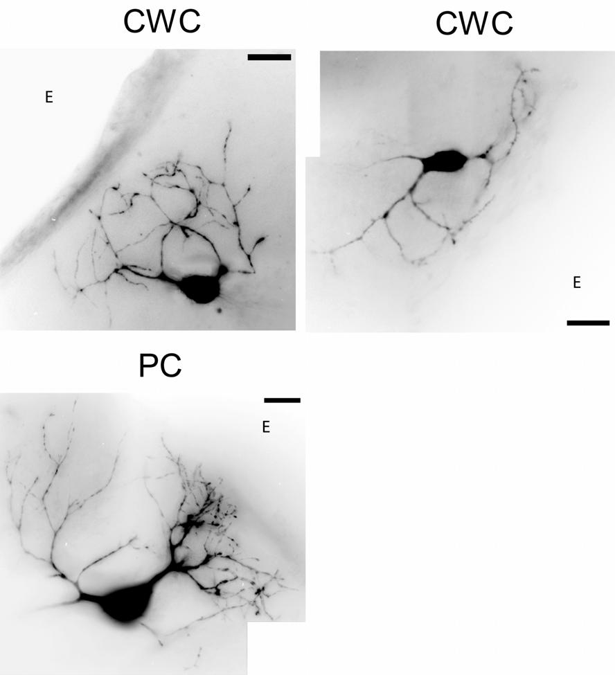
33 flattened into a single 2-D image using an algorithm that finds the most intense pixel along a column formed by all focal planes at any given X, Y coordinate. Montages of these flattened images were then assembled using Photoshop 7.0 (Adobe Systems, San Jose CA) to simultaneously display all visible anatomic features from different regions. CWCs were identified as medium-sized (long axis of soma µm) multipolar neurons with ovoid cell bodies and spiny dendrites typically limited to the superficial DCN. The spines characteristic of CWC dendrites were not always visible due to light scatter through the tissue sections; therefore, dendritic branching patterns were examined to further distinguish CWCs from other DCN neurons. CWC dendrites typically branch at wider angles, resulting in distal processes that sometimes curve around or toward the soma (Wouterlood and Mugnaini 1984), whereas the dendrites of other neurons in the superficial DCN, such as PCs and stellate cells, branch at smaller angles, resulting in dendritic processes that radiate outward from the soma (Fig. 2-2). A total of 102 out of 129 CWCs used in this study were positively identified as CWCs from morphologic reconstruction; no fluorescence images were obtained from the remaining 27 cells because of dye leakage following pipette removal. These cells were identified as CWCs based upon similarities in electrophysiologic responses, and from cell appearance under infrared video microscopy prior to recording Data analysis Isolation of SDPs Recordings of the membrane potential (V m ) time course were obtained during the presentation of brief (2 msec) depolarizing and hyperpolarizing current pulses whose 23
34 Figure 2-2: Fluorescence images of DCN neurons. Grayscale photomontages show the morphology of 2 CWCs and 1 PC. Scale bars: 25 um. CWCs have spiny dendrites that project into the molecular layer and curve back toward the cell body. PCs have two sets of dendrites that radiate away from the cell body; these include spiny apical dendrites that project into the molecular layer and smooth basal dendrites that project to deeper layers. E on each photo represents the epidemic surface. 24
35 25
36 amplitudes were varied in pa steps to elicit a range of subthreshold and suprathreshold V m responses. The V m time courses evoked by depolarizing and hyperpolarizing current pulses of the same magnitude were then added to isolate active events by eliminating the contribution of passive membrane properties that produce the characteristic exponential charging and discharging V m time course evoked by current pulses. In all figures, the results of this trace addition are shown immediately below the original V m responses to depolarizing and hyperpolarizing current pulses. The method used to isolate SDPs assumes that the membrane time constants governing the exponential time course of V m discharge following the offset of injected current pulses remain identical for depolarizing and hyperpolarizing stimuli. Because these time constants are determined in part by the membrane resistance (R m ), changes in R m that occur during the activation or deactivation of voltage-gated conductances will alter the time course of exponential discharge. Differences in the time course of exponential discharge can produce erroneous SDP magnitudes with the addition of V m traces elicited by depolarizing and hyperpolarizing stimuli. Potential sources of error in the SDP isolation procedure are illustrated in figure 2-3. In the absence of voltage-gated conductance changes, R m remains constant and addition of the V m time course in response to depolarizing and hyperpolarizing stimuli of the same amplitude will cancel to zero (Fig. 2-3, left column). The presence of voltagegated conductances that activate in response to suprathreshold depolarization, such as the Na +, K + and Ca 2+ conductances that generate APs and ADPs, will reduce R m and the discharge time constant, producing an erroneous hyperpolarization when the depolarizing and hyperpolarizing responses are added (Fig. 2-3, middle column). This will reduce the 26
37 Figure 2-3: Effects of membrane resistance (R m ) on the isolation of subthreshold depolarizations (SDPs). The voltage across a unit resistance in parallel with a unit capacitance was simulated in response to depolarizing and hyperpolarizing current pulses of equal magnitude. Left column: Addition of depolarizing and hyperpolarizing responses is zero when R m is the same under both conditions. Middle column: reducing the value of R m by 50% at the offset of the depolarizing pulse increases the rate of V m decay (compare solid and dotted lines in top row of traces) and results in a small subthreshold hyperpolarization following addition of the depolarizing and hyperpolarizing responses. Right column: reducing the value of R m by 50% at the offset of the hyperpolarizing pulse results in a small SDP following addition of the depolarizing and hyperpolarizing responses. 27
38 depol R m = hyperpol R m depol R m = hyperpol R m / 2 hyperpol R m = depol R m / 2 28
39 measured voltage deflection of APs and ADPs following the addition of responses to depolarizing and hyperpolarizing stimuli. However, this does not affect the interpretation of any results from the present study, in which AP and ADP magnitudes were reduced following the application of voltage-gated channel antagonists that would prevent a decrease of R m in response to a depolarizing stimulus. The presence of I h could also affect the SDP magnitudes obtained from the addition of depolarizing and hyperpolarizing V m traces. I h will activate in response to increasing hyperpolarization, reducing R m and the discharging time constant following the offset of a hyperpolarizing stimulus. A decrease in the time constant of the hyperpolarizing discharge will produce an erroneous depolarization when the depolarizing and hyperpolarizing responses are added (Fig. 2-3, right column). Thus, I h could erroneously augment SDP magnitudes because of the decreased R m and discharge time constant following the offset of the hyperpolarizing stimulus, and I h antagonists would therefore decrease SDP magnitudes by preventing this decrease in R m and discharge time constant. Instead, application of the I h antagonist ZD7288 had the opposite effect and augmented SDP magnitudes (Fig. 3-5 B-C). This apparent contradiction can be attributed to the slow time course of I h activation, which was inferred from the effects of ZD7288 on the time course of R m changes during the SDP (Fig. 3-5 D). These results show that ZD7288 produces little or no difference in R m values within the first 12 msec of the stimulus onset, where the majority of the decay during V m discharge would occur following the stimulus offset. Thus, the level of I h activation would not significantly change over the duration of the V m transients evoked by brief current pulses, and is not likely to produce the situation illustrated in the last 29
40 column of figure 2-3. However, during the experiments when both voltage-gated Ca 2+ channel antagonists, Ni 2+ and Cd 2+, and voltage-gated Na + channel antagonist, TTX were applied, the resting potentials of CWCs hyperpolarized dramatically (around -10 mv). The following hyperbolizing current pulses would probably activate more I h compared with those in control with much depolarized resting potentials. In that case, I h might contribute to the TTX-resistant SDPs (Fig. 3-8 D). Unfortunately, this hypothesis could not be tested experimentally because the whole-cell voltage-clamp recordings showed great instability of resting potentials during Ni 2+ /Cd 2+, TTX and ZD7288 (I h antagonist) application Voltage dependence of SDPs The voltage dependence of SDPs is shown by plotting SDP magnitudes obtained from the addition of V m responses to depolarizing and hyperpolarizing stimuli against the value of V m at the SDP peak during the response to a depolarizing stimulus. For example, if the response to a +1 na pulse from a holding V m of -60 mv produces a 7 mv deflection, and the response to a -1 na pulse produces a -2 mv deflection, then the SDP magnitude plotted on the ordinate would be 7 mv + -2 mv = 5 mv, and the value of V m plotted on the abscissa would be -60 mv + 7 mv = -53 mv. In figures where traces from the same cell are compared across different pharmacologic manipulations, traces were selected so that the V m values at the SDP peak were the most similar, and in many cases were evoked by different current amplitudes depending on the changes in R m produced by the pharmacologic manipulation. To normalize voltage dependence across cells, V m values plotted on the abscissa were offset so that V m = 0 mv at AP threshold, and the 30
41 SDP magnitudes were binned across a standardized voltage base (1 2.5 mv bin widths) prior to averaging across experiments (Fig. 3-3 A-B). No AP threshold could be detected in experiments utilizing TTX ACSF; therefore the original AP threshold under control conditions prior to application of TTX ACSF was used to calculate voltage dependence (Fig. 3-8 D) I h analysis on voltage-clamp recordings Recordings of current traces of I h with voltage clamp were fitted with a function of the form I t) = A + A exp( t / τ ) + A exp( t / ) (1) ( τ 2 using a Marquardt-Levenberg least-squares algorithm that minimized the errors over five parameters A 0, A 1, A2, τ 1, τ 2. To prevent artifacts from whole-cell capacitance, the initial 20 ~ 50 msec of each current trace was ignored by the fitting procedure. Activation and deactivation tail currents were used to construct the steady-state activation curve of I h (Fig. 3-7). Cells were generally held at -50 mv, stepped for 1.4 s to a group of test potentials ranging from around -150 mv to -40 mv to activate and deactivate I h, and then poststepped to -70mV for 0.5 s. Instantaneous tail currents (I tail (t = 0)) were computed by extrapolating the current traces back to the starting time of post step using the biexponential fit. Steady-state tail currents ( I tail (t = inf)) were set to the values of tail currents when test potential equaled to the level of post step potential, which is at -70 mv. Difference tail currents (I tail_diff ) were then calculated by subtracting steady-sate tail currents from instantaneous ones (I tail_diff = I tail (t = 0) - I tail (t = inf)). The steady-state activation function n inf (V) was obtained by a normalized equation as follows 31
42 n inf I tail _ diff ( V ) I tail _ diff ( Vmin ) ( V ) = (2) I ( V ) I ( V ) tail _ diff max tail _ diff where V max and V min represent membrane potentials corresponding to maximal and zero activation of g h. Finally, activation curves were fit by a Boltzmann function (see equation 6) to provide a functional relationship between steady-state activation and membrane potential V m. min 2.3. Simulation methods Simulation Model All simulation work was carried out using NEURON 5.7 (Hines and Carnevale 2001) under the Windows 2000 operating system. Based on experimental estimates, a one compartment model of a CWC was created to represent a cylindrical cell body approximately 31 µm in diameter and 15.4 µm in length. The specific membrane capacitance was set to 1 µf/cm 2, and the cytoplasmic resistivity 35.4 Ω cm. The leak conductance was set as 0.1 ms/cm 2, resting potential as -68 mv. The numerical integration used NEURON s backward-euler method with a time step of msec Active conductances The active conductances in the model used the Hodgkin-Huxley formulation to account for the voltage-dependence and time course of activation and inactivation kinetics (Hodgkin and Huxley 1952). In this formulation, each ionic current could be presented as Ohm s law I = g V V ) (3) ( rev 32
43 where g is the conductance of ionic current and (V-V rev ) the voltage driving force, the difference between the existing membrane potential V and reversal potential V rev, the membrane potential at which there would be no net electrochemical ionic current. The conductance g is voltage- and time-dependent, making ionic current voltage- and timedependent and presented by the following equations: p q I V, t) = G m( V, t) h( V, t) ( V V ) (4) ( max rev ds dt ( s inf s) = for s = m, h (5) τ s where I (V, t) is the ionic current as a function of time t; G max is the maximal conductance of current; m(v, t) and h(v, t) are activation and inactivation, respectively; p, q are the numbers of activation and inactivation gates. The variable s represents either of gating variables m or h. τ s is the first order time constant for m or h. The steady-state activation or inactivation s inf can be represented as a Boltzmann function = 1 1+ e for s = m, h (6) s inf ( V V / half ) K s where V half is the activation or inactivation mid-point voltage and K s the slope factor of the activation/inactivation curves Clamp protocols A simulated ideal two-electrode current-clamp was applied to the somatic compartment. During brief pulse current-clamp simulation, membrane potential was held at mv by applying zero injected currents for 200 msec to allow state variables to stabilize. It was then stepped to a series of test pulses for 2 msec to elicit subthreshold and suprathreshold responses. For long pulse current-clamp simulation, the model was 33
44 held at the same holding potential but the duration of test pulses was extended to 100 msec. 34
45 Chapter Three Experimental Results CWCs responded to suprathreshold current injection with complex spikes, or a simple spike followed by a characteristic afterdepolarization (ADP) that is mediated by Ca 2+ channels. CWCs responded to subthreshold stimuli with a slow active event, or a subthreshold depolarization (SDP), that appears as a characteristic deviation from the exponential decay of the membrane potential following the offset of the current pulse. SDPs were unaffected by the Ca 2+ channel antagonists Cd 2+ and Ni 2+, the anticonvulsants phenytoin and riluzole, but substantially depressed by TTX. Voltage-clamp recordings showed that a component of TTX-sensitive persistent Na + current existed in CWCs. SDPs were augmented by ZD7288, which blocked a hyperpolarization-activated inward current (I h ) in these cells that contributed to the repolarization of the membrane. Voltageclamp recordings confirmed the existence of I h in CWCs CWCs generate both complex spikes and simple spikes Recordings from 102 identified CWCs were obtained to investigate the ionic mechanisms underlying subthreshold and suprathreshold responses. Cells were initially 35
46 characterized using 100 msec current injections from a holding potential around -65 mv to -70 mv. There were two different responses to sustained current injection just above spike threshold. There were 23 complex-spiking CWCs responding to just above threshold depolarizing currents with multiple APs superimposed upon a slow underlying depolarization (Fig. 3-1, left column upper panel), as well as 94 simple-spiking CWCs responding with a simple spike (Fig. 3-1, right column upper panel). While given suprathreshold current injections, complex-spiking CWCs responded with one or more complex spikes (Fig. 3-1, left column middle panel) and simple-spiking CWCs responded with a train of simple spikes (Fig. 3-1, right column middle panel). In 65 of 94 simplespiking CWCs, complex spikes could be evoked from simple-spiking CWCs by increasing the amplitudes of injected currents (Fig. 3-1, inset of lower panel). Both complex-spiking and simple-spiking CWCs showed similar patterns of decreasing first spike latency (FSL) with increasing injection amplitudes. However, complex-spiking CWCs had short first inter-spike intervals (ISIs) that were constant over the whole range of suprathreshold stimuli while that simple-spiking CWCs showed long ISIs at the beginning of suprathreshold stimuli and shortened as amplitudes increased (Fig. 3-1, lower panel) Subthreshold responses of CWCs To isolate voltage-gated responses from passive membrane potential changes due to current injection, the electrophysiologic responses of CWCs were examined using 2 msec current pulses with amplitudes set just above (Fig. 3-2 A) or just below (Fig. 3-2 B) the threshold for AP initiation. CWCs respond to brief suprathreshold stimuli with one of 36
47 Figure 3-1: Suprathreshold responses of CWCs. Responses of 2 CWCs to suprathreshold current injection, one with AP bursts (Left column), one with regular AP trains (Right column). Upper panel: responses to just above threshold depolarizing current injection and corresponding hyperpolarizing stimulus. Middle panel: responses to current injection well above spike threshold and corresponding hyperpolarizing stimulus. Lower panel: First spike latency (FSL) and first interspike interval (ISI) plotted vs. amplitude of injected current. Increasing stimulus levels can elicit AP bursts in simple spiking CWCs (inset). 37
Supplementary Information
 Hyperpolarization-activated cation channels inhibit EPSPs by interactions with M-type K + channels Meena S. George, L.F. Abbott, Steven A. Siegelbaum Supplementary Information Part 1: Supplementary Figures
Is action potential threshold lowest in the axon?
 Supplementary information to: Is action potential threshold lowest in the axon? Maarten H. P. Kole & Greg J. Stuart Supplementary Fig. 1 Analysis of action potential (AP) threshold criteria. (a) Example
Dorsal Cochlear Nucleus. Amanda M. Lauer, Ph.D. Dept. of Otolaryngology-HNS
 Dorsal Cochlear Nucleus Amanda M. Lauer, Ph.D. Dept. of Otolaryngology-HNS May 30, 2016 Overview Structure Response properties Hypothesized roles in hearing Review of VCN-DCN circuits and projections Structure
The control of spiking by synaptic input in striatal and pallidal neurons
 The control of spiking by synaptic input in striatal and pallidal neurons Dieter Jaeger Department of Biology, Emory University, Atlanta, GA 30322 Key words: Abstract: rat, slice, whole cell, dynamic current
Supporting Online Material for
 www.sciencemag.org/cgi/content/full/317/5841/183/dc1 Supporting Online Material for Astrocytes Potentiate Transmitter Release at Single Hippocampal Synapses Gertrudis Perea and Alfonso Araque* *To whom
SUPPLEMENTARY INFORMATION. Supplementary Figure 1
 SUPPLEMENTARY INFORMATION Supplementary Figure 1 The supralinear events evoked in CA3 pyramidal cells fulfill the criteria for NMDA spikes, exhibiting a threshold, sensitivity to NMDAR blockade, and all-or-none
File name: Supplementary Information Description: Supplementary Figures, Supplementary Table and Supplementary References
 File name: Supplementary Information Description: Supplementary Figures, Supplementary Table and Supplementary References File name: Supplementary Data 1 Description: Summary datasheets showing the spatial
Sample Lab Report 1 from 1. Measuring and Manipulating Passive Membrane Properties
 Sample Lab Report 1 from http://www.bio365l.net 1 Abstract Measuring and Manipulating Passive Membrane Properties Biological membranes exhibit the properties of capacitance and resistance, which allow
Neurons of the Bed Nucleus of the Stria Terminalis (BNST)
") Neurons of the Bed Nucleus of the Stria Terminalis (BNST) Electrophysiological Properties and Their Response to Serotonin DONALD G. RAINNIE a Harvard Medical School and Department of Psychiatry, Brockton
Neurons of the Bed Nucleus of the Stria Terminalis (BNST) Electrophysiological Properties and Their Response to Serotonin DONALD G. RAINNIE a Harvard Medical School and Department of Psychiatry, Brockton
CYTOARCHITECTURE OF CEREBRAL CORTEX
 BASICS OF NEUROBIOLOGY CYTOARCHITECTURE OF CEREBRAL CORTEX ZSOLT LIPOSITS 1 CELLULAR COMPOSITION OF THE CEREBRAL CORTEX THE CEREBRAL CORTEX CONSISTS OF THE ARCHICORTEX (HIPPOCAMPAL FORMA- TION), PALEOCORTEX
Nature Methods: doi: /nmeth Supplementary Figure 1. Activity in turtle dorsal cortex is sparse.
 Supplementary Figure 1 Activity in turtle dorsal cortex is sparse. a. Probability distribution of firing rates across the population (notice log scale) in our data. The range of firing rates is wide but
Ivy/Neurogliaform Interneurons Coordinate Activity in the Neurogenic Niche
 Ivy/Neurogliaform Interneurons Coordinate Activity in the Neurogenic Niche Sean J. Markwardt, Cristina V. Dieni, Jacques I. Wadiche & Linda Overstreet-Wadiche Supplementary Methods. Animals We used hemizygous
BIONB/BME/ECE 4910 Neuronal Simulation Assignments 1, Spring 2013
 BIONB/BME/ECE 4910 Neuronal Simulation Assignments 1, Spring 2013 Tutorial Assignment Page Due Date Week 1/Assignment 1: Introduction to NIA 1 January 28 The Membrane Tutorial 9 Week 2/Assignment 2: Passive
Dep. Control Time (min)
") aa Control Dep. RP 1s 1 mv 2s 1 mv b % potentiation of IPSP 2 15 1 5 Dep. * 1 2 3 4 Time (min) Supplementary Figure 1. Rebound potentiation of IPSPs in PCs. a, IPSPs recorded with a K + gluconate pipette
aa Control Dep. RP 1s 1 mv 2s 1 mv b % potentiation of IPSP 2 15 1 5 Dep. * 1 2 3 4 Time (min) Supplementary Figure 1. Rebound potentiation of IPSPs in PCs. a, IPSPs recorded with a K + gluconate pipette
Electrophysiology. General Neurophysiology. Action Potentials
 5 Electrophysiology Cochlear implants should aim to reproduce the coding of sound in the auditory system as closely as possible, for best sound perception. The cochlear implant is in part the result of
Bursting dynamics in the brain. Jaeseung Jeong, Department of Biosystems, KAIST
 Bursting dynamics in the brain Jaeseung Jeong, Department of Biosystems, KAIST Tonic and phasic activity A neuron is said to exhibit a tonic activity when it fires a series of single action potentials
Intro. Comp. NeuroSci. Ch. 9 October 4, The threshold and channel memory
 9.7.4 The threshold and channel memory The action potential has a threshold. In figure the area around threshold is expanded (rectangle). A current injection that does not reach the threshold does not
Prolonged Synaptic Integration in Perirhinal Cortical Neurons
 RAPID COMMUNICATION Prolonged Synaptic Integration in Perirhinal Cortical Neurons JOHN M. BEGGS, 1 JAMES R. MOYER, JR., 1 JOHN P. MCGANN, 2 AND THOMAS H. BROWN 1 3 1 Department of Psychology, 2 Interdepartmental
Chapter 4 Neuronal Physiology
 Chapter 4 Neuronal Physiology V edit. Pg. 99-131 VI edit. Pg. 85-113 VII edit. Pg. 87-113 Input Zone Dendrites and Cell body Nucleus Trigger Zone Axon hillock Conducting Zone Axon (may be from 1mm to more
Introduction to Neurobiology
 Biology 240 General Zoology Introduction to Neurobiology Nervous System functions: communication of information via nerve signals integration and processing of information control of physiological and
Active Control of Spike-Timing Dependent Synaptic Plasticity in an Electrosensory System
 Active Control of Spike-Timing Dependent Synaptic Plasticity in an Electrosensory System Patrick D. Roberts and Curtis C. Bell Neurological Sciences Institute, OHSU 505 N.W. 185 th Avenue, Beaverton, OR
STRUCTURAL ELEMENTS OF THE NERVOUS SYSTEM
 STRUCTURAL ELEMENTS OF THE NERVOUS SYSTEM STRUCTURE AND MAINTENANCE OF NEURONS (a) (b) Dendrites Cell body Initial segment collateral terminals (a) Diagrammatic representation of a neuron. The break in
Nerve. (2) Duration of the stimulus A certain period can give response. The Strength - Duration Curve
 Duration of the stimulus A certain period can give response. The Strength - Duration Curve") Nerve Neuron (nerve cell) is the structural unit of nervous system. Nerve is formed of large numbers of nerve fibers. Types of nerve fibers Myelinated nerve fibers Covered by myelin sheath interrupted
Nerve Neuron (nerve cell) is the structural unit of nervous system. Nerve is formed of large numbers of nerve fibers. Types of nerve fibers Myelinated nerve fibers Covered by myelin sheath interrupted
Theme 2: Cellular mechanisms in the Cochlear Nucleus
 Theme 2: Cellular mechanisms in the Cochlear Nucleus The Cochlear Nucleus (CN) presents a unique opportunity for quantitatively studying input-output transformations by neurons because it gives rise to
EE 791 Lecture 2 Jan 19, 2015
 EE 791 Lecture 2 Jan 19, 2015 Action Potential Conduction And Neural Organization EE 791-Lecture 2 1 Core-conductor model: In the core-conductor model we approximate an axon or a segment of a dendrite
Firing Pattern Formation by Transient Calcium Current in Model Motoneuron
 Nonlinear Analysis: Modelling and Control, Vilnius, IMI, 2000, No 5 Lithuanian Association of Nonlinear Analysts, 2000 Firing Pattern Formation by Transient Calcium Current in Model Motoneuron N. Svirskienė,
Supporting Online Material for
 www.sciencemag.org/cgi/content/full/312/5779/1533/dc1 Supporting Online Material for Long-Term Potentiation of Neuron-Glia Synapses Mediated by Ca 2+ - Permeable AMPA Receptors Woo-Ping Ge, Xiu-Juan Yang,
Applied Neuroscience. Conclusion of Science Honors Program Spring 2017
 Applied Neuroscience Conclusion of Science Honors Program Spring 2017 Review Circle whichever is greater, A or B. If A = B, circle both: I. A. permeability of a neuronal membrane to Na + during the rise
MOLECULAR AND CELLULAR NEUROSCIENCE
 MOLECULAR AND CELLULAR NEUROSCIENCE BMP-218 November 4, 2014 DIVISIONS OF THE NERVOUS SYSTEM The nervous system is composed of two primary divisions: 1. CNS - Central Nervous System (Brain + Spinal Cord)
Simulation of myelinated neuron with focus on conduction speed and changeable excitability
 Simulation of myelinated neuron with focus on conduction speed and changeable excitability Pengfei Chen Sung Min Kim Abstract In this paper we focus on the two particular properties of myelinated neuron,
What is Anatomy and Physiology?
 Introduction BI 212 BI 213 BI 211 Ecosystems Organs / organ systems Cells Organelles Communities Tissues Molecules Populations Organisms Campbell et al. Figure 1.4 Introduction What is Anatomy and Physiology?
Processing in The Cochlear Nucleus
 Processing in The Cochlear Nucleus Alan R. Palmer Medical Research Council Institute of Hearing Research University Park Nottingham NG7 RD, UK The Auditory Nervous System Cortex Cortex MGB Medial Geniculate
CASE 48. What part of the cerebellum is responsible for planning and initiation of movement?
 CASE 48 A 34-year-old woman with a long-standing history of seizure disorder presents to her neurologist with difficulty walking and coordination. She has been on phenytoin for several days after having
NEURONS Chapter Neurons: specialized cells of the nervous system 2. Nerves: bundles of neuron axons 3. Nervous systems
 NEURONS Chapter 12 Figure 12.1 Neuronal and hormonal signaling both convey information over long distances 1. Nervous system A. nervous tissue B. conducts electrical impulses C. rapid communication 2.
ANATOMY AND PHYSIOLOGY OF NEURONS. AP Biology Chapter 48
 ANATOMY AND PHYSIOLOGY OF NEURONS AP Biology Chapter 48 Objectives Describe the different types of neurons Describe the structure and function of dendrites, axons, a synapse, types of ion channels, and
Current Clamp and Modeling Studies of Low-Threshold Calcium Spikes in Cells of the Cat s Lateral Geniculate Nucleus
 Current Clamp and Modeling Studies of Low-Threshold Calcium Spikes in Cells of the Cat s Lateral Geniculate Nucleus X. J. ZHAN, 1 C. L. COX, 1 J. RINZEL, 2 AND S. MURRAY SHERMAN 1 1 Department of Neurobiology,
Embryological origin of thalamus
 diencephalon Embryological origin of thalamus The diencephalon gives rise to the: Thalamus Epithalamus (pineal gland, habenula, paraventricular n.) Hypothalamus Subthalamus (Subthalamic nuclei) The Thalamus:
Chapter 11 Introduction to the Nervous System and Nervous Tissue Chapter Outline
 Chapter 11 Introduction to the Nervous System and Nervous Tissue Chapter Outline Module 11.1 Overview of the Nervous System (Figures 11.1-11.3) A. The nervous system controls our perception and experience
Arnaud Ruiz, Emilie Campanac, Ricardo Scott, Dmitri A. Rusakov, Dimitri M. Kullmann
 Presynaptic GABA A receptors enhance transmission and LTP induction at hippocampal mossy fiber synapses Arnaud Ruiz, Emilie Campanac, Ricardo Scott, Dmitri A. Rusakov, Dimitri M. Kullmann Supplementary
Dorsal Cochlear Nucleus September 14, 2005
 HST.722 Brain Mechanisms of Speech and Hearing Fall 2005 Dorsal Cochlear Nucleus September 14, 2005 Ken Hancock Dorsal Cochlear Nucleus (DCN) Overview of the cochlear nucleus and its subdivisions Anatomy
Chapter 7. The Nervous System: Structure and Control of Movement
 Chapter 7 The Nervous System: Structure and Control of Movement Objectives Discuss the general organization of the nervous system Describe the structure & function of a nerve Draw and label the pathways
Chapter 7. Objectives
 Chapter 7 The Nervous System: Structure and Control of Movement Objectives Discuss the general organization of the nervous system Describe the structure & function of a nerve Draw and label the pathways
Brief presynaptic bursts evoke synapse-specific retrograde inhibition mediated by endogenous cannabinoids
 Brief presynaptic bursts evoke synapse-specific retrograde inhibition mediated by endogenous cannabinoids Solange P Brown 1 3,Stephan D Brenowitz 1,3 & Wade G Regehr 1 Many types of neurons can release
Resurgent Sodium Current and Action Potential Formation in Dissociated Cerebellar Purkinje Neurons
 The Journal of Neuroscience, June 15, 1997, 17(12):4517 4526 Resurgent Sodium Current and Action Potential Formation in Dissociated Cerebellar Purkinje Neurons Indira M. Raman and Bruce P. Bean Vollum
The Nervous System. Nervous System Functions 1. gather sensory input 2. integration- process and interpret sensory input 3. cause motor output
 The Nervous System Nervous System Functions 1. gather sensory input 2. integration- process and interpret sensory input 3. cause motor output The Nervous System 2 Parts of the Nervous System 1. central
Chapter 3 subtitles Action potentials
 CELLULAR NEUROPHYSIOLOGY CONSTANCE HAMMOND Chapter 3 subtitles Action potentials Introduction (3:15) This third chapter explains the calcium current triggered by the arrival of the action potential in
Nervous System. Master controlling and communicating system of the body. Secrete chemicals called neurotransmitters
 Nervous System Master controlling and communicating system of the body Interacts with the endocrine system to control and coordinate the body s responses to changes in its environment, as well as growth,
Neuroscience 201A Problem Set #1, 27 September 2016
 Neuroscience 201A Problem Set #1, 27 September 2016 1. The figure above was obtained from a paper on calcium channels expressed by dentate granule cells. The whole-cell Ca 2+ currents in (A) were measured
Chapter 7 Nerve Cells and Electrical Signaling
 Chapter 7 Nerve Cells and Electrical Signaling 7.1. Overview of the Nervous System (Figure 7.1) 7.2. Cells of the Nervous System o Neurons are excitable cells which can generate action potentials o 90%
Large-conductance calcium-dependent potassium channels prevent dendritic excitability in neocortical pyramidal neurons
 Zurich Open Repository and Archive University of Zurich Main Library Strickhofstrasse 39 CH-857 Zurich www.zora.uzh.ch Year: 29 Large-conductance calcium-dependent potassium channels prevent dendritic
Supporting Information
 ATP from synaptic terminals and astrocytes regulates NMDA receptors and synaptic plasticity through PSD- 95 multi- protein complex U.Lalo, O.Palygin, A.Verkhratsky, S.G.N. Grant and Y. Pankratov Supporting
Contrasting Effects of the Persistent Na + Current on Neuronal Excitability and Spike Timing
 Neuron 49, 257 270, January 19, 2006 ª2006 Elsevier Inc. DOI 10.1016/j.neuron.2005.12.022 Contrasting Effects of the Persistent Na + Current on Neuronal Excitability and Spike Timing Koen Vervaeke, 1,2,4
SimNeuron. Current-and Voltage-Clamp Experiments in Virtual Laboratories. Tutorial
 SimNeuron Tutorial 1 Contents: SimNeuron Current-and Voltage-Clamp Experiments in Virtual Laboratories 1. Lab Design 2. Basic CURRENT-CLAMP Experiments Tutorial 2.1 Action Potential, Conductances and Currents.
Resonant synchronization of heterogeneous inhibitory networks
 Cerebellar oscillations: Anesthetized rats Transgenic animals Recurrent model Review of literature: γ Network resonance Life simulations Resonance frequency Conclusion Resonant synchronization of heterogeneous
10.1: Introduction. Cell types in neural tissue: Neurons Neuroglial cells (also known as neuroglia, glia, and glial cells) Dendrites.
 Dendrites.") 10.1: Introduction Copyright The McGraw-Hill Companies, Inc. Permission required for reproduction or display. Cell types in neural tissue: Neurons Neuroglial cells (also known as neuroglia, glia, and glial
10.1: Introduction Copyright The McGraw-Hill Companies, Inc. Permission required for reproduction or display. Cell types in neural tissue: Neurons Neuroglial cells (also known as neuroglia, glia, and glial
The Cerebellum. Outline. Lu Chen, Ph.D. MCB, UC Berkeley. Overview Structure Micro-circuitry of the cerebellum The cerebellum and motor learning
 The Cerebellum Lu Chen, Ph.D. MCB, UC Berkeley 1 Outline Overview Structure Micro-circuitry of the cerebellum The cerebellum and motor learning 2 Overview Little brain 10% of the total volume of the brain,
Lab 4: Compartmental Model of Binaural Coincidence Detector Neurons
 Lab 4: Compartmental Model of Binaural Coincidence Detector Neurons Introduction The purpose of this laboratory exercise is to give you hands-on experience with a compartmental model of a neuron. Compartmental
The Central Auditory System
 THE AUDITORY SYSTEM Each auditory nerve sends information to the cochlear nucleus. The Central Auditory System From there, projections diverge to many different pathways. The Central Auditory System There
Enhancement of synaptic transmission by cyclic AMP modulation of presynaptic I h channels. Vahri Beaumont and Robert S. Zucker
 Enhancement of synaptic transmission by cyclic AMP modulation of presynaptic I h channels Vahri Beaumont and Robert S. Zucker Background I h channels discovered in 1976 (Noma A. and Irisawa H.) Voltage-gated
Learning Rules for Spike Timing-Dependent Plasticity Depend on Dendritic Synapse Location
 10420 The Journal of Neuroscience, October 11, 2006 26(41):10420 10429 Cellular/Molecular Learning Rules for Spike Timing-Dependent Plasticity Depend on Dendritic Synapse Location Johannes J. Letzkus,
Neurobiology. Cells of the nervous system
 Neurobiology Cells of the nervous system Anthony Heape 2010 1 The nervous system Central nervous system (CNS) Peripheral nervous system (PNS) 2 Enteric nervous system (digestive tract, gall bladder and
Membrane Properties and Discharge Characteristics of Guinea Pig Dorsal Cochlear Nucleus Neurons Studied in vitro
 The Journal of Neuroscience, July 1990, M(7): 2338-2351 Membrane Properties and Discharge Characteristics of Guinea Pig Dorsal Cochlear Nucleus Neurons Studied in vitro Paul B. Manis Departments of Otolaryngology-Head
Chapter 11: Nervous System and Nervous Tissue
 Chapter 11: Nervous System and Nervous Tissue I. Functions and divisions of the nervous system A. Sensory input: monitor changes in internal and external environment B. Integrations: make decisions about
Central Synaptic Transmission
 Central Synaptic Transmission The main excitatory transmitter in the brain is the amino acid glutamate. The main inhibitory transmitters in the brain are the amino acids glycine (in the spinal cord) and
Ube3a is required for experience-dependent maturation of the neocortex
 Ube3a is required for experience-dependent maturation of the neocortex Koji Yashiro, Thorfinn T. Riday, Kathryn H. Condon, Adam C. Roberts, Danilo R. Bernardo, Rohit Prakash, Richard J. Weinberg, Michael
Astrocyte signaling controls spike timing-dependent depression at neocortical synapses
 Supplementary Information Astrocyte signaling controls spike timing-dependent depression at neocortical synapses Rogier Min and Thomas Nevian Department of Physiology, University of Berne, Bern, Switzerland
Neurons. Pyramidal neurons in mouse cerebral cortex expressing green fluorescent protein. The red staining indicates GABAergic interneurons.
 Neurons Pyramidal neurons in mouse cerebral cortex expressing green fluorescent protein. The red staining indicates GABAergic interneurons. MBL, Woods Hole R Cheung MSc Bioelectronics: PGEE11106 1 Neuron
Reciprocal inhibition controls the oscillatory state in thalamic networks
 Neurocomputing 44 46 (2002) 653 659 www.elsevier.com/locate/neucom Reciprocal inhibition controls the oscillatory state in thalamic networks Vikaas S. Sohal, John R. Huguenard Department of Neurology and
The mammalian cochlea possesses two classes of afferent neurons and two classes of efferent neurons.
 1 2 The mammalian cochlea possesses two classes of afferent neurons and two classes of efferent neurons. Type I afferents contact single inner hair cells to provide acoustic analysis as we know it. Type
Chapter 6 subtitles postsynaptic integration
 CELLULAR NEUROPHYSIOLOGY CONSTANCE HAMMOND Chapter 6 subtitles postsynaptic integration INTRODUCTION (1:56) This sixth and final chapter deals with the summation of presynaptic currents. Glutamate and
The action potential travels down both branches because each branch is a typical axon with voltage dependent Na + and K+ channels.
 BIO 360 - MIDTERM FALL 2018 This is an open book, open notes exam. PLEASE WRITE YOUR NAME ON EACH SHEET. Read each question carefully and answer as well as you can. Point values are shown at the beginning
Part 11: Mechanisms of Learning
 Neurophysiology and Information: Theory of Brain Function Christopher Fiorillo BiS 527, Spring 2012 042 350 4326, fiorillo@kaist.ac.kr Part 11: Mechanisms of Learning Reading: Bear, Connors, and Paradiso,
Basics of Computational Neuroscience: Neurons and Synapses to Networks
 Basics of Computational Neuroscience: Neurons and Synapses to Networks Bruce Graham Mathematics School of Natural Sciences University of Stirling Scotland, U.K. Useful Book Authors: David Sterratt, Bruce
Motor systems III: Cerebellum April 16, 2007 Mu-ming Poo
 Motor systems III: Cerebellum April 16, 2007 Mu-ming Poo Population coding in the motor cortex Overview and structure of cerebellum Microcircuitry of cerebellum Function of cerebellum -- vestibulo-ocular
Membrane Bistability in Thalamic Reticular Neurons During Spindle Oscillations
 J Neurophysiol 93: 294 304, 2005. First published August 25, 2004; doi:10.1152/jn.00552.2004. Membrane Bistability in Thalamic Reticular Neurons During Spindle Oscillations Pablo Fuentealba, 1 Igor Timofeev,
Supplementary Figure 1. Basic properties of compound EPSPs at
 Supplementary Figure 1. Basic properties of compound EPSPs at hippocampal CA3 CA3 cell synapses. (a) EPSPs were evoked by extracellular stimulation of the recurrent collaterals and pharmacologically isolated
Cellular Bioelectricity
 ELEC ENG 3BB3: Cellular Bioelectricity Notes for Lecture 24 Thursday, March 6, 2014 8. NEURAL ELECTROPHYSIOLOGY We will look at: Structure of the nervous system Sensory transducers and neurons Neural coding
Thalamo-Cortical Relationships Ultrastructure of Thalamic Synaptic Glomerulus
 Central Visual Pathways V1/2 NEUR 3001 dvanced Visual Neuroscience The Lateral Geniculate Nucleus () is more than a relay station LP SC Professor Tom Salt UCL Institute of Ophthalmology Retina t.salt@ucl.ac.uk
Physiology of synapses and receptors
 Physiology of synapses and receptors Dr Syed Shahid Habib Professor & Consultant Clinical Neurophysiology Dept. of Physiology College of Medicine & KKUH King Saud University REMEMBER These handouts will
Nervous System. 2. Receives information from the environment from CNS to organs and glands. 1. Relays messages, processes info, analyzes data
 Nervous System 1. Relays messages, processes info, analyzes data 2. Receives information from the environment from CNS to organs and glands 3. Transmits impulses from CNS to muscles and glands 4. Transmits
Supplementary Figure 1. ACE robotic platform. A. Overview of the rig setup showing major hardware components of ACE (Automatic single Cell
 2 Supplementary Figure 1. ACE robotic platform. A. Overview of the rig setup showing major hardware components of ACE (Automatic single Cell Experimenter) including the MultiClamp 700B, Digidata 1440A,
Dendritic Depolarization Efficiently Attenuates Low-Threshold Calcium Spikes in Thalamic Relay Cells
 The Journal of Neuroscience, May 15, 2000, 20(10):3909 3914 Dendritic Depolarization Efficiently Attenuates Low-Threshold Calcium Spikes in Thalamic Relay Cells X. J. Zhan, C. L. Cox, and S. Murray Sherman
Modeling and Simulation of the Auditory Pathway
 ModelingandSimulationoftheAuditory Pathway TECHNICALREPORT CalF.Rabang WeldonSchoolofBiomedicalEngineering crabang@purdue.edu EdwardL.Bartlett DepartmentsofBiologicalSciencesandBiomedicalEngineering ebartle@purdue.edu
PHY3111 Mid-Semester Test Study. Lecture 2: The hierarchical organisation of vision
 PHY3111 Mid-Semester Test Study Lecture 2: The hierarchical organisation of vision 1. Explain what a hierarchically organised neural system is, in terms of physiological response properties of its neurones.
Chapter 17 Nervous System
 Chapter 17 Nervous System 1 The Nervous System Two Anatomical Divisions Central Nervous System (CNS) Brain and Spinal Cord Peripheral Nervous System (PNS) Two Types of Cells Neurons Transmit nerve impulses
COGNITIVE SCIENCE 107A. Sensory Physiology and the Thalamus. Jaime A. Pineda, Ph.D.
 COGNITIVE SCIENCE 107A Sensory Physiology and the Thalamus Jaime A. Pineda, Ph.D. Sensory Physiology Energies (light, sound, sensation, smell, taste) Pre neural apparatus (collects, filters, amplifies)
Supplementary Information. Errors in the measurement of voltage activated ion channels. in cell attached patch clamp recordings
 Supplementary Information Errors in the measurement of voltage activated ion channels in cell attached patch clamp recordings Stephen R. Williams 1,2 and Christian Wozny 2 1 Queensland Brain Institute,
Intrinsic Firing Dynamics of Vestibular Nucleus Neurons
 The Journal of Neuroscience, March 15, 2002, 22(6):2083 2095 Intrinsic Firing Dynamics of Vestibular Nucleus Neurons Chris Sekirnjak and Sascha du Lac Systems Neurobiology Laboratories, The Salk Institute
Strick Lecture 3 March 22, 2017 Page 1
 Strick Lecture 3 March 22, 2017 Page 1 Cerebellum OUTLINE I. External structure- Inputs and Outputs Cerebellum - (summary diagram) 2 components (cortex and deep nuclei)- (diagram) 3 Sagittal zones (vermal,
The Journal of Physiology
 J Physiol 595.13 (217) pp 4431 4448 4431 Increased transient Na + conductance and action potential output in layer 2/3 prefrontal cortex neurons of the fmr1 /y mouse Brandy N. Routh, Rahul K. Rathour,
Cellular Neurobiology BIPN 140 Fall 2016 Problem Set #1
 Cellular Neurobiology BIPN 140 Fall 2016 Problem Set #1 1. (Antonia) You are studying a neuron with an arterial cuff under a microscope. However, you knock over a bunch of chemicals onto the plate because
Neocortex. Cortical Structures in the Brain. Neocortex Facts. Laminar Organization. Bark-like (cortical) structures: Shepherd (2004) Chapter 12
 structures: Shepherd (2004) Chapter 12") Neocortex Shepherd (2004) Chapter 12 Rodney Douglas, Henry Markram, and Kevan Martin Instructor: Yoonsuck Choe; CPSC 644 Cortical Networks Cortical Structures in the Brain Bark-like (cortical) structures:
Neocortex Shepherd (2004) Chapter 12 Rodney Douglas, Henry Markram, and Kevan Martin Instructor: Yoonsuck Choe; CPSC 644 Cortical Networks Cortical Structures in the Brain Bark-like (cortical) structures:
Short- and long-lasting consequences of in vivo nicotine treatment
 Short- and long-lasting consequences of in vivo nicotine treatment on hippocampal excitability Rachel E. Penton, Michael W. Quick, Robin A. J. Lester Supplementary Figure 1. Histogram showing the maximal
Model neurons!!!!synapses!
 Model neurons ynapses uggested reading: Chapter 5.8 in Dayan,. & Abbott, L., Theoretical Neuroscience, MIT ress, 200. Model neurons: ynapse Contents: ynapses ynaptic input into the RC-circuit pike-rate
Neural Basis of Motor Control. Chapter 4
 Neural Basis of Motor Control Chapter 4 Neurological Perspective A basic understanding of the physiology underlying the control of voluntary movement establishes a more comprehensive appreciation and awareness
Unit Three. I. General Functions of the Nervous System. I. General Functions of the Nervous System
 10 Refer to the following URLs. It is a good idea to print them and bring them to class. Be sure to study these along with your book. http://www.sirinet.net/~jgjohnso/nervous.html http://faculty.washington.edu/chudler/ap.html
Human Brain and Senses
 Human Brain and Senses Outline for today Levels of analysis Basic structure of neurons How neurons communicate Basic structure of the nervous system Levels of analysis Organism Brain Cell Synapses Membrane
Cellular Neurobiology BIPN 140 Fall 2016 Problem Set #1
 Cellular Neurobiology BIPN 140 Fall 2016 Problem Set #1 1. (Antonia) You are studying a neuron with an arterial cuff under a microscope. However, you knock over a bunch of chemicals onto the plate because