Neuromuscular Physiology. Skeletal muscle & Neuromuscular junction
|
|
|
- Edwina Joseph
- 5 years ago
- Views:
Transcription
1 Neuromuscular Physiology. Skeletal muscle & Neuromuscular junction Dr. Ana-Maria Zagrean Discipline of Physiology and Fundamental Neuroscience, Carol Davila University of Medicine and Pharmacy
2 Muscles: skeletal, cardiac, and smooth muscle The primary function of muscle is to generate force or movement in response to a physiological stimulus. Three different types of muscle adapted to specialized functions: 1. Skeletal muscle - voluntary movement of bones - locomotion and work production. - control of breathing cycle and pump function for the venous return through contraction of the diaphragm. 2. Cardiac muscle - specific to the heart - biomechanical pump driving the delivery of blood to the lungs and tissues. 3. Smooth muscle - mechanical control of organ systems: digestive, urinary, reproductive tracts, blood vessels of the circulatory system and airway passages of the respiratory system. Muscle properties: Excitability, Contractility, Extensibility, Elasticity

3 Skeletal muscle structure
4 Skeletal muscle structure
5 Skeletal muscle structure Most of the skeletal muscles fibers extend the entire length of the muscle and are innervated by only one nerve ending, located near the middle of the fiber. Sarcolemma - a plasma membrane that encloses a skeletal muscle fiber, and has an outer coat containing numerous thin collagen fibrils. At each end of the muscle fiber, sarcolemma fuses with a tendon fiber tendon fiber bundles muscle tendons that insert into the bones. Each muscle fiber contains several hundred to several thousand myofibrils, each composed of about 1500 adjacent myosin thick filaments and 3000 actin thin filaments (large polymerized protein molecules).
6 Skeletal muscle structure Myosin and actin filaments partially interdigitate cause light and dark bands in myofibrils that give skeletal and cardiac muscle their striated appearance Cross-bridges - small projections from the sides of the myosin filaments, that interact with actin filaments and causes contraction. Light I bands (isotropic to polarized light) contain actin filaments Dark A bands (anisotropic to polarized light) contain myosin filaments, as well as the ends of the actin filaments where they overlap the myosin Z disc - filamentous proteins passes crosswise across the myofibril and also crosswise from myofibril to myofibril, attaching the myofibrils to one another all the way across the muscle fiber; Z disc has the ends of the actin filaments attached to it, filaments that extend in both directions to interdigitate with the myosin filaments.
.")
7 Sarcomere = the portion of the myofibril /whole muscle fiber that lies between two successive Z discs. -muscle fiber is contracted the length of the sarcomere ~2 mm (actin filaments completely overlap the myosin filaments and the tips of the actin filaments are just beginning to overlap one another). Titin filamentous molecules. The side-by-side relationship between the myosin and actin filaments is maintained by a large number of filamentous molecules of a protein called titin. Titin: molecular weight ~ 3 million (one of the largest protein molecules in the body); it is filamentous and springy help holding the myosin and actin filaments in place so that the contractile machinery of the sarcomere will work.

8 Organization of proteins in a sarcomere. Each titin molecule extends from the Z disc to the M line. Part of the titin molecule is closely associated with the myosin thick filament, whereas the rest of the molecule is springy and changes length as the sarcomere contracts and relaxes.
9 Skeletal muscle structure Sarcoplasm - The intracellular fluid that fill in the spaces between the myofibrils, suspended side by side in the muscle fiber - Contain large quantities of K +, Mg 2+, and phosphate, plus multiple protein enzymes. - Contain tremendous numbers of mitochondria that lie parallel to the myofibrils supply the contracting myofibrils with large amounts of energy in the form of adenosine triphosphate (ATP)
10 Skeletal muscle structure Sarcoplasmic Reticulum -a specialized endoplasmic reticulum present in the sarcoplasm surrounding the myofibrils of each muscle fiber -the rapidly contracting types of muscle fibers have especially extensive sarcoplasmic reticula.
11 Skeletal Muscle fiber 1. Sarcolemma 2. Sarcoplasm 3. Myofibrils contractile elements a. Actin myofilament actin strands tropomyosin troponin (T, I, C) b. Myosin myofilament Skeletal muscle structure 4. Sarcomere arrangement of myofibrils a. Z disk attaches actin b. I band actin myofilament c. A band both actin and myosin H zone only myosin 5. T Tubules 6. Sarcoplasmic Reticulum (1) large terminal cisternae that abut the T tubules, (2) long longitudinal tubules that surround all surfaces of the actual contracting myofibrils.

12 Plasma membrane invaginations. A, T tubules B, Smooth muscle cells have invaginations of the plasma membrane, called caveolae, contacting the SR.
13 Myosin Filaments -are composed of multiple myosin molecules, 2 heavy chains (MW 200,000) and 4 light chains (MW 20,000). -The two heavy chains wrap spirally around each other to form a double helix = tail of the myosin molecule. -One end of each of these chains is folded bilaterally into a globular polypeptide structure called a myosin head 2 free heads at one end of the double-helix myosin molecule. -The light chains help control the function of the head during muscle contraction. Myosin filament (200 or more individual myosin molecules; 1.6 mm). - part of the body of each myosin molecule hangs to the side along with the head, thus providing an arm that extends the head outward from the body. - the protruding arms and heads together are called cross-bridges, flexible at two points called hinges one where the arm leaves the body of the myosin filament, and the other where the head attaches to the arm. - the hinged arms allow the heads to be either extended far outward from the body of the myosin filament or brought close to the body. The hinged heads in turn participate in the actual contraction process. - there are no cross-bridge heads in the center of the myosin filament for a distance of about 0.2 micrometer because the hinged arms extend away from the center. - cross-bridges extend in all directions around the filament. - the myosin head is an ATPase enzyme, essential for muscle contraction
14 Actin filaments = Actin + Tropomyosin + Troponin. Actin filament: -a double-stranded F-actin protein molecule of ~1 µm -each strand of the double F-actin helix is composed of polymerized G-actin molecules, with one molecule of ADP bound on the actin active sites/g-actin molecule. -the bases of the actin filaments are inserted strongly into the Z discs -the ends of the filaments protrude in both directions to lie in the spaces between the myosin molecules Tropomyosin Molecules: wrapped spirally around the sides of the F-actin helix. in the resting state, lie on top of the active sites of the actin strands so that attraction cannot occur between the actin and myosin filaments to cause contraction. Troponin = complex of 3 loosely bound protein subunits: troponin I - a strong affinity for actin troponin T- a strong affinity for tropomyosin troponin C - a strong affinity for calcium ions

15

16
17 Contraction = interaction of 1 myosin filament and 2 actin filaments in the presence of Ca 2+ -actin filament without the presence of the troponin-tropomyosin complex, in the presence of Mg 2+ and ATP, binds instantly and strongly with the heads of the myosin molecules. -the active sites on the normal actin filament of the relaxed muscle are inhibited or physically covered by the troponin-tropomyosin complex. -in the presence of large amounts of calcium ions, they combine with troponin C (up to 4 Ca 2+ /troponin C) troponin complex undergoes a conformational change and uncovers the active sites of the actin, thus allowing these to attract the myosin cross-bridge heads and cause contraction to proceed.

18 Interaction between the activated actin filament and the myosin cross-bridges - the walk-along theory of contraction.
19 The walk-along theory of contraction -a head of the cross-bridges from the myosin filaments attaches to an active site profound changes in the intramolecular forces between the head and arm of its cross-bridge new alignment of forces head tilt toward the arm and drag the actin filament along with it = power stroke. -immediately after tilting, the head automatically breaks away from the active site and returns to its extended direction then it combines with a new active site farther down along the actin filament head tilts again new power stroke actin filament moves another step. the heads of the cross-bridges bend back and forth and step by step walk along the actin filament, pulling the ends of two successive actin filaments toward the center of the myosin filament. -each cross-bridge operate independently of all others, attaching and pulling in a continuous repeated cycle the greater the number of cross-bridges in contact with the actin filament at any given time, the greater the force of contraction.
20 Chemical events in the motion of the myosin heads. Fenn effect - the greater the amount of work performed by the muscle, the greater the amount of ATP that is cleaved to ADP 1. Before contraction begins, the heads of the cross-bridges bind with ATP. The ATPase activity of the myosin head immediately cleaves the ATP but leaves the cleavage products, ADP plus phosphate ion, bound to the head the conformation of the head extends perpendicularly toward the actin filament but not yet attached to actin. 2. When the troponin-tropomyosin complex binds with Ca2+, active sites on the actin filament are uncovered and the myosin heads then bind with these. 3. The bond between the head of the cross-bridge and the active site of the actin filament causes a conformational change in the head, prompting the head to tilt toward the arm of the cross-bridge. This provides the power stroke for pulling the actin filament. The energy that activates the power stroke is the energy already stored, like a cocked spring, by the conformational change that occurred in the head when the ATP molecule was cleaved earlier. 4. Once the head of the cross-bridge tilts release of the ADP and phosphate ion that were previously attached to the head and at the site of release of the ADP, a new molecule of ATP binds. This binding of new ATP causes detachment of the head from the actin. 5. After the head has detached from the actin, the new molecule of ATP is cleaved to begin the next cycle, leading to a new power stroke the energy again cocks the head back to its perpendicular condition, ready to begin the new power stroke cycle.


21 The role of Ca 2+ in triggering the contraction of skeletal and cardiac muscle.

22 The cross-bridge cycle in skeletal and cardiac muscle. Each cycle advances the myosin head by two actin monomers, or 11 nm.

23

.")
24 Skeletal muscle Motor unit A typical skeletal muscle receives innervation from 100 somatic motor neurons. The whole assembly of muscle fibers innervated by the axon from one motor neuron is called a motor unit. When that axon fires, all of those myofibers (with one each motor end plate) are going to fire simultaneously and maximally. The amount of force generated in an anatomic muscle is dependent on how many motor units in it are "recruited" into use (hundreds/ thousands of motor units in any given muscle). Not all of them are "in use" at any given time; if the task requires little force, only a few may be "recruited" by the brain, and as these fatigue, they are shifted smoothly out of service and new motor units switched into use, to maintain the steady level of contraction that muscle has to have. Skeletal muscle; Iron hematoxylin stain, 100x

25 Nerve Skeletal Muscle Synapse: Neuromuscular junction / motor end plate - motor neuron (spinal cord) one axon branches into axon processes near the point of contact with the target muscle - one axon process innervate a separate muscular fiber through terminal arborizations, a small tree-like patch of unmyelinated nerve processes, ending into bulb-shaped terminals called boutons; Schwann cells intimately associate with the nerve terminal to form a cap over the face of the nerve membrane - the whole assembly of muscle fibers innervated by the axon from one motor neuron is called a motor unit. - presynaptic terminal boutons contain Ach vesicles (150 mm Ach, ,000 ACh molecules /vesicle = quantum ) quantum release into the synaptic cleft. - within a few sec. after each AP, "coated pits" appear in the presynaptic membrane, caused by contractile proteins in the nerve ending (clathrin); within ~ 20 sec. the proteins contract and cause the pits to break away to the interior of the membrane, thus forming new vesicles. - bouton reloads its discharged synaptic vesicles by resynthesizing ACh and transport it into the vesicles through an ACh-H + exchanger, working on a vacuolar H + -pump expense. Scanning electron micrograph showing termination of a single axon on a skeletal muscle cell.
26 Nerve Skeletal Muscle Synapse: Neuromuscular junction / motor end plate -synaptic cleft: 50 nm wide, filled with a meshwork of proteins and proteoglycans that are part of the extracellular matrix. -muscle basement membrane with a synaptic basal lamina: - contains proteins (e.g., collagen, laminin, agrin) that mediate adhesion of the neuromuscular junction and play important roles in synapse development and regeneration. - contains a high concentration of acetylcholinesterase (AChE), which ultimately terminates synaptic transmission by rapidly hydrolyzing free ACh to choline and acetate. - the postsynaptic membrane of the skeletal muscle fiber lying directly under the nerve terminal is characterized by extensive invaginations / infoldings = postjunctional folds (increase surface area of postsynaptic muscle plasma membr.) -postsynaptic nicotinic AChRs end-plate potential (EPP)/excitatory postsynaptic potential (EPSP).


27 Neuromuscular Junction: Nerve Muscle Synapse Collagen, laminin, agin AchE (~ 6,000 10,000 Ach molec.
28 Neuromuscular Junction AP When an AP spreads over the terminal, voltage-gated Ca channels open and allow calcium ions to diffuse from the synaptic space to the interior of the nerve terminal, where they exert an attractive influence on the ACh vesicles, drawing them to the presynaptic membrane. The vesicles then fuse with the neural membrane and empty their acetylcholine into the synaptic space by the process of exocytosis. Ach is removed rapidly (few msec) by: (1) acetylcholinesterase, attached mainly to the spongy layer of fine connective tissue in the synaptic space (2) diffusion out of the synaptic space (a smaller amount) ACh receptors in the muscle fiber membrane are ligand-gated ion channels. When opened, ACh channel has a diameter ~0.65 nm, large enough to allow positive ions (Na +, K +, Ca ++ ) to move easily. Na ions influx creates a local positive potential change of mv inside the muscle fiber membrane = end plate potential, that initiates an AP that spreads along the muscle membrane and thus causes muscle contraction.
29 End plate potentials Curara, a drug that blocks the gating action of ACh on the ACh channels by competing for the ACh receptor sites. curara normal botulinum Botulinum toxins A, E, C1 are bacterial poisons that decrease the quantity of ACh release by the nerve terminals by blocking the fusion of synaptic vesicles. End plate potentials (mv). A, Weakened end plate potential recorded in a curarized muscle, too weak to elicit an AP B, Normal end plate potential eliciting a muscle action potential. C, Weakened end plate potential caused by botulinum toxin that decreases end plate release of acetylcholine, again too weak to elicit a muscle action potential.
30 Miniature end-plate potentials reveal the quantal nature of Ach release from the presynaptic terminals AP in a presynaptic motor nerve axon produces a depolarizing postsynaptic end-plate potential (EPP), resulting from the release of ~ synaptic vesicles excess capacity of a neuromuscular junction [a single end plate is composed of ~ 1000 synaptic contacts (at the frog muscle end plate), each with an active zone that is lined with dozens of mature synaptic vesicles] The huge no. of available vesicles (>10 4 ), together with the ability to synthesize ACh and to package it into new vesicles, allows the neuromuscular junction to maintain a high rate of successful transmission without significant loss of function as a result of presynaptic depletion of vesicles or ACh. Safety factor for transmission at the neuromuscular junction. Fatigue of the junction - one impulse at the neuromuscular junction causes about 3x as much EPP as that required to stimulate the muscle fiber safety factor of the neuromuscular junction - stimulation of the nerve fiber at rates >100 times / sec. for several minutes often diminishes the number of ACh vesicles so much that impulses fail to pass into the muscle fiber fatigue of the neuromuscular junction -under normal functioning conditions, measurable fatigue of the neuromuscular junction occurs rarely, and even then only at the most exhausting levels of muscle activity.
31 Neuromuscular Junction: Nicotinic ACh Receptor Acetylcholine receptor of skeletal muscle is the best studied transmittergated ion channel of the chemical synapse ionotropic ligand-gated channel 1970 Nobel Prize in Physiology or Medicine for elucidating the mechanism of synaptic transmission at the neuromuscular junction - Bernard Katz. classified as a nonselective cation channel (permeable to Na+, Ca2+, K+, Cs+, Li+) its opening leads to a large influx of Na membrane depolarization muscle contraction


32 Acetylcholine Receptor Nicotinic
 i.e. it closes despite the presence of ACh.")
33 Three conformations of the acetylcholine receptor Binding of 2 ACh molecules opens the ligand-gated Na channel. Channel remains open until the ACh is hydrolyzed. Alternatively, if ACh is not hydrolyzed within 1 ms the channel inactivates (desensitizes) i.e. it closes despite the presence of ACh.

34 Pharmacology of the vertebrate neuromuscular junction. Prevent Depolarization Inhibit Repolarization Many of the proteins that are involved in synaptic transmission at the mammalian neuromuscular junction are the targets of naturally occurring or synthetic drugs. The antagonists are shown as minus signs highlighted in red. The agonists are shown as plus signs highlighted in green.
35 Drugs that enhance or block transmission at the neuromuscular junction Drugs That Stimulate the Muscle Fiber by Acetylcholine-Like Action. -methacholine, carbachol, and nicotine- these Ach agonists are not destroyed by cholinesterase or are destroyed so slowly that their action often persists for many minutes to several hours. -work by causing localized areas of depolarization of motor end plate every time the muscle fiber recovers from a previous contraction, these depolarized areas, by virtue of leaking ions, initiate a new action potential muscle spasm. Drugs That Stimulate the Neuromuscular Junction by Inactivating Acetylcholinesterase. -neostigmine, physostigmine, pyridostigmine and diisopropyl fluorophosphate inactivate AChE with each successive nerve impulse, additional ACh accumulates and stimulates the muscle fiber repetitively muscle spasm caused by minimum stimulation (it also can cause death due to laryngeal spasm). Neostigmine and physostigmine combine with AChE to reversible inactivate the AChE for up to several hours. Pyridostigmine is used in myasthenia gravis treatment. Diisopropyl fluorophosphate (powerful "nerve" gas poison) inactivates AChE for weeks, which makes this a particularly lethal poison. Transmission at the Neuromuscular Junction. -curariform drugs can prevent passage of impulses from the nerve ending into the muscle, by competing for the ACh receptor sites. For instance, D-tubocurarine blocks the action of ACh on the muscle fiber ACh receptors, thus preventing sufficient increase in permeability of the muscle membrane channels to initiate an action potential.
36 Myasthenia gravis = muscle weakness (from the Greek mys and asthenia) -acquired autoimmune disorder in which the spontaneous production of anti-achr antibodies results in progressive loss of muscle AChRs and degeneration of postjunctional folds. The most common target of these antibodies is a region of the AChR α subunit called MIR (main immunogenic region). -clinical: fatigue and weakness of skeletal muscle; severe cases - paralysis of the respiratory muscles death -treatment: 1) reduce the potency of the immunological attack (immunosuppressants - corticosteroids or plasmapheresis -removal of antibodies from the patient's serum) 2) enhance cholinergic activity within the synapse: AChE inhibitors -pyridostigmine; dosage of these drugs must be carefully monitored to prevent overexposure of the remaining AChRs to Ach overstimulation of the postsynaptic receptors, prolonged depolarization of the postsynaptic membrane, inactivation of neighboring Na + channels, and thus synaptic blockade. 3)Some patients with myasthenia gravis have a thymus gland tumor removal of the thymoma leads to clinical improvement in nearly 75% of the cases.
37 Differences between chemical transmission in CNS and at the neuromuscular junction (NMJ) at NMJ one muscle fiber receives input from only one axon at most CNS synapses, cell receives input from many, many axons of different cells (convergence) at NMJ one motoneuron axon innervates a small number of muscle fibers in CNS, one cell may project to a large number of target neurons (divergence) at NMJ area of synaptic contact is huge in comparison to area of contact at a CNS synapse at NMJ muscle fiber fires once in response to one a.p. in motoneuron in CNS, this is rarely the case (fewer vesicles released, many other influences are operative, etc.) at NMJ (striated mammalian muscle fibers), transmitter is excitatory in CNS, may be excitatory, inhibitory, or modulatory at NMJ, acetylcholine (Ach) is the transmitter in CNS, many substances in addition to Ach serve as transmitters at NMJ, receptor is part of channel some receptors in CNS are like this, but others are coupled to channels via complex metabolic pathways involving intracellular messengers
38 Muscle Action Potential 1. Resting membrane potential: about -80 to -90 millivolts in skeletal fibers-the same as in large myelinated nerve fibers. 2. Duration of action potential: 1 to 5 milliseconds in skeletal muscle-about five times as long as in large myelinated nerves. 3. Velocity of conduction: 3 to 5 m/sec-about 1/13 the velocity of conduction in the large myelinated nerve fibers that excite skeletal muscle.

39 Spread of the Action Potential to the Interior of the Muscle Fiber by Way of "Transverse Tubules" The skeletal muscle fiber is so large that AP spreading along its surface membrane cause almost no current flow deep within the fiber. APs are transmitted along transverse tubules (T tubules) that penetrate all the way through the muscle fiber from one side of the fiber to the other current penetrate deeply into the muscle fiber to the vicinity of the separate myofibrils to cause maximum muscle contraction The T tubule action potentials cause release of calcium ions inside the muscle fiber in the immediate vicinity of the myofibrils, and these calcium ions then cause contraction - excitation-contraction coupling

40 Excitation-Contraction Coupling When an AP spreads over a muscle fiber membrane, a potential change also spreads along the T tubules to the deep interior of the muscle fiber. A tetrad of four L-type Ca 2+ channels on the T tubules faces a single Ca 2+ -release channel of the SR, so that each L-type Ca 2+ channel interacts with the foot of one of the four subunits of the Ca 2+ -release channel. Half of the Ca 2+ -release channels lack associations with L-type Ca 2+ channels. DHP, dihydropyridine.

41 Excitation- Contraction Coupling
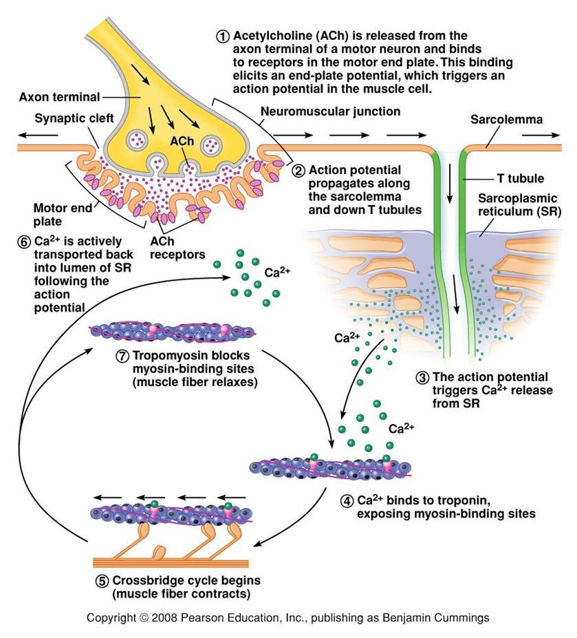
42 Mechanism of Muscle Contraction An AP travels along a motor nerve to its endings on muscle fibers Ach released. Ach acts on a local area of the muscle fiber membrane to open multiple ACh-gated" channels Ach nicotinic receptor. large quantities of Na+ diffuse to the interior of the muscle fiber membrane initiates an AP at the postsynaptic membrane. AP depolarizes the muscle membrane, and flows through the T tubes to the center of the muscle fiber causes the sarcoplasmic reticulum (SR) to release large quantities of Ca 2+ stored within SR (Ca 2+ bound in SR by calreticulin and calsequestrin). Ca 2+ initiate attractive forces between the actin and myosin filaments (generated by interaction of the cross-bridges from the myosin filaments with the actin filaments), causing them to slide alongside each other, which is the contractile process. Energy (ATP) is needed for the contractile process to proceed. After a fraction of a second, Ca 2+ are pumped back into SR by a Ca 2+ -membrane pump, and they remain stored until a new muscle AP comes along; this removal of Ca 2+ from the myofibrils causes the muscle contraction to cease (relaxation) and is an active process (ATP consume).
43 Muscular contraction mechanism Signal transmission 1. Motor neuron 2. Presynaptic terminal 3. Endplate region of skeletal fiber where synapse occurs 4. Nicotinic receptor Muscle Contraction 1. Action Potential sarcolemma T tubules 2. T tubules Sarcoplasmic Retic 3. Voltage gated Ca 2+ channels open 4. Ca 2+ sarcoplasm
44 Muscular contraction mechanism 5. Calcium binds to troponin (C) 6. Tropomyosin is deflected 7. Active sites of actin exposed 8. ATP attaches to myosin head 9. ATP is hydrolyzed (ADP & P) 10. Myosin head is phosphorylated & cocks 11. Myosin head binds to actin (cross bridge) 12. Myosin head dephosphorylates (head moves) & ADP released (power stroke) Muscle relaxation Calcium pumped into SR


45

46 Muscle cell must regenerate the ATP needed for muscle contraction - each round of the cross-bridge cycle consumes one molecule of ATP. - cellular store of ATP is sufficient to allow only a few seconds of continuous maximal contraction muscle cell must resynthesize ATP from ADP at a rate comparable to the rate of ATP consumption
47 Specialized energy stores in the muscle cell: The high-energy phosphate bond of phosphocreatine (its content in skeletal muscle is adequate to replenish the ATP pool several times, but it is still inadequate to sustain the energy needs of contracting muscle for more than 10 seconds): creatine kinase transfers the high-energy phosphate of phosphocreatine to ADP ATP Glycogen - more abundant energy source within skeletal muscle degradation to pyruvate is rapid and liberates energy that the cell invests in phosphorylating ADP to yield ATP (anaerobic metabolism). Pyruvate is further metabolized along with other foodstuffs by oxidative metabolism, which during the long term is the primary mechanism for the regeneration of ATP. The rate of ATP generation by oxidative metabolism is limited by the rate of oxygen delivery to the muscle. The pathway of muscle glycogen ensures that energy stores are sufficient to sustain muscle activity for nearly a minute even when oxygen is unavailable.


48 Single muscle twitch - instantaneous electrical excitation of the nerve to a muscle or a short electrical stimulus through the muscle itself, gives rise to a single, sudden contraction lasting for a fraction of a second. Muscle twitch Phases of muscle twitch 1. Lag/delay Phase from AP in motor neuron 2. Contraction Phase cross-bridge -> power stroke 3. Relaxation Phase calcium pumped into SR 4. Mechanical signal measured as tension

49 Stimulus vs contraction all or none response subthreshold stimulus -> no Threshold -> AP -> contraction increase Ca++ = increase force Stimulus frequency freq of AP = freq of contractions tetanus - calcium not pumped back Frequency summation of skeletal muscle twitches.
50 (1.65 µm) Length-tension diagram for a single fully contracted sarcomere, showing maximum strength of contraction when the sarcomere is 2.0 to 2.2 µm in length. At the upper right are the relative positions of the actin and myosin filaments at different sarcomere lengths from point A to point D.
51 Effect of muscle length on force of contraction in the whole intact muscle. When the muscle is at its normal resting length, which is at a sarcomere length of about 2 µm, it contracts upon activation with the approximate maximum force of contraction. The increase in tension that occurs during contraction = active tension decreases as the muscle is stretched beyond its normal length - that is, to a sarcomere length > 2.2 µm. Note that the whole muscle has a large amount of connective tissue in it; also, the sarcomeres in different parts of the muscle do not always contract the same amount.
52 Relation of load to velocity of contraction in a skeletal muscle with a cross section of 1 cm 2 and a length of 8 cm. A skeletal muscle contracts rapidly when it contracts against no load, to a state of full contraction in about 0.1 sec. for the average muscle. When loads are applied, the velocity of contraction becomes progressively less as the load increases. When the load increased to equal the maximum force that the muscle can exert, the velocity of contraction becomes zero and no contraction results, despite activation of the muscle fiber (a load on a contracting muscle is a reverse force that opposes the contractile force caused by muscle contraction).
53 Efficiency of muscle contraction. The efficiency of an engine: % energy input that is converted into work instead of heat. Low efficiency of muscle contraction: less than 25 % input energy to muscle (the chemical energy in nutrients) can be converted into work, the remainder becoming heat. Maximum efficiency of contraction is developed when the velocity of contraction is about 30 % of maximum.
54 Isometric Versus Isotonic Contraction Muscle contraction is -isotonic when it does shorten but the tension on the muscle remains constant throughout the contraction (the muscle shortens against a fixed load). -isometric when the muscle does not shorten during contraction (the muscle contracts against a force transducer without decreasing the muscle length);
55 Characteristics of isometric twitches recorded from different muscles. Duration of isometric contractions for different types of skeletal muscles, showing a latent period between the action potential (depolarization) and muscle contraction. Durations of contraction are adapted to the functions of the respective muscles: -ocular movements must be extremely rapid to maintain fixation of the eyes on specific objects to provide accuracy of vision -gastrocnemius muscle must contract moderately rapidly to provide sufficient velocity of limb movement for running and jumping -soleus muscle is concerned principally with slow contraction for continual, longterm support of the body against gravity.

56 The muscle fibers in each motor unit are not all bunched together in the muscle but overlap other motor units in microbundles of 3 to 15 fibers. This interdigitation allows the separate motor units to contract in support of one another rather than entirely as individual segments.

 by increasing the number of motor units contracting simultaneously, which is called")
57 Muscle contractions of different force - Force Summation. Summation = adding together of individual twitch contractions to increase the intensity of overall muscle contraction. Occurs: (1) by increasing the number of motor units contracting simultaneously, which is called multiple fiber summation, (2) by increasing the frequency of contraction, which is called frequency summation and can lead to tetanization.
58 Multiple Fiber Summation When the CNS sends a weak signal to contract a muscle, the smaller motor units of the muscle may be stimulated in preference to the larger motor units. The size principle: as the strength of the signal increases, larger and larger motor units begin to be excited as well (with the largest motor units - 50 times the contractile force of the smallest units) allows the gradations of muscle force during weak contraction to occur in small steps, whereas the steps become progressively greater when large amounts of force are required. The cause of this size principle is that the smaller motor units are driven by small motor nerve fibers, and the small motoneurons in the spinal cord are more excitable than the larger ones, so naturally they are excited first. The different motor units are driven asynchronously by the spinal cord, so contraction alternates among motor units one after the other, thus providing smooth contraction even at low frequencies of nerve signals.
59 Frequency Summation and Tetanization -as the frequency increases, there comes a point where each new contraction occurs before the preceding one is over one contraction is added partially to the previous one, so the total strength of contraction rises progressively with increasing frequency. -when the frequency reaches a critical level, the successive contractions eventually become so rapid that they fuse together and the whole muscle contraction appears to be completely smooth and continuous = tetanization. At a slightly higher frequency, the strength of contraction reaches its maximum, so any additional increase in frequency beyond that point has no further effect in increasing contractile force (calcium ions are maintained in the muscle sarcoplasm, even between APs, so that full contractile state is sustained without allowing any relaxation between APs).


60

61 Fast Versus Slow Muscle Fibers Muscles that react rapidly, including anterior tibialis, are composed mainly of fast fibers with only small numbers of the slow variety. Conversely, muscles such as soleus that respond slowly but with prolonged contraction are composed mainly of slow fibers. Slow- and fast-twitch fibers represent the extremes of a continuum of muscle fiber characteristics, each whole muscle is composed of fibers of each twitch type.
62 Slow Fibers (Type 1, Red muscle) Smaller fibers. Also innervated by smaller nerve fibers. More extensive blood vessel system and capillaries to supply extra amounts of oxygen. Greatly increased no. of mitochondria to support high levels of oxidative metabolism Fibers contain large amounts of myoglobin, an iron-containing protein similar to hemoglobin in red blood cells. Myoglobin combines with oxygen and stores it until needed; this also greatly speeds oxygen transport to the mitochondria. The myoglobin gives the slow muscle a reddish appearance and the name red muscle. Oxidative metabolism is slow but efficient, making these fibers resistant to fatigue. Fast Fibers (Type II, White muscle) Large fibers for great strength of contraction. Extensive sarcoplasmic reticulum for rapid release of calcium ions to initiate contraction. Large amounts of glycolytic enzymes for rapid release of energy by the glycolytic process more easily fatigable. Less extensive blood supply because oxidative metabolism is of secondary importance. Fewer mitochondria, also because oxidative metabolism is secondary. A deficit of red myoglobin in fast muscle gives it the name white muscle.
63 Fatigued muscle produces less force and has a reduced velocity of shortening. Muscle fatigue - inability to maintain a desired power output-resulting from muscle contraction against a load-with a decline in both force and velocity of shortening that results from - reduction in the number of active cross-bridges - reduction of the force produced per cross-bridge. As fatigue develops, the production of force usually declines earlier and to a greater extent than shortening velocity. Other characteristics of fatigued skeletal muscle are lower rates of both force production and relaxation, owing to impaired release and reuptake of Ca 2+ from the sarcoplasmic reticulum (SR). As a result, fast movements become difficult or impossible, and athletic performance suffers accordingly. Fatigue may serve an important protective role in allowing contractions at reduced rates and lower forces while preventing extreme changes in cell composition that could cause damage. Muscle fatigue is reversible with rest, which contrasts with muscle damage or weakness, in which even muscles that are well rested are compromised in their ability to develop force.
64 Factors contributing to fatigue Changes in the CNS produce central fatigue: altered input from muscle sensory nerve fibers, reduced excitatory input to motor control centers of the brain and spinal cord, and altered excitability of α and γ motor neurons Impaired excitability and impaired Ca 2+ release can produce peripheral fatigue. Fatigue can result from ATP depletion, lactic acid accumulation, and glycogen depletion.
Chapter 9 Muscle. Types of muscle Skeletal muscle Cardiac muscle Smooth muscle. Striated muscle
 Chapter 9 Muscle Types of muscle Skeletal muscle Cardiac muscle Smooth muscle Striated muscle Chapter 9 Muscle (cont.) The sliding filament mechanism, in which myosin filaments bind to and move actin
About This Chapter. Skeletal muscle Mechanics of body movement Smooth muscle Cardiac muscle Pearson Education, Inc.
 About This Chapter Skeletal muscle Mechanics of body movement Smooth muscle Cardiac muscle Skeletal Muscle Usually attached to bones by tendons Origin: closest to the trunk or to more stationary bone Insertion:
Skeletal Muscle Contraction 4/11/2018 Dr. Hiwa Shafiq
 Skeletal Muscle Contraction 4/11/2018 Dr. Hiwa Shafiq Skeletal Muscle Fiber About 40 per cent of the body is skeletal muscle, and 10 per cent is smooth and cardiac muscle. Skeletal muscles are composed
Ch. 6: Contraction of Skeletal Muscle Physiological Anatomy of Skeletal Muscle
 Ch. 6: Contraction of Skeletal Muscle 40% skeletal muscle + 10% smooth and cardiac muscle Ch. 7: Excitation of Skeletal Muscle Ch. 9: Contraction and Excitation of Smooth Muscle Physiological Anatomy of
Muscle and Muscle Tissue
 Muscle and Muscle Tissue Make up about half of total body mass Exerts force by converting chemical energy, ATP, to mechanical energy Muscle tissue is classified based on Shape Number and position of nuclei
Skeletal Muscle and the Molecular Basis of Contraction. Lanny Shulman, O.D., Ph.D. University of Houston College of Optometry
 Skeletal Muscle and the Molecular Basis of Contraction Lanny Shulman, O.D., Ph.D. University of Houston College of Optometry Like neurons, all muscle cells can be excited chemically, electrically, and
Skeletal Muscle Qiang XIA (
 Skeletal Muscle Qiang XIA ( 夏强 ), PhD Department of Physiology Rm C518, Block C, Research Building, School of Medicine Tel: 88208252 Email: xiaqiang@zju.edu.cn Course website: http://10.71.121.151/physiology
Skeletal Muscle Contraction 5/11/2017 Dr. Hiwa Shafiq
 Skeletal Muscle Contraction 5/11/2017 Dr. Hiwa Shafiq Skeletal Muscle Fiber About 40 per cent of the body is skeletal muscle, and 10 per cent is smooth and cardiac muscle. Skeletal muscles are composed
Muscle and Neuromuscular Junction. Peter Takizawa Department of Cell Biology
 Muscle and Neuromuscular Junction Peter Takizawa Department of Cell Biology Types and structure of muscle cells Structural basis of contraction Triggering muscle contraction Skeletal muscle consists of
2/19/2018. Learn and Understand:
 Muscular System with Special Emphasis on Skeletal Muscle Anatomy and Physiology Learn and Understand: The definition of cell changes again The contractile unit of muscle is the sarcomere. ATP and Ca 2+
Chapter 8: Skeletal Muscle: Structure and Function
 Chapter 8: Skeletal Muscle: Structure and Function Objectives Draw & label the microstructure of skeletal muscle Outline the steps leading to muscle shortening Define the concentric and isometric Discuss:
MUSCLE TISSUE (MUSCLE PHYSIOLOGY) PART I: MUSCLE STRUCTURE
 PART I: MUSCLE STRUCTURE") PART I: MUSCLE STRUCTURE Muscle Tissue A primary tissue type, divided into: skeletal muscle cardiac muscle smooth muscle Functions of Skeletal Muscles Produce skeletal movement Maintain body position Support
PART I: MUSCLE STRUCTURE Muscle Tissue A primary tissue type, divided into: skeletal muscle cardiac muscle smooth muscle Functions of Skeletal Muscles Produce skeletal movement Maintain body position Support
Concept 50.5: The physical interaction of protein filaments is required for muscle function
 Concept 50.5: The physical interaction of protein filaments is required for muscle function Muscle activity is a response to input from the nervous system The action of a muscle is always to contract Vertebrate
Chapter 10 Muscle Tissue Lecture Outline
 Chapter 10 Muscle Tissue Lecture Outline Muscle tissue types 1. Skeletal muscle = voluntary striated 2. Cardiac muscle = involuntary striated 3. Smooth muscle = involuntary nonstriated Characteristics
Skeletal Muscle. Connective tissue: Binding, support and insulation. Blood vessels
 Chapter 12 Muscle Physiology Outline o Skeletal Muscle Structure o The mechanism of Force Generation in Muscle o The mechanics of Skeletal Muscle Contraction o Skeletal Muscle Metabolism o Control of Skeletal
Muscle Tissue- 3 Types
 AN INTRODUCTION TO MUSCLE TISSUE Muscle Tissue- 3 Types Skeletal muscle (focus on these) Cardiac muscle Smooth muscle FUNCTIONS OF SKELETAL MUSCLES Produce movement of the skeleton Maintain posture and
Neuromuscular Physiology
 Neuromuscular Physiology Dr. Ana-Maria Zagrean Discipline of Physiology and Neuroscience, Carol Davila University of Medicine and Pharmacy, Bucharest The motor unit of the skeletal muscle All skeletal
Organismic Biology Bio 207. Lecture 6. Muscle and movement; sliding filaments; E-C coupling; length-tension relationships; biomechanics. Prof.
 Organismic Biology Bio 207 Lecture 6 Muscle and movement; sliding filaments; E-C coupling; length-tension relationships; biomechanics Prof. Simchon Today s Agenda Skeletal muscle Neuro Muscular Junction
Ch 12: Muscles sarcolemma, t-tubules, sarcoplasmic reticulum, myofibrils, myofilaments, sarcomere...
 Ch 12: Muscles Review micro-anatomy of muscle tissue Terminology examples: sarcolemma, t-tubules, sarcoplasmic reticulum, myofibrils, myofilaments, sarcomere... SLOs Differentiate levels of muscle structure:
Skeletal Muscle : Structure
 1 Skeletal Muscle : Structure Dr.Viral I. Champaneri, MD Assistant Professor Department of Physiology 2 Learning objectives 1. Gross anatomy of the skeletal muscle 2. Myofilaments & their molecular structure
Chapter 10: Muscles. Vocabulary: aponeurosis, fatigue
 Chapter 10: Muscles 37. Describe the structural components of skeletal muscle tissue from the molecular to the organ level. 38. Describe the structure, function, and importance of sarcomeres. 39. Identify
Chapter 9 - Muscle and Muscle Tissue
 Chapter 9 - Muscle and Muscle Tissue I. Overview of muscle tissue A. Three muscle types in the body: B. Special characteristics 1. Excitability: able to receive and respond to a stimulus 2. Contractility:
Muscle Physiology. Dr. Ebneshahidi Ebneshahidi
 Muscle Physiology Dr. Ebneshahidi Skeletal Muscle Figure 9.2 (a) Functions of the muscular system 1. Locomotion body movements are due to skeletal muscle contraction. 2. Vasoconstriction and vasodilatation
Muscle Physiology. Introduction. Four Characteristics of Muscle tissue. Skeletal Muscle
 Muscle Physiology Introduction Muscle = tissue capable of forceful shortening or contraction Converts chemical energy (ATP) into mechanical energy Important in: Respiration Urine collection & flow Gastrointestinal
Chapter 10 Muscle Tissue and Physiology Chapter Outline
 Chapter 10 Muscle Tissue and Physiology Chapter Outline Module 10.1 Overview of muscle tissue (Figures 10.1 10.2) A. Types of Muscle Tissue (Figure 10.1) 1. The three types of cells in muscle tissue are,,
Muscles and Muscle Tissue
 1 Muscles and Muscle Tissue Chapter 9 2 Overview of Muscle Tissues Compare and Contrast the three basic types of muscle tissue List four important functions of muscle tissue 3 Muscle Terminology Muscle
Anatomy and Physiology 1 Chapter 10 self quiz Pro, Dima Darwish,MD.
 Anatomy and Physiology 1 Chapter 10 self quiz Pro, Dima Darwish,MD. 1) Which of the following is a recognized function of skeletal muscle? A) produce movement B) maintain posture C) maintain body temperature
Muscle Tissue. PowerPoint Lecture Presentations prepared by Jason LaPres. Lone Star College North Harris Pearson Education, Inc.
 10 Muscle Tissue PowerPoint Lecture Presentations prepared by Jason LaPres Lone Star College North Harris An Introduction to Muscle Tissue Muscle Tissue A primary tissue type, divided into: Skeletal muscle
Nerve Muscle Relationship and Neural Muscular Junction Quiz. Remember, you need to know the structure and the function!
 Nerve Muscle Relationship and Neural Muscular Junction Quiz Remember, you need to know the structure and the function! What is this called? What is this? Schwann cell What is this called? Basal lamina
Hole s Human Anatomy and Physiology Eleventh Edition. Mrs. Hummer. Chapter 9 Muscular System
 Hole s Human Anatomy and Physiology Eleventh Edition Mrs. Hummer Chapter 9 Muscular System 1 Chapter 9 Muscular System Skeletal Muscle usually attached to bones under conscious control striated Three Types
BIOH111. o Cell Module o Tissue Module o Integumentary system o Skeletal system o Muscle system o Nervous system o Endocrine system
 BIOH111 o Cell Module o Tissue Module o Integumentary system o Skeletal system o Muscle system o Nervous system o Endocrine system Endeavour College of Natural Health endeavour.edu.au 1 TEXTBOOK AND REQUIRED/RECOMMENDED
Chapter 8 Notes. Muscles
 Chapter 8 Notes Muscles 8.1 Intro Three muscle types Skeletal Smooth cardiac 8.2 Structure of Skeletal Muscle Composition Skeletal muscle tissue Nervous tissue Blood Connective tissue Connective tissue
Principles of Anatomy and Physiology
 Principles of Anatomy and Physiology 14 th Edition CHAPTER 10 Muscular Tissue Introduction The purpose of the chapter is to: 1. Learn about the structure and function of the 3 types of muscular tissue
Ch.10 Muscle Tissue. Copyright 2009, John Wiley & Sons, Inc.
 Ch.10 Muscle Tissue Preview Chapter 10 In groups we will define the following terms 1. Skeletal muscle 2. Smooth muscle 3. Cardiac muscle 4. Sarcomere 5. Myofibril 6. Myofilament 7. Sarcoplasmic reticulum
Fig Copyright McGraw-Hill Education. Permission required for reproduction or display. Nucleus. Muscle fiber. Endomysium. Striations.
 Fig. 11.1 Nucleus Muscle fiber Endomysium Striations Ed Reschke 1 Fig. 11.2 Muscle fiber Nucleus I band A band Z disc Mitochondria Openings into transverse tubules Sarcoplasmic reticulum Triad: Terminal
MODULE 6 MUSCLE PHYSIOLOGY
 MODULE 6 MUSCLE PHYSIOLOGY III SEMESTER BOTANY Syllabi: Striated, Non striated and Cardiac muscle, Ultra structure of striated muscle fibre, Mechanism of muscle contraction, Threshold and spike potential,
1/4/2017. Introduction. Connective Tissue Coverings. 9.1: Structure of a Skeletal Muscle. Skeletal Muscle Fibers. Connective Tissue Coverings
 Introduction Chapter 09 Lecture Outline See separate PowerPoint slides for all figures and tables preinserted into PowerPoint without notes. Copyright McGraw-Hill Education. Permission required for reproduction
PSK4U THE NEUROMUSCULAR SYSTEM
 PSK4U THE NEUROMUSCULAR SYSTEM REVIEW Review of muscle so we can see how the neuromuscular system works This is not on today's note Skeletal Muscle Cell: Cellular System A) Excitation System Electrical
CHAPTER 6 2/9/2016. Learning Objectives List the four traits that all muscle types have in common.
 Learning Objectives List the four traits that all muscle types have in common. CHAPTER 6 The Muscular System Demonstrate and explain the use of antagonistic muscle pairs. Describe the attachment of muscle
Muscle Tissue. Alternating contraction and relaxation of cells. Chemical energy changed into mechanical energy
 Know these muscles Muscle Tissue Alternating contraction and relaxation of cells Chemical energy changed into mechanical energy 3 Types of Muscle Tissue Skeletal muscle attaches to bone, skin or fascia
Muscle Dr. Ted Milner (KIN 416)
") Muscle Dr. Ted Milner (KIN 416) Muscles are biological motors which actively generate force and produce movement through the process of contraction. The molecular mechanism responsible for muscle contraction
Muscle Dr. Ted Milner (KIN 416) Muscles are biological motors which actively generate force and produce movement through the process of contraction. The molecular mechanism responsible for muscle contraction
Session 3-Part 2: Skeletal Muscle
 Session 3-Part 2: Skeletal Muscle Course: Introduction to Exercise Science-Level 2 (Exercise Physiology) Presentation Created by Ken Baldwin, M.ED, ACSM-H/FI Copyright EFS Inc. All Rights Reserved. Skeletal
Contraction of Skeletal Muscle
 chapter 6 Contraction of Skeletal Muscle Unit II About 4 percent of the body is skeletal muscle, and perhaps another 1 percent is smooth and cardiac muscle. Some of the same basic principles of contraction
Musculoskeletal Systems. Anatomy: Arrangement of Cells Physiology: Contractions
 Musculoskeletal Systems Anatomy: Arrangement of Cells Physiology: Contractions Characteristics of all muscle Contractile: it shortens Excitable: receives & responds to electrical signals Extensible: stretches
MUSCULAR TISSUE. Dr. Gary Mumaugh
 MUSCULAR TISSUE Dr. Gary Mumaugh MUSCLE OVERVIEW The three types of muscle tissue are skeletal, cardiac, and smooth These types differ in structure, location, function, and means of activation FUNCTIONAL
Skeletal muscle in the light of its structure
 Mechanism of contraction of Skeletal muscle in the light of its structure By Dr. Mudassar Ali Roomi (MBBS, M. Phil) Muscle Tissue Skeletal Muscle Cardiac Muscle Smooth Muscle Skeletal Muscle Long cylindrical
Smooth Cardiac Skeletal Location Around tubes Heart tissue attached to skeleton Moves stuff thru Heart beat pumps Moves body parts
 Biology 067 - Muscular system A. Type of muscles: Smooth Cardiac Skeletal Location Around tubes Heart tissue attached to skeleton Function Moves stuff thru Heart beat pumps Moves body parts tubes blood
Muscle Cells & Muscle Fiber Contractions. Packet #8
 Muscle Cells & Muscle Fiber Contractions Packet #8 Skeletal muscle is attached to bones and is responsible for movement. Introduction Introduction II Skeletal muscle is composed of bundles of muscle fibers
The organization of skeletal muscles. Excitation contraction coupling. Whole Skeletal Muscles contractions. Muscle Energetics
 Muscle and Movement The organization of skeletal muscles Excitation contraction coupling Whole Skeletal Muscles contractions Muscle Energetics The molecular bases of movement Muscular cells use molecular
Microanatomy of Muscles. Anatomy & Physiology Class
 Microanatomy of Muscles Anatomy & Physiology Class Three Main Muscle Types Objectives: By the end of this presentation you will have the information to: 1. 2. 3. 4. 5. 6. Describe the 3 main types of muscles.
#1 20. physiology. Muscle tissue 30/9/2015. Ahmad Adel Sallal. Mohammad Qudah
 # 20 physiology Muscle tissue Ahmad Adel Sallal 30/9/205 Mohammad Qudah MUSCLES PHYSIOLOGY Awn, welcome to the first physiology lecture in the MSS, I wish you a perfect exams with high grades, and never
10 - Muscular Contraction. Taft College Human Physiology
 10 - Muscular Contraction Taft College Human Physiology Muscular Contraction Sliding filament theory (Hanson and Huxley, 1954) These 2 investigators proposed that skeletal muscle shortens during contraction
CLASS SET Unit 4: The Muscular System STUDY GUIDE
 NPHS Anatomy & Physiology Questions to answer: 1) List three functions of the muscular system. 1) movement 2) thermogenesis (generates heat) 3) posture & body/joint support CLASS SET Unit 4: The Muscular
Human Anatomy. Muscle Tissue and Organization. DR.SADIQ ALI (K.E Medalist) 10-1
 10-1") Human Anatomy Muscle Tissue and Organization DR.SADIQ ALI (K.E Medalist) 10-1 Tissue and Organization Over 700 skeletal muscles have been named. Form the muscular system. Muscle tissue is distributed almost
Human Anatomy Muscle Tissue and Organization DR.SADIQ ALI (K.E Medalist) 10-1 Tissue and Organization Over 700 skeletal muscles have been named. Form the muscular system. Muscle tissue is distributed almost
The Musculoskeletal System. Chapter 46
 The Musculoskeletal System Chapter 46 Types of Skeletal Systems Changes in movement occur because muscles pull against a support structure Zoologists recognize three types: 1. Hydrostatic skeletons a fluid
Skeletal Muscle Tissue
 Functions of Skeletal Muscle Skeletal Muscle Tissue Keri Muma Bio 6 Movement muscles attach directly or indirectly to bone, pull on bone or tissue when they contract Maintain posture / body position muscles
Successive magnifications
 Successive magnifications Skeletal ("striated" = striped) muscle cell ("fiber" = cell) 10-100 microns (huge) and long (tendon to tendon) There are smaller units within fiber called "myofibrils" (1-2 microns
Nerve Cell (aka neuron)
") Nerve Cell (aka neuron) Neuromuscular Junction Nerve cell Muscle fiber (cell) The Nerve Stimulus and Action Potential The Nerve Stimulus and Action Potential Skeletal muscles must be stimulated by a motor
Nerve Cell (aka neuron) Neuromuscular Junction Nerve cell Muscle fiber (cell) The Nerve Stimulus and Action Potential The Nerve Stimulus and Action Potential Skeletal muscles must be stimulated by a motor
Ch 12 can be done in one lecture
 Ch 12 can be done in one lecture Developed by John Gallagher, MS, DVM Chapter 12: Muscles Review muscle anatomy (esp. microanatomy of skeletal muscle) Terminology: sarcolemma t-tubules sarcoplasmic reticulum
Lecture Overview. Muscular System. Marieb s Human Anatomy and Physiology. Chapter 9 Muscles and Muscle Tissue Lecture 16
 Marieb s Human Anatomy and Physiology Marieb Hoehn Chapter 9 Muscles and Muscle Tissue Lecture 16 1 Lecture Overview Types, characteristics, functions of muscle Structure of skeletal muscle Mechanism of
BIOLOGY - CLUTCH CH.49 - MUSCLE SYSTEMS.
 !! www.clutchprep.com BIOLOGY - CLUTCH Muscle system organ system that includes skeletal, cardiac, and smooth muscle Muscle tissue capable of contracting through the interaction of actin and myosin proteins
Muscular System. This chapter will focus on muscle cells and tissues. Muscle tissue has several functions:
 Muscular System Slide 2 This chapter will focus on muscle cells and tissues. Muscle tissue has several functions: Movement: Muscles work as pulleys on bones to help create changes in body position. Muscles
1-Recognize the meaning of summation of contraction and its types. 2-detrmine the effect of changing length on skeletal muscle tension.
 Lec7 Physiology Dr.HananLuay Objectives 1-Recognize the meaning of summation of contraction and its types. 2-detrmine the effect of changing length on skeletal muscle tension. 3-Differntiate between the
Chapter 10 -Muscle Tissue
 Chapter 10 -Muscle Tissue Muscles: 1. Overview of Muscle Tissue A. Review 5 functions of muscle tissue. B. Review the 5 properties of muscle tissue. WHICH do they share with nervous tissue? (2, plus the
Outline. Bio 105: Muscular System. Muscular System. Types of Muscles. Smooth Muscle. Cardiac Muscle 4/6/2016
 Outline Bio 105: Muscular System Lecture 11 Chapter 6 Characteristics of muscles 3 types of muscles Functions of muscles Structure of skeletal muscles Mechanics of muscle contraction Energy sources for
Muscular Tissue. Functions of Muscular Tissue. Types of Muscular Tissue. Skeletal Muscular Tissue. Properties of Muscular Tissue
 Muscular Tissue Functions of Muscular Tissue Muscle makes up a large percentage of the body s weight (40-50%) Their main functions are to: Create motion muscles work with nerves, bones, and joints to produce
Baraa Ayed. Mohammad khatatbeh. 1 P a g e
 4 Baraa Ayed أسامة الخض Mohammad khatatbeh 1 P a g e Today we want to talk about these concepts: Excitation-Contraction coupling Smooth muscles (Generally speaking) Excitation-Contraction coupling Excitation-Contraction
Connective tissue MUSCLE TISSUE
 Connective tissue MUSCLE TISSUE Part 1 General features of MT Develop from mesoderm Many cells, less intercellular matrix Function contraction (shortening) Skeletal (striated, voluntary) Types of MT Cardiac
Skeletal Muscle. Skeletal Muscle
 Skeletal Muscle Skeletal Muscle Types of muscle Skeletal muscle-moves the skeleton by pulling on the tendons that are connected to the bones Cardiac muscle-pumps blood through the heart and blood vessels
Chapter 50. You re on your own for: Sensory Reception Mechanoreceptors Gravity, Hearing and Equilibrium. Chemoreception taste and smell
 1 Sensory and Motor Mechanisms 2 Chapter 50 You re on your own for: Sensory Reception Mechanoreceptors Gravity, Hearing and Equilibrium Chemoreception taste and smell Photoreceptors vision It s interesting.
Muscle Tissue. Dr. Heba Kalbouneh Associate Professor of Anatomy and Histology
 Muscle Tissue Dr. Heba Kalbouneh Associate Professor of Anatomy and Histology Functions of muscle tissue Movement Maintenance of posture Joint stabilization Heat generation Tendon Belly Tendon Types of
Nerve regeneration. Somatic nervous system
 Somatic nervous system Signals from CNS are sent to skeletal muscles. Final result is a muscle contraction. Motor neuron starts in CNS and its axon ends at a muscle cell. Alpha motor neuron Alpha motor
Nerve meets muscle. Nerve regeneration. Somatic nervous system
 Somatic nervous system Signals from CNS are sent to skeletal muscles. Final result is a muscle contraction. Alpha motor neurons branch into several terminals (can be over 1000), each contacting a separate
1. Locomotion. 2. Repositioning. 3. Internal movement
 MUSCLE and MOVEMENT Chapters 20, 8, 21 1. Locomotion A. Movement B. 2. Repositioning A. 3. Internal movement A. 1 Muscle Cells 1. Contractile 2. Myocytes 3. Striated A. Skeletal B. Cardiac 4. Smooth 5.
1. Locomotion. 2. Repositioning. 3. Internal movement
 MUSCLE and MOVEMENT Chapters 20, 8, 21 1. Locomotion A. Movement B. 2. Repositioning A. 3. Internal movement A. Muscle Cells 1. Contractile 2. Myocytes 3. Striated A. Skeletal B. Cardiac 4. Smooth 5. Striated
Muscle Tissue. Alternating contraction and relaxation of cells Chemical energy changed into mechanical energy 10:32
 Muscle Tissue Alternating contraction and relaxation of cells Chemical energy changed into mechanical energy 1 Properties of Muscle Tissue Excitability responds to chemical messengers (neurotransmitters)
Human Anatomy and Physiology - Problem Drill 09: The Muscular System
 Human Anatomy and Physiology - Problem Drill 09: The Muscular System Question No. 1 of 10 The muscular system of the human body fulfills many different roles. Which of the following statements about the
Muscle Tissue. Muscle Tissue Outline. General Function of Muscle Tissue
 Muscle Tissue Muscle Tissue Outline General Functions of Muscle Tissue Characteristics of Muscle Tissue Classification of Muscle Tissue Skeletal Muscle Structure and Function Muscle Energetics Muscle Mechanics
The All-or-None Principle Motor units also comply to a rule known as the all-ornone principle (or law).
.") The All-or-None Principle Motor units also comply to a rule known as the all-ornone principle (or law). This principle stipulates that, when a motor unit is stimulated to contract, it will do so to its
The All-or-None Principle Motor units also comply to a rule known as the all-ornone principle (or law). This principle stipulates that, when a motor unit is stimulated to contract, it will do so to its
Anatomy & Physiology. Unit Two. Muscular System URLs Frog Dissection
 Anatomy & Physiology 9 Muscular System URLs Frog Dissection http://curry.edschool.virginia.edu/go/frog/home.html Cat Dissection http://www.mhhe.com/biosci/ap/cat_dissect/index.htm List of Muscles http://www.meddean.luc.edu/lumen/meded/
Skeletal Muscle. Bởi: OpenStaxCollege
 Bởi: OpenStaxCollege The best-known feature of skeletal muscle is its ability to contract and cause movement. Skeletal muscles act not only to produce movement but also to stop movement, such as resisting
Physiology of the skeletal muscle
 THE EFFECTORS Physiology of the skeletal muscle About 40 percent of the body is skeletal muscle, and perhaps another 10 percent is smooth and cardiac muscle. Anatomy of skeletal muscle All skeletal muscles
Muscles & Motor Locomotion Why Do We Need All That ATP?
 Muscles & Motor Locomotion Why Do We Need All That ATP? 2006-2007 Animal Locomotion What are the advantages of locomotion? sessile motile Lots of ways to get around Lots of ways to get around mollusk mammal
Muscle Cell Anatomy & Function (mainly striated muscle tissue)
") Muscle Cell Anatomy & Function (mainly striated muscle tissue) General Structure of Muscle Cells (skeletal) several nuclei (skeletal muscle) skeletal muscles are formed when embryonic cells fuse together
Muscle Cell Anatomy & Function (mainly striated muscle tissue) General Structure of Muscle Cells (skeletal) several nuclei (skeletal muscle) skeletal muscles are formed when embryonic cells fuse together
Chapter Skeletal Muscle Structure and Function
 Chapter 10.2 Skeletal Muscle Structure and Function Introduction to Muscle Physiology Movement is a fundamental characteristic of all living things All muscle cells (skeletal, cardiac, and smooth) are
On which skeletal muscle filament is troponin located? What is the function of the sarcoplasmic reticulum (SR)?
?") CASE 6 A 21-year-old man presents to a rural emergency center with a 1-day history of progressive stiffness of the neck and jaw, difficulty swallowing, stiff shoulders and back, and a rigid abdomen. Upon
CASE 6 A 21-year-old man presents to a rural emergency center with a 1-day history of progressive stiffness of the neck and jaw, difficulty swallowing, stiff shoulders and back, and a rigid abdomen. Upon
The Muscular System. Objective: The student will become familiar with the structure and function of the muscular system
 The Muscular System Objective: The student will become familiar with the structure and function of the muscular system 1 Question of the day: What moves you? Composition: The musclar system makes up 40-50%
BCH 450 Biochemistry of Specialized Tissues. V. Muscle Tissues
 BCH 450 Biochemistry of Specialized Tissues V. Muscle Tissues Nomenclature Sarcolemma = plasma membrane Sarcoplasmic reticulum = endoplasmic reticulum Muscle fiber = cell Myofibril = subcellular fibers
Muscle Tissue. C h a p t e r. PowerPoint Lecture Slides prepared by Jason LaPres Lone Star College - North Harris
 C h a p t e r 10 Muscle Tissue PowerPoint Lecture Slides prepared by Jason LaPres Lone Star College - North Harris Copyright 2009 Pearson Education, Inc., publishing as Pearson Benjamin Cummings An Introduction
AP Biology
 Chapter 49. Animal Locomotion What are the advantages of locomotion? sessile motile Muscles & Motor Locomotion Muscle voluntary, striated involuntary, striated auto-rhythmic involuntary, non-striated 1
Chapter 49. Muscles & Motor Locomotion. AP Biology
 Chapter 49. Muscles & Motor Locomotion Animal Locomotion What are the advantages of locomotion? sessile motile Muscle voluntary, striated involuntary, striated auto-rhythmic involuntary, non-striated
Functions of Muscle Tissue
 The Muscular System Functions of Muscle Tissue Movement Facilitation Thermogenesis Postural Support Regulation of Organ Volume Protects Internal Organs Pumps Blood (HEART) Characteristics of Muscle Tissue
Chapter 10: Muscle Tissue
 Chapter 10: Muscle Tissue Muscle is one of the 4 primary types of tissue. It is subdivided into skeletal, cardiac and smooth muscle. I. Skeletal Muscle Tissue and the Muscular System, p. 284 Objective
AnS SI 214 Practice Exam 2 Nervous, Muscle, Cardiovascular
 AnS SI 214 Practice Exam 2 Nervous, Muscle, Cardiovascular Select the best answer choice in the questions below. 1) On the electrocardiogram, repolarization of the atria is represented by the: A) P wave
B108 BC Muscle Contraction and Locomotion *
 OpenStax-CNX module: m62420 1 B108 BC Muscle Contraction and Locomotion * Melodye Gold Based on Human Biology Chapter 16: Muscle Contraction and Locomotion by OpenStax Willy Cushwa This work is produced
I. Overview of Muscle Tissues
 I. Overview of Muscle Tissues A. Types of Muscle Tissue 1. Terminology 1. Muscle fibers = muscle cells are greatly elongated therefore known as fibers; true for skeletal and smooth muscles only 2. Myo
Neuromuscular Junction (NMJ) Skeletal Muscle Structure. Motor Unit. Motor Units. Chapter 12 Outline
 Skeletal Muscle Structure. Motor Unit. Motor Units. Chapter 12 Outline") Chapter 12 Outline Skeletal Muscles Mechanisms of Contraction Contractions of Skeletal Muscle Energy Requirements of Skeletal Muscle Neural Control of Skeletal Muscles Cardiac and Smooth Muscle Muscle
Chapter 12 Outline Skeletal Muscles Mechanisms of Contraction Contractions of Skeletal Muscle Energy Requirements of Skeletal Muscle Neural Control of Skeletal Muscles Cardiac and Smooth Muscle Muscle
Chapter 10. The Muscular System 11-1
 Chapter 10 The Muscular System 11-1 The Muscular System Structural and functional organization of muscles Muscles of the head and neck Muscles of the trunk Muscles acting on the shoulder and upper limb
Chapter 10! Chapter 10, Part 2 Muscle. Muscle Tissue - Part 2! Pages !
 ! Chapter 10, Part 2 Muscle Chapter 10! Muscle Tissue - Part 2! Pages 308-324! SECTION 10-5! Sarcomere shortening and muscle fiber stimulation produce tension! 2! Tension Production - Muscle FIBER! All-or-none
What effect would an AChE inhibitor have at the neuromuscular junction?
 CASE 4 A 32-year-old woman presents to her primary care physician s office with difficulty chewing food. She states that when she eats certain foods that require a significant amount of chewing (meat),