Neuro-ophthalmologyophthalmology. Marek Michalec, MD.
|
|
|
- Homer Benson
- 5 years ago
- Views:
Transcription
1 Neuro-ophthalmologyophthalmology Marek Michalec, MD.
2 Neuro-ophthalmology Study integrating ophthalmology and neurology Disorders affecting parts of CNS devoted to vision or eye: Afferent system (visual pathway, incl. optic nerve) Efferent system (ocular motor control, pupillary function)
3 Part I Neuro-ophthalmologic Examination
4 Examination History Eye examination (visual acuity, tonometry, anterior segment examination, funduscopic examination) Perimetry Color vision, contrast sensitivity, electrophysiology (ERG, VEP) MRI of brain, Neurologic examination
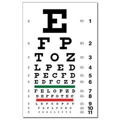
5 Visual acuity Each eye separately Distance and near vision Using of corrective lenses, pinhole Using Snellen chart (20 feet) normal 20/20 Count fingers, hand motion, light perception, no light perception


 100")
6 Color vision Each eye separately Comparison between eyes Examination: pseudoisochromatic plates (Ishihara) 100 Hue test (Farnsworth-Munsell)


7 Farnsworth-Munsell 100 Hue test Ordering the color tiles as patient sees it

8 Contrast sensitivity Examining spatial frequency Decreased in some optic nerve disorders (typically optic neuritis)

9 Perimetry To assess the quality of visual field Characteristic visual field defect =location of possible intracranial lesions

10 Perimetry Automated static perimetry


11 Perimetry Goldmann kinetic perimetry
12 Electrophysiologic examination ERG = Electroretinography Access possible functional pathology of retina (scotopic, photopic and central part) Flash ERG (activity of bipolar cells as an answer to stimation of photosensitive cells rods, cones) Pattern ERG (activity of gaglionar cell as a response to stimulation of cones in macula) VEP = Visual evoked potentials (responses) Access the capability of anterior visual pathways optic nerve Major use: diagnosis/confirm of optic neuritis

13 Electrophysiologic examination
14 Electroretinography
15 Visual evoked potentials

16 Multifocal ERG, Multifocal VEP Mostly experimental use, not standard in clinical medical practice here
17 Part II Pathology of Afferent system
")
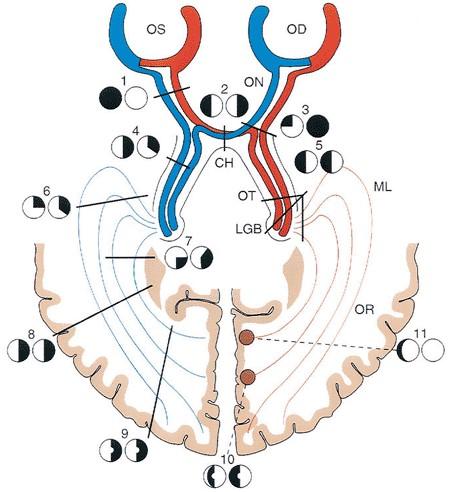
18 Afferent system Retina (cones, rods, bipolar and ganglion cells) Optic nerve Optic chiasm Optic tract Lateral geniculate body Optic radiation Visual cortex (V1 = Brodmann area 17)
19 Pathologies of Afferent Visual System Papilledema Optic Neuritis Optic Neuropathy Optic Atrophy
20 Papilledema Not a disease - sing secondary due to elevated intracranial pressure (ICP) Unspecific sign Require immediate diagnosis = increased ICP is a lifethreatening situation!!! 60% of cases = increased ICP caused by intracranial tumor!!! Other possible causes: hydrocephalus, meningitis, encephalitis, brain abscess...


21 Papilledema Clinical picture Early Margins are obscured Optic cup initially preserved Hyperemic disc Acute Elevation of disc Radial hemorrhages Grayish-white exudates Chronic Disc edema Obiterated optic cup
22 Optic neuritis Inflammation of the optic nerve Intraocular within the globe Retrobulbar posteriot to the globe Usually unilateral Tendency to repeat Etiology Often associated with multiple sclerosis (MS) = demyelinating optic neuritis (20% = first sign of MS) Other possible inflammatory causes: Lyme disease, syphilis, inflammation from orbit, paranasal sinuses...
23 Optic neuritis Symptoms Sudden vision loss within several hours (mild blurring/light perception) Central, paracentral scotoma Retrobulbar/parabulbar pain Present afferent pupillary defect Prognosis depends on underlying disorders MS = usually good significant spontaneous improvement (several weeks) Some permanent disturbances of vision are possible (color vision decreasing, scotoma)
24 Anterior Ischemic Optic Neuropathy Etiology Acute disruption of blood supply (due to vascular changes, infarction) Symptoms Sudden unilateral loss of vision Altitudinal or wedge-shaped visual field defect Present afferent pupillary defect Clinical picture Edema of optic disc Segmental obscuration of margins (correlation with visual field defect)
 eye within days/ weeks!")
25 Anterior ischemic optic neuropathy 2 forms Benign: Nonarteritic AION Malign: Arteritic AION Arteritic AION Association with systemic vasculitis (giant cell arteritis) Diagnosis: sedimentation rate, biopsy of temporal artery High risk of affection of contralateral (fellow) eye within days/ weeks!!! Need for immediate therapy with high dose intravenous corticoids!!!
26 AION forms Arteritic form Non-arteritic form % of cases AION 10 % 90% age 70 years 60 years Sex Female > male Female = male Systemic disease association Giant cell arteritis (Horton disease) idiopathic Prognosis Very rare mild Fellow eye affection Diagnostics: Sedimentation (FW) treatment often (50-90%) Very high High dosage of systemic corticoids rare (10-20%) normal Not available
27 Optic Atrophy Irreversible loss of axons as a result to damage of optic nerve Etiology Primary due to trauma, direct pressure by tumor Secondary due to affection of optic nerve (optic neuritis...) Glaucomatous due to glaucomatic damage Pathogenesis Ascending - lesion located anterior to the lamina cribrosa Descending lesion located posteriot to the lamina cribrosa

 Compressive (orbital/intracranial mass) Traumatic")
 Congenital/hereditary (LHON, Kjer atrophy) Systemic")
28 Optic Atrophy Clinical picture Total/partial pale optic disc Well defined / blurred margins Constricted / reduced retinal vessels Etiology Vascular (AION, RAO) Inflammation (optic neuritis, neuroinfections) Compressive (orbital/intracranial mass) Traumatic (avulsion, bone fracture) Toxic (methyl alcohol, various poisons, cytostatics) Congenital/hereditary (LHON, Kjer atrophy) Systemic (hematooncological diseases)
29 Part III Pathology of Efferent system
30 Efferent system 1) Cranial neuropathies (III, IV, VI) 2) Pupillary abnormalities
31 Ocular motility produced by extraocular muscles 4 rectus muscles (lateral, medial, superior, inferior) 2 oblique muscles (superior, inferior) Eye movement
32 Cranial neuropathies Signs Oculomotor nerve palsy Diplopia Multiple muscle paralysis Ptosis Anisocoria Trochlear nerve palsy Vertical diplopia Abnormal head tilt Abducens nerve palsy Horizontal diplopia in the gaze palsy
33 Cranial neuropathies Etiology Ischemic (diabetes, hypertension, hyperlipidemia) Demyelinating disease (MS) Compressive (tumor, aneurysm) Elevated ICP Multiple cranial neuropathies = suspect lesion in the posterior orbit or cavenrous sinus region

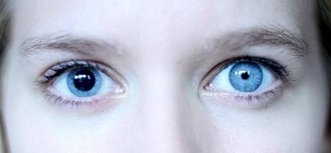
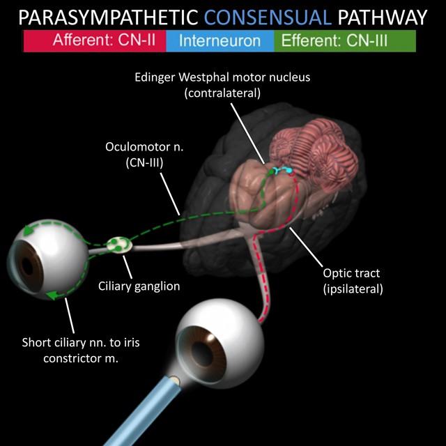
34 Pupil Miosis parasympathetic nervous system Mydriasis sympathetic nervous system
35
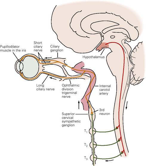
36 Sympathetic pathway
37 Anisocoria Pupillary abnormalities inequality of pupil size May be physiologic Possible accidental discovery May be isolated / associated with eyelid or ocular motility abnormalities Diagnosis Direct shine at pupil Test near response (miosis with accomodation) Pupil sizes in light and dark
 Ptosis Pseudo-enophthalmus")
 Etiology Trauma, internal carotid")

38 Signs Horner s Syndrome Miosis (pupil does not dilate in dark) Ptosis Pseudo-enophthalmus Anhidrosis (diminished sweating) Heterochromia (if congenital) Etiology Trauma, internal carotid artery dissection, brain stem strokes, MS, brain tumor, syringomyelia, apical lung tumor, goiter, thyroid carcinoma...
39 Adie s Pupil Signs No present / slow miosis to light Present miosis to accomodation Pupil is larger with light/near dissociation Etiology Inflammation (viral or bacterial infection) Therapy Pilocarpine drops, thoracic sympathectomy
40 Thank you for your attention!
OCT : retinal layers. Extraocular muscles. History. Central vs Peripheral vision. History: Temporal course. Optical Coherence Tomography (OCT)
") Optical Coherence Tomography (OCT) OCT : retinal layers 7 Central vs Peripheral vision Extraocular muscles RPE E Peripheral Vision: Rods (95 million) 30% Ganglion cells Central Vision: Cones (5 million)
Optical Coherence Tomography (OCT) OCT : retinal layers 7 Central vs Peripheral vision Extraocular muscles RPE E Peripheral Vision: Rods (95 million) 30% Ganglion cells Central Vision: Cones (5 million)
3/16/2018. Optic Nerve Examination. Hassan Eisa Swify FRCS Ed (Ophthalmology) Air Force Hospital
 Air Force Hospital") Optic Nerve Examination Hassan Eisa Swify FRCS Ed (Ophthalmology) Air Force Hospital 1 Examination Structure ( optic disc) Function Examination of the optic disc The only cranial nerve (brain tract) which
Optic Nerve Examination Hassan Eisa Swify FRCS Ed (Ophthalmology) Air Force Hospital 1 Examination Structure ( optic disc) Function Examination of the optic disc The only cranial nerve (brain tract) which
Role Of Various Factors In The Treatment Of Optic Neuritis----A Study Abstract Aim: Materials & Methods Discussion: Conclusion: Key words
 IOSR Journal of Dental and Medical Sciences (IOSR-JDMS) e-issn: 2279-0853, p-issn: 2279-0861.Volume 15, Issue 9 Ver. X (September). 2016), PP 51-57 www.iosrjournals.org Role Of Various Factors In The Treatment
Pupil Exams and Visual Fields
 Pupil Exams and Visual Fields A Closer Look at Cranial Nerves No Financial Interests Amy Jost does not have any financial interests related to this presentation AMY JOST, BS, COMT, CCRC, OSC CINCINNATI
Alan G. Kabat, OD, FAAO (901)
") THE SWOLLEN OPTIC DISC: EMERGENCY OR ANOMALY? Alan G. Kabat, OD, FAAO (901) 252-3691 Memphis, Tennessee alan.kabat@alankabat.com Course description: The swollen disc presents a diagnostic dilemma. While
THE SWOLLEN OPTIC DISC: EMERGENCY OR ANOMALY? Alan G. Kabat, OD, FAAO (901) 252-3691 Memphis, Tennessee alan.kabat@alankabat.com Course description: The swollen disc presents a diagnostic dilemma. While
NEURO-OPHTHALMIC ASSESSMENT DR. B. C. UGWU
 CLINICAL VIGNETTE 2019; 5:1 NEURO-OPHTHALMIC ASSESSMENT DR. B. C. UGWU Editor-in-Chief: Prof Olufemi Idowu Neurological surgery Division, Department of Surgery, LASUCOM/LASUTH, Ikeja, Lagos, Nigeria. Copyright-
Neuro-Ocular Grand Rounds
 Neuro-Ocular Grand Rounds Anthony B. Litwak,OD, FAAO VA Medical Center Baltimore, Maryland Dr. Litwak is on the speaker and advisory boards for Alcon and Zeiss Meditek COMMON OPTIC NEUROPATHIES THAT CAN
Index. Note: Page numbers of article titles are in boldface type.
 Index Note: Page numbers of article titles are in boldface type. A Acetazolamide, in idiopathic intracranial hypertension, 49 52, 60 Angiography, computed tomography, in cranial nerve palsy, 103 107 digital
Neuro-Ocular Grand Rounds Anthony B. Litwak,OD, FAAO VA Medical Center Baltimore, Maryland
 Neuro-Ocular Grand Rounds Anthony B. Litwak,OD, FAAO VA Medical Center Baltimore, Maryland Dr. Litwak is on the speaker and advisory boards for Alcon and Zeiss Meditek COMMON OPTIC NEUROPATHIES THAT CAN
I have nothing to disclose but I
 OPTIC NEUROPATHIES Robert L. Tomsak MD PhD Professor of Ophthalmology and Neurology Wayne State t University it Sh School of Mdii Medicine I have nothing to disclose but I wish I did. dd Road map for this
C19. Pediatric Neuro-ophthalmology: Dilemmas in clinical practice. 12 June, :15 15:45. Room 115 HAND-OUTS
 C19 Pediatric Neuro-ophthalmology: Dilemmas in clinical practice 12 June, 2017 14:15 15:45 Room 115 HAND-OUTS Is this strabismus really harmful? Karl Golnik, MD, MEd University of Cincinnati, USA Childhood
Learn Connect Succeed. JCAHPO Regional Meetings 2015
 Learn Connect Succeed JCAHPO Regional Meetings 2015 OPTIC NEUROPATHY AS EASY AS 1,2,3,4 OPTIC NERVE ANATOMY M. Tariq Bhatti, MD Departments of Ophthalmology and Neurology Duke Eye Center and Duke University
REVIEW OF HEAD AND NECK CRANIAL NERVES AND EVERYTHING ELSE
 REVIEW OF HEAD AND NECK CRANIAL NERVES AND EVERYTHING ELSE OLFACTORY NERVE CN I ANTERIOR CRANIAL FOSSA CRISTA GALLI OF ETHMOID OLFACTORY FORAMINA IN CRIBIFORM PLATE OF ETHMOID BONE CN I OLFACTORY NERVE
LOOKING AT BLINDNESS FROM NEUROLOGIST S PERSPECTIVE
 Vet Times The website for the veterinary profession https://www.vettimes.co.uk LOOKING AT BLINDNESS FROM NEUROLOGIST S PERSPECTIVE Author : LAURENT S GAROSI Categories : Vets Date : June 23, 2008 LAURENT
Pearls, Pitfalls and Advances in Neuro-Ophthalmology
 Pearls, Pitfalls and Advances in Neuro-Ophthalmology Nancy J. Newman, MD Emory University Atlanta, GA Consultant for Gensight Biologics, Santhera Data Safety Monitoring Board for Quark AION Study Medical-legal
Non-arteritic anterior ischemic optic neuropathy (NAION) with segmental optic disc edema. Jonathan A. Micieli, MD Valérie Biousse, MD
 with segmental optic disc edema. Jonathan A. Micieli, MD Valérie Biousse, MD") Non-arteritic anterior ischemic optic neuropathy (NAION) with segmental optic disc edema Jonathan A. Micieli, MD Valérie Biousse, MD A 75 year old white woman lost vision in the inferior part of her visual
Non-arteritic anterior ischemic optic neuropathy (NAION) with segmental optic disc edema Jonathan A. Micieli, MD Valérie Biousse, MD A 75 year old white woman lost vision in the inferior part of her visual
Anterior Ischemic Optic Neuropathy (AION)
") Anterior Ischemic Optic Neuropathy (AION) Your doctor thinks you have suffered an episode of anterior ischemic optic neuropathy (AION). This is the most common cause of sudden decreased vision in patients
Anterior Ischemic Optic Neuropathy (AION) Your doctor thinks you have suffered an episode of anterior ischemic optic neuropathy (AION). This is the most common cause of sudden decreased vision in patients
Optic Nerve Disorders: Structure and Function and Causes
 Optic Nerve Disorders: Structure and Function and Causes Using Visual Fields, OCT and B-scan Ultrasound to Diagnose and Follow Optic Nerve Visual Losses Ohio Ophthalmological Society and Ophthalmic Tech
Carotid Cavernous Fistula
 Chief Complaint: Double vision. Carotid Cavernous Fistula Alex W. Cohen, MD, PhD; Richard Allen, MD, PhD May 14, 2010 History of Present Illness: A 46 year old female patient presented to the Oculoplastics
Dr/ Marwa Abdellah EOS /16/2018. Dr/ Marwa Abdellah EOS When do you ask Fluorescein angiography for optic disc diseases???
 When do you ask Fluorescein angiography for optic disc diseases??? 1 NORMAL OPTIC DISC The normal optic disc on fluorescein angiography is fluorescent due to filling of vessels arising from the posterior
A Case of Carotid-Cavernous Fistula
 A Case of Carotid-Cavernous Fistula By : Mohamed Elkhawaga 2 nd Year Resident of Ophthalmology Alexandria University A 19 year old male patient came to our outpatient clinic, complaining of : -Severe conjunctival
EYE INJURIES OBJECTIVES COMMON EYE EMERGENCIES 7/19/2017 IMPROVE ASSESSMENT OF EYE INJURIES
 EYE INJURIES BRITTA ANDERSON D.O. DMC PRIMARY CARE SPORTS MEDICINE ASSOCIATE TEAM PHYSICIAN DETROIT TIGERS OBJECTIVES IMPROVE ASSESSMENT OF EYE INJURIES UNDERSTAND WHAT IS CONSIDERED AN EMERGENCY DEVELOP
Jacqueline Theis, O.D., F.A.A.O.
 Neuro-Ophthalmological Emergencies Presenting in Primary Care Optometry Describes the symptoms, signs, and management of neuro-ophthalmological emergencies. Signs/Symptoms to be Concerned about (especially
VISUAL REFLEXES. B. The oculomotor nucleus, Edinger-Westphal nucleus, and oculomotor nerve at level of the superior colliculus.
 Neuroanatomy Suzanne Stensaas February 24, 2011, 10:00-12:00 p.m. Reading: Waxman Ch. 15 HyperBrain: Ch 7 with quizzes and or Lab 7 videotape http://www-medlib.med.utah.edu/kw/hyperbrain/anim/reflex.html
Arielle Bokhour, class of 2017
 Arielle Bokhour, class of 2017 Objectives 1. Understand the actions and innervation of the extrinsic and intrinsic eye muscles 2. Describe the pathways for pupillary constriction and dilation 3. Understand
Identify the choice that best completes the statement or answers the question.
 Chapter 5. The Eye Multiple Choice Identify the choice that best completes the statement or answers the question. 1. The most common type of eye disorder is: A. Refractive errors B. Macular conditions
CNS 2 Physiology lab
 It should be noted that the doctor emphasized that this material is also considered as continuation of the theory material and is INCLUDED IN THE THEORY EXAM. Presbiopia: is decrease in accommodation of
GNK485 The eye and related structures. Prof MC Bosman 2012
 GNK485 The eye and related structures Prof MC Bosman 2012 Surface anatomy Bony orbit Eyeball and Lacrimal apparatus Extra-ocular muscles Movements of the eye Innervation Arterial supply and venous drainage
Making headway: problem-oriented approaches to neurological disease
 Vet Times The website for the veterinary profession https://www.vettimes.co.uk Making headway: problem-oriented approaches to neurological disease Author : Mark Lowrie Categories : Vets Date : July 4,
Sequential non-arteritic anterior ischemic optic neuropathy (NAION) Jonathan A. Micieli, MD Valérie Biousse, MD
 Jonathan A. Micieli, MD Valérie Biousse, MD") Sequential non-arteritic anterior ischemic optic neuropathy (NAION) Jonathan A. Micieli, MD Valérie Biousse, MD A 68 year old white woman had a new onset of floaters in her right eye and was found to have
Sequential non-arteritic anterior ischemic optic neuropathy (NAION) Jonathan A. Micieli, MD Valérie Biousse, MD A 68 year old white woman had a new onset of floaters in her right eye and was found to have
LECTURE # 7 EYECARE REVIEW: PART III
 LECTURE # 7 EYECARE REVIEW: PART III HOW TO TRIAGE EYE EMERGENCIES STEVE BUTZON, O.D. EYECARE REVIEW: HOW TO TRIAGE EYE EMERGENCIES FOR PRIMARY CARE PHYSICIANS Steve Butzon, O.D. Member Director IDOC President
THE SWOLLEN DISC. Valerie Biousse, MD Emory University School of Medicine Atlanta, GA
 THE SWOLLEN DISC Valerie Biousse, MD Emory University School of Medicine Atlanta, GA Updated from: Neuro-Ophthalmology Illustrated. Biousse V, Newman NJ. Thieme, New-York,NY. 2 nd Ed, 2016. Edema of the
Imaging Orbit/Periorbital Injury
 Imaging Orbit/Periorbital Injury 9 th Nordic Trauma Radiology Course 2016 Stuart E. Mirvis, M.D., FACR Department of Radiology University of Maryland School of Medicine Fireworks Topics to Cover Struts
Professor Helen Danesh-Meyer. Eye Institute Auckland
 Professor Helen Danesh-Meyer Eye Institute Auckland Bitten by Ophthalmology Emergencies Helen Danesh-Meyer, MBChB, MD, FRANZCO Sir William and Lady Stevenson Professor of Ophthalmology Head of Glaucoma
Learn Connect Succeed. JCAHPO Regional Meetings 2015
 Learn Connect Succeed JCAHPO Regional Meetings 2015 VISUAL FIELDS No financial conflicks Florida Society of Ophthalmology 2015 Gary Schemmer, MD Definition of Visual Field The area in space perceived by
Emergency Ocular Motility Disorders Hassan Eisa Swify FRCS Ed (Ophthalmology) Air Force Hospital
 Air Force Hospital") Emergency Ocular Motility Disorders Hassan Eisa Swify FRCS Ed (Ophthalmology) Air Force Hospital 1 Emergency Ocular Motility Disorders Cranial nerves palsies (oculomotor, Trochlear & abducent) Orbital
Emergency Ocular Motility Disorders Hassan Eisa Swify FRCS Ed (Ophthalmology) Air Force Hospital 1 Emergency Ocular Motility Disorders Cranial nerves palsies (oculomotor, Trochlear & abducent) Orbital
Vision I. Steven McLoon Department of Neuroscience University of Minnesota
 Vision I Steven McLoon Department of Neuroscience University of Minnesota 1 Eye Cornea Sclera Conjunctiva 2 Eye The conjunctiva lines the inner surface of the eyelids and outer surface of the sclera. 3
Lecture Content. Disorders of optic nerve and retina Chiasmal and retrochiasmal disorders Pupil disorders Motility disorders
 Neuro-Ophthalmology Celia H. Chang MD Department of Neurology MIND Institute University of California, Davis, Health System celia.chang@ucdmc.ucdavis.edu Lecture Content Disorders of optic nerve and retina
Divine Intervention Episode 58 Neurology Clerkship Shelf Review Part 7. Some PGY1
 Divine Intervention Episode 58 Neurology Clerkship Shelf Review Part 7 Some PGY1 1 Discussion of the pathway/information carried by the 3 HY spinal cord tracts (DCMLS, STT, CST). Description of the Romberg
UC SF. g h. Eye Trauma. Martha Neighbor, MD Emergency Services San Francisco General Hospital University of California
 UC SF Eye Trauma sf g h Martha Neighbor, MD Emergency Services San Francisco General Hospital University of California Goals Recognize vision threatening eye emergencies Treat them when we can Know when
Objectives. Unexplained Vision Loss: Where Do I Go From Here. History. History. Drug Induced Vision Loss
 Objectives Unexplained Vision Loss: Where Do I Go From Here Denise Goodwin, OD, FAAO Coordinator, Neuro-ophthalmic Disease Clinic Pacific University College of Optometry goodwin@pacificu.edu Know the importance
Learn Connect Succeed. JCAHPO Regional Meetings 2017
 Learn Connect Succeed JCAHPO Regional Meetings 2017 NO FINANCIAL DISCLOSURES Technician s Role in Neuro-Ophthalmology Workup Beth Koch COT, ROUB Cleveland 9/16/2017 What Tests Are You Expected To Perform?
Ischaemic optic neuropathy: the Singapore scene
 O r i g i n a l A r t i c l e Singapore Med J 2007; 48 (4) : 281 Ischaemic optic neuropathy: the Singapore scene Cullen J F, Por Y M Abstract The commonest cause of an optic neuropathy in Singapore is
NEURO 101 NEURO SYMPTOMS NEURO SIGNS. Transient Visual Obscurations AMAUROSIS FUGAX WORK-UP FOR AMAUROSIS
 NEURO 101 Jill Autry, O.D., R.Ph. Eye Center of Texas, Houston drjillautry@tropicalce.com NEURO SYMPTOMS Sudden or gradual vision loss/visual field loss AION, optic neuritis, compressive lesion Transient
Electroretinographic abnormalities and advanced multiple sclerosis
 Electroretinographic abnormalities and advanced multiple sclerosis James Pitzer Gills, Jr. Reduced electroretinographic responses were present in patients with advanced multiple sclerosis. The observed
ISCHEMIC OPTIC neuropathy (ION)
") OBSERVATION Ischemic Optic Neuropathy Associated With Internal Carotid Artery Dissection Valérie Biousse, MD; Monique Schaison, MD; Pierre-Jean Touboul, MD; Jacques D Anglejan-Chatillon, MD; Marie-Germaine
OBSERVATION Ischemic Optic Neuropathy Associated With Internal Carotid Artery Dissection Valérie Biousse, MD; Monique Schaison, MD; Pierre-Jean Touboul, MD; Jacques D Anglejan-Chatillon, MD; Marie-Germaine
9/11/11. Temporal Arteritis. Background. Background. Richard E. Castillo, OD, DO NORTHEASTERN STATE UNIVERSITY Director, Ophthalmic Surgery Service
 Temporal Arteritis Richard E. Castillo, OD, DO NORTHEASTERN STATE UNIVERSITY Director, Ophthalmic Surgery Service 1 Background Giant Cell Arteritis Temporal Arteritis Cranial Arteritis Granulomatous Arteritis
Headache Assessment In Primary Eye Care
 Headache Assessment In Primary Eye Care Spencer Johnson, O.D., F.A.A.O. Northeastern State University Oklahoma College of Optometry johns137@nsuok.edu Course Objectives Review headache classification Understand
Cavernous sinus thrombosis: Departmental guidelines
 Michele Long Division of Otorhinolaryngology Faculty of Health Sciences Tygerberg Campus, University of Stellenbosch Cavernous sinus thrombosis: Departmental guidelines Anatomy- cavernous sinus 2cm in
Rashed Al-Jomard. Alanood Bostanji
 Anatomy #2 The Orbit & Cranial Nerve III, IV, VI Rashed AlJomard Alanood Bostanji 1 P a g e The Orbit & Cranial nerves III,IV&VI ** Some notes about the last lec & first MM : Lens :: "just clarify for
CHAPTER 13 CLINICAL CASES INTRODUCTION
 2 CHAPTER 3 CLINICAL CASES INTRODUCTION The previous chapters of this book have systematically presented various aspects of visual field testing and is now put into a clinical context. In this chapter,
Re-Double. Ron Teed, M.D. 12 January 2007 Vanderbilt Eye Institute. Alfred Bielschowsky
 Re-Double Ron Teed, M.D. 12 January 2007 Vanderbilt Eye Institute Alfred Bielschowsky Patient History I cc: vertical binocular diplopia 63 yo male with 4 week history of diplopia; first intermittent, then
Sudden Vision Loss. Brendan Girschek, MD, FRCSC, FACS Vitreoretinal Surgery Cedar Valley Medical Specialists
 Sudden Vision Loss Brendan Girschek, MD, FRCSC, FACS Vitreoretinal Surgery Cedar Valley Medical Specialists My Credentials -Residency in Ophthalmology at the LSU Eye Center in New Orleans, LA -Fellowship
HEAD AND NECK ANATOMY PRACTICE QUESTIONS
 HEAD AND NECK ANATOMY PRACTICE QUESTIONS 1. A patient complains that he has lost sensation on his face and that the skin of his face feels numb. The physician tests tactile acuity by touching the forehead
Temporal arteritis. Occurrence of ocular complications 7 years after diagnosis. University of Edinburgh, and Royal Infirmary of Edinburgh
 Brit. J. Ophthal. (I 972) 56, 584 Temporal arteritis Occurrence of ocular complications 7 years after diagnosis JAMES F. CULLEN Department of Ophthalmology, University of Edinburgh, and Royal Infirmary
Evaluation of ONH Pallor in Glaucoma Patients and Suspects. Leticia Rousso, O.D. Joseph Sowka, O.D
 Evaluation of ONH Pallor in Glaucoma Patients and Suspects Leticia Rousso, O.D Joseph Sowka, O.D I. Abstract This case report will evaluate a young glaucoma suspect with unilateral sectoral optic nerve
Herpes Zoster Ophthalmicus and Lateral Rectus Palsy in an Elderly Patient
 This is an Open Access article licensed under the terms of the Creative Commons Attribution-NonCommercial-NoDerivs 3.0 License (www.karger.com/oa-license), applicable to the online version of the article
Pathway from the eye to the cortex
 Vision: CNS 2017 Pathway from the eye to the cortex Themes of this lecture Visual information is analyzed in more complicated ways than in the retina. One major pathway from the eye leads to the striate
THE SPECIAL SENSES. Introduction Vision
 THE SPECIAL SENSES Introduction Vision RECEPTORS Structures designed to respond to stimuli Variable complexity RECEPTORS: GENERAL PROPERTIES Transducers Receptor Potential Generator Potential RECEPTORS
Paola Diaz, O.D. Ocular Disease Resident University of Houston College of Optometry
 Paola Diaz, O.D. Ocular Disease Resident University of Houston College of Optometry Cedar Springs Eye Clinic Abnormal Pupils Why? Test the neurological integrity Aid in the determination for vision loss
Ophthalmic Trauma Update
 Ophthalmic Trauma Update Richard S. Davidson, M.D. Professor of Ophthalmology Vice Chair for Quality and Clinical Affairs UCHealth Eye Center University of Colorado School of Medicine August 5, 2017 Financial
Normal Pupil. The normal pupil is 2 mm to 6 mm in diameter. In ordinary ambient light the pupils are usually 3 mm to 4 mm in diameter.
 Normal Pupil The normal pupil is 2 mm to 6 mm in diameter. In ordinary ambient light the pupils are usually 3 mm to 4 mm in diameter. Normal Pupil The pupils are small and poorly reactive at birth and
Pupil-Involving Third Cranial Nerve Palsy: Think the Worst First By Annie Stuart, Contributing Writer
 38 june 2012 ALFRED T. KAMAJIAN hat do a drooping eyelid, a dilated pupil, an in-turned eye, a sore scalp, jaw pain, and growing feet have in common? They are among the possible symptoms or signs of neuroophthalmic
Faculty Financial Disclosure. Learning Objectives: Office Ophthalmology. Basic Eye Exam: What s in your pocket/office? Office Ophthalmology
 Faculty Financial Disclosure Office Ophthalmology Lynn K. Gordon, MD, PhD, has no financial relationships to disclose. Lynn K. Gordon, MD, PhD Professor and Vernon O Underwood Family Chair Department of
Examination and Diseases of Cranial Nerves
 Cranial nerve evaluation is an important part of a neurologic exam. There are some differences in the assessment of cranial nerves with different species but the general principles are the same. Going
THE EYE: RETINA AND GLOBE
 Neuroanatomy Suzanne Stensaas February 24, 2011, 10:00-12:00 p.m. Reading: Waxman Ch. 15. Your histology and gross anatomy books should be useful. Reading: Histology of the Eye from any histology book
Speaker Disclosure Statement. " Dr. Tim Maillet and Dr. Vladimir Kozousek have no conflicts of interest to disclose.
 Speaker Disclosure Statement Dr. Tim Maillet and Dr. Vladimir Kozousek have no conflicts of interest to disclose. Diabetes Morbidity Diabetes doubles the risk of stroke. Diabetes quadruples the risk of
The Orbit. The Orbit OCULAR ANATOMY AND DISSECTION 9/25/2014. The eye is a 23 mm organ...how difficult can this be? Openings in the orbit
 The eye is a 23 mm organ...how difficult can this be? OCULAR ANATOMY AND DISSECTION JEFFREY M. GAMBLE, OD COLUMBIA EYE CONSULTANTS OPTOMETRY & UNIVERSITY OF MISSOURI DEPARTMENT OF OPHTHALMOLOGY CLINICAL
Neuro-Ophthalmic Masqueraders
 Neuro-Ophthalmic Masqueraders Leonid Skorin, Jr., OD, DO, MS, FAAO, FAOCO Mayo Clinic Health System in Albert Lea Denise Goodwin, OD, FAAO Pacific University College of Optometry Please silence all mobile
Brain and spinal nerve. By: shirin Kashfi
 Brain and spinal nerve By: shirin Kashfi Nervous system: central nervous system (CNS) peripheral nervous system (PNS) Brain (cranial) nerves Spinal nerves Ganglions (dorsal root ganglions, sympathetic
Lab Activity 19 & 20. Cranial Nerves General Senses. Portland Community College BI 232
 Lab Activity 19 & 20 Cranial Nerves General Senses Portland Community College BI 232 Cranial Nerves Nerves that originate from the brain rather than the spinal cord Part of the peripheral nervous system
Neuroanatomy, Text and Atlas (J. H. Martin), 3 rd Edition Chapter 7, The Visual System, pp ,
, 3 rd Edition Chapter 7, The Visual System, pp ,") Normal CNS, Special Senses, Head and Neck TOPIC: FACULTY: LECTURE: READING: RETINA and CENTRAL VISUAL PATHWAYS P. Hitchcock, Ph.D. Department Cell and Developmental Biology Kellogg Eye Center Friday, 20
Normal CNS, Special Senses, Head and Neck TOPIC: FACULTY: LECTURE: READING: RETINA and CENTRAL VISUAL PATHWAYS P. Hitchcock, Ph.D. Department Cell and Developmental Biology Kellogg Eye Center Friday, 20
The orbit-1. Dr. Heba Kalbouneh Assistant Professor of Anatomy and Histology
 The orbit-1 Dr. Heba Kalbouneh Assistant Professor of Anatomy and Histology Orbital plate of frontal bone Orbital plate of ethmoid bone Lesser wing of sphenoid Greater wing of sphenoid Lacrimal bone Orbital
10/27/2013. Optic Red Herrings
 Optic Red Herrings 1 Optic neuropathy Compressive Inflammatory Toxic Glaucomatous Ischemic Post traumatic GLAUCOMATOUS OPTIC NEUROPATHY Glaucoma: Traditionally defined as a progressive optic neuropathy
LISC-322 Neuroscience. Visual Field Representation. Visual Field Representation. Visual Field Representation. Visual Field Representation
 LISC-3 Neuroscience THE VISUAL SYSTEM Central Visual Pathways Each eye sees a part of the visual space that defines its visual field. The s of both eyes overlap extensively to create a binocular. eye both
Radiation Chiasma Neuropathy after Radiotherapy for Treatment of Paranasal Sinus lymphoma
 Radiation Chiasma Neuropathy after Radiotherapy for Treatment of Paranasal Sinus lymphoma Mohammad Pakravan, MD 1 Bagher Hosseiny, MD 2 Mostafa Soltan-Sanjari, MD 3 Abstract Purpose: To present a patient
Required Slide. Session Objectives
 Vision: CNS 2018 Required Slide Session Objectives Visual system: CNS At the end of this session, students will be able to: 1. Understand how axons from the eyes travel through the optic nerves and tracts
Clinical Characteristics of Optic Neuritis in Koreans Greater than 50 Years of Age
 pissn: 1011-8942 eissn: 2092-9382 Korean J Ophthalmol 2012;26(2):111-115 http://dx.doi.org/10.3341/kjo.2012.26.2.111 Original Article Clinical Characteristics of Optic Neuritis in Koreans Greater than
4/22/16. Eye. External Anatomy of Eye. Accessory Structures. Bio 40B Dr. Kandula
 Eye Bio 40B Dr. Kandula External Anatomy of Eye Accessory Structures l Eyebrows l Levator Palpebrae Superioris - opens eye l Eyelashes l Ciliary glands modified sweat glands l Small sebaceous glands l
Picture of patient with apparent lid retraction and poor elevation. Shows you Orbital CT-Scan with muscle involvement including IR and asks is this
 NEUROLOGY Q: MENINGIOMAS AND SKULL (*2) Real skull is given, and you are asked to point to tuberculum sella What kind of meningioma occurs at this location? Where is the anterior clinoid process? Where
Fundus Autofluorescence. Jonathan A. Micieli, MD Valérie Biousse, MD
 Fundus Autofluorescence Jonathan A. Micieli, MD Valérie Biousse, MD The retinal pigment epithelium (RPE) has many important functions including phagocytosis of the photoreceptor outer segments Cone Rod
IDIOPATHIC INTRACRANIAL HYPERTENSION
 IDIOPATHIC INTRACRANIAL HYPERTENSION ASSESSMENT OF VISUAL FUNCTION AND PROGNOSIS FOR VISUAL OUTCOME Doctor of Philosophy thesis Anglia Ruskin University, Cambridge Fiona J. Rowe Department of Orthoptics,
Band atrophy of the optic nerve: A report on different anatomical locations in three patients
 Saudi Journal of Ophthalmology (2013) 27, 65 69 Case Report Band atrophy of the optic nerve: A report on different anatomical locations in three patients Alberto Gálvez-Ruiz, MD a,b, ; Nawal Arishi, MD
Midbrain Infarction Causing Diplopia and Atypical Neurological Symptoms: An Abducens Palsy Review
 Midbrain Infarction Causing Diplopia and Atypical Neurological Symptoms: An Abducens Palsy Review A 68 year old white male reports distance horizontal diplopia immediately following a cerebrovascular accident.
Brain Injuries. Presented By Dr. Said Said Elshama
 Brain Injuries Presented By Dr. Said Said Elshama Types of head injuries 1- Scalp injuries 2- Skull injuries 3- Intra Cranial injuries ( Brain ) Anatomical structure of meninges Intra- Cranial Injuries
Shared embryology Eye and brain develop from neuro-ectoderm
 The Patient with Visual Loss: Localization of Neuropathologic Disease and Select Diseases of Neuropathologic Interest Steven A. Kane, M.D., Ph.D. The Edward S. Harkness Eye Institute Shared embryology
OCCLUSIVE VASCULAR DISORDERS OF THE RETINA
 OCCLUSIVE VASCULAR DISORDERS OF THE RETINA Learning outcomes By the end of this lecture the students would be able to Classify occlusive vascular disorders (OVD) of the retina. Correlate the clinical features
The Prevalence of diabetic optic neuropathy in type 2 diabetes mellitus
 The Prevalence of diabetic optic neuropathy in type 2 diabetes mellitus Received: 25/4/2016 Accepted: 8/12/2016 Introduction Diabetic papillopathy is an atypical form of non-arteritic anterior ischemic
213: HUMAN FUNCTIONAL ANATOMY: PRACTICAL CLASS 12 Cranial cavity, eye and orbit
 213: HUMAN FUNCTIONAL ANATOMY: PRACTICAL CLASS 12 Cranial cavity, eye and orbit OSTEOLOGY Identify the bones which comprise the walls of the orbit: maxilla, zygomatic, ethmoid, lachrymal, frontal, and
Acquired Color Deficiency in Various Diseases
 84th Annual AsMA Scientific Meeting Acquired Color Deficiency in Various Diseases Jeff Rabin,1,2 Michael Castro,1 Daniel Ewing,1 Hayley George,1 Paul Lau,1 Shannon Leon,1 Andrew Yoder,1 John Gooch2 and
Unit VIII Problem 8 Anatomy: Orbit and Eyeball
 Unit VIII Problem 8 Anatomy: Orbit and Eyeball - The bony orbit: it is protecting our eyeball and resembling a pyramid: With a base directed: anterolaterally. And an apex directed: posteromedially. Notes:
Pathologies of postchiasmatic visual pathways and visual cortex
 Pathologies of postchiasmatic visual pathways and visual cortex Optic radiation: anatomy Pathologies of the postchiamsatic visual pathways and visual cortex Characterized by homonymous hemianopsia. This
Blindness In An Elderly Woman
 Blindness In An Elderly Woman A 74 y/o woman with a chief complaint of: a cloud in front of my right eye and I can t t see through it Symptoms began 24 hours prior to examination. Visual loss was painless
Eye Movements. Geometry of the Orbit. Extraocular Muscles
 Eye Movements Geometry of the Orbit The eye (oculus) is located in the anterior aspect of the orbit: the equator of the eye (defined by a coronal plane passing through its middle) lies at the margin of
NANOS Patient Brochure
 NANOS Patient Brochure Anisocoria Copyright 2016. North American Neuro-Ophthalmology Society. All rights reserved. These brochures are produced and made available as is without warranty and for informational
ASSESSING THE EYES. Structures. Eyelids Extraocularmuscles Eyelashes Lacrimal glands: Lacrimal ducts Cornea Conjunctiva Sclera Pupils Iris.
 ASSESSING THE EYES Structures External Eyelids Extraocularmuscles Eyelashes Lacrimal glands: Lacrimal ducts Cornea Conjunctiva Sclera Pupils Iris 1 2 Structures Internal Optic disc Physiological cup Retinal
Bleeding in the anterior chamber, obstructing vision Caused by surgery, injury, coagulopathy, sickle cell or idiopathic Needs urgent care to prevent
 Bleeding in the anterior chamber, obstructing vision Caused by surgery, injury, coagulopathy, sickle cell or idiopathic Needs urgent care to prevent long-term vision loss TX by elevating head of bed, reducing
The NIHSS score is 4 (considering 2 pts for the ataxia involving upper and lower limbs.
 Neuroscience case 5 1. Speech comprehension, ability to speak, and word use were normal in Mr. Washburn, indicating that aphasia (cortical language problem) was not involved. However, he did have a problem
Electrodiagnostics Alphabet Soup
 Nathan Lighthizer, O.D., F.A.A.O Assistant Professor, NSUOCO Chief of Specialty Care Clinics Chief of Electrodiagnostics Clinic What is electrodiagnostics testing? Visual Pathway Basic Understanding VEP