Imaging of Moya Moya Disease
|
|
|
- Lorena Norman
- 5 years ago
- Views:
Transcription
1 Abstract Imaging of Moya Moya Disease Pages with reference to book, From 181 To 185 Rashid Ahmed, Hurnera Ahsan ( Liaquat National Hospital, Karachi. ) Moya Moya disease is a rare disease causing occlusion of the supraclinoid portions of the internal carotid arteries. The CT, MRI and Angiography findings of four patients of Moya Moya disease are presented. CT revealed presence of infarction in the bilateral cerebral hemispheres and atrophy in all patients who had CT. CT was also able to detect abnormal vessels at basal ganglia in one patient. MRI was more informative and besides showing the infarctions and atrophy it also, revealed abnormal Moya Moya vessels in all patients. Cerebral angiography is the most definitive method of diagnosis. It showed occlusion of supra clinoid portion of internal carotid arteries on both sides in three patients and on one side in one patient. It also showed pressure of Moya Moya vessels in all cases. It also showed collaterals from meningeal and ophthalmic arteries in all cases. Imaging findings of Moya Moya disease are very specific and provide early diagnosis (JPMA 47:181,1997). Introduction Moya Moya disease is a rare cerebrovascular occlusive disease of unknown origin 1,2. Although it is morn commonly seen in Japan, cases have also been reported elsewhere 3,4. We present CT, MRI and Angiographic finding in four patients with Moya Moya disease. Patients and Methods Four patients of Moya Moya disease were studied from 1992 to Three were females and one was male. Age ranged from 3 years to 8 years. In three patients CT, MRI and Angiography was performed while in one patient only angiography was done. CT scans were obtained on Shimadzu Scanner SCT- 5000T. Ten mm axial images were obtained with and without contrast. MRI were obtained on HITACHI 0.2 Tesla units. Both TI and T2 weighted images and proton density images were obtained in axial and coronal planes. Contrast was not used in any of the patients. Angiograms were done on Philips DSA unit. Ionic contrast was used in all patients. A catheter was introduced through transfemoral route and bilateral carotid and right or left vertebral angiograms were obtained. Results Results of CT MRI and Angiographic findings are summarized in Table I, II and III.
2

3 The CT scans and MRI imaging findings were reviewed for signs of infarctions, hemorrhages, atrophy, ventricular size, visualization of circle of Willis and Moya Moya vessels. Angiographic findings were reviewed for Steno-occlusive disease involving the internal carotid, anterior cerebral, middle cerebral and posteriorcerebral vessels, presence of dilated tortuous vessels at basal ganglia (Moya Moya vessels), evidence of medullary arteries, evidence of collaterals through branches of middle meningeal vessels and ophthalmic artery and for presence of aneurysms. Infarctions in bilateral cerebral hemispheres were present in all patients who had CT scan or MRI (Figure 1).
.")
4 Atrophy and ventricular dilatation was moderate in two patients and mild in one patient. Moya Moya vessels were seen on CT scan only in one patient (Figure 2).

5 Circle of Willis was not visualized in all three patients who had CT scan. MRJ was able to detect small dilated tortuous signal void areas in the basal ganglia region in three patients (Figure 3).
.")
6 Among four patients studied, three patients showed complete occlusion of supraclinoid portion of internal carotid artery on both sides (Figure 4).
.")
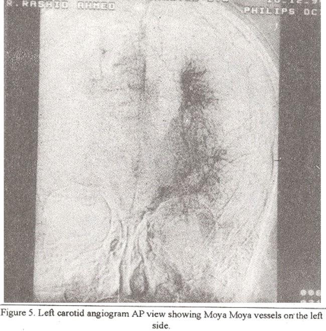
7 One patient showed occlusion of supra clinoid portion of internal carotid artery only on one side (left side). On angiography Moya Moya vessels were seen profusely in one case (Figures 5 and 6).
8

9 Moderate visualization was evident in two cases. In one case Moya Moya vessels were only present on one side. Collateral from meningeal and ophthalmic artery was seen in all cases. Filling ofmoyamoya vessels from posterior circulation was seen in all four cases (Figure 7).

10 Involvement of vertebral artery was seen in three cases, while it was spared in one case. Small aneurysms were seen within the posterior circulation and collateral vessels in two cases (Figure 8).
 from the supra-seller cistern to the basal ganglia 5.")
11 Discussion Moya Moya disease is characterized by progressive narrowing of the bilateral internal carotid bifurcation, followed by development of extensive parenchymal vessels known as MoyaMoyavessels (MMV S) from the supra-seller cistern to the basal ganglia 5. The cause of MoyaMoya disease is still
12 unknown and it occurs most frequently in young patients. Stroke in children occurs either as a known complication of an already established disease such as cardiopathy, neurofibromatosis type I, homocystinurea, meningitis or sickle cell anaemia or occurs in isolation. One of the cases (case No. 3) was a diagnosed case of sickle cell disease. In all other cases no cause for cerebral occlusion was found. In sickle cell disease stroke occurs in 6-9% of the cases. Both small and large vessel occlusion occurs and multiple intracranial anewysm have been reported. The pathogenesis of sickle cell disease causing stroke is controversial. Recently it is thought that degenerative insult initiated by endothelial cell injury from adhesions of sickled RBC is responsible for occlusion. In children the principle clinical symptoms result from brain ischaemia and consist of hemiplegia, monoplegia, paresthesia involuntary movements, headache and convulsions. In adults most common symptom is intracranial hemorrhage either intracerebral or subarachnoid 6. The application of CT to the study of this condition has revealed areas of low density in the basal ganglia and cortices with cerebral atrophy, presence of abnonnal vessels and poor visualization of the proximal portions of the anterior and middle cerebral arteries, The demonstration of these findings by CT is important in making a diagnosis 5. All cases studied by CT in our series showed presence of multiple low density areas in bilateral cerebral hemispheres representing infarctions. MRI was also able to show the Moya Moya vessels in the basal ganglia region in addition to the infarcts and brain atrophy in 3 cases. Cerebral angiography is the only definitive method of diagnosis. The main angiographic findings consist of narrowing or occlusion of the supm clinoid portion of the internal carotid artery with involvement of the anterior and middle cerebral arteries. This is usually. bilateral. In later stages of the disease the posterior communicating, posterior cerebral and basilar arteries become involved and extensive abnormal vessels (Moya Moya vessels) develop at the base of the brain. The individual vessels of Moya Moya vessels cannot be clearly discerned and its extent is vaguely outlined on routine cerebral angiograms, explaining the name MoyaMoya which is Japanese forpuff of smoke or Hazy 7. Collateral circulation develops between external and internal carotid system through branches of middle meningeal and ophthalmic arteries and anterior and middle cerebral arteries. Medullary arteries provide communication between the branches of middle cerebral arteries and MMV (dilated tlialamostriate vessels). MR angiography is a new technology inwhichthevessels canbe imaged with MR signal without the use of contrast agents. Recently there are reports on the role of MR angiography for evaluation of MoyaMoya disease which are vety promising. Diagnosis of Moya Moya disease is most thorough with MRI angiography and MRI imaging. In patients with Moya Moya disease the CT, MRI and Angiographic findings are very specific and they can readily diagnose and evaluate the extent of the disease. Due to its high resolution and contrast MRI was found to be more informative than CT in detection of abnormal vessels (MMV). Angiography has been the most accurate method for detection of Moya disease but as it is an invasive procedure, it has inherent risks. Due to its non- invasive quality, MRI and MR angiograhy are now preferred methods of investigation for MoyaMoya disease. References 1. Kudo, T. Spontaneous occlusion of the circle of Willis. Neurology, 1968 ; 18 : Suzuki, T. and Takaku, A. Cerebrovascular Moya Moya disease showing abnormal net like vessels in base ofbrain. ARCH. Neurol., 1969,20: Traveras, J.M. Multiple progressive intracranial arterial occlusion: A syndrome of children andyoung adults. Am.J,Radiol,, 1969;106: Pecker, LT., Simn. J. and Gyls-Herry, J.F. Nishimoto s disease: Significance of its angiographic appearance. Neuro-radiology. 1973;5 : Takahashi, M., Miyauch, T. and Masayoshi Konada. Computed tomography of Moya Moya disease.
13 Demonstration of occluded arteries and collateralvessels as important diagnostic signs. Radiology, 1980;134: Ichiro Yamada, I., Yoshihare Mat, S. and Sushima. Moya Moya disease comparison of assessment with MR. Angiography verses conventional angiography. Radiology, 1995;196: Takahashi, M Magnification angiography in Moya Moya disease. Radiology, 1980;136:
Although moyamoya disease, a rare cerebrovascular occlusive
 Renal Artery Lesions in Patients With Moyamoya Disease Angiographic Findings Ichiro Yamada, MD; Yoshiro Himeno, MD; Yoshiharu Matsushima, MD; Hitoshi Shibuya, MD Background and Purpose Renal artery lesions
Moyamoya by magnetic resonance imaging scan
 www.edoriumjournals.com CLINICAL IMAGES OPEN ACCESS Moyamoya by magnetic resonance imaging scan Caroline Edward Ayad, Ahmed Alamin Alnoor, Aymen El-Mesallamy ABSTRACT Abstract is not required for Clinical
Moyamoya Syndrome with contra lateral DACA aneurysm: First Case report with review of literature
 Romanian Neurosurgery Volume XXXI Number 3 2017 July-September Article Moyamoya Syndrome with contra lateral DACA aneurysm: First Case report with review of literature Ashish Kumar Dwivedi, Pradeep Kumar,
Moyamoya Disease A Vasculopathy and an Uncommon Cause of Recurrent Cerebrovascular Accidents
 Moyamoya Disease A Vasculopathy and an Uncommon Cause of Recurrent Cerebrovascular Accidents Yasmin S. Hamirani, Md 1 *, Mohammad Valikhani, Md 2, Allison Sweney, Ms Iii 2, Hafsa Khan, Md 2, Mohammad Pathan,
Longitudinal anterior-to-posterior shift of collateral channels in patients with moyamoya disease: an implication for its hemorrhagic onset
 CLINICAL ARTICLE Longitudinal anterior-to-posterior shift of collateral channels in patients with moyamoya disease: an implication for its hemorrhagic onset Shusuke Yamamoto, MD, Satoshi Hori, MD, PhD,
INSTITUTE OF NEUROSURGERY & DEPARTMENT OF PICU
 CEREBRAL BYPASS An Innovative Treatment for Arteritis INSTITUTE OF NEUROSURGERY & DEPARTMENT OF PICU CASE 1 q 1 year old girl -recurrent seizure, right side limb weakness, excessive cry and irritability.
Essentials of Clinical MR, 2 nd edition. 14. Ischemia and Infarction II
 14. Ischemia and Infarction II Lacunar infarcts are small deep parenchymal lesions involving the basal ganglia, internal capsule, thalamus, and brainstem. The vascular supply of these areas includes the
Moyamoya disease is an unusual form of chronic, occlusive
 Angiographic Dilatation and Branch Extension of the Anterior Choroidal and Posterior Communicating Arteries Are Predictors of Hemorrhage in Adult Moyamoya Patients Motohiro Morioka, MD; Jun-Ichiro Hamada,
Diagnosis of Middle Cerebral Artery Occlusion with Transcranial Color-Coded Real-Time Sonography
 Diagnosis of Middle Cerebral Artery Occlusion with Transcranial Color-Coded Real-Time Sonography Kazumi Kimura, Yoichiro Hashimoto, Teruyuki Hirano, Makoto Uchino, and Masayuki Ando PURPOSE: To determine
STROKE - IMAGING. Dr RAJASEKHAR REDDY 2nd Yr P.G. RADIODIAGNOSIS KIMS,Narkatpalli.
 STROKE - IMAGING Dr RAJASEKHAR REDDY 2nd Yr P.G. RADIODIAGNOSIS KIMS,Narkatpalli. STROKE Describes a clinical event that consists of sudden onset of neurological symptoms Types Infarction - occlusion of
Moyamoya disease (MMD) is a chronic, progressive cerebrovascular. Clinical and Angiographic Features and Stroke Types in Adult Moyamoya Disease
 is a chronic, progressive cerebrovascular. Clinical and Angiographic Features and Stroke Types in Adult Moyamoya Disease") ORIGINAL RESEARCH BRAIN Clinical and Angiographic Features and Stroke Types in Adult Moyamoya Disease D.-K. Jang, K.-S. Lee, H.K. Rha, P.-W. Huh, J.-H. Yang, I.S. Park, J.-G. Ahn, J.H. Sung, and Y.-M.
ORIGINAL RESEARCH BRAIN Clinical and Angiographic Features and Stroke Types in Adult Moyamoya Disease D.-K. Jang, K.-S. Lee, H.K. Rha, P.-W. Huh, J.-H. Yang, I.S. Park, J.-G. Ahn, J.H. Sung, and Y.-M.
Moyamoya syndrome associated with cocaine abuse Case report
 Neurosurg Focus 5 (5):Article 7, 1998 Moyamoya syndrome associated with cocaine abuse Case report Marc S. Schwartz, M.D., and R. Michael Scott, M.D. Division of Neurosurgery, Albany Medical College, Albany,
Case Report 1. CTA head. (c) Tele3D Advantage, LLC
 Tele3D Advantage, LLC") Case Report 1 CTA head 1 History 82 YEAR OLD woman with signs and symptoms of increased intra cranial pressure in setting of SAH. CT Brain was performed followed by CT Angiography of head. 2 CT brain Extensive
Case Report 1 CTA head 1 History 82 YEAR OLD woman with signs and symptoms of increased intra cranial pressure in setting of SAH. CT Brain was performed followed by CT Angiography of head. 2 CT brain Extensive
Ruptured aberrant internal carotid artery pseudoaneurysm presenting with spontaneous massive ear bleeding following a single sneeze: a case report
 Case eport JNET 7:312-316, 2013 uptured aberrant internal carotid artery pseudoaneurysm presenting with spontaneous massive ear bleeding following a single sneeze: a case report Seiichiro HIONO 1) Eiichi
PTA 106 Unit 1 Lecture 3
 PTA 106 Unit 1 Lecture 3 The Basics Arteries: Carry blood away from the heart toward tissues. They typically have thicker vessels walls to handle increased pressure. Contain internal and external elastic
The central nervous system
 Sectc.qxd 29/06/99 09:42 Page 81 Section C The central nervous system CNS haemorrhage Subarachnoid haemorrhage Cerebral infarction Brain atrophy Ring enhancing lesions MRI of the pituitary Multiple sclerosis
Original Article Ischemic and hemorrhagic moyamoya disease in adults: CT findings
 Int J Clin Exp Med 2015;8(11):21351-21357 www.ijcem.com /ISSN:1940-5901/IJCEM0009621 Original Article Ischemic and hemorrhagic moyamoya disease in adults: CT findings Anming Xie 1*, Li Luo 2*, Yaojun Ding
CT and MR Imaging in Young Stroke Patients
 CT and MR Imaging in Young Stroke Patients Ashfaq A. Razzaq,Behram A. Khan,Shahid Baig ( Department of Neurology, Aga Khan University Hospital, Karachi. ) Abstract Pages with reference to book, From 66
Basilar artery stenosis with bilateral cerebellar strokes on coumadin
 Qaisar A. Shah, MD Patient Profile 68 years old female with a history of; Basilar artery stenosis with bilateral cerebellar strokes on coumadin Diabetes mellitus Hyperlipidemia Hypertension She developed
Delineation of unruptured cerebral computerized angiotomography
 J Neurosurg57:527-534,1982 Delineation of unruptured cerebral computerized angiotomography aneurysms by SYOJI ASARI, M.D., Tonu SATOH, M.D., MASARU SAKURAI, M.D., YuJI YAMAMOTO, M.D., AND KAZUHIKO SADAMOTO,
Emergency Department Stroke Registry Indicator Specifications 2018 Report Year (07/01/2017 to 06/30/2018 Discharge Dates)
") 2018 Report Year (07/01/2017 to 06/30/2018 Discharge Dates) Summary of Changes I62.9 added to hemorrhagic stroke ICD-10-CM diagnosis code list (table 3) Measure Description Methodology Rationale Measurement
2018 Report Year (07/01/2017 to 06/30/2018 Discharge Dates) Summary of Changes I62.9 added to hemorrhagic stroke ICD-10-CM diagnosis code list (table 3) Measure Description Methodology Rationale Measurement
Overview Blood supply of the brain What is moyamoya disease? > 1
 Moyamoya Disease Overview Moyamoya disease is caused by blocked arteries at the base of the brain. The name "moyamoya" means "puff of smoke" in Japanese and describes the appearance of tiny vessels that
A CASE OF RECURRENT ALTERNATING TRANSIENT HEMIPARESIS Dr. Shunmuga Arumugasamy.S DNB Resident Railway Hospital, Perambur.
 A CASE OF RECURRENT ALTERNATING TRANSIENT HEMIPARESIS Dr. Shunmuga Arumugasamy.S DNB Resident Railway Hospital, Perambur. 6 year old school going child. Apparently normal till 3 yrs when she developed
Occlusion of All Four Extracranial Vessels With Minimal Clinical Symptomatology. Case Report
 Occlusion of All Four Extracranial Vessels With Minimal Clinical Symptomatology. Case Report BY JIRI J. VITEK, M.D., JAMES H. HALSEY, JR., M.D., AND HOLT A. McDOWELL, M.D. Abstract: Occlusion of All Four
CT angiography and its role in the investigation of intracranial haemorrhage
 CT angiography and its role in the investigation of intracranial haemorrhage RD Magazine, 39, 458, 29-30 Dr M Igra Radiology SPR Leeds General Infirmary Dr I Djoukhadar Research fellow Wolfson Molecular
Neuroscience. Journal. Moyamoya disease a review and case illustration. P A L M E T T O H E A L T H Vol. 2 Issue 3 Summer 2016
 Neuroscience P A L M E T T O H E A L T H Vol. 2 Issue 3 Summer 2016 Journal Moyamoya disease a review and case illustration pg. 5 Choroid Plexus Papilloma in adults pg. 8 As physician co-leaders of Palmetto
Ruptured Aneurysm of the Accessory Middle Cerebral Artery Associated with Moyamoya Disease A Case Report
 Case Report 541 Ruptured Aneurysm of the Accessory Middle Cerebral Artery Associated with Moyamoya Disease A Case Report Cheng-Chi Lee, MD; Zhuo-Hao Liu, MD; Shih-Ming Jung 1, MD; Tao-Chieh Yang, MD The
Cerebral Hemodynamic Change in the Child and the Adult With Moyamoya Disease
 272 Cerebral Hemodynamic Change in the Child and the Adult With Moyamoya Disease Yasuo Kuwabara, MD, Yuichi Ichiya, MD, Makoto Otsuka, MD, Takashi Tahara, MD, Ranjan Gunasekera, MD, Kanehiro Hasuo, MD,
Congenital Absence of the Internal Carotid Artery
 Journal of Soonchunhyang Medical Science 16(1) p.09~15 June 2010 9 Congenital bsence of the Internal Carotid rtery Sun Hye Jeong, Hyun Sook Hong, Sung-Il Park, Dae Ho Kim, Hae Kyung Lee Department of Radiology,
Moya Moya syndrome : how to diagnose?
 Moya Moya syndrome : how to diagnose? Poster No.: C-1504 Congress: ECR 2016 Type: Educational Exhibit Authors: A. Cherif, A. Berrich, A. Ben Abdallah, K. KADRI, N. Mama, H. Jemni, K. Tlili; Sousse/TN Keywords:
Multiple Progressive Intracranial Arterial Occlusions
 Multiple Progressive Intracranial Arterial Occlusions BY ANGELINE R. MASTRI, M.D.,* PAUL M. SILVERSTEIN, M.D.,f LAWRENCE GOLD, M.D.,* AND ERIK P. ESELIUS, M.D. Abstract: Multiple Progressive Intracranial
Vivek R. Deshmukh, MD Director, Cerebrovascular and Endovascular Neurosurgery Chairman, Department of Neurosurgery Providence Brain and Spine
 Vivek R. Deshmukh, MD Director, Cerebrovascular and Endovascular Neurosurgery Chairman, Department of Neurosurgery Providence Brain and Spine Institute The Oregon Clinic Disclosure I declare that neither
[(PHY-3a) Initials of MD reviewing films] [(PHY-3b) Initials of 2 nd opinion MD]
![[(PHY-3a) Initials of MD reviewing films] [(PHY-3b) Initials of 2 nd opinion MD]](/thumbs/89/98619893.jpg "[(PHY-3a) Initials of MD reviewing films] [(PHY-3b) Initials of 2 nd opinion MD]") 2015 PHYSICIAN SIGN-OFF (1) STUDY NO (PHY-1) CASE, PER PHYSICIAN REVIEW 1=yes 2=no [strictly meets case definition] (PHY-1a) CASE, IN PHYSICIAN S OPINION 1=yes 2=no (PHY-2) (PHY-3) [based on all available
2015 PHYSICIAN SIGN-OFF (1) STUDY NO (PHY-1) CASE, PER PHYSICIAN REVIEW 1=yes 2=no [strictly meets case definition] (PHY-1a) CASE, IN PHYSICIAN S OPINION 1=yes 2=no (PHY-2) (PHY-3) [based on all available
Bilateral Carotid and Vertebral Rete Mirabile Presenting with a Prominent Anterior Spinal Artery Mimicking a Spinal Dural AV Fistula at MRI
 Case Report http://dx.doi.org/10.3348/kjr.2011.12.6.740 pissn 1229-6929 eissn 2005-8330 Korean J Radiol 2011;12(6):740-744 Bilateral Carotid and Vertebral Rete Mirabile Presenting with a Prominent Anterior
Occlusio Supra Occlusionem: Intracranial Occlusions Following Carotid Thrombosis as Diagnosed by Cerebral Angiography
 Occlusio Supra Occlusionem: Intracranial Occlusions Following Carotid Thrombosis as Diagnosed by Cerebral Angiography BY B. ALBERT RING, M.D. Abstract: Occlusio Supra Occlusionem: Intracranial Occlusions
MMD is a rare cerebrovascular disease first described by
 ORIGINAL RESEARCH M.A. Mogensen P. Karzmark P.D. Zeifert J. Rosenberg M. Marks G.K. Steinberg L.J. Dorfman Neuroradiologic Correlates of Cognitive Impairment in Adult Moyamoya Disease BACKGROUND AND PURPOSE:
Blood Supply. Allen Chung, class of 2013
 Blood Supply Allen Chung, class of 2013 Objectives Understand the importance of the cerebral circulation. Understand stroke and the types of vascular problems that cause it. Understand ischemic penumbra
Hemodynamics in the Anterior Part of the Circle of Willis in Patients with Intracranial Aneurysms : A Study by Cerebral Angiography
 Tohoku J. exp. Med., 1980, 132, 69-73 Hemodynamics in the Anterior Part of the Circle of Willis in Patients with Intracranial Aneurysms : A Study by Cerebral Angiography RYUNGCHAN KWAK, HIROSHI NIIZUMA
Cerebral aneurysms A case study
 August 2001 Cerebral aneurysms A case study Heather L. Hinds, Harvard Medical School Year III Our Patient 57yr old woman History of migraines Presents with persistent headache several months duration different
Carotid artery stenting for long CTO and pseudo occlusion of carotid artery -2 case reports-
 Carotid artery stenting for long CTO and pseudo occlusion of carotid artery -2 case reports- Katsutoshi Takayama, MD, Ph.D Department of Radiology and Interventional Neuroradiology Ishinkai Yao General
Posterior Cerebral Artery Aneurysms with Common Carotid Artery Occlusion: A Report of Two Cases
 Journal of Neuroendovascular Therapy 2017; 11: 371 375 Online March 3, 2017 DOI: 10.5797/jnet.cr.2016-0114 Posterior Cerebral Artery Aneurysms with Common Carotid Artery Occlusion: A Report of Two Cases
Surface Appearance of the Vertebrobasilar Artery Revealed on Basiparallel Anatomic Scanning (BPAS) MR Imaging: Its Role for Brain MR Examination
 MR Imaging: Its Role for Brain MR Examination") AJNR Am J Neuroradiol 26:2508 2513, November/December 2005 Surface Appearance of the Vertebrobasilar Artery Revealed on Basiparallel Anatomic Scanning (BPAS) MR Imaging: Its Role for Brain MR Examination
AJNR Am J Neuroradiol 26:2508 2513, November/December 2005 Surface Appearance of the Vertebrobasilar Artery Revealed on Basiparallel Anatomic Scanning (BPAS) MR Imaging: Its Role for Brain MR Examination
Anatomic Evaluation of the Circle of Willis: MR Angiography versus Intraarterial Digital Subtraction Angiography
 Anatomic Evaluation of the Circle of Willis: MR Angiography versus Intraarterial Digital Subtraction Angiography K. W. Stock, S. Wetzel, E. Kirsch, G. Bongartz, W. Steinbrich, and E. W. Radue PURPOSE:
Brain AVM with Accompanying Venous Aneurysm with Intracerebral and Intraventricular Hemorrhage
 Cronicon OPEN ACCESS EC PAEDIATRICS Case Report Brain AVM with Accompanying Venous Aneurysm with Intracerebral and Intraventricular Hemorrhage Dimitrios Panagopoulos* Neurosurgical Department, University
Moyamoya disease in the midwestern United States
 Neurosurg Focus 5 (5):Article 1, 1998 Moyamoya disease in the midwestern United States Nicholas M. Wetjen, B.S., P. Charles Garell, M.D., Nicholas V. Stence, and Christopher M. Loftus, M.D. Division of
Treatment of Unruptured Vertebral Artery Dissecting Aneurysms
 33 Treatment of Unruptured Vertebral Artery Dissecting Aneurysms Isao NAITO, M.D., Shin TAKATAMA, M.D., Naoko MIYAMOTO, M.D., Hidetoshi SHIMAGUCHI, M.D., and Tomoyuki IWAI, M.D. Department of Neurosurgery,
MOYA Moya disease is a rare idiopathic
 Research Papers Moya Moya Cases Treated with Encephaloduroarteriosynangiosis Parimal Tripathi, Varsha Tripathi, Ronak J. Naik and Jaimin M. Patel From Gujarat Cancer & Research Institute, Ahmedabad; Sterling
Vasculopathie cérébrale après greffe S. VERLHAC Washington 2007
 CEREBRAL VASCULOPATHY OUTCOME AFTER STEM-CELL TRANSPLANTATION FOR SICKLE CELL DISEASE S Verlhac*, F Bernaudin, C Galeotti, M Benkerrou, I Thuret, M de Montalembert, t A Kandem, M Vasile, G Sebag and the
Moyamoya Disease: Comparison of Assessment with 3.0T MR Angiography and MR Imaging versus Conventional
 Moyamoya Disease: Comparison of Assessment with 3.0T MR Angiography and MR Imaging versus Conventional Angiography Poster No.: C-2771 Congress: ECR 2010 Type: Scientific Exhibit Topic: Neuro Authors: Q.
Deborah K. Mann & Jennifer Bash. Coding Documentation and Education Managers
 Deborah K. Mann & Jennifer Bash Coding Documentation and Education Managers OBJECTIVES Review the basics of Diagnostic, CT, & MRI documentation Risk areas in radiology associated with Diagnostic, CT, &
Cryptogenic Enlargement Of Bilateral Superior Ophthalmic Veins
 ISPUB.COM The Internet Journal of Radiology Volume 18 Number 1 Cryptogenic Enlargement Of Bilateral Superior Ophthalmic Veins K Kragha Citation K Kragha. Cryptogenic Enlargement Of Bilateral Superior Ophthalmic
CMS Limitations Guide - Radiology Services
 CMS Limitations Guide - Radiology Services Starting October 1, 2015, CMS will update their existing medical necessity limitations on tests and procedures to correspond to ICD-10 codes. This limitations
Head CT Scan Interpretation: A Five-Step Approach to Seeing Inside the Head Lawrence B. Stack, MD
 Head CT Scan Interpretation: A Five-Step Approach to Seeing Inside the Head Lawrence B. Stack, MD Five Step Approach 1. Adequate study 2. Bone windows 3. Ventricles 4. Quadrigeminal cistern 5. Parenchyma
Brain Attack. Strategies in the Management of Acute Ischemic Stroke: Neuroscience Clerkship. Case Medical Center
 Brain Attack Strategies in the Management of Acute Ischemic Stroke: Neuroscience Clerkship Stroke is a common and devastating disorder Third leading antecedent of death in American men, and second among
NEURO IMAGING 2. Dr. Said Huwaijah Chairman of radiology Dep, Damascus Univercity
 NEURO IMAGING 2 Dr. Said Huwaijah Chairman of radiology Dep, Damascus Univercity I. EPIDURAL HEMATOMA (EDH) LOCATION Seventy to seventy-five percent occur in temporoparietal region. CAUSE Most likely caused
Principles Arteries & Veins of the CNS LO14
 Principles Arteries & Veins of the CNS LO14 14. Identify (on cadaver specimens, models and diagrams) and name the principal arteries and veins of the CNS: Why is it important to understand blood supply
Neuro-Vascular Intervention AAPC Regional Conference Springfield, MA
 Neuro-Vascular Intervention AAPC Regional Conference Springfield, MA October 8, 2010 1 Presented by: David Zielske, MD,CIRCC, CPC H, CCC, CCS, RCC General Recommendations for Physician Dictations State
Reduced Caliber of the Internal Carotid Artery: A Normal Finding with Ipsilateral Absence or Hypoplasia of the A1 Segment
 Reduced Caliber of the Internal Carotid Artery: A Normal Finding with Ipsilateral Absence or Hypoplasia of the A1 Segment Arthur G. Kane, William P. Dillon, A. James Barkovich, David Norman, Christopher
NEURORADIOLOGY Part I
 NEURORADIOLOGY Part I Vörös Erika University of Szeged Department of Radiology SZEGED BRAIN IMAGING METHODS Plain film radiography Ultrasonography (US) Computer tomography (CT) Magnetic resonance imaging
IMAGING IN ACUTE ISCHEMIC STROKE
 IMAGING IN ACUTE ISCHEMIC STROKE Timo Krings MD, PhD, FRCP (C) Professor of Radiology & Surgery Braley Chair of Neuroradiology, Chief and Program Director of Diagnostic and Interventional Neuroradiology;
Acute stroke imaging
 Acute stroke imaging Aims Imaging modalities and differences Why image acute stroke Clinical correlation to imaging appearance What is stroke Classic definition: acute focal injury to the central nervous
Multidetector computed tomographic (CT) angiography : FREQUENTLY ANATOMICAL VARIATIONS OF THE CIRCLE WILLIS ICONOGRAPHIC REVIEW
 angiography : FREQUENTLY ANATOMICAL VARIATIONS OF THE CIRCLE WILLIS ICONOGRAPHIC REVIEW") Multidetector computed tomographic (CT) angiography : FREQUENTLY ANATOMICAL VARIATIONS OF THE CIRCLE WILLIS ICONOGRAPHIC REVIEW Dra. Ximena González Larramendi Dr. Fernando Landó Baison ABSTRACT: Objetives:
Multidetector computed tomographic (CT) angiography : FREQUENTLY ANATOMICAL VARIATIONS OF THE CIRCLE WILLIS ICONOGRAPHIC REVIEW Dra. Ximena González Larramendi Dr. Fernando Landó Baison ABSTRACT: Objetives:
Bilateral Carotid and Vertebral Rete Mirabile Presenting with Subarachnoid Hemorrhage Caused by the Rupture of Spinal Artery Aneurysm
 Tohoku J. Exp. Med., 2013, 230, 205-209 Carotid and Vertebral Rete Mirabile Presenting with SAH 205 Bilateral Carotid and Vertebral Rete Mirabile Presenting with Subarachnoid Hemorrhage Caused by the Rupture
Moyamoya disease presenting as acute onset cortical blindness: a case report
 Romanian Neurosurgery Volume XXX Number 1 2016 January-March Article Moyamoya disease presenting as acute onset cortical blindness: a case report Dudi Maniram, Bansal Rajeev, Srivastava Trilochan, Sardana
Endosaccular aneurysm occlusion with Guglielmi detachable coils for obstructive hydrocephalus caused by a large basilar tip aneurysm Case report
 Neurosurg Focus 7 (4):Article 5, 1999 Endosaccular aneurysm occlusion with Guglielmi detachable coils for obstructive hydrocephalus caused by a large basilar tip aneurysm Case report Akira Watanabe, M.D.,
Recent Advances in Neurology Difficult Cases
 Patient X: History Part 1 Recent Advances in Neurology Difficult Cases Heather J. Fullerton, MD, MAS Professor of Neurology & Pediatrics Director, Pediatric Brain Center Previously healthy 14-year old
Advances in Neuro-Endovascular Care for Acute Stroke
 Advances in Neuro-Endovascular Care for Acute Stroke Ciarán J. Powers, MD, PhD, FAANS Associate Professor Program Director Department of Neurological Surgery Surgical Director Comprehensive Stroke Center
Assessment Of Collateral Pathways In Acute Ischemic Cerebrovascular Stroke Using A Mansour Grading Scale; A New Scale, A Pilot Study
 ISPUB.COM The Internet Journal of Interventional Medicine Volume 3 Number 1 Assessment Of Collateral Pathways In Acute Ischemic Cerebrovascular Stroke Using A Mansour Grading Scale; A New Scale, A Pilot
IMAGING IN ACUTE ISCHEMIC STROKE
 IMAGING IN ACUTE ISCHEMIC STROKE Timo Krings MD, PhD, FRCP (C) Professor of Radiology & Surgery Braley Chair of Neuroradiology, Chief and Program Director of Diagnostic and Interventional Neuroradiology;
Cerebro-vascular stroke
 Cerebro-vascular stroke CT Terminology Hypodense lesion = lesion of lower density than the normal brain tissue Hyperdense lesion = lesion of higher density than normal brain tissue Isodense lesion = lesion
Tutorials. By Dr Sharon Truter
 Tutorials By Dr Sharon Truter To the Tutorials By Dr Sharon Truter What to expect from the Tutorials What to expect from these tutorials Outlines, structure, guided reading, explanations, mnemonics Begin
Acute Complications of Sickle Cell Disease Case Study 5 year old girl with Hemoglobin SS, weakness and slurred speech
 Acute Complications of Sickle Cell Disease Case Study 5 year old girl with Hemoglobin SS, weakness and slurred speech Beatrice E. Gee, MD Medical Director, Sickle Cell and Hematology Program Children s
Any vascular studies performed should be as a result of, or to complement, a thorough patient evaluation and neurological examination.
 National Imaging Associates, Inc. Clinical guidelines NON-INVASIVE CEREBROVASCULAR ARTERIALS TUDIES Original Date: October 2015 Page 1 of 8 FOR CMS (MEDICARE) MEMBERS ONLY CPT4 Codes: Please refer to page
Subtraction CT Angiography with Controlled- Orbit Helical Scanning for Detection of Intracranial Aneurysms
 AJNR Am J Neuroradiol 19:291 295, February 1998 Subtraction CT Angiography with Controlled- Orbit Helical Scanning for Detection of Intracranial Aneurysms Satoshi Imakita, Yoshitaka Onishi, Tokihiro Hashimoto,
Vascular Disorders. Nervous System Disorders (Part B-1) Module 8 -Chapter 14. Cerebrovascular disease S/S 1/9/2013
 Module 8 -Chapter 14. Cerebrovascular disease S/S 1/9/2013") Nervous System Disorders (Part B-1) Module 8 -Chapter 14 Overview ACUTE NEUROLOGIC DISORDERS Vascular Disorders Infections/Inflammation/Toxins Metabolic, Endocrinologic, Nutritional, Toxic Neoplastic Traumatic
Nervous System Disorders (Part B-1) Module 8 -Chapter 14 Overview ACUTE NEUROLOGIC DISORDERS Vascular Disorders Infections/Inflammation/Toxins Metabolic, Endocrinologic, Nutritional, Toxic Neoplastic Traumatic
Collateral Circulation of the Brain. -With Special Reference to Atherosclerosis of the. Major Cervical and Cerebral Arteries- Masakuni Kameyama
 Collateral Circulation of the Brain -With Special Reference to Atherosclerosis of the Major Cervical and Cerebral Arteries- Masakuni Kameyama The Third Department of Internal Medicine, Faculty of Medicine,
Al Am een J Med Sci 2016; 9(2): US National Library of Medicine enlisted journal ISSN
: US National Library of Medicine enlisted journal ISSN") Al Am een J Med Sci 2016; 9(2):101-106 US National Library of Medicine enlisted journal ISSN 0974-1143 ORIGI NAL ARTICLE C O D E N : A A J MB G Cerebrovascular ischemic changes associated with fetal posterior
Al Am een J Med Sci 2016; 9(2):101-106 US National Library of Medicine enlisted journal ISSN 0974-1143 ORIGI NAL ARTICLE C O D E N : A A J MB G Cerebrovascular ischemic changes associated with fetal posterior
lek Magdalena Puławska-Stalmach
 lek Magdalena Puławska-Stalmach tytuł pracy: Kliniczne i radiologiczne aspekty tętniaków wewnątrzczaszkowych a wybór metody leczenia Summary An aneurysm is a localized, abnormal distended lumen of the
A Case of Carotid-Cavernous Fistula
 A Case of Carotid-Cavernous Fistula By : Mohamed Elkhawaga 2 nd Year Resident of Ophthalmology Alexandria University A 19 year old male patient came to our outpatient clinic, complaining of : -Severe conjunctival
ISCHEMIC STROKE IMAGING
 ISCHEMIC STROKE IMAGING ผศ.พญ พญ.จ ร ร ตน ธรรมโรจน ภาคว ชาร งส ว ทยา คณะแพทยศาสตร มหาว ทยาล ยขอนแก น A case of acute hemiplegia Which side is the abnormality, right or left? Early Right MCA infarction
2D Cine Phase-Contrast MRI for Volume Flow Evaluation of the Brain-Supplying Circulation in Moyamoya Disease
 MRI in Moyamoya Disease Neuroradiology Original Research A C D E M N E U T R Y L I A M C A I G O F I N G K. Wolfgang Neff 1 Peter Horn 2 Peter Schmiedek 2 Christoph Düber 1 Dietmar J. Dinter 1 Neff KW,
In cerebral embolism, recanaiization occurs very
 680 Case Reports Recanaiization of Intracranial Carotid Occlusion Detected by Duplex Carotid Sonography Haruhiko Hoshino, MD, Makoto Takagi, MD, Ikuo Takeuchi, MD, Tsugio Akutsu, MD, Yasuyuki Takagi, MD,
Comparison of Five Major Recent Endovascular Treatment Trials
 Comparison of Five Major Recent Endovascular Treatment Trials Sample size 500 # sites 70 (100 planned) 316 (500 planned) 196 (833 estimated) 206 (690 planned) 16 10 22 39 4 Treatment contrasts Baseline
HAAD quality KPI; waiting time
 Type: Waiting Time Indicator Indicator Number: WT001 Primary Care Appointment- Outpatient Setting Time to see a HAAD licensed family physician or member of their team (GP) Time of request (walk-in or by
IV. Cerebrovascular diseases
 IV. Cerebrovascular diseases - Cerebrovascular disease denotes brain disorders caused by pathologic processes involving the blood vessels. - The three main pathogenic mechanisms are: 1. Thrombotic occlusion
JAWDA Guidelines for Pre-hospital Emergency Medical Service (EMS)
") JAWDA Guidelines for Pre-hospital Emergency Medical Service (EMS) January 2019 Page 1 of 17 Table of Contents Executive Summary... 3 About this Guidance... 4 Emergency Medical Service Performance Indicators...
JAWDA Guidelines for Pre-hospital Emergency Medical Service (EMS) January 2019 Page 1 of 17 Table of Contents Executive Summary... 3 About this Guidance... 4 Emergency Medical Service Performance Indicators...
Spontaneous Recanalization after Complete Occlusion of the Common Carotid Artery with Subsequent Embolic Ischemic Stroke
 Original Contribution Spontaneous Recanalization after Complete Occlusion of the Common Carotid Artery with Subsequent Embolic Ischemic Stroke Abstract Introduction: Acute carotid artery occlusion carries
/ / / / / / Hospital Abstraction: Stroke/TIA. Participant ID: Hospital Code: Multi-Ethnic Study of Atherosclerosis
 Multi-Ethnic Study of Atherosclerosis Participant ID: Hospital Code: Hospital Abstraction: Stroke/TIA History and Hospital Record 1. Was the participant hospitalized as an immediate consequence of this
History of revascularization
 History of revascularization Author (year) Kredel, 1942 Woringer& Kunlin, 1963 Donaghy& Yasargil, 1968 Loughheed 1971 Kikuchini & Karasawa1973 Karasawa, 1977 Story, 1978 Sundt, 1982 EC/IC bypass study
Table 1.Summary of 12 Patients with Brain Death and Deep Coma: Clinical Findings Patients No. Age/Sex Underlying Cause Study No.
 3 9 5 Table 1.Summary of 12 Patients with Brain Death and Deep Coma: Clinical Findings Patients No. Age/Sex Underlying Cause Study No. Brain Stem Reflex EEG Clinical Diagnosis 01 40/M Trauma 01 ECS Brain
Enhancement of Cranial US: Utility of Supplementary Acoustic Windows and Doppler Harriet J. Paltiel, MD
 Enhancement of Cranial US: Utility of Supplementary Acoustic Windows and Doppler Harriet J. Paltiel, MD Boston Children s Hospital Harvard Medical School None Disclosures Conventional US Anterior fontanelle
Computerised axial tomography in patients with severe migraine: a preliminary report
 Journal ofneurology, Neurosurgery, and Psychiatry, 1976, 39, 990-994 Computerised axial tomography in patients with severe migraine: a preliminary report G. D. HUNGERFORD', G. H. du BOULAY2, AND K. J.
SAMPLE EDITION PELVIC AND LOWER EXTREMITY ARTERIES WITH ENDOVASCULAR REVASCULARIZATION. Cardiovascular Illustrations and Guidelines
 Cardiovascular Illustrations and Guidelines PELVIC AND LOWER EXTREMITY ARTERIES WITH ENDOVASCULAR REVASCULARIZATION ANGIOPLASTY INTRAVASCULAR STENT PLACEMENT ATHERECTOMY For Fem-Pop Territory Angioplasty
Diagnosis of Subarachnoid Hemorrhage (SAH) and Non- Aneurysmal Causes
 and Non- Aneurysmal Causes") Diagnosis of Subarachnoid Hemorrhage (SAH) and Non- Aneurysmal Causes By Sheila Smith, MD Swedish Medical Center 1 Disclosures I have no disclosures 2 Course Objectives Review significance and differential
Diagnosis of Subarachnoid Hemorrhage (SAH) and Non- Aneurysmal Causes By Sheila Smith, MD Swedish Medical Center 1 Disclosures I have no disclosures 2 Course Objectives Review significance and differential
Overview of imaging modalities for cerebral aneurysms
 Overview of imaging modalities for cerebral aneurysms Soroush Zaghi BIDMC PCE: Radiology August 2008 (Images from BIDMC, PACS.) Our Patient: Presentation Our patient is a 57 y/o woman who reports blowing
Cerebral Vascular Diseases. Nabila Hamdi MD, PhD
 Cerebral Vascular Diseases Nabila Hamdi MD, PhD Outline I. Stroke statistics II. Cerebral circulation III. Clinical symptoms of stroke IV. Pathogenesis of cerebral infarcts (Stroke) 1. Ischemic - Thrombotic
CEREBROVASCULAR DISEASES. By: Shifaa AlQa qa
 CEREBROVASCULAR DISEASES By: Shifaa AlQa qa Cerebrovascular diseases Brain disorders caused by pathologic processes involving blood vessels 3 pathogenic mechanisms (1) thrombotic occlusion, (2) embolic
Brain Arteriovenous Malformations Endovascular Therapy and Associated Therapeutic Protocols Jorge Guedes Cabral de Campos
 Endovascular Therapy and Associated Therapeutic Protocols Jorge Guedes Cabral de Campos Neuroradiology Department Hospital de Santa Maria University of Lisbon CEREBRAL AVM CLINICAL / EPIDEMIOLOGY Brain
Bilateral blunt carotid artery injury: A case report and review of the literature
 CASE REPORT Bilateral blunt carotid artery injury: A case report and review of the literature S Cheddie, 1 MMed (Surg), FCS (SA); B Pillay, 2 FCS (SA), Cert Vascular Surgery; R Goga, 2 FCS (SA) 1 Department
Attenuation value in HU From -500 To HU From -10 To HU From 60 To 90 HU. From 200 HU and above
 Brain Imaging Common CT attenuation values Structure Air Fat Water Brain tissue Recent hematoma Calcifications Bone Brain edema and infarction Normal liver parenchyma Attenuation value in HU From -500