Velocity, strain and strain rate: Doppler and Non-Doppler methods. Thoraxcentre, Erasmus MC,Rotterdam
|
|
|
- Stanley Robbins
- 5 years ago
- Views:
Transcription
1 Velocity, strain and strain rate: Doppler and Non-Doppler methods J Roelandt J. Roelandt Thoraxcentre, Erasmus MC,Rotterdam
2 Basics of tissue Doppler imaging
3 Instantaneous annular velocity profiles IVCT IVRT Sa ejection filling Ea Aa
4 Instantaneous myocardial velocity profiles Color Doppler myocardial imaging Hatle L Pulsed Doppler Color M-mode

5 Validation of tissue Doppler velocities against MRI Ajmone N et al, Heart 2009
6 Pulsed-wave vs color-coded tissue Doppler imaging
7 Clinical role of TDI(TVI) Annular velocities: Complement assessment of global function - Reduced E-velocity indicates abnormal relaxation - Mitral E/e : estimation of LV filling pressure

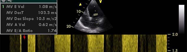
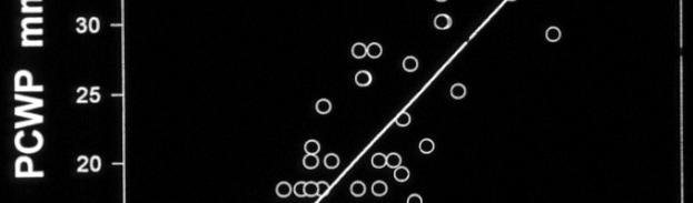
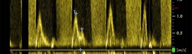

8 LV filling pressure estimation E: 108 Nagueh et al, 1997 e : e: 4 E/e : 27
9 Risk stratification in chronic systolic heart ffailure Transmitral flow and MV annulus velocity Dini L. Frank, Eur J Echocardiogr 2009
10 Role of TDI(TVI) in LV Function assessment Annular velocities Complements assessment of global l function - Reduced E-velocity indicates abnormal relaxation - Mitral E / e : estimation of LV filling pressure Myocardial velocities, strain, strainraterate Essential for assessment of regional function - Ischemia - Hypertrophy - Cardiomyopathies JR/1353
11 Color-coded TDI and dyssynchrony imaging
12 Myocardial strain (stretching): deformation imaging X1 Y1 Y2 depth L1 (10 mm) L2 (14 mm) L2 - L1 x 100 = Strain (in %) L1 lengthening
13 Myocardial strain rate expressed as a velocity gradient X1 L1 Y1 t1 depth V1 V2 t2 X2 Y2 depth Strain (%) = L2 L2 -L1 (y2 - x2)-(y1 - x1) (y2 - y1)-(x2 - x1) = = L1 (y1 - x1) (y1 - x1) Strain rate (sec 1 ) = (y2 - y1)/dt - (x2 - x1)/dt V2 - V1 = (y1 - x1) L1
14 Strain rate imaging L1 V2 V1 SR = SR = V2 - V1 V2 - V1 SR Local gradient of velocity (cm/s.cm -1 L1= sec -1 ) Voigt J et al g L1


15 Definition of strain rate Compression = shortening = contraction No change Stretching = lengthening = relaxation
16 Doppler measurement of regional velocities Spatial velocity gradient Temporal integral Strain rate Strain
17 All tissue Doppler parameters are derived from myocardial velocities Motion Deformation
18 Strain and strain rate measurements by Doppler Only velocity vectors along the interrogating beam Longitudinal lengthening and shortening Radial thickening and thinning Circumferential lengthening and shortening
19 Strain/strain rate measurement Problems with Doppler methods Poor inter-observer & inter-study reproducibility Acquisition highly operator dependent Dependence on high frame rates No automatic tracking of sampled site Off-line processing difficult interpretation One-dimensional: velocities along sound beam Solution: 2D and speckle tracking!

 in the next frame, the motion of the kernel can be tracked from frame to frame")
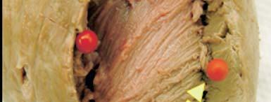
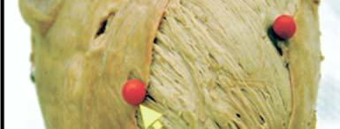
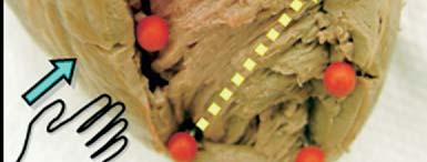
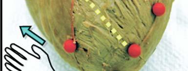
20 Speckle pattern The randomness of the speckle pattern ensures that each region of the myocardium has its own rather unique speckle pattern, that can helps differentiating one region from another The speckle pattern remains relatively stable, and speckles follow myocardial motion. By defining a region of speckles(kernels) in one frame and identifying a similar By defining a region of speckles(kernels) in one frame and identifying a similar kernel (with the same size and shape) in the next frame, the motion of the kernel can be tracked from frame to frame
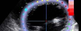
21 Speckle tracking natural acoustic markers Leitman M, J Am Soc Echocardiogr 2004; 17:
22 Experimental validation of speckle tracking Baseline Apical Basal Dobutamine Apical Basal Acute ischemia Apical Basal echocardio ography (º º) Ro otation by 10º 5º -10º -5º 5º 10º -5º r = 0.97 p < y = 0.92x º Rotation by sonomicrometry (º) Helle-Valle T et al, Circulation 2005; 112:
23 Improvements as compared to current techniques Amundsen et al. European Journal of Echocardiography 2009;


24 LONGITUDINAL STRAIN RADIAL STRAIN
25 Speckle tracking: radial and circumferential strain

26 RADIAL STRAIN CIRCUMFERENTIAL STRAIN
27


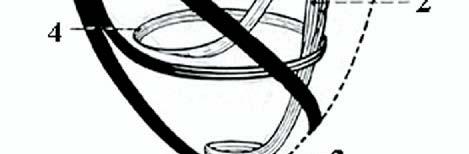
28 Helices in the heart CW CCW Sengupta et al. J Am Soc Echocardiogr 2007; 20:
29 Helices in the heart Endocard 15% fiber shortening of each myocyte Myocard circumferential spiral Epicard EF 30%
30 Nomenclature CW Rotation ( ) -3 LV Twist ( ) ((difference in rotation) distance (mm) CCW Rotation ti ( ) +8 LV Torsion ( /mm) (twist normalized for distance)


31 Basal and apical LV rotation Basal clockwise rotation Apical counterclockwise rotation
32 LV rotation and ageing LV rotatio on, deg grees 10 apical >55 years years <35 years msec basal Van Dalen et al. Am J Physiol 2008; 295: H
33 New parameters - LV twist and untwist - pathology Aortic stenosis Van Dalen et al. Int J Cardiol. 2010; in press
34 Clinical applications of LV twist 8 LV Rotation in Controls o 4 LV base LV apex LV Rotation in DCM LV base LV apex 8 LV Rotation in NCCM LV base LV apex
35 Clinical applications of LV twist Basal clockwise rotation Apical counterclockwise rotation
36 Non-Doppler (speckle tracking) velocity, strain/strain rate measurement 2D on-line frame-by-frame cross-correlation Radio-frequency data; angle-independent Less temporal resolution higher heart rates Less noise, improved quantification Automated analysis of strain and strain rate and displacement in all myocardial segments Longitudinal & radial & circumferential function
37 Non-Doppler (speckle tracking) velocity, strain/strain rate measurement Clinical indications Whenever segmental function is important e.g. All established coronary syndromes Whenever strain or strain rate is more sensitive s e than myocardial a velocities es e.g. Cardiomyopathies (HCM, amyloid, Systemic sclerosis.) Choose diagnostic modality based on available clinical outcome data in future
38 Many thanks!
How To Perform Strain Imaging; Step By Step Approach. Maryam Bo Khamseen Echotechnoligist II EACVI, ARDMS, RCS King Abdulaziz Cardiac Center- Riyadh
 How To Perform Strain Imaging; Step By Step Approach Maryam Bo Khamseen Echotechnoligist II EACVI, ARDMS, RCS King Abdulaziz Cardiac Center- Riyadh Outlines: Introduction Describe the basic of myocardium
2/2/2011. Strain and Strain Rate Imaging How, Why and When? Movement vs Deformation. Doppler Myocardial Velocities. Movement. Deformation.
 Strain and Strain Rate Imaging How, Why and When? João L. Cavalcante, MD Advanced Cardiac Imaging Fellow Cleveland Clinic Foundation Disclosures: No conflicts of interest Movement vs Deformation Movement
Strain and Strain Rate Imaging How, Why and When?
 Strain and Strain Rate Imaging How, Why and When? João L. Cavalcante, MD Advanced Cardiac Imaging Fellow Cleveland Clinic Foundation Disclosures: No conflicts of interest Movement vs Deformation Movement
Strain/Untwisting/Diastolic Suction
 What Is Diastole and How to Assess It? Strain/Untwisting/Diastolic Suction James D. Thomas, M.D., F.A.C.C. Cardiovascular Imaging Center Department of Cardiology Cleveland Clinic Foundation Cleveland,
DISCLOSURE. Myocardial Mechanics. Relevant Financial Relationship(s) Off Label Usage
 Off Label Usage") 7th Annual Team Echocardiography: The Heart of Cardiovascular Medicine Tissue Doppler, Strain, Speckle: What? How? Christopher J Kramer RDCS Aurora Medical Group Advanced Cardiovascular Services, Aurora
7th Annual Team Echocardiography: The Heart of Cardiovascular Medicine Tissue Doppler, Strain, Speckle: What? How? Christopher J Kramer RDCS Aurora Medical Group Advanced Cardiovascular Services, Aurora
22 nd Annual Conference of the Saudi Heart Association Riyadh, Saudi Arabia
 22 nd Annual Conference of the Saudi Heart Association Riyadh, Saudi Arabia New Echocardiographic Modalities to Evaluate Ventricular Function in Congenital Heart Disease: Tissue Doppler & Strain Rate Imaging
Tissue Doppler and Strain Imaging. Steven J. Lester MD, FRCP(C), FACC, FASE
, FACC, FASE") Tissue Doppler and Strain Imaging Steven J. Lester MD, FRCP(C), FACC, FASE Relevant Financial Relationship(s) None Off Label Usage None a. Turn the wall filters on and turn down the receiver gain. b. Turn
Tissue Doppler and Strain Imaging Steven J. Lester MD, FRCP(C), FACC, FASE Relevant Financial Relationship(s) None Off Label Usage None a. Turn the wall filters on and turn down the receiver gain. b. Turn
Tissue Doppler Imaging in Congenital Heart Disease
 Tissue Doppler Imaging in Congenital Heart Disease L. Youngmin Eun, M.D. Department of Pediatrics, Division of Pediatric Cardiology, Kwandong University College of Medicine The potential advantage of ultrasound
Tissue Doppler and Strain Imaging
 Tissue Doppler and Strain Imaging Steven J. Lester MD, FRCP(C), FACC, FASE Relevant Financial Relationship(s) None Off Label Usage None 1 Objective way with which to quantify the minor amplitude and temporal
Nancy Goldman Cutler, MD Beaumont Children s Hospital Royal Oak, Mi
 Nancy Goldman Cutler, MD Beaumont Children s Hospital Royal Oak, Mi Identify increased LV wall thickness (WT) Understand increased WT in athletes Understand hypertrophic cardiomyopathy (HCM) Enhance understanding
LV FUNCTION ASSESSMENT: WHAT IS BEYOND EJECTION FRACTION
 LV FUNCTION ASSESSMENT: WHAT IS BEYOND EJECTION FRACTION Jamilah S AlRahimi Assistant Professor, KSU-HS Consultant Noninvasive Cardiology KFCC, MNGHA-WR Introduction LV function assessment in Heart Failure:
Tissue Doppler and Strain Imaging
 Tissue Doppler and Strain Imaging Steven J. Lester MD, FRCP(C), FACC, FASE Relevant Financial Relationship(s) None Off Label Usage None 1 Objective way with which to quantify the minor amplitude and temporal
Advanced Multi-Layer Speckle Strain Permits Transmural Myocardial Function Analysis in Health and Disease:
 Advanced Multi-Layer Speckle Strain Permits Transmural Myocardial Function Analysis in Health and Disease: Clinical Case Examples Jeffrey C. Hill, BS, RDCS Echocardiography Laboratory, University of Massachusetts
Diastology Disclosures: None. Dias2011:1
 Diastology 2011 James D. Thomas, M.D., F.A.C.C. Cardiovascular Imaging Center Department of Cardiology Cleveland Clinic Foundation Cleveland, Ohio, USA Disclosures: None Dias2011:1 Is EVERYBODY a member!?!
We are IntechOpen, the world s leading publisher of Open Access books Built by scientists, for scientists. International authors and editors
 We are IntechOpen, the world s leading publisher of Open Access books Built by scientists, for scientists 4,100 116,000 120M Open access books available International authors and editors Downloads Our
Myocardial Strain Imaging in Cardiac Diseases and Cardiomyopathies.
 Myocardial Strain Imaging in Cardiac Diseases and Cardiomyopathies. Session: Cardiomyopathy Tarun Pandey MD, FRCR. Associate Professor University of Arkansas for Medical Sciences Disclosures No relevant
Three-dimensional Wall Motion Tracking:
 Three-dimensional Wall Motion Tracking: A Novel Echocardiographic Method for the Assessment of Ventricular Volumes, Strain and Dyssynchrony Jeffrey C. Hill, BS, RDCS, FASE Jennifer L. Kane, RCS Gerard
Novel echocardiographic modalities: 3D echo, speckle tracking and strain rate imaging. Potential roles in sports cardiology. Stefano Caselli, MD, PhD
 Novel echocardiographic modalities: 3D echo, speckle tracking and strain rate imaging. Potential roles in sports cardiology. Stefano Caselli, MD, PhD Ospedale San Pietro Fatebenefratelli Rome, Italy Differential
Incorporating the New Echo Guidelines Into Everyday Practice
 Incorporating the New Echo Guidelines Into Everyday Practice Clinical Case RIGHT VENTRICULAR FAILURE Gustavo Restrepo MD President Elect Interamerican Society of Cardiology Director Fellowship Training
Left Ventricular Dyssynchrony in Patients Showing Diastolic Dysfunction without Overt Symptoms of Heart Failure
 ORIGINAL ARTICLE DOI: 10.3904/kjim.2010.25.3.246 Left Ventricular Dyssynchrony in Patients Showing Diastolic Dysfunction without Overt Symptoms of Heart Failure Jae Hoon Kim, Hee Sang Jang, Byung Seok
Cardiac Chamber Quantification by Echocardiography
 Cardiac Chamber Quantification by Echocardiography Maryam Bokhamseen, RCS, RCDS, EACVI Echotechnologist ǁ, Non invasive Cardiac Laboratory King Abdulaziz Cardiac Center. Outline: Introduction. Background
3D-stress echocardiography Bernard Cosyns, MD, PhD
 3D-stress echocardiography Bernard Cosyns, MD, PhD No Disclosure The Pro-Technology bias Sicari et al. Cardiovascular Ultrasound 2006, 4:11 Overview 2D stress echocardiography: main limitations 3D echocardiography:
DECLARATION OF CONFLICT OF INTEREST. None
 DECLARATION OF CONFLICT OF INTEREST None Hot Topics in Echocardiography: The position of the EAE EAE / ASE recommendation about Echo Assessment of Cardiac Mechanics Jens-Uwe Voigt Dpt. of Cardiovascular
VECTORS OF CONTRACTION
 1/3/216 Strain, Strain Rate, and Torsion: Myocardial Mechanics Simplified and Applied VECTORS OF CONTRACTION John Gorcsan, MD University of Pittsburgh, Pittsburgh, PA Shortening Thickening Twisting No
The rapid evolution of echocardiography during the past 25 years
 Evaluation of Myocardial Mechanics in the Fetus by Velocity Vector Imaging Adel K. Younoszai, MD, David E. Saudek, MD, Stephen P. Emery, MD, and James D. Thomas, MD, Denver, Colorado; Cleveland, Ohio;
Heart disease and left ventricular rotation a systematic review and quantitative summary
 Phillips et al. BMC Cardiovascular Disorders 2012, 12:46 RESEARCH ARTICLE Heart disease and left ventricular rotation a systematic review and quantitative summary Aaron A Phillips 1,2, Anita T Cote 2,
MYOCARDIAL DEFORMATION IMAGING ON EXERCISE IN CHRONIC PRIMARY MITRAL REGURGITATION
 MYOCARDIAL DEFORMATION IMAGING ON EXERCISE IN CHRONIC PRIMARY MITRAL REGURGITATION A thesis submitted to the University of Manchester for the degree of Doctor of Medicine in the Faculty of Medical and
RIGHT VENTRICULAR SIZE AND FUNCTION
 RIGHT VENTRICULAR SIZE AND FUNCTION Edwin S. Tucay, MD, FPCC, FPCC, FPSE Philippine Society of Echocardiography Quezon City, Philippines Echo Mission, BRTTH, Legaspi City, July 1-2, 2016 NO DISCLOSURE
Strain imaging in children: from Tissue Doppler to 3 D
 Strain imaging in children: from Tissue Doppler to 3 D Mark kk. Friedberg Fi Outline Deformation in the fetus and neonate Deformation in pediatric cardiomyopathy y (briefly!) Deformation in Congenital
Tissue Doppler Imaging
 Cronicon OPEN ACCESS Hesham Rashid* Tissue Doppler Imaging CARDIOLOGY Editorial Department of Cardiology, Benha University, Egypt *Corresponding Author: Hesham Rashid, Department of Cardiology, Benha University,
HYPERTROPHY: Behind the curtain. V. Yotova St. Radboud Medical University Center, Nijmegen
 HYPERTROPHY: Behind the curtain V. Yotova St. Radboud Medical University Center, Nijmegen Disclosure of interest: none Relative wall thickness (cm) M 0.22 0.42 0.43 0.47 0.48 0.52 0.53 F 0.24 0.42 0.43
Strain rate imaging: fundamental principles and progress so far
 review Strain rate imaging: fundamental principles and progress so far Echocardiography remains the modality of choice for diagnosing heart disease in routine clinical practice. However, to date, the clinical
Diastolic Function: What the Sonographer Needs to Know. Echocardiographic Assessment of Diastolic Function: Basic Concepts 2/8/2012
 Diastolic Function: What the Sonographer Needs to Know Pat Bailey, RDCS, FASE Technical Director Beaumont Health System Echocardiographic Assessment of Diastolic Function: Basic Concepts Practical Hints
An Integrated Approach to Study LV Diastolic Function
 An Integrated Approach to Study LV Diastolic Function Assoc. Prof. Adriana Ilieşiu, FESC University of Medicine Carol Davila Bucharest, Romania LV Diastolic Dysfunction impaired relaxation (early diastole)
Alicia Armour, MA, BS, RDCS
 Alicia Armour, MA, BS, RDCS No disclosures Review 2D Speckle Strain (briefly) Discuss some various patient populations & disease pathways where Strain can be helpful Discuss how to acquire images for Strain
10/7/2013. Systolic Function How to Measure, How Accurate is Echo, Role of Contrast. Thanks to our Course Director: Neil J.
 Systolic Function How to Measure, How Accurate is Echo, Role of Contrast Neil J. Weissman, MD MedStar Health Research Institute & Professor of Medicine Georgetown University Washington, D.C. No Disclosures
Characteristics of Myocardial Deformation and Rotation in Subjects With Diastolic Dysfunction Without Diastolic Heart Failure
 ORIGINAL ARTICLE DOI.4070 / kcj.09.39.12.532 Print ISSN 1738-55 / On-line ISSN 1738-5555 Copyright c 09 The Korean Society of Cardiology Open Access Characteristics of Myocardial Deformation and Rotation
Evaluation of Left Ventricular Diastolic Dysfunction by Doppler and 2D Speckle-tracking Imaging in Patients with Primary Pulmonary Hypertension
 ESC Congress 2011.No 85975 Evaluation of Left Ventricular Diastolic Dysfunction by Doppler and 2D Speckle-tracking Imaging in Patients with Primary Pulmonary Hypertension Second Department of Internal
We are IntechOpen, the world s leading publisher of Open Access books Built by scientists, for scientists. International authors and editors
 We are IntechOpen, the world s leading publisher of Open Access books Built by scientists, for scientists 4,000 116,000 120M Open access books available International authors and editors Downloads Our
MYOCARDIAL STRAIN MEASUREMENTS WITH MRI USING FEATURE TRACKING
 MYOCARDIAL STRAIN MEASUREMENTS WITH MRI USING FEATURE TRACKING Mercy Kataike Victoria MSc in Physics Submission date: May 2016 Supervisor: Pål Erik Goa, IFY Norwegian University of Science and Technology
The Value of 2D Strain Imaging during Stress Testing
 The Value of 2D Strain Imaging during Stress Testing Marie Moonen, M.D., Patrizio Lancellotti, M.D., Ph.D., Dimitrios Zacharakis, M.D., and Luc Pierard, M.D., Ph.D., F.E.S.C., F.A.C.C. CHU Sart Tilman,
L ecocardiografia nello Scompenso Cardiaco Acuto e cronico: vecchi dogmi e nuovi trends.
 V SESSIONE SCOMPENSO CARDIACO 2015 Genova, 13-14 Novembre 2015 L ecocardiografia nello Scompenso Cardiaco Acuto e cronico: vecchi dogmi e nuovi trends. Gian Paolo Bezante, MD, FACC UOC Clinica di Malattie
Aortic valve Stenosis: Insights in the evaluation of LV function. Erwan DONAL Cardiologie CHU Rennes
 Aortic valve Stenosis: Insights in the evaluation of LV function Erwan DONAL Cardiologie CHU Rennes erwan.donal@chu-rennes.fr Preload Afterload Myocardial Fiber Shortening Circumferential Longitudinal
Echo assessment of the failing heart
 Echo assessment of the failing heart Mark K. Friedberg, MD The Labatt Family Heart Center The Hospital for Sick Children Toronto, Ontario, Canada Cardiac function- definitions Cardiovascular function:
Carlos Eduardo Suaide Silva, Luiz Darcy Cortez Ferreira, Luciana Braz Peixoto, Claudia Gianini Monaco, Manuel Adán Gil, Juarez Ortiz
 Silva et al Original Article Arq Bras Cardiol Study of the Myocardial Contraction and Relaxation Velocities through Doppler Tissue Imaging Echocardiography. A New Alternative in the Assessment of the Segmental
Implementing New Technology
 Implementing New Technology PP16 Imaging Conference Bicol Hospital, Legaspi City, Philippines July 2016 David Adams, ACS, RCS, RDCS, FASE Duke University Medical Center Echo Evolution By Dr. Zoghbi Handheld/POC
Evalua&on)of)Le-)Ventricular)Diastolic) Dysfunc&on)by)Echocardiography:) Role)of)Ejec&on)Frac&on)
of)Le-)Ventricular)Diastolic) Dysfunc&on)by)Echocardiography:) Role)of)Ejec&on)Frac&on)") Evalua&on)of)Le-)Ventricular)Diastolic) Dysfunc&on)by)Echocardiography:) Role)of)Ejec&on)Frac&on) N.Koutsogiannis) Department)of)Cardiology) University)Hospital)of)Patras)! I have no conflicts of interest
Evalua&on)of)Le-)Ventricular)Diastolic) Dysfunc&on)by)Echocardiography:) Role)of)Ejec&on)Frac&on) N.Koutsogiannis) Department)of)Cardiology) University)Hospital)of)Patras)! I have no conflicts of interest
Fetal cardiac function: what to use and does it make a difference?
 17 th International Conference on Prenatal Diagnosis and Therapy Lisbon, June 2013 Fetal cardiac function: what to use and does it make a difference? Fàtima Crispi Department of Maternal-Fetal Medicine,
Altered left ventricular geometry and torsional mechanics in high altitude-induced pulmonary hypertension:
 Altered left ventricular geometry and torsional mechanics in high altitude-induced pulmonary hypertension: a 3-D echocardiographic study B.W. De Boeck,* S. Kiencke, C. Dehnert, K. Auinger, # M. Maggiorini,
Diastole is Not a Single Entity Four Components of Diastolic Dysfunction
 Physiology of Diastolic Function Made Easy James D. Thomas, MD, FACC, FASE Director, Center for Heart Valve Disease Bluhm Cardiovascular Institute Professor of Medicine, Feinberg School of Medicine, Northwestern
DOPPLER HEMODYNAMICS (1) QUANTIFICATION OF PRESSURE GRADIENTS and INTRACARDIAC PRESSURES
 QUANTIFICATION OF PRESSURE GRADIENTS and INTRACARDIAC PRESSURES") THORAXCENTRE DOPPLER HEMODYNAMICS (1) QUANTIFICATION OF PRESSURE GRADIENTS and INTRACARDIAC PRESSURES J. Roelandt DOPPLER HEMODYNAMICS Intracardiac pressures and pressure gradients Volumetric measurement
THORAXCENTRE DOPPLER HEMODYNAMICS (1) QUANTIFICATION OF PRESSURE GRADIENTS and INTRACARDIAC PRESSURES J. Roelandt DOPPLER HEMODYNAMICS Intracardiac pressures and pressure gradients Volumetric measurement
Outline. EuroScore II. Society of Thoracic Surgeons Score. EuroScore II
 SURGICAL RISK IN VALVULAR HEART DISEASE: WHAT 2D AND 3D ECHO CAN TELL YOU AND WHAT THEY CAN'T Ernesto E Salcedo, MD Professor of Medicine University of Colorado School of Medicine Director of Echocardiography
Acute impairment of basal left ventricular rotation but not twist and untwist are involved in the pathogenesis of acute hypertensive pulmonary oedema
 Acute impairment of basal left ventricular rotation but not twist and untwist are involved in the pathogenesis of acute hypertensive pulmonary oedema A.D. Margulescu 1,2, R.C. Sisu 1,2, M. Florescu 2,
Assessing Function by Echocardiography in VHD Asymptomatic Severe Organic MR. Dr. Julien Magne, PhD Sart Tilman Liège, BELGIUM
 Assessing Function by Echocardiography in VHD Asymptomatic Severe Organic MR Dr. Julien Magne, PhD Sart Tilman Liège, BELGIUM Conflict of Interest Disclosure None Why to assess LV function in asymptomatic
How does the heart pump? From sarcomere to ejection volume
 How does the heart pump? From sarcomere to ejection volume Piet Claus Cardiovascular Imaging and Dynamics Department of Cardiovascular Diseases University Leuven, Leuven, Belgium Course on deformation
Introduction. Cardiac Imaging Modalities MRI. Overview. MRI (Continued) MRI (Continued) Arnaud Bistoquet 12/19/03
 MRI (Continued) Arnaud Bistoquet 12/19/03") Introduction Cardiac Imaging Modalities Arnaud Bistoquet 12/19/03 Coronary heart disease: the vessels that supply oxygen-carrying blood to the heart, become narrowed and unable to carry a normal amount
Introduction Cardiac Imaging Modalities Arnaud Bistoquet 12/19/03 Coronary heart disease: the vessels that supply oxygen-carrying blood to the heart, become narrowed and unable to carry a normal amount
Highlights from EuroEcho 2009 Echo in cardiomyopathies
 Highlights from EuroEcho 2009 Echo in cardiomyopathies Bogdan A. Popescu University of Medicine and Pharmacy, Bucharest, Romania ESC Congress 2010 Hypertrophic cardiomyopathy To determine the differences
Left ventricular rotational and strain analysis by three-dimensional speckle tracking echocardiography in cardiomyopathies.
 1 2nd Department of Medicine and Cardiology Center, Medical Faculty, Albert Szent-Györgyi Clinical Center, University of Szeged Left ventricular rotational and strain analysis by three-dimensional speckle
Basic Approach to the Echocardiographic Evaluation of Ventricular Diastolic Function
 Basic Approach to the Echocardiographic Evaluation of Ventricular Diastolic Function J A F E R A L I, M D U N I V E R S I T Y H O S P I T A L S C A S E M E D I C A L C E N T E R S T A F F C A R D I O T
Left Ventricular Rotation and Twist: Why Should We Learn?
 online ML Comm DOI: 10.4250/jcu.2011.19.1.1 REVIEW J Cardiovasc Ultrasound 2011;19(1):1-6 pissn 1975-4612/ eissn 2005-9655 Copyright 2011 Korean Society of Echocardiography www.kse-jcu.org Left Ventricular
Validation of echocardiographic 2-dimensional. speckle tracking longitudinal strain imaging for
 Chapter 10 Validation of echocardiographic 2-dimensional speckle tracking longitudinal strain imaging for viability assessment in patients with chronic ischemic left ventricular dysfunction and comparison
Left atrial function. Aliakbar Arvandi MD
 In the clinic Left atrial function Abstract The left atrium (LA) is a left posterior cardiac chamber which is located adjacent to the esophagus. It is separated from the right atrium by the inter-atrial
Ultrasound 10/1/2014. Basic Echocardiography for the Internist. Mechanical (sector) transducer Piezoelectric crystal moved through a sector sweep
 transducer Piezoelectric crystal moved through a sector sweep") Ultrasound Basic Echocardiography for the Internist Carol Gruver, MD, FACC UT Erlanger Cardiology Mechanical wave of compression and rarefaction Requires a medium for transmission Ultrasound frequency
Ultrasound Basic Echocardiography for the Internist Carol Gruver, MD, FACC UT Erlanger Cardiology Mechanical wave of compression and rarefaction Requires a medium for transmission Ultrasound frequency
The road to successful CRT implantation: The role of echo
 The road to successful CRT implantation: The role of echo Tae-Ho Park Dong-A University Hospital, Busan, Korea Terminology Cardiac Resynchronization Therapy (CRT) = Biventricular pacing (BiV) = Left ventricular
Imaging in Heart Failure: A Multimodality Approach. Thomas Ryan, MD
 Imaging in Heart Failure: A Multimodality Approach Thomas Ryan, MD Heart Failure HFrEF HFpEF EF50% Lifetime risk 20% Prevalence 6M Americans Societal costs - $30B 50% 5-year survival 1 Systolic
Left ventricular diastolic function and filling pressure in patients with dilated cardiomyopathy
 Left ventricular diastolic function and filling pressure in patients with dilated cardiomyopathy Bogdan A. Popescu University of Medicine and Pharmacy Bucharest, Romania My conflicts of interest: I have
Two-Dimensional Ultrasonic Strain Rate Measurement of the Human Heart in Vivo
 Two-Dimensional Ultrasonic Strain Rate Measurement of the Human Heart in Vivo Jan D hooge 1, Fadi Jamal 2, Bart Bijnens 2, Jan Thoen 3, Frans Van de Werf 2, George R. Sutherland 2, and Paul Suetens 1 1
Myocardial performance index, Tissue Doppler echocardiography
 Value of Measuring Myocardial Performance Index by Tissue Doppler Echocardiography in Normal and Diseased Heart Tarkan TEKTEN, 1 MD, Alper O. ONBASILI, 1 MD, Ceyhun CEYHAN, 1 MD, Selim ÜNAL, 1 MD, and
The importance of left atrium in LV diastolic function
 II Baltic Heart Failure Meeting and Congress of Latvian Society of Cardiology The importance of left atrium in LV diastolic function Dr. Artem Kalinin Eastern Clinical University Hospital Riga 30.09.2010.
Strain Imaging: Myocardial Mechanics Simplified and Applied
 9/28/217 Strain Imaging: Myocardial Mechanics Simplified and Applied John Gorcsan III, MD Professor of Medicine Director of Clinical Research Division of Cardiology VECTORS OF CONTRACTION Shortening Thickening
A Practical Approach to Using Strain Echocardiography to Evaluate the Left Ventricle
 Circulation Journal Official Journal of the Japanese Circulation Society http://www.j-circ.or.jp REVIEW A Practical Approach to Using Strain Echocardiography to Evaluate the Left Ventricle Harvey Feigenbaum,
LV geometric and functional changes in VHD: How to assess? Mi-Seung Shin M.D., Ph.D. Gachon University Gil Hospital
 LV geometric and functional changes in VHD: How to assess? Mi-Seung Shin M.D., Ph.D. Gachon University Gil Hospital LV inflow across MV LV LV outflow across AV LV LV geometric changes Pressure overload
Assessment of cardiac function with 3D echocardiography. Đánh giá chức năng tim bằng siêu âm tim 3D
 Assessment of cardiac function with 3D echocardiography Đánh giá chức năng tim bằng siêu âm tim 3D TS. BS. Nguyễn Thị Thu Hoài Viện Tim Mạch Quốc Gia Việt Nam TỪ SIÊU ÂM M-mode ĐẾN SIÊU ÂM 3D TỪ SIÊU ÂM
Quantitation of right ventricular dimensions and function
 SCCS Basics of cardiac assessment Quantitation of right ventricular dimensions and function Tomasz Kukulski, MD PhD Dept of Cardiology, Congenital Heart Disease and Electrotherapy Silesian Medical University
Mechanisms of False Positive Exercise Electrocardiography: Is False Positive Test Truly False?
 Mechanisms of False Positive Exercise Electrocardiography: Is False Positive Test Truly False? Masaki Izumo a, Kengo Suzuki b, Hidekazu Kikuchi b, Seisyo Kou b, Keisuke Kida b, Yu Eguchi b, Nobuyuki Azuma
The Patient with Atrial Fibrilation
 Assessment of Diastolic Function The Patient with Atrial Fibrilation Assoc. Prof. Adriana Ilieşiu, FESC University of Medicine Carol Davila Bucharest, Romania Associated Conditions with Atrial Fibrillation
Vevo 2100 System Cardio Measurements. Dieter Fuchs, PhD FUJIFILM VisualSonics, Inc.
 Vevo 2100 System Cardio Measurements Dieter Fuchs, PhD FUJIFILM VisualSonics, Inc. dfuchs@visualsonics.com Instructions This document is a guideline on how to assess cardiac function in rodents imaged
Echo is Still Strong in Myocardial Viability Wook-Jin Chung, MD, PhD
 Echo is Still Strong in Myocardial Viability Wook-Jin Chung, MD, PhD Noninvasive CV Imaging Lab, HF & PAH Clinic Gachon University Gil Hospital Incheon, Korea LV dysfunction Necrosis Ischemia Stunned Hibernating
Global and Regional Myocardial Function Quantification by Two-Dimensional Strain Application in Hypertrophic Cardiomyopathy
 Journal of the American College of Cardiology Vol. 47, No. 6, 2006 2006 by the American College of Cardiology Foundation ISSN 0735-1097/06/$32.00 Published by Elsevier Inc. doi:10.1016/j.jacc.2005.10.061
New approaches in small animal echocardiography: imaging the sounds of silence
 New approaches in small animal echocardiography: imaging the sounds of silence Rashmi Ram, Deanne M. Mickelsen, Catherine Theodoropoulos and Burns C. Blaxall Am J Physiol Heart Circ Physiol 301:H1765-H1780,
OPTIMIZING ECHO ACQUISTION FOR STRAIN AND DIASTOLOGY
 OPTIMIZING ECHO ACQUISTION FOR STRAIN AND DIASTOLOGY October 8, 2017 Deborah Agler, ACS, RDCS, FASE Coordinator of Education and Training Cleveland Clinic General Principles Diastology Clinical Data Heart
Quantification of Cardiac Chamber Size
 2017 KSE 2017-11-25 Quantification of Cardiac Chamber Size Division of Cardiology Keimyung University Dongsan Medical Center In-Cheol Kim M.D., Ph.D. LV size and function Internal linear dimensions PLX
Stephen Glen ISCHAEMIC HEART DISEASE AND LEFT VENTRICULAR FUNCTION
 Stephen Glen ISCHAEMIC HEART DISEASE AND LEFT VENTRICULAR FUNCTION Overview Coronary arteries Terminology to describe contractility Measuring ventricular function Systolic dysfunction Practice cases- LV
New aspects of Echocardiography in Hypertensive Heart Disease. Fausto J. Pinto, MD, PhD, FESC, FACC, FASE
 New aspects of Echocardiography in Hypertensive Heart Disease Fausto J. Pinto, MD, PhD, FESC, FACC, FASE Progressive increase in cardiovascular morbidity (left) and all-cause mortality (right) rates
Evaluation of Systolic Function of the Left Ventricle
 Evaluation of Systolic Function of the Left Ventricle Roxy Senior MD DM FRCP FESC FACC and Vinay Kumar Bhatia PhD MRCP Department of Cardiovascular Medicine, Northwick Park Hospital and Institute for Medical
Echo-Doppler evaluation of left ventricular diastolic function. Michel Slama Amiens France
 Echo-Doppler evaluation of left ventricular diastolic function Michel Slama Amiens France Left ventricular pressure Pressure A wave [ LVEDP LVEDP préa Congestive cardiac failure with preserved systolic
Measuring cardiac tissue motion and strain
 Ultrasound Measuring cardiac tissue motion and strain Automated Cardiac Motion Quantification A.I. (acmq A.I. ) David Prater, MS, Clinical Scientist, Philips Jane Vogel, MD, Senior Product Manager, Philips
Mechanisms of heart failure with normal EF Arterial stiffness and ventricular-arterial coupling. What is the pathophysiology at presentation?
 Mechanisms of heart failure with normal EF Arterial stiffness and ventricular-arterial coupling What is the pathophysiology at presentation? Ventricular-arterial coupling elastance Central arterial pressure
LA Function analysis Marcia Barbosa Vice Presidente - Brazilian Soc of Cardiology President-elect - Interamerican Soc of Cardiology
 LA Function analysis Marcia Barbosa Vice Presidente - Brazilian Soc of Cardiology President-elect - Interamerican Soc of Cardiology Belo Horizonte Brazil DECLARATION OF CONFLICT OF INTEREST Nothing to
Global left ventricular circumferential strain is a marker for both systolic and diastolic myocardial function
 Global left ventricular circumferential strain is a marker for both systolic and diastolic myocardial function Toshinari Onishi 1, Samir K. Saha 2, Daniel Ludwig 1, Erik B. Schelbert 1, David Schwartzman
CLINICAL/ORIGINAL PAPERS. Brage H. Amundsen 1 *, Jonas Crosby 1, Per Arvid Steen 2, Hans Torp 1, Stig A. Slørdahl 1,3, and Asbjørn Støylen 1,3
 European Journal of Echocardiography (2009) 10, 229 237 doi:10.1093/ejechocard/jen201 CLINICAL/ORIGINAL PAPERS Regional myocardial long-axis strain and strain rate measured by different tissue Doppler
Echocardiography for the Electrophysiologist: Day-to-day practice. Emmanuel Fares, MD
 Echocardiography for the Electrophysiologist: Day-to-day practice Emmanuel Fares, MD EP and pacing service, Department of Cardiovascular Medicine, Cairo University Agenda Role of echo in arrhythmia management:
Imaging to Measure Cardiac Contractility: Current and Future. Safety Pharmacology Society 2012 Jon Heyen on behalf of Bob Coatney
 Imaging to Measure Cardiac Contractility: Current and Future Safety Pharmacology Society 2012 Jon Heyen on behalf of Bob Coatney Background / Context / Scope How do we bridge? Contractility = Force generated
Strain Imaging in Pediatrics
 Strain Imaging in Pediatrics Dr Moceri Pamela CHU Nice Hôpitaux pédiatriques de Nice Lenval None Disclosures Myocardial deformabon imaging Study of the myocardial mobon Longitudinal deformabon Radial deformabon
Feasibility and reproducibility of left ventricular rotation parameters measured by speckle tracking echocardiography
 European Journal of Echocardiography (2009) 10, 669 676 doi:10.1093/ejechocard/jep036 Feasibility and reproducibility of left ventricular rotation parameters measured by speckle tracking echocardiography
How NOT to miss Hypertrophic Cardiomyopathy? Adaya Weissler-Snir, MD University Health Network, University of Toronto
 How NOT to miss Hypertrophic Cardiomyopathy? Adaya Weissler-Snir, MD University Health Network, University of Toronto Introduction Hypertrophic cardiomyopathy is the most common genetic cardiomyopathy,
Quantification of Left Ventricular Systolic Function by Tissue Doppler Echocardiography
 Quantification of Left Ventricular Systolic Function by Tissue Doppler Echocardiography Added Value of Measuring Pre- and Postejection Velocities in Ischemic Myocardium Thor Edvardsen, MD; Stig Urheim,
Value of echocardiography in chronic dyspnea
 Value of echocardiography in chronic dyspnea Jahrestagung Schweizerische Gesellschaft für /Schweizerische Gesellschaft für Pneumologie B. Kaufmann 16.06.2016 Chronic dyspnea Shortness of breath lasting
Review Article Transthoracic Echocardiography in Children and Young Adults with Congenital Heart Disease
 International Scholarly Research Network ISRN Pediatrics Volume 2012, Article ID 753481, 15 pages doi:10.5402/2012/753481 Review Article Transthoracic Echocardiography in Children and Young Adults with
Reproducibility in echocardiographic assessment of the left ventricular global and regional function, the HUNT study
 European Journal of Echocardiography (2010) 11, 149 156 doi:10.1093/ejechocard/jep188 Reproducibility in echocardiographic assessment of the left ventricular global and regional function, the HUNT study