An Extracardiac Unruptured Right Sinus of Valsalva Aneurysm Complicated with
|
|
|
- Ira Fowler
- 6 years ago
- Views:
Transcription
1 Page of An Extracardiac Unruptured Right Sinus of Valsalva Aneurysm Complicated with Atherothrombosis Jun Zhang, MD, Yani Liu, MD, PhD, Ligang Liu, MD, PhD, Youbin Deng, MD, PhD. Department of Medical Ultrasound, Tongji Hospital, Tongji Medical College, Huazhong University of Science and Technology, Wuhan, 000, China Department of Cardiothoracic Surgery, Tongji Hospital, Tongji Medical College, Huazhong University of Science and Technology, Wuhan, 000, China 0 Correspondence should be addressed to Yani Liu. yani.liu@.com 0 Summary We present quite a rare case of extracardiac unruptured right sinus of Valsalva aneurysm (SVA) complicated with atherothrombosis in a young adult man. A -year-old male was diagnosed as having giant unruptured SVA arising from the right coronary sinus (RCS) with extracardiac protrusion by echocardiography. Contrast-enhanced computed tomography (CT) scan revealed a huge calcified aneurysm with mural thrombi originating from the aortic root, and right coronary artery (RCA) was involved as about 0% stenosis at the initial segment. Intraoperative exploration demonstrated a giant unruptured aneurysm arising from the RCS. Different from other SVAs reported before, this aneurismal wall appeared thick and atheromatous-like. In this aneurysm, there was a small localized intima tearing and mural thrombosis. And the orifice of the RCA was almost blocked. This patient underwent surgical patch repair to prevent aneurysm rupture and coronary artery bypass grafting for RCA revascularization. In conclusion, the pathological examination demonstrated marked foam cells, inflammatory cells and thrombosis in the aneurismal wall. Learning points Characteristics of echocardiogram of Sinus of Valsalva aneurysm (SVA)
2 Page of Diagnostic evaluation of extracardiac unruptured SVA Pathology of rare SVA 0 Background Sinus of Valsalva aneurysm (SVA) is an unusual cardiovascular anomaly, most commonly rupturing or protruding into upper portion of right ventricular outflow tract. Extracardiac unruptured SVA was rarely reported. Since it is usually asymptomatic, SVA is often incidentally revealed by echocardiography or other imaging modalities for other indications. SVA is either congenital or secondary to infectious diseases, aortic trauma and degenerative diseases, such as connective tissue disorders, or cystic medial necrosis. Giving rise to coronary flow obstruction causing myocardial ischemic is one of the most common complications of unruptured SVAs -. With a literature review of some cases of giant extracardiac unruptured SVAs, the pathological manifestations included absence of medial elastic fibers, mucoid degeneration, myxomatous degeneration and medial infiltration of the eosinophils -. Few of case revealed a SVA complicated with atherothrombosis only located on the aneurismal wall. In this report, we present an extremely rare case of a giant extracardiac unruptured SVA arising from the right coronary sinus (RCS) with localized atherothrombosis in a young adult man. 0 Case presentation A -year-old male was referred to our hospital with chest distress. At admission, physical examination revealed a blood pressure of 0/0 mmhg, a regular pulse rate of 0 beats/min, and normal temperature. There was no murmur in auscultation of the heart and the lungs. Both electrocardiogram and chest X-ray were non-specific. There were no abnormalities in whole blood lipid analysis and renal function examination. Considering the high blood pressure, secondary hypertension was further ruled out after laboratory investigations,
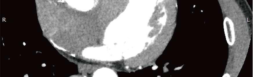
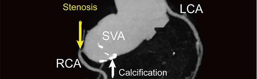
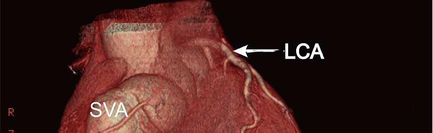
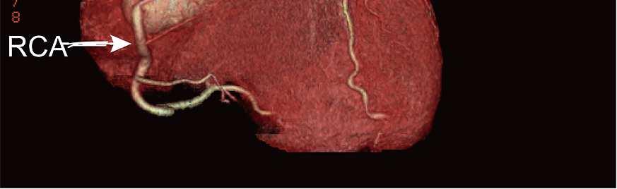
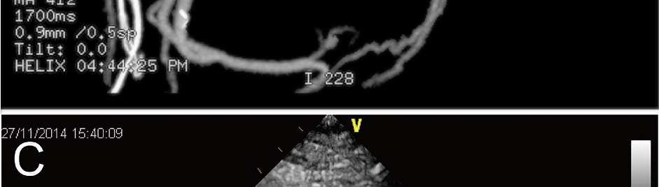
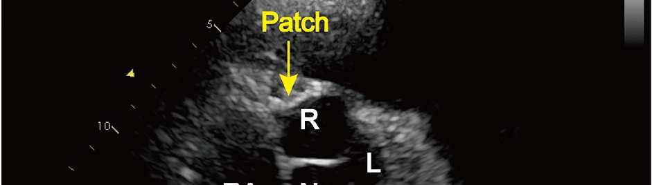
3 Page of 0 computed tomography (CT) scan and renal angiography. Moreover, standard serological test for syphilis showed Treponema pallidum negative and CRP, ESR, platelet and other indicators of serum fluid were normal,. Investigations Two-dimensional echocardiography on admission incidentally revealed a giant aneurysm located in RCS, approximately mm in size. On the parasternal long-axis and short axis views, a flap-like appearance can be visualized in the aneurysm (Figure ). Multiple views indicated this giant SVA extracardiac protruded outward without right atrium and right ventricle compression. There were no other intracardiac anomalies including aortic regurgitation and ventricular septal defect in color doppler imaging (Figure ). Cardiac multislice CT demonstrated a giant unruptured extracardiac aneurysm arising from the RCS, complicated with calcification and mural thrombi in it. In addition, CT coronary angiography revealed right coronary artery (RCA) originated from this SVA and there was 0% to 0% stenosis at the initial segment of the RCA (Figure ). 0 Treatment and outcomes Although there were no indications of SVA rupture, immediate SVA patch repair, combined with RCA bypass grafting was performed to prevent potentially life-threatening complications. The procedure was done via median sternotomy with cardiopulmonary bypass. The intraoperative examination demonstrated a large aneurysm of the RCS (Figure ). After dissecting the epicardial fat around the aneurysm and aortic clamping, a longitudinal incision was made on the aneurysm. The aneurismal wall appeared very thick and filled with yellow atheromatous necrosis materials (Figure ). On the inner side of the aneurysm, a small localized intima tearing and mural thrombosis were detected. The orifice of the aneurysm was located in the RCS and demonstrated an oval measuring mm (Figure ). The ostium of the RCA was seen close to the orifice and almost totally blocked. The aortic valve was intact, and the other sinuses were normal. Patch closure of the orifice of this SVA was porformed
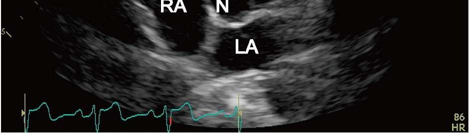
4 Page of using the-right-size prosthetic vascular patch (Figure ). After this, the ostium of the RCA was closed. And right great saphenous vein graft was used to connect the ascending aorta to the proximal RCA with bridge connection. Finally, the incision of the aneurismal wall was folded closed and the patient was easily weaned from cardiopulmonary bypass. The pathological examination with hematoxylin/eosin staining demonstrated significant foam cells, inflammatory cells and thrombosis in the aortic wall (Figure ). The postoperative echocardiography and CT demonstrated successful reconstruction of the RCS and RCA revascularization (Figure ). Three months after surgery, the patient currently 0 recovered well and follow-up echocardiography demonstrated that there was no aortic regurgitation. 0 Discussion SVA is dilatation of one of the three aortic sinuses between the sinotubular junctions and the aortic valve annuluses supra-aortic ridge, which commonly involving the right or noncoronary sinus. As a kind of rare cardiac anomalies, SVA is mostly detected in the situation of complication of a rupture. Patients with ruptured SVAs have symptoms such as dyspnea, chest pain, cough, or peripheral edema. Physical examination could reveal continuous, mechanical-sounding murmur. In contrast, an unruptured SVA usually remains asymptomatic and undetected unless expanding SVA affecting the adjacent tissues. In patients without any obvious complications, SVAs may only be accidentally detected by examinations -. Currently, echocardiography is the initial imaging choice to detect SVA in suspected patients. On echocardiograms, SVA commonly had a thin-walled saccular lesion arising from the aortic root in continuation with the aortic annulus. According to the echocardiographic features including origin, protruding position, and whether ruptured or not, SVAs could be classified into different patterns. In previously reported patients, a few unruptured SVA arising from RCS with extracardiac protrusion was detected. In fact, it was easily confused with dilation of
5 Page of 0 RCA for inexperienced doctors since echocardiography was not always satisfactory in imprecisely delineating the anatomic relations of the aneurysm and its associated lesions. Further CT coronary angiography and D reconstruction was helpful in confirming the RCA arising from the remote of the aneurysm. Different from common simple SVAs, prominent mural thrombi and calcification were found in the aneurysm. In addition, this young patient was complicated with severe RCA stenosis. Compressing coronary arteries to cause a myocardial ischemic event is severe complication of unruptured SVA, which can even result in myocardial infarction or angina -. In fact, the mechanism of coronary flow obstacle is different between the left and right sinus aneurysms. Multiple case reports described unruptured left SVA compressing the LCA to give rise to LCA stenosis, and a few right SVA could also cause the proximal part of RCA compression. However, coronary flow obstruction due to right SVA is mainly because of involvement of RCA ostium, More specifically, RCA may be occluded by a thrombus in the aneurysm or surrounded by a haematoma, leading to a stenosis in the RCA ostium. Polat et al. reported a right SVA causing acute myocardial infarction, They presented a thrombotic right SVA associated with RCA occluded at ostium with the thrombus, which led to acute myocardial infarction and ischemic stroke. In our case, the localized mural thrombi and calcification that obstructed the ostium of RCA may be the cause of the clinical complaint of this particular patient. 0 These imaging results coincided with the final surgical exploration except a localized intima tearing in the aneurysm. Although there was a flap-like appearance in the aneurysm on echocardiographic views, we couldn t decide whether it indicated the intima tearing or not. This patient report demonstrated imaging modalities sometimes could not make a comprehensive diagnosis especially in the SVA with rare complications. Therefore, surgical exploration was quite important in confirming the final diagnosis. SVA can be congenital in origin or may be acquired through infection, degenerative diseases, trauma or atherosclerosis. In our case, the pathological finding was atherosclerotic change.
6 Page of The patient denied any history of trauma and infections. And laboratory examination results could rule out the possibility of infective disease such as Kawasaki or Lues. Although in the absence of atherosclerosis anywhere else, the aneurismal wall presented as marked foam cells and inflammatory cells infiltration. Most of previous SVA cases were complicated with coronary arterial atherosclerosis which caused coronary arteries obstruction. In our case, unruptured extracardiac SVA with atherothrombosis involving the SVA wall was observed. Moreover, the reason for almost obliterative RCA was secondary to localized atherosclerosis of the aneurysm not compression of SVA. To our knowledge, this is the first case report of such a condition. 0 In present reported case, conventional surgical patch repair was performed to prevent aneurysm rupture and thromboembolic events. Meanwhile, coronary artery bypass grafting was also performed for RCA revascularization. Three months after surgery, the patient recovered well and imaging examinations showed successful repair. In summary, we present an extracardiac unruptured SVA complicated with localized atherothrombosis and obstruction of RCA ostium. SVA should be exactly diagnosed by echocardiography, CT coronary angiography and even surgical exploration and pathological examination. Surgery repair is main treatment for SVA. 0 Funding statement This research is supported by grants (No. 00 and No. ) from the National Natural Science Foundation of China. Declaration of interest The authors declare there is no conflict of interest that could be perceived as prejudicing the impartiality of the research reported.
7 Page of Patient consent Written consent was obtained. Author contribution statement Jun Zhang collected images, wrote the manuscript and completed a literature review. Yani Liu and Youbin Deng reviewed the manuscript prior to submission and assisted with review of the literature and included images. Ligang Liu provided the clinical information of the patient and performed the operation. 0 0 References. Cheng TO, Yang YL, Xie MX, Wang XF, Dong NG, Su W, Lü Q, He L, Lu XF, Wang J, Li L & Yuan L. Echocardiographic diagnosis of sinus of Valsalva aneurysm: A -year ( 0) experience of surgically treated patients from one single medical center in China. Int J Cardiol Ott DA. Aneurysm of the sinus of Valsalva. Semin Thorac Cardiovasc Surg Pediatr Card Surg Annu Feldman DN & Roman MJ. Aneurysms of sinuses of valsalva. Cardiology Cottogni M, Antretter H & Wicke K. Subtotal rarefication of one aortic leaflet in a bicuspid aortic valve due to large aneurysm of left Valsalva's sinus. Scand J Thorac Cardiovasc Surg -0.. Yasuda F, Shimono T, Adachi K, Onoda K, Tani K & Yada I. Surgical repair of extracardiac unruptured acquired Valsalva aneurysms. Ann Thorac Surg Nakagiri K, Kadowaki T, Morimoto N, Murakami H, Yoshida M & Mukohara N. Aortic root reimplantation for isolated sinus of valsalva aneurysm in the patient with Marfan's syndrome. Ann Thorac Surg 0 e-e.. Lu S, Sun X, Wang C, Hong T, Xu D, Zhao W & Liu X. Surgical correction of giant extracardiac unruptured aneurysm of the right coronary sinus of Valsalva: case report and
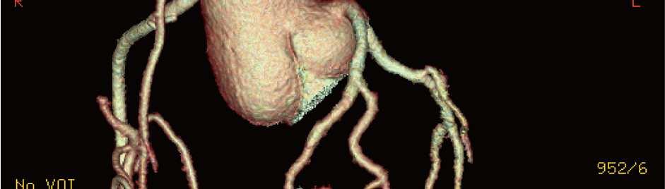
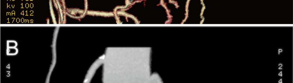
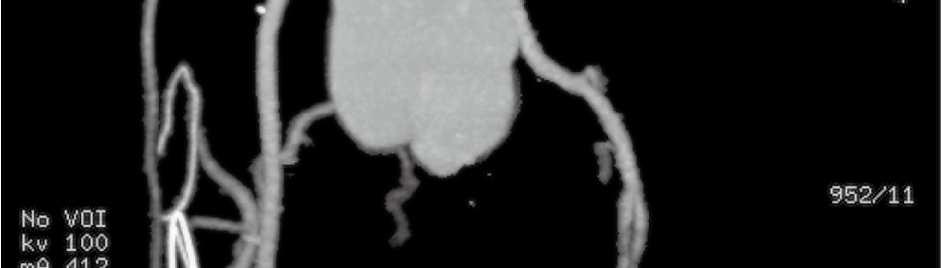
8 Page of review of the literature. Gen Thorac Cardiovasc Surg Brandt J, Jögi P & Lührs C. Sinus of Valsalva aneurysm obstructing coronary arterial flow: case report and collective review of the literature. Eur Heart J Polat N, Yildiz A, Yuksel M, Acet H & Alan S. A thrombotic right sinus of valsalva aneurysm causing acute myocardial infarction and ischemic stroke. Echocardiography Legends to figures Figure Images of echocardiography before surgery. A. In transthoracic echocardiography, the parasternal long axis view showing a cystic mass located in the right coronary sinus with a flap-like appearance in the aneurysm (Arrow). B. On parasternal short axis view, a giant saccular aneurysm located in the right coronary sinus, approximately mm in size. A weak-echogenic flap-like appearance can be seen in the aneurysm (Arrow). C & D On parasternal long axis and short axis views, color doppler imaging showing no other intracardiac anomalies including aortic regurgitation and ventricular septal defect. SVA= sinus of Valsalva aneurysm; LV= left ventricle; LA= left atrium; RV= right ventricle; RA= right atrium; AO= aorta; L= left coronary sinus; N= noncoronary sinus. Figure Contrast-enhanced CT and three dimensional reconstruction demonstrating a giant unruptured aneurysm arising from the RCS with extracardiac protrusion. A. A view showing the SVA complicated with mural thrombi. B. CT coronary angiography confirming RCA originated from this SVA with 0% to 0% stenosis (Yellow arrow) at the initial segment. C. Three dimensional reconstruction showing the SVA with LCA and RCA.
9 Page of 0 0 SVA= sinus of Valsalva aneurysm; LCA= left coronary artery; RCA= right coronary artery. Figure Images of intraoperative examination. A. Intraoperative examination demonstrating a large SVA with extracardiac protrusion. B. After a longitudinal incision on the aneurysm, the aneurismal wall appearing very thick and filled with yellow atheromatous necrosis materials. C. Intraoperative view of the orifice of the aneurysm of the right sinus of Valsalva. The ostium of the right coronary artery close to the orifice and explored almost totally blocked (Arrow). D. Patch closure of the aneurysm was performed. Figure Hematoxylin/eosin staining of the aneurysm wall. A. Extensive foam cells and inflammatory cells infiltration in the aortic wall with deposition of calcium salt. Hematoxylin/eosin staining,. B. Mural thrombosis in this aneurysm. Hematoxylin/eosin staining,. C. At high magnification, network of platelets and fibrin containing red blood cells. Hematoxylin/eosin staining, 0. Figure Echocardiography and CT coronary angiography images after surgery. A. Postoperative echocardiography: RCS is remodeled with patch repair (Yellow arrow). B. Postoperative CT three dimensional reconstruction: RCS was remolded with patch repair. C. Postoperative CT coronary angiography: the GSV graft connecting the ascending aorta and the proximal RCA. LA= left atrium; RA= right atrium; R= right coronary sinus; L= left coronary sinus; N= noncoronary sinus; GSV= great saphenous vein.
10 Figure Images of echocardiography before surgery. xmm (00 x 00 DPI) Page 0 of

11 Page of Figure Contrast-enhanced CT and three dimensional reconstruction demonstrating a giant unruptured aneurysm arising from the RCS with extracardiac protrusion. xmm (00 x 00 DPI)
12 Figure Images of intraoperative examination. xmm (00 x 00 DPI) Page of
13 Page of Figure Hematoxylin/eosin staining of the aneurysm wall. xmm (00 x 00 DPI)
14 Figure Echocardiography and CT coronary angiography images after surgery. xmm (00 x 00 DPI) Page of
An aneurysm is a localized abnormal dilation of a blood vessel or the heart Types: 1-"true" aneurysm it involves all three layers of the arterial
 An aneurysm is a localized abnormal dilation of a blood vessel or the heart Types: 1-"true" aneurysm it involves all three layers of the arterial wall (intima, media, and adventitia) or the attenuated
Coronary Artery from the Wrong Sinus of Valsalva: A Physiologic Repair Strategy
 Coronary Artery from the Wrong Sinus of Valsalva: A Physiologic Repair Strategy Tom R. Karl, MS, MD he most commonly reported coronary artery malformation leading to sudden death in children and young
PROSTHETIC VALVE BOARD REVIEW
 PROSTHETIC VALVE BOARD REVIEW The correct answer D This two chamber view shows a porcine mitral prosthesis with the typical appearance of the struts although the leaflets are not well seen. The valve
Giant Right Atrial Mass: An Unusual Presentation Of Unruptured Sinus Of Valsalva Aneurysm
 ISPUB.COM The Internet Journal of Cardiology Volume 10 Number 2 Giant Right Atrial Mass: An Unusual Presentation Of Unruptured Sinus Of Valsalva Aneurysm S Chandraprakasam, L Stickley Citation S Chandraprakasam,
Adult Echocardiography Examination Content Outline
 Adult Echocardiography Examination Content Outline (Outline Summary) # Domain Subdomain Percentage 1 2 3 4 5 Anatomy and Physiology Pathology Clinical Care and Safety Measurement Techniques, Maneuvers,
14 Valvular Stenosis
 14 Valvular Stenosis 14-1. Valvular Stenosis unicuspid valve FIGUE 14-1. This photograph shows severe valvular stenosis as it occurs in a newborn. There is a unicuspid, horseshoe-shaped leaflet with a
Outcomes of Mitral Valve Repair for Mitral Regurgitation Due to Degenerative Disease
 Outcomes of Mitral Valve Repair for Mitral Regurgitation Due to Degenerative Disease TIRONE E. DAVID, MD ; SEMIN THORAC CARDIOVASC SURG 19:116-120c 2007 ELSEVIER INC. PRESENTED BY INTERN 許士盟 Mitral valve
Imaging by multislice CT of a large aortico-left ventricular tunnel mimicking as ventricular septal defect
 Case Report Page 1 of 5 Imaging by multislice CT of a large aortico-left ventricular tunnel mimicking as ventricular septal defect Sarv Priya 1, Gurpreet S. Gulati 1, Anita Saxena 2, Balram Airan 3 1 Department
Left ventricle pseudoaneurysm as late postoperative complication of a large apical aneurysm
 CASE REPORT Left ventricle pseudoaneurysm as late postoperative complication of a large apical aneurysm Mariana M. Floria 1, 4, Carmen Elena Pleșoianu 2, 4, Michel Buche 3, Baudouin Marchandise 4, Erwin
Very late recurrence of sinus of Valsalva aneurysm rupture after patch repair
 Lin et al. BMC Surgery 2014, 14:73 CASE REPORT Open Access Very late recurrence of sinus of Valsalva aneurysm rupture after patch repair Ting-Tse Lin 1, Hsiao-En Tsai 2, Lin Lin 1, Tsung-Yan Chen 2, Cheng-Pin
Journal of Pediatric Sciences
 Journal of Pediatric Sciences Rupture of sinus of valsalva aneurysm: earliest presentation in association with ventricular septal defect and aortic regurgitation Binoy Shankar, Sanjay Kumar, Dinesh Kumar
Calcified Aortic Sinotubular Ridge: A Source of Coronary Ostial Stenosis or Embolism
 1510 JACC Vol. 12, No, 6 December 1988:1510--4 Calcified Aortic Sinotubular Ridge: A Source of Coronary Ostial Stenosis or Embolism KEVIN J. TVETER, MD, JESSE E. EDWARDS, MD, FACC St, Paul, Minnesota This
Case 47 Clinical Presentation
 93 Case 47 C Clinical Presentation 45-year-old man presents with chest pain and new onset of a murmur. Echocardiography shows severe aortic insufficiency. 94 RadCases Cardiac Imaging Imaging Findings C
Unusual Causes of Aortic Regurgitation. Case 1
 Unusual Causes of Aortic Regurgitation Judy Hung, MD Cardiology Division Massachusetts General Hospital Boston, MA No Disclosures Case 1 54 year old female with h/o cerebral aneurysm and vascular malformation
SURGICAL INTERVENTION IN AORTOPATHIES ZOHAIR ALHALEES, MD RIYADH, SAUDI ARABIA
 SURGICAL INTERVENTION IN AORTOPATHIES ZOHAIR ALHALEES, MD RIYADH, SAUDI ARABIA In patients born with CHD, dilatation of the aorta is a frequent feature at presentation and during follow up after surgical
Functional anatomy of the aortic root. ΔΡΟΣΟΣ ΓΕΩΡΓΙΟΣ Διεσθσνηής Καρδιοθωρακοτειροσργικής Κλινικής Γ.Ν. «Γ. Παπανικολάοσ» Θεζζαλονίκη
 Functional anatomy of the aortic root ΔΡΟΣΟΣ ΓΕΩΡΓΙΟΣ Διεσθσνηής Καρδιοθωρακοτειροσργικής Κλινικής Γ.Ν. «Γ. Παπανικολάοσ» Θεζζαλονίκη What is the aortic root? represents the outflow tract from the LV provides
Bicuspid aortic root spared during ascending aorta surgery: an update of long-term results
 Short Communication Bicuspid aortic root spared during ascending aorta surgery: an update of long-term results Marco Russo, Guglielmo Saitto, Paolo Nardi, Fabio Bertoldo, Carlo Bassano, Antonio Scafuri,
In 1980, Bex and associates 1 first introduced the initial
 Technique of Aortic Translocation for the Management of Transposition of the Great Arteries with a Ventricular Septal Defect and Pulmonary Stenosis Victor O. Morell, MD, and Peter D. Wearden, MD, PhD In
Surgery for Congenital Heart Disease. Surgical treatment of giant coronary artery aneurysm
 Surgical treatment of giant coronary artery aneurysm Dianyuan Li, MD, a * Qingyu Wu, MD, a * Lizhong Sun, MD, a Yunhu Song, MD, a Wei Wang, MD, a Shiwei Pan, MD, a Guohua Luo, MD, a Yongmin Liu, MD, a
Giant aneurysm of the left main coronary artery with fistulous communication to the right atrium
 Zhu et al. Journal of Cardiothoracic Surgery (2015) 10:117 DOI 10.1186/s13019-015-0324-8 CASE REPORT Giant aneurysm of the left main coronary artery with fistulous communication to the right atrium Zhicheng
Aortic root false aneurysm from gelatin-resorcinolformaldehyde GRF glue following surgical treatment for type A dissection
 Jichi Medical University Journal Aortic root false aneurysm from gelatin-resorcinolformaldehyde GRF glue following surgical treatment for type A dissection Yasuhito Sakano, Tsutomu Saito, Yoshio Misawa
Aneurysms & a Brief Discussion on Embolism
 Aneurysms & a Brief Discussion on Embolism Aneurysms, overview = congenital or acquired dilations of blood vessels or the heart True aneurysms -involve all three layers of the artery (intima, media, and
Diseases of the Aorta
 Diseases of the Aorta ASE Review 2018 Susan E Wiegers, MD, FASE, FACC Professor of Medicine My great friend Dr. Roberto Lang Disclosure None related to this presentation 1 Objectives Aneurysm Dissection
Overview of cardiac and paracardiac aneurysms/pseudoaneurysms: Radiologist`s perspective. Presenting Authors. Ameya J Baxi, MD Carlos Restrepo, MD
 Overview of cardiac and paracardiac aneurysms/pseudoaneurysms: Radiologist`s perspective Presenting Authors Ameya J Baxi, MD Carlos Restrepo, MD Co-authors S. Martinez Jiminez, MD Disclaimer: We do not
Detailed Order Request Checklists for Cardiology
 Next Generation Solutions Detailed Order Request Checklists for Cardiology 8600 West Bryn Mawr Avenue South Tower Suite 800 Chicago, IL 60631 www.aimspecialtyhealth.com Appropriate.Safe.Affordable 2018
Surgical Procedures and Complications
 Radiological Society of North America, RSNA 2013 Refresher Course Program: Vascular Track Surgical Procedures and Complications Learning objectives Outline RC 112 : Key Concepts: Surgical Procedures and
New Cardiovascular Devices and Interventions: Non-Contrast MRI for TAVR Abhishek Chaturvedi Assistant Professor. Cardiothoracic Radiology
 New Cardiovascular Devices and Interventions: Non-Contrast MRI for TAVR Abhishek Chaturvedi Assistant Professor Cardiothoracic Radiology Disclosure I have no disclosure pertinent to this presentation.
Med. J. Malaysia Vol. 46 No. 4 December 1991
 Med. J. Malaysia Vol. 46 No. 4 December 1991 aneurysms ofthe sinus of valsalva R. J eyamalar. MBBS, IvIRCP. Lecturer P. Kannan, MBBS,MRCP. Associate Professor Dept. of Medicine, University Hospital, 59100
Imaging in TAVI. Jeroen J Bax Dept of Cardiology Leiden Univ Medical Center The Netherlands Davos, feb 2013
 Imaging in TAVI Jeroen J Bax Dept of Cardiology Leiden Univ Medical Center The Netherlands Davos, feb 2013 Research grants: Medtronic, Biotronik, Boston Scientific, St Jude, BMS imaging, GE Healthcare,
Looking Outside the Box: Incidental Extracardiac Finding in Echo
 Looking Outside the Box: Incidental Extracardiac Finding in Echo Dr. Aijaz Shah Head of Division, Adult Echocardiography Laboratory Prince Sultan Cardiac Centre Riyadh Case 1 17 year old boy presented
Cardiac Imaging Tests
 Cardiac Imaging Tests http://www.medpagetoday.com/upload/2010/11/15/23347.jpg Standard imaging tests include echocardiography, chest x-ray, CT, MRI, and various radionuclide techniques. Standard CT and
Case Report Preoperative Assessment of Anomalous Right Coronary Artery Arising from the Main Pulmonary Artery
 Case Reports in Medicine Volume 2011, Article ID 642126, 4 pages doi:10.1155/2011/642126 Case Report Preoperative Assessment of Anomalous Right Coronary Artery Arising from the Main Pulmonary Artery Marshall
Multimodality Imaging of Anomalous Left Coronary Artery from the Pulmonary
 1 IMAGES IN CARDIOVASCULAR ULTRASOUND 2 3 4 Multimodality Imaging of Anomalous Left Coronary Artery from the Pulmonary Artery 5 6 7 Byung Gyu Kim, MD 1, Sung Woo Cho, MD 1, Dae Hyun Hwang, MD 2 and Jong
ADVANCED CARDIOVASCULAR IMAGING. Medical Knowledge. Goals and Objectives PF EF MF LF Aspirational
 Medical Knowledge Goals and Objectives PF EF MF LF Aspirational Know the basic principles of magnetic resonance imaging (MRI) including the role of the magnetic fields and gradient coil systems, generation
Diseases of the aorta
 Diseases of the aorta Aneurysm, dissection and aortitis are the main pathologies (Fig. 18.79 ). data:text/html;charset=utf-8,%3ch2%20id%3d%22cc5a0836d6aa490ca26dd7c15632b559%22%20style%3d%22margin%3a%201.3em%200px%200.5em%3b%20padding%3a%200px%3b%20border%3a%200px%3b%20font-fa
Reviews. Sinus of Valsalva Aneurysms: Review of the Literature and an Update on Management
 Reviews Sinus of Valsalva Aneurysms: Review of the Literature and an Update on Management Michael Weinreich, MD, MPH; Pey-Jen Yu, MD; Biana Trost, MD Department of Medicine (Weinreich), Department of Cardiovascular
We present the case of an asymptomatic, 75-year-old
 Images in Cardiovascular Medicine Asymptomatic Rupture of the Left Ventricle Lech Paluszkiewicz, MD; Stefan Ożegowski, MD; Mohammad Amin Parsa, MD; Jan Gummert, PhD, MD We present the case of an asymptomatic,
HISTORY. Question: What category of heart disease is suggested by this history? CHIEF COMPLAINT: Heart murmur present since early infancy.
 HISTORY 18-year-old man. CHIEF COMPLAINT: Heart murmur present since early infancy. PRESENT ILLNESS: Although normal at birth, a heart murmur was heard at the six week check-up and has persisted since
Adult Cardiac Surgery
 Adult Cardiac Surgery Mahmoud ABU-ABEELEH Associate Professor Department of Surgery Division of Cardiothoracic Surgery School of Medicine University Of Jordan Adult Cardiac Surgery: Ischemic Heart Disease
UNUSUAL PRESENTATION OF MULTIPLE ANEURYSMS OF THE ASCENDING
 UNUSUAL PRESENTATION OF MULTIPLE ANEURYSMS OF THE ASCENDING AORTA. A CASE REPORT. Sergio Francisco dos Santos Junior, Marcelo Luiz Peixoto Sobral, Anderson da Silva Terrazas, Gilmar Geraldo dos Santos,
Utility of CT angiography for pre-operative evaluation of robotic-assisted minimally invasive mitral valve surgery.
 Utility of CT angiography for pre-operative evaluation of robotic-assisted minimally invasive mitral valve surgery. Poster No.: C-2214 Congress: ECR 2014 Type: Educational Exhibit Authors: M. Muthuvelu,
Case 9799 Stanford type A aortic dissection: US and CT findings
 Case 9799 Stanford type A aortic dissection: US and CT findings Accogli S, Aringhieri G, Scalise P, Angelini G, Pancrazi F, Bemi P, Bartolozzi C Department of Diagnostic and Interventional Radiology, University
Congenital heart disease involving the coronary artery
 Anomalous Coronary Artery With Aortic Origin and Course Between the Great Arteries: Improved Diagnosis, Anatomic Findings, and Surgical Treatment Eldad Erez, MD, Vincent K. H. Tam, MD, Nancy A. Doublin,
An anterior aortoventriculoplasty, known as the Konno-
 The Konno-Rastan Procedure for Anterior Aortic Annular Enlargement Mark E. Roeser, MD An anterior aortoventriculoplasty, known as the Konno-Rastan procedure, is a useful tool for the cardiac surgeon. Originally,
Aorta-to-Left Atrial Fistula Caused by Air Gun Pellet Cardiac Injury
 Cardiol Ther (2014) 3:67 71 DOI 10.1007/s40119-014-0026-7 CASE REPORT Aorta-to-Left Atrial Fistula Caused by Air Gun Pellet Cardiac Injury Mustafa K. Avsar Serafettin Demir İbrahim Özgür Önsel Huseyin
E J Meijboom (Lausanne, CH) Which athlete can re-enter his active sports career? After re-implantation of an abnormal origin of a coronary artery
 Which athlete can re-enter his active sports career? After re-implantation of an abnormal origin of a coronary artery") E J Meijboom (Lausanne, CH) Which athlete can re-enter his active sports career? After re-implantation of an abnormal origin of a coronary artery Coronary Anomalies Congenital and Isolated Angiographic
E J Meijboom (Lausanne, CH) Which athlete can re-enter his active sports career? After re-implantation of an abnormal origin of a coronary artery Coronary Anomalies Congenital and Isolated Angiographic
Department of Cardiology, Heidelberg University, Im Neuenheimer Feld 410, Heidelberg, Germany 2
 Volume 2012, Article ID 524526, 4 pages doi:10.1155/2012/524526 Case Report Giant Dilatation of the Right Coronary Aortic Bulb with Compression of the Right Ventricular Outflow Tract Mimicking a Ventricular
12 th Annual West Virginia ACC Meeting April 8, 2017
 12 th Annual West Virginia ACC Meeting April 8, 2017 Rameez Sayyed, M.D., FACC, FSCAI Associate professor of Medicine Program Director for interventional cardiology Marshall University Joan C. Edwards
Case Report Subacute Staphylococcusepidermidis Bacterial Endocarditis Complicated by Mitral-Aortic Intervalvular Fibrosa Pseudoaneurysm
 Case Reports in Cardiology Volume 2012, Article ID 467210, 4 pages doi:10.1155/2012/467210 Case Report Subacute Staphylococcusepidermidis Bacterial Endocarditis Complicated by Mitral-Aortic Intervalvular
The Edge-to-Edge Technique f For Barlow's Disease
 The Edge-to-Edge Technique f For Barlow's Disease Ottavio Alfieri, Michele De Bonis, Elisabetta Lapenna, Francesco Maisano, Lucia Torracca, Giovanni La Canna. Department of Cardiac Surgery, San Raffaele
Common Codes for ICD-10
 Common Codes for ICD-10 Specialty: Cardiology *Always utilize more specific codes first. ABNORMALITIES OF HEART RHYTHM ICD-9-CM Codes: 427.81, 427.89, 785.0, 785.1, 785.3 R00.0 Tachycardia, unspecified
Pathophysiology of Cardiovascular System. Dr. Hemn Hassan Othman, PhD
 Pathophysiology of Cardiovascular System Dr. Hemn Hassan Othman, PhD hemn.othman@univsul.edu.iq What is the circulatory system? The circulatory system carries blood and dissolved substances to and from
The production of murmurs is due to 3 main factors:
 Heart murmurs The production of murmurs is due to 3 main factors: high blood flow rate through normal or abnormal orifices forward flow through a narrowed or irregular orifice into a dilated vessel or
Aneurysm of the Aorta in Children*
 Aneurysm of the Aorta in Children* Frederick T. Fricker, M.D.; Sang C. Park, M.D.; William H. Neches, M.D.; 00 Robert A.!lfathews, M.D.; and David B. Lerlwrg, M.D., F.C.C.P. Seven children with aortic
Aortic Regurgitation & Aorta Evaluation
 VALVULAR HEART DISEASE Regurgitation Valvular Lessions 2017 Aortic Regurgitation & Aorta Evaluation Jorge Eduardo Cossío-Aranda MD, FACC Chairman of Outpatient Care Department Instituto Nacional de Cardiología
Echocardiography in Adult Congenital Heart Disease
 Echocardiography in Adult Congenital Heart Disease Michael Vogel Kinderherz-Praxis München CHD missed in childhood Subsequent lesions after repaired CHD Follow-up of cyanotic heart disease CHD missed in
pulmonary valve on, 107 pulmonary valve vegetations on, 113
 INDEX Adriamycin-induced cardiomyopathy, 176 Amyloidosis, 160-161 echocardiographic abnormalities in, 160 intra-mural tumors similar to, 294 myocardial involvement in, 160-161 two-dimensional echocardiography
Late presentation of anomalous origin of the left coronary artery from the pulmonary artery (ALCAPA) was confused with coronary artery fistula.
 was confused with coronary artery fistula.") Case Report http://www.alliedacademies.org/annals-of-cardiovascular-and-thoracic-surgery/ Late presentation of anomalous origin of the left coronary artery from the pulmonary artery (ALCAPA) was confused
Case Report http://www.alliedacademies.org/annals-of-cardiovascular-and-thoracic-surgery/ Late presentation of anomalous origin of the left coronary artery from the pulmonary artery (ALCAPA) was confused
Appropriate Use Criteria for Initial Transthoracic Echocardiography in Outpatient Pediatric Cardiology (scores listed by Appropriate Use rating)
") Appropriate Use Criteria for Initial Transthoracic Echocardiography in Outpatient Pediatric Cardiology (scores listed by Appropriate Use rating) Table 1: Appropriate indications (median score 7-9) Indication
Appropriate Use Criteria for Initial Transthoracic Echocardiography in Outpatient Pediatric Cardiology (scores listed by Appropriate Use rating) Table 1: Appropriate indications (median score 7-9) Indication
AORTIC DISSECTION. DISSECTING ANEURYSMS OF THE AORTA or CLASSIFICATION
 DISSECTING ANEURYSMS OF THE AORTA or AORTIC DISSECTION CLASSIFICATION DeBakey classified aortic dissections into types I, II, and III :- Type I dissection the tear site originates in the ascending aorta,
HISTORY. Question: What category of heart disease is suggested by the fact that a murmur was heard at birth?
 HISTORY 23-year-old man. CHIEF COMPLAINT: Decreasing exercise tolerance of several years duration. PRESENT ILLNESS: The patient is the product of an uncomplicated term pregnancy. A heart murmur was discovered
HISTORY. Question: How do you interpret the patient s history? CHIEF COMPLAINT: Dyspnea of two days duration. PRESENT ILLNESS: 45-year-old man.
 HISTORY 45-year-old man. CHIEF COMPLAINT: Dyspnea of two days duration. PRESENT ILLNESS: His dyspnea began suddenly and has been associated with orthopnea, but no chest pain. For two months he has felt
The production of murmurs is due to 3 main factors:
 Heart murmurs The production of murmurs is due to 3 main factors: high blood flow rate through normal or abnormal orifices forward flow through a narrowed or irregular orifice into a dilated vessel or
How to Perform a Valve Sparing Root Replacement Joseph S. Coselli, M.D.
 How to Perform a Valve Sparing Root Replacement Joseph S. Coselli, M.D. AATS International Cardiovascular Symposium 2017 Session 6: Technical Aspects of Open Surgery on the Aortic Valve Sao Paulo, Brazil
Anomalous origin of left circumflex coronary artery: An easy pick on transthoracic echocardiography
 www.edoriumjournals.com CLINICAL IMAGES PEER REVIEWED OPEN ACCESS Anomalous origin of left circumflex coronary artery: An easy pick on transthoracic echocardiography Keyur Vora, Alok Ranjan ABSTRACT Abstract
Management of Ascending Aortic
 Management of Ascending Aortic Aneurysm Complicating Coarctation of the Aorta Ramanathan Sampath, M.D., William N. O'Connor, M.D., Jacqueline A. Noonan, M.D., and Edward P. Todd, M.D., Ph.D. ABSTRACT Four
Doppler-echocardiographic findings in a patient with persisting right ventricular sinusoids
 Zurich Open Repository and Archive University of Zurich Main Library Strickhofstrasse 39 CH-8057 Zurich www.zora.uzh.ch Year: 1990 Doppler-echocardiographic findings in a patient with persisting right
Delayed Infective Endocarditis with Mycotic Aneurysm Rupture below the Mechanical Valved Conduit after the Bentall Procedure
 Case Report Acta Cardiol Sin 2014;30:341 345 Delayed Infective Endocarditis with Mycotic Aneurysm Rupture below the Mechanical Valved Conduit after the Bentall Procedure Mei-Ling Chen, 1,3 Michael Y. Chen,
CT angiography in type I acute aortic dissection complicated with malperfusion - a visual review of obstruciton patterns
 CT angiography in type I acute aortic dissection complicated with malperfusion - a visual review of obstruciton patterns Eneva M. St. Ekaterna University Hospital Report objectives 1. Review malperfusion
Diversion of the inferior vena cava following repair of atrial septal defect causing hypoxemia
 Marshall University Marshall Digital Scholar Internal Medicine Faculty Research Spring 5-2004 Diversion of the inferior vena cava following repair of atrial septal defect causing hypoxemia Ellen A. Thompson
Coronary Arteriovenous Malformation presenting as Acute Myocardial Infarction. Choon Ta NG, Aaron WONG, Foong-Koon CHEAH, Chi Keong CHING
 Coronary Arteriovenous Malformation presenting as Acute Myocardial Infarction Choon Ta NG, Aaron WONG, Foong-Koon CHEAH, Chi Keong CHING The patient 49 year old Male presented with Chest tightness x 1
Unruptured Sinus of Valsalva Aneurysm, With Dissection into the
 Case Report Unruptured Sinus of Valsalva Aneurysm, With Dissection into the Interventricular Septum Udora NC, Ejim EC *, Okoye JU Department of Medicine, University of Nigeria, Teaching Hospital, Ituku-
Index. Note: Page numbers of article titles are in boldface type.
 Index Note: Page numbers of article titles are in boldface type. A Acute coronary syndrome(s), anticoagulant therapy in, 706, 707 antiplatelet therapy in, 702 ß-blockers in, 703 cardiac biomarkers in,
By the end of this session, the student should be able to:
 Valvular Heart disease HVD By Dr. Ashraf Abdelfatah Deyab VHD- Objectives By the end of this session, the student should be able to: Define and classify valvular heart disease. Enlist the causes of acquired
Isolated right ventricular hypoplasia caused by giant aneurysm of right coronary artery to left ventricle fistula in an adult: a case report
 Zhu et al. Journal of Cardiothoracic Surgery (2016) 11:93 DOI 10.1186/s13019-016-0494-z CASE REPORT Open Access Isolated right ventricular hypoplasia caused by giant aneurysm of right coronary artery to
Echocardiography as a diagnostic and management tool in medical emergencies
 Echocardiography as a diagnostic and management tool in medical emergencies Frank van der Heusen MD Department of Anesthesia and perioperative Care UCSF Medical Center Objective of this presentation Indications
Complete Proximal Occlusion of All Three Main Coronary Arteries Complicated With a Left Main Coronary Aneurysm: A Case Report
 J Cardiol 2004 Nov; 44 5 : 201 205 Complete Proximal Occlusion of All Three Main Coronary Arteries Complicated With a Left Main Coronary Aneurysm: A Case Report Takatoshi Hiroshi Akira Takahiro Masayasu
The application of autologous pulmonary artery in surgical correction of complicated aortic arch anomaly
 Original Article The application of autologous pulmonary artery in surgical correction of complicated aortic arch anomaly Shusheng Wen, Jianzheng Cen, Jimei Chen, Gang Xu, Biaochuan He, Yun Teng, Jian
Acute Aortic Syndromes
 Acute Aortic Syndromes Carole J. Dennie, MD Acute Thoracic Aortic Syndromes Background Non-Traumatic Acute Thoracic Aortic Syndromes Carole Dennie MD FRCPC Associate Professor of Radiology and Cardiology
Aortic CT: Intramural Hematoma. Leslie E. Quint, M.D.
 Aortic CT: Intramural Hematoma Leslie E. Quint, M.D. 43 M Mid back pain X several months What type of aortic disease? A. Aneurysm with intraluminal thrombus B. Chronic dissection with thrombosed false
Echocardiographic Cardiovascular Risk Stratification: Beyond Ejection Fraction
 Echocardiographic Cardiovascular Risk Stratification: Beyond Ejection Fraction October 4, 2014 James S. Lee, M.D., F.A.C.C. Associates in Cardiology, P.A. Silver Spring, M.D. Disclosures Financial none
Descending aorta replacement through median sternotomy
 Descending aorta replacement through median sternotomy Mitrev Z, Anguseva T, Belostotckij V, Hristov N. Special hospital for surgery Filip Vtori Skopje - Makedonija June, 2010 Cardiosurgery - Skopje 1
Aortic valve repair: When and how to employ this novel approach?
 Aortic valve repair: When and how to employ this novel approach? Konstadinos A Plestis, MD System Chief of Cardiac Thoracic and Vascular Surgery Main Line Health Care System Professor Sidney Kimmel Medical
HISTORY. Question: What type of heart disease is suggested by this history? CHIEF COMPLAINT: Decreasing exercise tolerance.
 HISTORY 15-year-old male. CHIEF COMPLAINT: Decreasing exercise tolerance. PRESENT ILLNESS: A heart murmur was noted in childhood, but subsequent medical care was sporadic. Easy fatigability and slight
Cardiac MRI in ACHD What We. ACHD Patients
 Cardiac MRI in ACHD What We Have Learned to Apply to ACHD Patients Faris Al Mousily, MBChB, FAAC, FACC Consultant, Pediatric Cardiology, KFSH&RC/Jeddah Adjunct Faculty, Division of Pediatric Cardiology
IMAGING the AORTA. Mirvat Alasnag FACP, FSCAI, FSCCT, FASE June 1 st, 2011
 IMAGING the AORTA Mirvat Alasnag FACP, FSCAI, FSCCT, FASE June 1 st, 2011 September 11, 2003 Family is asking $67 million in damages from two doctors Is it an aneurysm? Is it a dissection? What type of
Operative Strategy. Operative Technique
 Domingo Liotta, M.D.; Christian Cabrol, M.D; Miguel del Rio, M.D; Armando Diluch, M.D; Adriano Malusardi, M.D. Figure 11 Acute dissected aortic root and ascending aorta with valvular regurgitation. -Replacement
Saccular Coronary Artery Aneurysm and Fistula with Organized Thrombi
 Case Report Print ISSN 1738-5520 On-line ISSN 1738-5555 Korean Circulation Journal Saccular Coronary rtery neurysm and Fistula with Organized Thrombi Eun Haeng Jeong, MD 1, yung Jin Kim, MD 1, Ki ae ang,
Congenital Coronary Anomalies
 Chapter 50 Congenital Coronary Anomalies S. Adil Husain, Brett C. Sheridan, and Michael R. Mill Congenital coronary anomalies may have a significant impact on myocardial perfusion and secondary ischemia,
Research Presentation June 23, Nimish Muni Resident Internal Medicine
 Research Presentation June 23, 2009 Nimish Muni Resident Internal Medicine Research Question In adult patients with repaired Tetralogy of Fallot, how does Echocardiography compare to MRI in evaluating
, David Stultz, MD. Aortic Dissection. David Stultz, MD October 7, 2003
 Aortic Dissection David Stultz, MD October 7, 2003 Background Incidence of 1 in 2000 in US Early mortality of 1%/hour for proximal dissection Two theories of formation Breach of intimal layer of aorta
From Valve to Arch: How s Your Aorta? March 7, 2011
 From Valve to Arch: How s Your Aorta? March 7, 2011 Susan Housholder-Hughes, RN, MSN, ANP-BC, FAHA, AACC Nurse Practitioner, Multidisciplinary Aortic Program Cardiovascular Center Adjunct Clinical Instructor,
Right Coronary Artery With Anomalous Origin and Slit Ostium
 Right Coronary Artery With Anomalous Origin and Slit Ostium Raul Garcia Rinaldi, MO, Jorge Carballido, MO, Richard Giles, MO, Emilio Del Taro, MO, and Raul Porro, MO Departments of Cardiovascular Surgery
Echo in Asymptomatic Mitral and Aortic Regurgitation
 2017 ASE Florida Orlando, FL October 9, 2017 10:40 11:00 PM 20 min Grand Harbor Ballroom South Echo in Asymptomatic Mitral and Aortic Regurgitation Muhamed Sarić MD, PhD, MPA Director of Noninvasive Cardiology
Low-dose prospective ECG-triggering dual-source CT angiography in infants and children with complex congenital heart disease: first experience
 Low-dose prospective ECG-triggering dual-source CT angiography in infants and children with complex congenital heart disease: first experience Ximing Wang, M.D., Zhaoping Cheng, M.D., Dawei Wu, M.D., Lebin
7. Echocardiography Appropriate Use Criteria (by Indication)
") Criteria for Echocardiography 1133 7. Echocardiography Criteria (by ) Table 1. TTE for General Evaluation of Cardiac Structure and Function Suspected Cardiac Etiology General With TTE 1. Symptoms or conditions
Criteria for Echocardiography 1133 7. Echocardiography Criteria (by ) Table 1. TTE for General Evaluation of Cardiac Structure and Function Suspected Cardiac Etiology General With TTE 1. Symptoms or conditions
New ASE Guidelines: What you must know
 New ASE Guidelines: What you must know Federico M Asch MD, FASE, FACC Chair, ASE Guidelines and Standards Committee Medstar Washington Hospital Center Medstar Health Research Institute Georgetown University
Uncommon Doppler Echocardiographic Findings of Severe Pulmonic Insufficiency
 Uncommon Doppler Echocardiographic Findings of Severe Pulmonic Insufficiency Rahul R. Jhaveri, MD, Muhamed Saric, MD, PhD, FASE, and Itzhak Kronzon, MD, FASE, New York, New York Background: Two-dimensional
Our Experiences With Adult Type Aortic Coarctation
 ISPUB.COM The Internet Journal of Thoracic and Cardiovascular Surgery Volume 7 Number 2 Our Experiences With Adult Type Aortic Coarctation E Duran, S Canbaz, M Acipayam, O Gur, O Karaca Citation E Duran,
New York Valves Patient focused evidence-based approach. New York City: 6 December Antonio Colombo
 New York Valves 2018 Patient focused evidence-based approach New York City: 6 December 2018 Antonio Colombo Speaker 7 EMO GVM Centro Cuore Columbus Milan, Italy No conflicts to report Vascular complications