Normal LV Ejection Fraction Limits Using 4D-MSPECT: Comparisons of Gated Perfusion and Gated Blood Pool SPECT Data with Planar Blood Pool
|
|
|
- Belinda Bishop
- 6 years ago
- Views:
Transcription

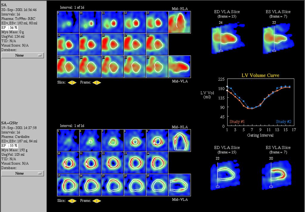
1 Normal LV Ejection Fraction Limits Using 4D-MSPECT: Comparisons of Gated Perfusion and Gated Blood Pool SPECT Data with Planar Blood Pool EP Ficaro, JN Kritzman, JR Corbett University of Michigan Health System, Ann Arbor, MI
2 Conflict of Interest The University of Michigan receives royalties from the sale of the cardiac quantification software, 4D- MSPECT.
3 Objective The objective of this study was to estimate the difference in left ventricular ejection fractions (LVEF) measured in normals studied with gated perfusion and blood pool (GBP) SPECT quantified using 4D-MSPECT. A comparison with conventional planar blood pool processing was also conducted in the same patient population.
4 Patient Population The study consisted of 63 patients (42M, 59±15yo) who had undergone gated myocardial perfusion SPECT, gated blood pool SPECT (GBPS), and gated planar (LAO projection) blood pool imaging. Each of these patients had Low ( 5%) pre-test likelihood for cardiac disease No perfusion or wall motion abnormalities
5 Methods Planar Blood Pool Processing 16 frame planar image at LAO projection. The LVEFs were measured using standard methods from background subtracted end-diastolic (ED) and end-systolic (ES) regions of interest (Marconi Odyssey software). Planar data was processed blinded to the results of the other data sets.
6 Methods Myocardial Perfusion SPECT 16 frame, uncorrected FBP reconstructions from RAO to LPO with 5mm voxels. LVEF, EDv and ESv values were determined automatically with 4D-MSPECT v2.1.
7 Methods GBPS Surface Algorithm 16 frame, uncorrected FBP reconstructions from RAO to LPO with 5mm voxels. The algorithm incorporates standard image processing tools Gradient operators Segmentation and morphologic operators Weighted spline interpolators
8 Methods GBPS Surface Algorithm Cylindrical/Spherical Sampling System Line profile analysis from mid-hla is used to delineate RV from LV. Thresholding the mid-hla gradient image for first frame is used to delineate the LV from the left atrium (LA). Surfaces are iteratively determined from 2D gradient LA images. Weighted spline interpolators (LA, SA and temporal) are employed for smooth contours throughout the cardiac cycle. The final LV basal positions at ED and ES are determined from information extracted from the segmented gradient images, stroke images, and phase analysis. Using the ED and ES limits, a periodic spline is used to define the location for all frames in the cardiac cycle.
9 Results Means and Linear Correlations Mean LVEF Perf SPECT: (70 ± 7)% GBP SPECT: (72 ± 7)% GBP Planar: (68 ± 8)% Linear Correlations GBP SPECT vs GBP Planar: y = 1.05x , r=0.95 GBP SPECT vs Perf SPECT: y = 0.97x + 3.6, r=0.98
10 Results Mean LV Ejection Fractions Perf SPECT: (70 ± 8)% p=0.02 GBP SPECT: (68 ± 7)% p=ns p=0.001 GBP Planar: (71 ± 8)%
11
12 EDv Correlation: GBPS vs. Perfusion SPECT 250 y(ml) = 0.73x + 20, r= GBPS EDv (ml) Perfusion EDv (ml)
13 EDv Bland-Altman: GBPS vs. Perfusion SPECT Residual (GBPS-Perf) EDv (ml) Mean: (-13 ± 15) ml Perfusion EDv (ml)
14 ESv Correlation: GBPS vs. Perfusion SPECT 100 y = 0.74x + 10, r=0.87 GBPS ESv (ml) Perfusion ESv (ml)
15 ESv Bland-Altman: GBPS vs. Perfusion SPECT 40 Mean: (-1.0 ± 9.1) ml Residual (GBPS-Perf) ESv Perfusion ESv (ml)
16 Summary Results from this population demonstrated good accuracy for LVEF for GBPS compared to myocardial perfusion SPECT and planar blood pool imaging. While the LVEF differences were small (2-4%) they should be known when serial comparisons are required. For EDv and ESv, there was good correlation between GBPS and perfusion SPECT. However, the GBPS volume estimates tended to be lower than those estimate from perfusion SPECT data.
TW Hamilton, EP Ficaro, TA Mitchell, JN Kritzman, JR Corbett University of Michigan Health System, Ann Arbor, MI
 Accuracy and Variability of of 3D-MSPECT for Estimating the Left Ventricular Ejection Fraction as as a Function of of Gating Frames and Reconstruction Filters TW Hamilton, EP Ficaro, TA Mitchell, JN Kritzman,
Integrated Report Generation for Myocardial Perfusion SPECT: Efficiency Comparison to Report Dictation
 Integrated Report Generation for Myocardial Perfusion SPECT: Efficiency Comparison to Report Dictation C Bui, EP Ficaro, JN Kritzman, G Wu, JR Corbett University of Michigan Health System, Ann Arbor, MI
Measurement of Ventricular Volumes and Function: A Comparison of Gated PET and Cardiovascular Magnetic Resonance
 BRIEF COMMUNICATION Measurement of Ventricular Volumes and Function: A Comparison of Gated PET and Cardiovascular Magnetic Resonance Kim Rajappan, MBBS 1,2 ; Lefteris Livieratos, MSc 2 ; Paolo G. Camici,
Disclosure of Interests. No financial relationships to disclose concerning the content of this presentation or session.
 Comparison of Free Breathing Cardiac MRI Radial Technique to the Standard Multi Breath-Hold Cine SSFP CMR Technique For the Assessment of LV Volumes and Function Shimon Kolker, Giora Weisz, Naama Bogot,
The consequences of a new software package for the quantification of gated-spect myocardial perfusion studies
 Eur J Nucl Med Mol Imaging (2010) 37:1736 1744 DOI 10.1007/s00259-010-1465-6 ORIGINAL ARTICLE The consequences of a new software package for the quantification of gated-spect myocardial perfusion studies
Impaired Regional Myocardial Function Detection Using the Standard Inter-Segmental Integration SINE Wave Curve On Magnetic Resonance Imaging
 Original Article Impaired Regional Myocardial Function Detection Using the Standard Inter-Segmental Integration Ngam-Maung B, RT email : chaothawee@yahoo.com Busakol Ngam-Maung, RT 1 Lertlak Chaothawee,
ORIGINAL ARTICLE. Koichi Okuda, PhD 1) and Kenichi Nakajima, MD 2) Annals of Nuclear Cardiology Vol. 3 No
 and Kenichi Nakajima, MD 2) Annals of Nuclear Cardiology Vol. 3 No") Annals of Nuclear Cardiology Vol. 3 No. 1 29-33 ORIGINAL ARTICLE Normal Values and Gender Differences of Left Ventricular Functional Parameters with CardioREPO Software: Volume, Diastolic Function, and
Annals of Nuclear Cardiology Vol. 3 No. 1 29-33 ORIGINAL ARTICLE Normal Values and Gender Differences of Left Ventricular Functional Parameters with CardioREPO Software: Volume, Diastolic Function, and
Automatic cardiac contour propagation in short axis cardiac MR images
 International Congress Series 1281 (2005) 351 356 www.ics-elsevier.com Automatic cardiac contour propagation in short axis cardiac MR images G.L.T.F. Hautvast a,b, T, M. Breeuwer a, S. Lobregt a, A. Vilanova
Electrocardiographically (ECG) gated planar radionuclide
 gated planar radionuclide") Electrocardiographically Gated Blood-Pool SPECT and Left Ventricular Function: Comparative Value of 3 Methods for Ejection Fraction and Volume Estimation Doumit Daou, François Harel, Badia O. Helal, Thierry
Electrocardiographically Gated Blood-Pool SPECT and Left Ventricular Function: Comparative Value of 3 Methods for Ejection Fraction and Volume Estimation Doumit Daou, François Harel, Badia O. Helal, Thierry
Effects of Motion and Tissue Weighting on PET Myocardial Blood Flow Estimates
 Effects of Motion and Tissue Weighting on PET Myocardial Blood Flow Estimates Benjamin C. Lee 1, Jonathan B. Moody 1, Venkatesh L. Murthy 2, James R. Corbett 2, Edward P. Ficaro 2 1 INVIA Medical Imaging
The accurate estimation of right ventricular (RV) ejection
 ejection") Accuracy of 4 Different Algorithms for the Analysis of Tomographic Radionuclide Ventriculography Using a Physical, Dynamic 4-Chamber Cardiac Phantom Pieter De Bondt, MD 1,2 ; Tom Claessens MScCivE 3 ;
Accuracy of 4 Different Algorithms for the Analysis of Tomographic Radionuclide Ventriculography Using a Physical, Dynamic 4-Chamber Cardiac Phantom Pieter De Bondt, MD 1,2 ; Tom Claessens MScCivE 3 ;
Multiple Gated Acquisition (MUGA) Scanning
 Scanning") Multiple Gated Acquisition (MUGA) Scanning Dmitry Beyder MPA, CNMT Nuclear Medicine, Radiology Barnes-Jewish Hospital / Washington University St. Louis, MO Disclaimers/Relationships Standard of care research
Multiple Gated Acquisition (MUGA) Scanning Dmitry Beyder MPA, CNMT Nuclear Medicine, Radiology Barnes-Jewish Hospital / Washington University St. Louis, MO Disclaimers/Relationships Standard of care research
LEFT VENTRICLE SEGMENTATION AND MEASUREMENT Using Analyze
 LEFT VENTRICLE SEGMENTATION AND MEASUREMENT Using Analyze 2 Table of Contents 1. Introduction page 3 2. Segmentation page 4 3. Measurement Instructions page 11 4. Calculation Instructions page 14 5. References
Gated SPECT SPECT. small heart small heart 2. R-R SPECT. MBq Marconi/Shimadzu 3 R-R SPECT R-R R-R. (OdysseyFX) 16 8 QGS (Quantitative Gated SPECT)
 16 8 QGS (Quantitative Gated SPECT)") 97 Gated SPECT * * SPECT R-R R-R 20 ml small heart SPECT ( 39: 97 102, 2002) 1. SPECT SPECT R-R R-R * 13 11 21 1715 (0 270 1694) small heart small heart 2. R-R 12 48 99m Tc-tetrofosmin 740 MBq Marconi/Shimadzu
97 Gated SPECT * * SPECT R-R R-R 20 ml small heart SPECT ( 39: 97 102, 2002) 1. SPECT SPECT R-R R-R * 13 11 21 1715 (0 270 1694) small heart small heart 2. R-R 12 48 99m Tc-tetrofosmin 740 MBq Marconi/Shimadzu
Coupling of left ventricular (LV) myocardial perfusion
 myocardial perfusion") Performance of OSEM and Depth-Dependent Resolution Recovery Algorithms for the Evaluation of Global Left Ventricular Function in 201 Tl Gated Myocardial Perfusion SPECT Doumit Daou, MD 1 ; Isabelle Pointurier,
Performance of OSEM and Depth-Dependent Resolution Recovery Algorithms for the Evaluation of Global Left Ventricular Function in 201 Tl Gated Myocardial Perfusion SPECT Doumit Daou, MD 1 ; Isabelle Pointurier,
SPECT. quantitative gated SPECT (QGS) II. viability RH-2 QGS. Butterworth. 14% 10% 0.43 cycles/cm ( 39: 21 27, 2002) ( )
 II. viability RH-2 QGS. Butterworth. 14% 10% 0.43 cycles/cm ( 39: 21 27, 2002) ( )") 21 201 Tl SPECT * * * * * * * * SPECT QGS 99m Tc 201 Tl QGS 20 QGS Butterworth Butterworth 0.39 cycles/cm 14% 10% 0.43 cycles/cm ( r = 0.80 r = 0.86 r = 0.80) 201 Tl QGS ( 39: 21 27, 2002) I. quantitative
21 201 Tl SPECT * * * * * * * * SPECT QGS 99m Tc 201 Tl QGS 20 QGS Butterworth Butterworth 0.39 cycles/cm 14% 10% 0.43 cycles/cm ( r = 0.80 r = 0.86 r = 0.80) 201 Tl QGS ( 39: 21 27, 2002) I. quantitative
QCVC Committees Scientific Activities Central Hall General Information FAC. SPECT tomography has the advantage of quantifying biventricular volumes.
 QCVC Committees Scientific Activities Central Hall General Information FAC Thematic Units Arrhythmias and Electrophysiology Basic Research Bioengineering and Medical Informatics Cardiac Surgical Intensive
05/02/ CPT Preauthorization Groupings Effective May 2, Computerized Tomography (CT) Abdomen 6. CPT Description SEGR CT01
 Abdomen 6. CPT Description SEGR CT01") Computerized Tomography (CT) 6 & 101 5 Upper Extremity 11 Lower Extremity 12 Head 3 Orbit 1 Sinus 2 Neck 4 7 Cervical Spine 8 Thoracic Spine 9 Lumbar Spine 10 Colon 13 CPT Preauthorization Groupings CPT
Computerized Tomography (CT) 6 & 101 5 Upper Extremity 11 Lower Extremity 12 Head 3 Orbit 1 Sinus 2 Neck 4 7 Cervical Spine 8 Thoracic Spine 9 Lumbar Spine 10 Colon 13 CPT Preauthorization Groupings CPT
AIM 2014 CPT Radiology & Cardiac Codes Requiring Review
 AIM 2014 CPT Radiology & Cardiac Codes Requiring Review Modality Body Part CT Head 1 70480 CT orbit, sella or posterior fossa; w/o contrast 1 CT Head 1 70481 CT orbit, sella or posterior fossa; with CT
2012 CPT Radiology Codes Requiring Review Blue Cross and Blue Shield of Louisiana
 2012 CPT Radiology Codes Requiring Review Blue Cross and Blue Shield of Louisiana CT Head 70480 CT orbit, sella or posterior fossa; w/o CT Head 70481 CT orbit, sella or posterior fossa; with CT Head 70482
Evaluation of new data processing algorithms for planar gated ventriculography (MUGA)
") JOURNAL OF APPLIED CLINICAL MEDICAL PHYSICS, VOLUME 10, NUMBER 3, Summer 2009 Evaluation of new data processing algorithms for planar gated ventriculography (MUGA) Joanna R. Fair, Philip H. Heintz, a Robert
JOURNAL OF APPLIED CLINICAL MEDICAL PHYSICS, VOLUME 10, NUMBER 3, Summer 2009 Evaluation of new data processing algorithms for planar gated ventriculography (MUGA) Joanna R. Fair, Philip H. Heintz, a Robert
ORIGINAL ARTICLE. Takahiro HIGUCHI,*, ** Junichi TAKI,* Kenichi NAKAJIMA,* Seigo KINUYA,* Masatoshi IKEDA,*** Masanobu NAMURA*** and Norihisa TONAMI*
 ORIGINAL ARTICLE Annals of Nuclear Medicine Vol. 18, No. 6, 507 511, 2004 Left ventricular ejection and filling rate measurement based on the automatic edge detection method of ECG-gated blood pool single-photon
ORIGINAL. Keywords : multidetector-row computed tomography, myocardial infarction, cardiac function
 ORIGINAL Department of Digestive and Cardiovascular Medicine, Institute of Health Biosciences, The University of Tokushima Graduate School, Tokushima, Japan, Faculty of Integrated Art and Sciences, Department
Gated SPECT (gspect) offers the possibility of simultaneous
 offers the possibility of simultaneous") Calculation of the Left Ventricular Ejection Fraction Without Edge Detection: Application to Small Hearts Bing Feng, MS 1 ; Arkadiusz Sitek, PhD 1,2 ; and Grant T. Gullberg, PhD 1 1 Radiology Department,
Calculation of the Left Ventricular Ejection Fraction Without Edge Detection: Application to Small Hearts Bing Feng, MS 1 ; Arkadiusz Sitek, PhD 1,2 ; and Grant T. Gullberg, PhD 1 1 Radiology Department,
AMERICAN IMAGING MANAGEMENT
 2012 CPT Codes Computerized Tomography (CT) CPT Description Abdomen 74150 CT abdomen; w/o 74160 CT abdomen; with 74170 CT abdomen; w/o followed by Chest 71250 CT thorax; w/o 71260 CT thorax; with 71270
AMERICAN IMAGING MANAGEMENT
 2010 BCBS of Georgia CPT Codes With Grouper Numbers Computerized Tomography (CT) CPT Description Abdomen 74150 CT abdomen; w/o contrast 6 74160 CT abdomen; with contrast 74170 CT abdomen; w/o contrast
MOLINA HEALTHCARE OF MICHIGAN PRIOR AUTHORIZATION / PRE-SERVICE REVIEW GUIDE IMAGING CODES REQUIRING PRIOR AUTHORIZATION EFFECTIVE 1/1/2014
 70336 MRI MRI, temporomandibular joint(s) 70450 CT/CTA CT, head or brain; without contrast material 70460 CT/CTA CT, head or brain; with contrast material(s) 70470 CT/CTA CT, head or brain; without contrast
Quantification of Right Ventricular Function in Pulmonary Hypertension using Cardiac PET Images
 Quantification of Right Ventricular Function in Pulmonary Hypertension using Cardiac PET Images By Simisani Takobana A thesis submitted to the Faculty of Graduate and Postdoctoral Affairs in partial fulfillment
Abnormal, Autoquant Adenosine Myocardial Perfusion Heart Imaging. ID: GOLD Date: Age: 46 Sex: M John Doe Phone (310)
") Background: Reason: preoperative assessment of CAD, Shortness of Breath Symptom: atypical chest pain Risk factors: hypertension Under influence: a beta blocker Medications: digoxin Height: 66 in. Weight:
Background: Reason: preoperative assessment of CAD, Shortness of Breath Symptom: atypical chest pain Risk factors: hypertension Under influence: a beta blocker Medications: digoxin Height: 66 in. Weight:
Evidence and the extent of regional and global cardiac
 Quantification of Left Ventricular Volumes and Ejection Fraction from Gated 99m Tc-MIBI SPECT: MRI Validation and Comparison of the Emory Cardiac Tool Box with QGS and 4D-MSPECT Wolfgang M. Schaefer, MD,
MPI Overview. Artifacts and Pitfalls in MPI. Acquisition and Processing. Peeyush Bhargava MD, MBA
 Artifacts and Pitfalls in MPI MPI Overview Peeyush Bhargava MD, MBA www.nuclearmd.com ~ 40% of all Nuclear Medicine Procedures ~ 9 million procedures every year in US >95% are SPECT,
3/27/2014. Introduction.
 Introduction. Myocardial perfusion & contractility becomes abnormal immediately after the onset of ischaemia, even before the development of the symptoms & ST segment changes. 1 Myocardial Wall Motion
Measuring cardiac tissue motion and strain
 Ultrasound Measuring cardiac tissue motion and strain Automated Cardiac Motion Quantification A.I. (acmq A.I. ) David Prater, MS, Clinical Scientist, Philips Jane Vogel, MD, Senior Product Manager, Philips
Noncoronary Cardiac MDCT
 Noncoronary Cardiac MDCT David A. Bluemke, M.D., Ph.D. Professor, of Radiology and Medicine Johns Hopkins University School of Medicine Baltimore, Maryland Toshiba Disclosures Grant support Noncoronary
ARVD/C and the athlete s heart: Application of revised Task Force Criteria
 ARVD/C and the athlete s heart: Application of revised Task Force Criteria T. Luijkx MD, B.K. Velthuis MD PhD, N.H.J. Prakken MD PhD, M.G.P.J. Cox MD, R.N.W. Hauer MD PhD, M.J.M. Cramer MD PhD Stockholm,
Diagnostic Imaging Utilization Management and Consultation Management Programs Imaging Code Listing for Connecticut, Maine and New Hampshire
 Diagnostic Imaging Utilization Management and Consultation Management Programs Imaging Code Listing for Connecticut, Maine and New Hampshire The grid below contains the CPT * codes that are subject to
Quantification of left ventricular regional functions using ECG-gated myocardial perfusion SPECT Validation of left ventricular systolic functions
 ORIGINAL ARTICLE Annals of Nuclear Medicine Vol. 20, No. 7, 449 456, 2006 Quantification of left ventricular regional functions using ECG-gated myocardial perfusion SPECT Validation of left ventricular
Comparison of Cardiac MDCT with MRI and Echocardiography in the Assessement of Left Ventricular Function
 Comparison of Cardiac MDCT with MRI and Echocardiography in the Assessement of Left Ventricular Function Poster No.: C-0969 Congress: ECR 2012 Type: Scientific Exhibit Authors: B. Kara, Y. Paksoy, C. Erol,
2014 CPT Radiology Codes Requiring Review
 CT Head 1 70480 CT orbit, sella or posterior fossa; w/o contrast 1 CT Head 1 70481 CT orbit, sella or posterior fossa; with CT orbit, sella or posterior fossa; w/o contrast CT Head 1 70482 followed by
Left ventricular ejection fraction estimation using mutual information on technetium 99m multiple gated SPECT scans
 DOI 10.1186/s12938-015-0117-2 BioMedical Engineering OnLine RESEARCH Open Access Left ventricular ejection fraction estimation using mutual information on technetium 99m multiple gated SPECT scans Shih
Conflict of Interests
 The Left Ventricle: How Should We Quantify Its Size and Function; Is It Time for 3D in Everyone? Roberto M Lang, MD Conflict of Interests Philips Medical Imaging Research Grants Speakers bureau Advisory
Alicia Armour, MA, BS, RDCS
 Alicia Armour, MA, BS, RDCS No disclosures Review 2D Speckle Strain (briefly) Discuss some various patient populations & disease pathways where Strain can be helpful Discuss how to acquire images for Strain
Click here for Link to References: CMS Website HOPPS CY 2018 Final Rule. CMS Website HOPPS CY2018 Final Rule Updated November 2017.
 Final Compared to 3Q 2017 Rates Medicare Hospital Outpatient Prospective Payment System HOPPS () Nuclear Cardiology Procedures, Radiopharmaceuticals, and Drugs Click here for Link to References: CMS Website
Introduction Myocardial perfusion single photon emission tomography (SPET), (MPS) has been
, (MPS) has been") Application of the R0-R3 formulas using the ECToolbox software to calculate left ventricular ejection fraction in myocardial perfusion SPET and comparison with equilibrium radionuclide ventriculography.
Application of the R0-R3 formulas using the ECToolbox software to calculate left ventricular ejection fraction in myocardial perfusion SPET and comparison with equilibrium radionuclide ventriculography.
INTRAVENOUS ADENOSINE MYOCARDIAL PERFUSION STUDY (rest/pharmacologic stress SPECT with gated SPECT wall motion studies at rest and post-stress)
") nucware.com, LLC Product Demo Anytown Cardiac Specialists, Inc. Janet Jones, MD, FACC Ed Wilson, MD, FACC Tom Smith, MD, FACC Jim Wilson, MD, FACC John Womack, MD, FACC JOHNSON, VICTOR DOB: 09/06/1938
nucware.com, LLC Product Demo Anytown Cardiac Specialists, Inc. Janet Jones, MD, FACC Ed Wilson, MD, FACC Tom Smith, MD, FACC Jim Wilson, MD, FACC John Womack, MD, FACC JOHNSON, VICTOR DOB: 09/06/1938
Objectives. CMR Volumetric Analysis 8/25/11. CMR Volumetric Analysis Technique. Cardiac imaging plane acquisition. CMR Volumetric Analysis
 Objectives Cynthia K. Rigsby Children s Memorial Hospital Chicago, IL CMR volumetric analysis Techniques Normalized data Sources of error CMR phase contrast flow analysis Techniques What we can do with
411.1 INTERMED CORONARY SYNDROME 412 OLD MYOCARDIAL INFARCT ANGINA PECTORIS OT/UNSPEC CORONARY ATHRSCL UNS VESSEL
 78451 Myocardial perfusion imaging, tomographic (SPECT) (including attenuation correction, qualitative NUC 1073 410.91 AMI UNSP INITIAL EPISODE 410.92 AMI UNSP SUBSEQUENT EPISODE 414.8 CHR ISCHEMIC HRT
1. LV function and remodeling. 2. Contribution of myocardial ischemia due to CAD, and
 1 The clinical syndrome of heart failure in adults is commonly associated with the etiologies of ischemic and non-ischemic dilated cardiomyopathy, hypertrophic cardiomyopathy, hypertensive heart disease,
CHAPTER. Quantification in cardiac MRI. This chapter was adapted from:
 CHAPTER Quantification in cardiac MRI This chapter was adapted from: Quantification in cardiac MRI Rob J. van der Geest, Johan H.C. Reiber Journal of Magnetic Resonance Imaging 1999, Volume 10, Pages 602-608.
MEDVISO WHITE PAPER ON STRAIN ANALYSIS IN TAGGED MR IMAGES
 Purpose of document The purpose of this document is to document validation of the Strain analysis module in Segment software packages. Intended audience The intended audiences of this document are: Engineering
Global left ventricular circumferential strain is a marker for both systolic and diastolic myocardial function
 Global left ventricular circumferential strain is a marker for both systolic and diastolic myocardial function Toshinari Onishi 1, Samir K. Saha 2, Daniel Ludwig 1, Erik B. Schelbert 1, David Schwartzman
Cardiac Magnetic Resonance in pregnant women
 Cardiac Magnetic Resonance in pregnant women Chen SSM, Leeton L, Dennis AT Royal Women s Hospital and The University of Melbourne, Parkville, Australia alicia.dennis@thewomens.org.au Quantification of
Gated blood pool ventriculography: Is there still a role in myocardial viability?
 Gated blood pool ventriculography: Is there still a role in myocardial viability? Oliver C. Alix, MD Adult Clinical and Nuclear Cardiology St. Luke s Medical Centre - Global City Case Presentation A 62-year-old
Velocity Vector Imaging as a new approach for cardiac magnetic resonance: Comparison with echocardiography
 Velocity Vector Imaging as a new approach for cardiac magnetic resonance: Comparison with echocardiography Toshinari Onishi 1, Samir K. Saha 2, Daniel Ludwig 1, Erik B. Schelbert 1, David Schwartzman 1,
Previous MI with no intervention
 Previous MI with no intervention F. Mut, M. Beretta Nuclear Medicine Service, Asociacion Española Montevideo, Uruguay Clinical history Woman 68 y.o. Recent acute MI (3 weeks) with no intervention. Discharged
2019 Qualified Clinical Data Registry (QCDR) Performance Measures
 Performance Measures") 2019 Qualified Clinical Data Registry (QCDR) Performance Measures Description: This document contains the 18 performance measures approved by CMS for inclusion in the 2019 Qualified Clinical Data Registry
2019 Qualified Clinical Data Registry (QCDR) Performance Measures Description: This document contains the 18 performance measures approved by CMS for inclusion in the 2019 Qualified Clinical Data Registry
Fundamentals of Nuclear Cardiology. Terrence Ruddy, MD, FRCPC, FACC
 Fundamentals of Nuclear Cardiology Terrence Ruddy, MD, FRCPC, FACC Objectives To understand the Principles of Nuclear Cardiac Imaging Radiotracers Image acquisition and processing Stress protocols To appreciate
Altered left ventricular geometry and torsional mechanics in high altitude-induced pulmonary hypertension:
 Altered left ventricular geometry and torsional mechanics in high altitude-induced pulmonary hypertension: a 3-D echocardiographic study B.W. De Boeck,* S. Kiencke, C. Dehnert, K. Auinger, # M. Maggiorini,
Diagnostic Imaging Prior Review Code List 2 nd Quarter 2018
 Computerized Tomography (CT) Abdomen 6 Abdomen/Pelvis Combination 101 Service 74150 CT abdomen; w/o 74160 CT abdomen; with 74170 CT abdomen; w/o followed by 74176 Computed tomography, abdomen and pelvis;
BlueAdvantage SM. & BlueChoice SM Radiology Prior Authorization Program Code List CPT /HCPS
 BlueAdvantage SM & BlueChoice SM Radiology Prior Authorization Program Code List CPT /HCPS 70336 MRI TMJ 70450 CT Head Without Contrast 70460 CT Head With Contrast 70470 CT Head Without & With Contrast
Assessment of cardiac function with 3D echocardiography. Đánh giá chức năng tim bằng siêu âm tim 3D
 Assessment of cardiac function with 3D echocardiography Đánh giá chức năng tim bằng siêu âm tim 3D TS. BS. Nguyễn Thị Thu Hoài Viện Tim Mạch Quốc Gia Việt Nam TỪ SIÊU ÂM M-mode ĐẾN SIÊU ÂM 3D TỪ SIÊU ÂM
Mechanisms of heart failure with normal EF Arterial stiffness and ventricular-arterial coupling. What is the pathophysiology at presentation?
 Mechanisms of heart failure with normal EF Arterial stiffness and ventricular-arterial coupling What is the pathophysiology at presentation? Ventricular-arterial coupling elastance Central arterial pressure
Advanced Multi-Layer Speckle Strain Permits Transmural Myocardial Function Analysis in Health and Disease:
 Advanced Multi-Layer Speckle Strain Permits Transmural Myocardial Function Analysis in Health and Disease: Clinical Case Examples Jeffrey C. Hill, BS, RDCS Echocardiography Laboratory, University of Massachusetts
MRI (AND CT) FOR REPAIRED TETRALOGY OF FALLOT
 FOR REPAIRED TETRALOGY OF FALLOT") MRI (AND CT) FOR REPAIRED TETRALOGY OF FALLOT Linda B Haramati MD, MS Departments of Radiology and Medicine Bronx, New York OUTLINE Pathogenesis Variants Initial surgical treatments Basic MR protocols
MRI (AND CT) FOR REPAIRED TETRALOGY OF FALLOT Linda B Haramati MD, MS Departments of Radiology and Medicine Bronx, New York OUTLINE Pathogenesis Variants Initial surgical treatments Basic MR protocols
Cardiac PET is a noninvasive diagnostic method that is
 Model-Based Analysis of Electrocardiography- Gated Cardiac 18 F-FDG PET Images to Assess Left Ventricular Geometry and Contractile Function Aliasghar Khorsand, PhD 1 ; Senta Graf, MD 1 ; Herbert Frank,
Typical chest pain with normal ECG
 Typical chest pain with normal ECG F. Mut, C. Bentancourt, M. Beretta Nuclear Medicine Service, Asociacion Española Montevideo, Uruguay Clinical history Male 41 y.o. Overweight, hypertension, high cholesterol,
THE first objective of this thesis was to explore possible shape parameterizations
 8 SUMMARY Columbus is not the only person who has discovered a new continent. So too have I. Anak Semua Bangsa (Child of All Nations) PRAMOEDYA ANANTA TOER 8.1 Myocardial wall motion modeling THE first
Echocardiographic Assessment of the Left Ventricle
 Echocardiographic Assessment of the Left Ventricle Theodora Zaglavara, MD, PhD, BSCI/BSCCT Department of Cardiovascular Imaging INTERBALKAN EUROPEAN MEDICAL CENTER 2015 The quantification of cardiac chamber
High Tech Imaging Quick Reference Guide
 High Tech Imaging Quick Reference Guide 1 High Tech Imaging Authorizations may now be requested through our secure provider portal, BlueAccess. Getting Started Step 1: Log into BlueAccess from www.bcbst.com
evicore cardiology procedures and services requiring prior authorization
 evicore cardiology procedures and services requiring prior authorization Moda Health Commercial Group and Individual Members* *Check EBT to verify member enrollment in evicore program Radiology Advanced
Myocardial Delineation via Registration in a Polar Coordinate System
 Myocardial Delineation via Registration in a Polar Coordinate System Nicholas M.I. Noble, Derek L.G. Hill, Marcel Breeuwer 2, JuliaA. Schnabel, David J. Hawkes, FransA. Gerritsen 2, and Reza Razavi Computer
Prof. JL Zamorano Hospital Universitario Ramón y Cajal
 Prof. JL Zamorano Hospital Universitario Ramón y Cajal Fully Automated Quantification Software Adaptive analytical algorithm consists in knowledge-based identification of global shape and specific adaptation
The Role of Ventricular Electrical Delay to Predict Left Ventricular Remodeling With Cardiac Resynchronization Therapy
 The Role of Ventricular Electrical Delay to Predict Left Ventricular Remodeling With Cardiac Resynchronization Therapy Results from the SMART-AV Trial Michael R. Gold, MD, PhD, Ulrika Birgersdotter-Green,
Managing Hypertrophic Cardiomyopathy with Imaging. Gisela C. Mueller University of Michigan Department of Radiology
 Managing Hypertrophic Cardiomyopathy with Imaging Gisela C. Mueller University of Michigan Department of Radiology Disclosures Gadolinium contrast material for cardiac MRI Acronyms Afib CAD Atrial fibrillation
Imaging and heart failure
 Imaging and heart failure Jeroen J Bax Dept of Cardiology Leiden Univ Medical Center The Netherlands Davos, feb 2013 Research grants: Medtronic, Biotronik, Boston, St Jude, BMS imaging, GE Healthcare,
Scisense ADV500. Pressure-Volume Measurement System for Cardiac Function Research in All Sizes of Hearts. Pressure-Volume
 Pressure-Volume Scisense ADV500 Pressure-Volume Measurement System for Cardiac Function Research in All Sizes of Hearts True volume in real-time with Admittance technology Variable Segment Length (VSL)
Use of Cardiac Computed Tomography for Ventricular Volumetry in Late Postoperative Patients with Tetralogy of Fallot
 Korean J Thorac Cardiovasc Surg 2017;50:71-77 ISSN: 2233-601X (Print) ISSN: 2093-6516 (Online) CLINICAL RESEARCH https://doi.org/10.5090/kjtcs.2017.50.2.71 Use of Cardiac Computed Tomography for Ventricular
Fransson, Helen; Hedeer, Fredrik; Arévalo, Carmen; Carlsson, Marcus; Engblom, Henrik; Ubachs, Joey; Arheden, Håkan; Heiberg, Einar
 Development and validation of a new automatic algorithm for quantification of left ventricular volumes and function in gated myocardial perfusion SPECT using cardiac magnetic resonance as reference standard.
LV geometric and functional changes in VHD: How to assess? Mi-Seung Shin M.D., Ph.D. Gachon University Gil Hospital
 LV geometric and functional changes in VHD: How to assess? Mi-Seung Shin M.D., Ph.D. Gachon University Gil Hospital LV inflow across MV LV LV outflow across AV LV LV geometric changes Pressure overload
Load and Function - Valvular Heart Disease. Tom Marwick, Cardiovascular Imaging Cleveland Clinic
 Load and Function - Valvular Heart Disease Tom Marwick, Cardiovascular Imaging Cleveland Clinic Indications for surgery in common valve lesions Risks Operative mortality Failed repair - to MVR Operative
Normal values for cardiovascular magnetic resonance in adults and children
 Kawel-Boehm et al. Journal of Cardiovascular Magnetic Resonance (2015) 17:29 DOI 10.1186/s12968-015-0111-7 REVIEW Normal values for cardiovascular magnetic resonance in adults and children Nadine Kawel-Boehm
BUSINESS. Articles? Grades Midterm Review session
 BUSINESS Articles? Grades Midterm Review session REVIEW Cardiac cells Myogenic cells Properties of contractile cells CONDUCTION SYSTEM OF THE HEART Conduction pathway SA node (pacemaker) atrial depolarization
A structured report for assessment of Tetralogy of Fallot by Cardiac MRI according to recent guidelines
 A structured report for assessment of Tetralogy of Fallot by Cardiac MRI according to recent guidelines Poster No.: C-0125 Congress: ECR 2016 Type: Educational Exhibit Authors: N. Stagnaro, G. Trocchio,
BIOAUTOMATION, 2009, 13 (4), 89-96
, 89-96") Preliminary Results оf Assessment of Systolic and Diastolic Function in Patients with Cardiac Syndrome X Using SPECT CT Tsonev Sv. 1, Donova T. 1, Garcheva M. 1, Matveev M. 2 1 Medical University Sofia
Preliminary Results оf Assessment of Systolic and Diastolic Function in Patients with Cardiac Syndrome X Using SPECT CT Tsonev Sv. 1, Donova T. 1, Garcheva M. 1, Matveev M. 2 1 Medical University Sofia
PRESENTER DISCLOSURE INFORMATION. There are no potential conflicts of interest regarding current presentation
 PRESENTER DISCLOSURE INFORMATION There are no potential conflicts of interest regarding current presentation Better synchrony and diastolic function for septal versus apical right ventricular permanent
Ejection across stenotic aortic valve requires a systolic pressure gradient between the LV and aorta. This places a pressure load on the LV.
 Valvular Heart Disease Etiology General Principles Cellular and molecular mechanism of valve damage Structural pathology Functional pathology - stenosis/regurgitation Loading conditions - pressure/volume
Chamber Quantitation Guidelines: What is New?
 Chamber Quantitation Guidelines: What is New? Roberto M Lang, MD J AM Soc Echocardiogr 2005; 18:1440-1463 1 Approximately 10,000 citations iase in itune Cardiac Chamber Quantification: What is New? Database
presenters 2010 Sameh Sabet Ain Shams University
 Guidelines for PCI in late STEMI presenters 2010 Sameh Sabet Assistant Professor of Cardiology Ain Shams University 29% of MI patients have STEMI. NRMI 4 (Fourth National Registry of Myocardial Infarction),
Influence of Location and Size of Myocardial Infarction on Post-infarction Remodelling
 Influence of Location and Size of Myocardial Infarction on Post-infarction Remodelling Masci PG, MD ; Ganame J, MD ; Francone M, MD, PhD # ; Desmet W, MD ; Iacucci I, MD # ; Barison A, MD ; Carbone I,
Challenge on Endocardial Three-dimensional Ultrasound Segmentation (CETUS)
") Challenge on Endocardial Three-dimensional Ultrasound Segmentation (CETUS) Olivier Bernard 1,BrechtHeyde 2, Martino Alessandrini 2, Daniel Barbosa 3, Sorina Camarasu-Pop 1, Frederic Cervenansky 1, Sebastien
Challenge on Endocardial Three-dimensional Ultrasound Segmentation (CETUS) Olivier Bernard 1,BrechtHeyde 2, Martino Alessandrini 2, Daniel Barbosa 3, Sorina Camarasu-Pop 1, Frederic Cervenansky 1, Sebastien
Cardiac Chamber Quantification by Echocardiography
 Cardiac Chamber Quantification by Echocardiography Maryam Bokhamseen, RCS, RCDS, EACVI Echotechnologist ǁ, Non invasive Cardiac Laboratory King Abdulaziz Cardiac Center. Outline: Introduction. Background
Experimental Assessment of Infarct Lesion Growth in Mice using Time-Resolved T2* MR Image Sequences
 Experimental Assessment of Infarct Lesion Growth in Mice using Time-Resolved T2* MR Image Sequences Nils Daniel Forkert 1, Dennis Säring 1, Andrea Eisenbeis 2, Frank Leypoldt 3, Jens Fiehler 2, Heinz Handels
Cardiovascular hemodynamics in the stress echo lab with open-source software
 Cardiovascular hemodynamics in the stress echo lab with open-source software T. Bombardini, D. Cini, E. Picano Institute of Clinical Physiology of CNR, Pisa, Italy no conflict of interest Background Stress
LV FUNCTION ASSESSMENT: WHAT IS BEYOND EJECTION FRACTION
 LV FUNCTION ASSESSMENT: WHAT IS BEYOND EJECTION FRACTION Jamilah S AlRahimi Assistant Professor, KSU-HS Consultant Noninvasive Cardiology KFCC, MNGHA-WR Introduction LV function assessment in Heart Failure:
4D Auto LAQ (Left Atrial Quantification)
") 4D Auto LAQ (Left Atrial Quantification) Introduction There has been an increased interest in quantification of the left atrium (LA) for various types of diseases; e.g. heart failure, heart valve diseases,
4D Auto LAQ (Left Atrial Quantification) Introduction There has been an increased interest in quantification of the left atrium (LA) for various types of diseases; e.g. heart failure, heart valve diseases,
Contrast-enhanced echocardiography improves agreement on the assessment of ejection fraction and left ventricular function. A multicentre study
 Eur J Echocardiography 7 Suppl. 2 (2006) S16 S21 Contrast-enhanced echocardiography improves agreement on the assessment of ejection fraction and left ventricular function. A multicentre study Rainer Hoffmann*
A Snapshot on Nuclear Cardiac Imaging
 Editorial A Snapshot on Nuclear Cardiac Imaging Khalil, M. Department of Physics, Faculty of Science, Helwan University. There is no doubt that nuclear medicine scanning devices are essential tool in the
Ejection across stenotic aortic valve requires a systolic pressure gradient between the LV and aorta. This places a pressure load on the LV.
 Valvular Heart Disease General Principles Etiology Cellular and molecular mechanism of valve damage Structural pathology Functional pathology - stenosis/regurgitation Loading conditions - pressure/volume
The use of Cardiac CT and MRI in Clinical Practice
 The use of Cardiac CT and MRI in Clinical Practice Matthew W. Martinez, MD Assistant Professor of Medicine LVPG - Lehigh Valley Heart Specialists Lehigh Valley Health Network Oct. 3, 2009 DISCLOSURE Relevant
10/7/2013. Systolic Function How to Measure, How Accurate is Echo, Role of Contrast. Thanks to our Course Director: Neil J.
 Systolic Function How to Measure, How Accurate is Echo, Role of Contrast Neil J. Weissman, MD MedStar Health Research Institute & Professor of Medicine Georgetown University Washington, D.C. No Disclosures