Magnetic Resonance Angiography
|
|
|
- Maryann Parker
- 6 years ago
- Views:
Transcription

1 Magnetic Resonance Angiography 1

2 Magnetic Resonance Angiography exploits flow enhancement of GR sequences saturation of venous flow allows arterial visualization saturation of arterial flow allows venous visualization 2


3 MR Angiography MRA is not a look at the actual anatomy. MRA is a physiological record of blood flow. If there is no blood flow there will be no MR angiographic visualization of the vessel. 3


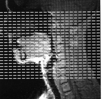
4 MRA MRA ARTERIOGRAM MRA is a record of blood flow. Any disturbance in blood flow will be recorded in the image. Small arrow - faster flowing blood. Large arrow/lighter signal - slower flowing blood. 4
5 Introduction MR angiography can provide screening of the vascular anatomy of the head, neck, body, and periphery. Clinical information should be supplemented with conventional MR images 5
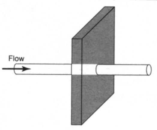
6 Time-of-Flight Time-of-Flight A vascular imaging technique usually for fast flow. The slab or slice (2D vs 3D) is saturated by repeated RF excitation. The saturation of the spins cause tissues to be darkened. Unsaturated blood entering the slab or slice will not be saturated and therefore will be bright. This is also called a rephase technique. 6
7 Time-of-Flight TOF is the time it takes for blood to flow through an imaging slice and the effect it has on the signal from blood. When using spin-echo pulses time-of-flight blood is dark. When using gradient echo sequences time-of-flight bright blood results. Time-of-flight refers to the effect of allowing blood to flow into or out of a slice between excitations. 7
8 Time-of-Flight 8
9 2D TOF GR images used short TR (~ msec) very short TE shortest TE times minimize intravoxel dephasing resulting in maximum flow effects small to medium flip angles 9
10 2D Time Of Flight (TOF) Benefits Both arteries and veins can be visualized. Shorter acquisitions times lend itself to abdominal vascular imaging using breath-hold techniques. Background tissue suppression is superior to 3D TOF imaging. Extremely slow flow velocity blood can be adequately visualized. Large area of coverage 10
11 2D Time Of Flight (TOF) Disadvantages Resolution limitations (smaller available slice thickness) cause an increase in partial volume effects. MIP projections tend to suffer due to thicker individual slices. Longer TE times and larger voxel volumes may result in an inappropriate decreased in signal following an area of stenosis. If the vessel is not quite perpendicular to the slice, or if it changes directions within the slice, saturation of the vascular signal occurs. 11
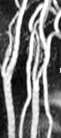
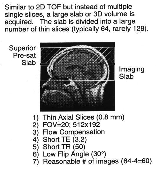
12 3D TOF 3D TOF imaging volumetrically acquires contiguous thin slices by the usual 3DFT method. 3D TOF imaging is the method of choice for imaging fast flowing blood that does not particularly follow a straight course. The TE times tend to be shorter than with the 2D TOF method resulting in minimized flow-turbulent related artifacts. In addition, high resolution images can be acquired lending easily to high resolution MIP images. 12

13 3D Time-of Flight 13
14 3D Time-of-Flight 14
15 3D TOF Benefits High resolution vascular imaging High SNR Rapid acquisition of 3-5 cm area High resolution MIP images possible 15
16 3D TOF Disadvantages Blood must traverse the volume quickly in order not to become saturated along with the background tissue (saturation of in-plane flow). Short T1 tissues in the background may not become saturated completely thereby simulating a vessel. Coverage 16
17 Phase Contrast Phase Contrast A vascular imaging technique where sets of images are encoded with different phase velocities and are then subtracted. Background subtraction is complete. This technique is used for slow flow as in the extremities. This is also known as phase shift. Available 2D and 3D imaging 17

18 Phase Contrast / PSI Angiography The greater the velocity of motion, the more phase shift, and the brighter the appearance of blood in the image. Therefore, complete suppression of stationary tissue can be achieved with PC. 18
19 Phase Contrast Angiography In phase contrast (PC) MRA, signal is based on the phase gained (or lost) as the spins move through a magnetic field gradient. 19


20 Phase Contrast Angiography Spins that are moving in the same direction as a magnetic field gradient develop a phase shift that is proportional to the velocity of the spins. This is the basis of phase-contrast angiography. 20
21 Phase Contrast Angiography This phase is subtracted from the background phase to determine the portion of the phase that is only due to motion. 21
22 Velocity Encoding (VENC) The velocity of blood flow is defined by the distance traveled between the positive and negative lobe of the velocity-encoding gradient (a 180 phase shift will produce maximum signal intensity). It is shown in cm/sec. 22
23 VENC The VENC value chosen will cause the blood flow traveling at exactly that value, in the direction of the positive lobe, to be assigned the maximum pixel value. Blood flow in the opposite direction will be dark. Choose a VENC value slightly higher than the predicted velocity of blood being observed to reduce the possibility of aliasing. 23
24 Phase Contrast Advantages Sensitive to flow within FOV Background suppression is superior Can evaluate blood flow velocity Can evaluate blood flow direction (magnitude and phase images) 24
25 Phase Contrast Disadvantages Scan times are long due to acquisition methods (2 additional sets of images acquired). Less sensitive to unpredictable flow 25
26 Contrast enhanced MRA Often in-plane flow and motion artifacts can degrade MRA images. Contrast MRA has therefore become the standard in MRA imaging. These sequences are generated with T1 weighted gradient echo images with bolus injections of gadolinium. Dynamic imaging are acquired before, during and after injection timed based on the vascular consideration. 26

27 Blood Flow 27

28 LAMINAR FLOW Blood flow that has a parabolic profile, in which the velocity of the protons in the center of the vessel is greater than the velocity of protons moving adjacent to the vessels walls. 28


29 Laminar Flow Laminar flow is usually found in veins and small arteries. MRA most accurately represents laminar flow. 29

30 Scan Parameters GRE sequences are required for bright blood Thin slices decrease the velocity necessary for 100% inflow of unsaturated blood as well as improve resolution Flow compensation refocuses motion related dephasing resulting in a brighter signal 30
31 Scan Parameter Optimization TR TR must be kept short to minimize acquisition time. However, TR and flip angle are complimentary and cannot exceed a maximum value without compromising the time of flight effect. 31
32 TE s Scan Parameter Optimization Must be kept short to avoid loss of signal from turbulence. If different TE choices are available, TE will be chosen by size of vessel to be imaged and pathology expected. 32
33 Scan Parameter Optimization Flip Angle Flip angle and TR for adequate suppression of background tissues. Flip Angle also will affect the saturation of the inflowing blood. The longer the vessel must remain unsaturated, the shorter the FA will need to be (e.g., the coronal or para-coronal acquisition of the proximal carotid arteries. If the FA is too large, the time of flight effect will be reduced. 33
34 Voxel size Scan Condition Optimization The voxel is the cubic portion of data as prescribed by the slice thickness and matrix. Smaller voxels reduce the turbulent signal loss as well as provide higher resolution of the vessels. 34
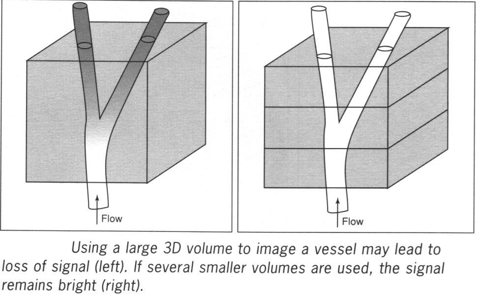
35 Slab thickness Scan Condition Optimization The slab thickness should never be thicker than the vessel segment of interest. The size of the slab is also related to the direction of flow. If the slab is perpendicular to the direction of flow in the vessel of interest, the distance the blood must flow is an essential consideration. Signal from blood flow will be saturated out of the slab if too thick. 35
36 Multiple Overlapping Thin Slice or Slab Acquisition (MOTSA) Multiple 2 to 3 cm thick 3D-TOF slabs covering the anatomy of interest. MOTSA combines the best features of 2D- and 3D-TOF MRA, because it has the unlimited coverage of 2D-TOF and the high spatial resolution of 3D-TOF. 36
 of interest.")
37 Maximum Intensity Projection Algorithm The maximum intensity projection algorithm is responsible for projecting the brightest pixels, from an anatomical stack of 2D or 3D base images, onto a plane, to generate an image of the projected view of the vessel(s) of interest. 37

38 Maximum Intensity Projection (MIP) 38
39 MRA Optimization Saturation bands are planar regions parallel to and adjacent to imaging planes or slabs. Within them blood that we do not want to see is saturated with repeated RF signals before it enters the imaging volume, emitting no signal. 39
40 Spatial Saturation With MRA To limit a study to either the arteries or veins, a saturation pulse may be added to the MRA pulse sequence to eliminate the inflowing blood from irrelevant vessels. 40
41 Saturation Bands To saturate Venous Flow - from heart If below heart: inferior presaturation band If above heart: superior presaturation band To saturate Arterial Flow - from heart If below heart: superior presaturation band If above heart: inferior presaturation band 41
42 Saturation Bands Walking saturation bands Places a new saturation band adjacent to each new imaging plane or slab as it is acquired in sequence so that each scan has been presaturated. More sophisticated placements include use of multiple bands and placement at oblique angles to optimize presaturation and exclusion of signals. 42

43 MRA Optimization Two main areas that require great care when producing MRA images are the carotid bifurcation and the circle of Willis. The common carotid artery on the left is usually the second major vessel originating from the aortic arch, whereas on the right side, the common carotid arises from the proximal innominate artery. The common carotid arteries usually bifurcate into the external and internal carotid arteries. 43


44 MRA Optimization - Carotids The bifurcations are important to visualize as the internal carotid arteries provide major blood flow to the brain. Turbulence and highvelocity blood flow effects produce complex flow patterns that can make MRA difficult. 44

45 Circle of Willis The best way to visualize the circle of Willis is to position it entirely within the middle slab of a three-slab MOTSA sequence. Avoid placing the interslab interface cross the circle of Willis. Any time-of-flight apparatus can produce a circle of Willis study without MOTSA using a stack of 2D or a 3D slab acquisition. Phase-contrast MRA may be used to show anatomic relationships and can provide information about the direction and velocity of flow (normal and abnormal) in the COW. 45

46 MOTSA through Circle of Willis MOTSA through a normal Circle of Willis. The vessels that form the circle of Willis include: two internal carotid arteries horizontal segments of the proximal and anterior cerebral arteries anterior cerebral arteries two posterior communicating arteries basilar artery 46
MR Advance Techniques. Vascular Imaging. Class II
 MR Advance Techniques Vascular Imaging Class II 1 Vascular Imaging There are several methods that can be used to evaluate the cardiovascular systems with the use of MRI. MRI will aloud to evaluate morphology
Essentials of Clinical MR, 2 nd edition. 99. MRA Principles and Carotid MRA
 99. MRA Principles and Carotid MRA As described in Chapter 12, time of flight (TOF) magnetic resonance angiography (MRA) is commonly utilized in the evaluation of the circle of Willis. TOF MRA allows depiction
1Pulse sequences for non CE MRA
 MRI: Principles and Applications, Friday, 8.30 9.20 am Pulse sequences for non CE MRA S. I. Gonçalves, PhD Radiology Department University Hospital Coimbra Autumn Semester, 2011 1 Magnetic resonance angiography
非對比劑與對比劑增強 MRA. 血管攝影與對比劑 A Course of MRI. 本週課程內容 -MR Angiography (MRA) Unenhanced MRA
 Unenhanced MRA") 本週課程內容 -MR Angiography (MRA) 血管攝影與對比劑 A Course of MRI 盧家鋒助理教授國立陽明大學物理治療暨輔助科技學系 alvin4016@ym.edu.tw 非對比劑增強 MRA(Unenhanced MRA) Time-of-flight (TOF) angiography Phase-contrast (PC) angiography 對比劑增強 MRA(Contrast-enhanced
本週課程內容 -MR Angiography (MRA) 血管攝影與對比劑 A Course of MRI 盧家鋒助理教授國立陽明大學物理治療暨輔助科技學系 alvin4016@ym.edu.tw 非對比劑增強 MRA(Unenhanced MRA) Time-of-flight (TOF) angiography Phase-contrast (PC) angiography 對比劑增強 MRA(Contrast-enhanced
Department of Radiology University of California San Diego. MR Angiography. Techniques & Applications. John R. Hesselink, M.D.
 Department of Radiology University of California San Diego MR Angiography Techniques & Applications John R. Hesselink, M.D. Vascular Imaging Arterial flow void Flow enhancement Gadolinium enhancement Vascular
MR Imaging with the CCSVI or Haacke protocol
 MR Imaging with the CCSVI or Haacke protocol Reports from the Haacke protocol are often made available to the patients. The report consists of four major components: 1. anatomical images of major neck
Non Contrast MRA. Mayil Krishnam. Director, Cardiovascular and Thoracic Imaging University of California, Irvine
 Non Contrast MRA Mayil Krishnam Director, Cardiovascular and Thoracic Imaging University of California, Irvine No disclosures Non contrast MRA-Why? Limitations of CTA Radiation exposure Iodinated contrast
Magnetic Resonance Imaging. Basics of MRI in practice. Generation of MR signal. Generation of MR signal. Spin echo imaging. Generation of MR signal
 Magnetic Resonance Imaging Protons aligned with B0 magnetic filed Longitudinal magnetization - T1 relaxation Transverse magnetization - T2 relaxation Signal measured in the transverse plane Basics of MRI
MR Flow Imaging in Vascular Malformations Using Gradient Recalled Acquisition
 637 MR Flow Imaging in Vascular Malformations Using Gradient Recalled Acquisition William M. Needell 1 Kenneth R. Maravilla Twenty patients with known or suspected intracranial vascular lesions were evaluated
Clinical Applications
 C H A P T E R 16 Clinical Applications In selecting pulse sequences and measurement parameters for a specific application, MRI allows the user tremendous flexibility to produce variations in contrast between
Cerebral MR Venography: Normal Anatomy and Potential Diagnostic Pitfalls
 AJNR Am J Neuroradiol 21:74 78, January 2000 Cerebral MR Venography: Normal Anatomy and Potential Diagnostic Pitfalls R. H. Ayanzen, C. R. Bird, P. J. Keller, F. J. McCully, M. R. Theobald, and J. E. Heiserman
Non-Contrast MRA. How and When 1996! Why Non-Contrast MRA? Angiography: What are our goals? Inflow Techniques Differences in excitation hx
 A major teaching hospital of Harvard Medical School Angiography: What are our goals? Non-Contrast MRA: How and When Neil M. Rofsky, M.D. Professor of Radiology, Harvard Medical School Director of MRI &
Sung Hong Park. M.S. in Electrical Engineering, KAIST, South Korea, Submitted to the Graduate Faculty of
 NONINVASIVE IMAGING OF BRAIN VASCULATURE WITH HIGH RESOLUTION BLOOD OXYGENATION LEVEL DEPENDENT VENOGRAPHY IN MAGNETIC RESONANCE IMAGING: APPLICATIONS TO FUNCTIONAL AND CLINICAL STUDIES by Sung Hong Park
T Clinical Magnetic Resonance Angiography
 MRI for Technologists 4712-204T Clinical Magnetic Resonance Angiography PROGRAM INFORMATION MRI for Technologists is a training program designed to meet the needs of radiologic technologists entering or
How I do it: Non Contrast-Enhanced MR Angiography (syngo NATIVE)
") Clinical How-I-do-it Cardiovascular How I do it: Non Contrast-Enhanced MR Angiography (syngo NATIVE) Manuela Rick, Nina Kaarmann, Peter Weale, Peter Schmitt Siemens Healthcare, Erlangen, Germany Introduction
Clinical How-I-do-it Cardiovascular How I do it: Non Contrast-Enhanced MR Angiography (syngo NATIVE) Manuela Rick, Nina Kaarmann, Peter Weale, Peter Schmitt Siemens Healthcare, Erlangen, Germany Introduction
MR Angiography in the evaluation of Lower Extremity Arterial Disease
 March 2001 MR Angiography in the evaluation of Lower Extremity Arterial Disease Ted Mau, Harvard Medical School Year III Objectives We will cover: Indications for Magnetic Resonance Angiography (MRA) Basic
Neuroradiology MR Protocols
 Neuroradiology MR Protocols Brain protocols N 1: Brain MRI without contrast N 2: Pre- and post-contrast brain MRI N 3 is deleted N 4: Brain MRI without or pre-/post-contrast (seizure protocol) N 5: Pre-
Anatomic Evaluation of the Circle of Willis: MR Angiography versus Intraarterial Digital Subtraction Angiography
 Anatomic Evaluation of the Circle of Willis: MR Angiography versus Intraarterial Digital Subtraction Angiography K. W. Stock, S. Wetzel, E. Kirsch, G. Bongartz, W. Steinbrich, and E. W. Radue PURPOSE:
Department of Radiology, Division of MRI, University of Michigan, 1500 E. Medical Center Drive, Ann Arbor, MI , USA
 Abdom Imaging 23:469 484 (1998) Abdominal Imaging Springer-Verlag New York Inc. 1998 Contrast-enhanced MR angiography J. H. Maki, T. L. Chenevert, M. R. Prince Department of Radiology, Division of MRI,
A Magnetic Resonance Imaging Method for
 Journal of Cardiovascular Magnetic Resonance, 1(1), 59-64 (1999) INVITED PAPER Use of MRI in ASD Asessment A Magnetic Resonance Imaging Method for Evaluating Atrial Septa1 Defects Godtfred Holmvang Cardiac
6/23/2009. Inversion Recovery (IR) Techniques and Applications. Variations of IR Technique. STIR, FLAIR, TI and TI Null. Applications of IR
 Techniques and Applications. Variations of IR Technique. STIR, FLAIR, TI and TI Null. Applications of IR") The Anatomy of Basic R Pulse Sequences Inversion Recovery () Techniques and Applications Chen Lin, PhD Indiana University School of edicine & Clarian Health Partners agnetization Preparation Section Chemical
The Anatomy of Basic R Pulse Sequences Inversion Recovery () Techniques and Applications Chen Lin, PhD Indiana University School of edicine & Clarian Health Partners agnetization Preparation Section Chemical
Methods. Yahya Paksoy, Bülent Oğuz Genç, and Emine Genç. AJNR Am J Neuroradiol 24: , August 2003
 AJNR Am J Neuroradiol 24:1364 1368, August 2003 Retrograde Flow in the Left Inferior Petrosal Sinus and Blood Steal of the Cavernous Sinus Associated with Central Vein Stenosis: MR Angiographic Findings
RECENT ADVANCES IN CLINICAL MR OF ARTICULAR CARTILAGE
 In Practice RECENT ADVANCES IN CLINICAL MR OF ARTICULAR CARTILAGE By Atsuya Watanabe, MD, PhD, Director, Advanced Diagnostic Imaging Center and Associate Professor, Department of Orthopedic Surgery, Teikyo
NEURO PROTOCOLS MRI NEURO PROTOCOLS (SIEMENS SCANNERS)
") Page 1 NEURO PROTOCOLS Brain Stroke Brain Brain with contrast Brain for seizures Brain for MS Brain for Pineal gland Sella FAST Scan for hydrocephalus MRA/MRV Brain MRA carotids 8 th nerve Cranial nerves
Page 1 NEURO PROTOCOLS Brain Stroke Brain Brain with contrast Brain for seizures Brain for MS Brain for Pineal gland Sella FAST Scan for hydrocephalus MRA/MRV Brain MRA carotids 8 th nerve Cranial nerves
Speed, Comfort and Quality with NeuroDrive
 Speed, Comfort and Quality with NeuroDrive Echelon Oval provides a broad range of capabilities supporting fast, accurate diagnosis of brain conditions and injuries. From anatomical depiction to vascular
Previous talks. Clinical applications for spiral flow imaging. Clinical applications. Clinical applications. Coronary flow: Motivation
 for spiral flow imaging Joao L. A. Carvalho Previous talks Non-Cartesian reconstruction (2005) Spiral FVE (Spring 2006) Aortic flow Carotid flow Accelerated spiral FVE (Fall 2006) 2007? Department of Electrical
Perforating arteries originating from the posterior communicating artery: a 7.0-Tesla MRI study
 Eur Radiol (2009) 19: 2986 2992 DOI 10.1007/s00330-009-1485-4 MAGNETIC RESONANCE Mandy M. A. Conijn Jeroen Hendrikse Jaco J. M. Zwanenburg Taro Takahara Mirjam I. Geerlings Willem P. Th. M. Mali Peter
Raja Muthupillai, PhD. Department of Diagnostic and Interventional Radiology St. Luke s Episcopal Hospital. Research Support: Philips Healthcare
 3D Cardiac Imaging Raja Muthupillai, PhD Department of Diagnostic and Interventional Radiology St. Luke s Episcopal Hospital Houston, TX Disclosures Research Support: Philips Healthcare This presentation
MRI protocol for post-repaired TOF
 2012 NASCI MRI protocol for post-repaired TOF Taylor Chung, M.D. Associate Director, Body and Cardiovascular Imaging Department of Diagnostic Imaging Children s Hospital & Research Center Oakland Oakland,
MR Advance Techniques. Cardiac Imaging. Class IV
 MR Advance Techniques Cardiac Imaging Class IV Heart The heart is a muscular organ responsible for pumping blood through the blood vessels by repeated, rhythmic contractions. Layers of the heart Endocardium
Extracranial Carotid Arteries: Evaluation with Black Blood MR Angiography
 Robert R. Edelman, MD #{149} Heinrich P. Mattle, MD #{149} Bernd Wallner, MD #{149} Richard Bajakian, MD #{149} Jonathan Kleefield, MD #{149} Craig Kent, MD #{149} John J. Skillman, MD Jeffrey B. Mendel,
MRI Abdomen Protocol Pancreas/MRCP with Contrast
 MRI Abdomen Protocol Pancreas/MRCP with Contrast Reviewed By: Brett Mollard, MD; Anna Ellermeier, MD Last Reviewed: July 2018 Contact: (866) 761-4200 Standard uses: 1. Characterization of cystic and solid
High-Sensitivity Coil Array for Head and Neck Imaging: Technical Note
 AJNR Am J Neuroradiol 22:1881 1886, November/December 2001 Technical Note High-Sensitivity Coil Array for Head and Neck Imaging: Technical Note Roland G. Henry, Nancy J. Fischbein, William P. Dillon, Daniel
Normal MRI Appearance and Motion-Related Phenomena of CSF
 Lisanti et al. MRI of CSF MR Imaging Pictorial Essay Christopher Lisanti 1 Carrie Carlin 1 Kevin P. anks 2 David Wang 3 Lisanti C, Carlin C, anks KP, Wang D Keywords: CNS, CSF, MRI DOI:10.2214/JR.05.0003
MR Advance Techniques. Cardiac Imaging. Class III
 MR Advance Techniques Cardiac Imaging Class III Black Blood Imaging & IR Blue= O2 poor blood Red=O2 rich blood Inversion pulses can produce black blood imaging in GRE pulse sequences. Specially on the
Objectives 8/17/2011. Challenges in Cardiac Imaging. Challenges in Cardiac Imaging. Basic Cardiac MRI Sequences
 8/17/2011 Traditional Protocol Model for Tomographic Imaging Cardiac MRI Sequences and Protocols Frandics Chan, M.D., Ph.D. Stanford University Medical Center Interpretation Lucile Packard Children s Hospital
ρ = 4(νp)2 Scale -200 to 200 V = m/s Grad = 34 mmhg V = 1.9 m/s Grad = 14 mmhg Types
2 Scale -200 to 200 V = m/s Grad = 34 mmhg V = 1.9 m/s Grad = 14 mmhg Types") Pre and Post Operative Evaluation of the Aorta and Aortic Valve Andrew J. Bierhals, MD The Pre and Post-Operative Evaluation of the Aorta and Aortic Valve Andrew Bierhals, MD, MPH Mallinckrodt Institute
Pre and Post Operative Evaluation of the Aorta and Aortic Valve Andrew J. Bierhals, MD The Pre and Post-Operative Evaluation of the Aorta and Aortic Valve Andrew Bierhals, MD, MPH Mallinckrodt Institute
Field Strength. Regional Perfusion Imaging (RPI) matches cerebral arteries to flow territories
 matches cerebral arteries to flow territories") Field Strength Changing how the world looks at MR. Regional Perfusion Imaging (RPI) matches cerebral arteries to flow territories Research groups in Utrecht, Baltimore and Singapore collaborate on this
Field Strength Changing how the world looks at MR. Regional Perfusion Imaging (RPI) matches cerebral arteries to flow territories Research groups in Utrecht, Baltimore and Singapore collaborate on this
Three-Dimensional Time-of-Flight MR Angiography in the Evaluation of Intracranial Aneurysms Treated by Endovascular Balloon Occlusion
 Three-Dimensional Time-of-Flight MR Angiography in the Evaluation of Intracranial Aneurysms Treated by Endovascular Balloon Occlusion JayS. Tsuruda, Robert J. Sevick, 1 and Van V. Halbach Summary: The
High Field MR of the Spine
 Department of Radiology University of California San Diego 3T for MR Applications Advantages High Field MR of the Spine Increased signal-to-noise Better fat suppression Increased enhancement with gadolinium
Cardiovascular MR Imaging at 3 T: Opportunities, Challenges, and Solutions 1
 TECHNICAL ADVANCEMENTS IN CARDIAC MR IMAGING 1612 Cardiovascular MR Imaging at 3 T: Opportunities, Challenges, and Solutions 1 Prabhakar Rajiah, MD, FRCR Michael A. Bolen, MD Abbreviations: BOLD = blood
Tips and Tricks of State of the art MRA
 Tips and Tricks of State of the art MRA Mayil Krishnam, MD,MBA, MRCP,FRCR(UK) Professor of Radiology Director, Cardiovascular and Thoracic Imaging University of California, Irvine Objectives Technical
Can SCMR CMR protocol recommendations
 Can SCMR CMR protocol recommendations V1.3 - April 2009 CanSCMR CMR Protocol and SOP Recommendation 2009 (15 minutes) 2 Planning of LV fct. real time multiple axes Realtime 3 cine long axis 6 long axes
PHYSICS OF MRI ACQUISITION. Alternatives to BOLD for fmri
 PHYSICS OF MRI ACQUISITION Quick Review for fmri HST-583, Fall 2002 HST.583: Functional Magnetic Resonance Imaging: Data Acquisition and Analysis Harvard-MIT Division of Health Sciences and Technology
Fulfilling the Promise
 Fulfilling the Promise of Cardiac MR Non-contrast, free-breathing technique generates comprehensive evaluation of the coronary arteries By Maggie Fung, MR Cardiovascular Clinical Development Manager; Wei
Introduction. Cardiac Imaging Modalities MRI. Overview. MRI (Continued) MRI (Continued) Arnaud Bistoquet 12/19/03
 MRI (Continued) Arnaud Bistoquet 12/19/03") Introduction Cardiac Imaging Modalities Arnaud Bistoquet 12/19/03 Coronary heart disease: the vessels that supply oxygen-carrying blood to the heart, become narrowed and unable to carry a normal amount
Introduction Cardiac Imaging Modalities Arnaud Bistoquet 12/19/03 Coronary heart disease: the vessels that supply oxygen-carrying blood to the heart, become narrowed and unable to carry a normal amount
The Use and Pitfalls of Intracranial Vessel Wall Imaging: How We Do It 1
 This copy is for personal use only. To order printed copies, contact reprints@rsna.org Reviews and Commentary n How I Do It Arjen Lindenholz, MD Anja G. van der Kolk, MD, PhD Jaco J. M. Zwanenburg, PhD
Time-Of-Flight MRA. Faculty Disclosures Vincent B. Ho, M.D. Presentation Objectives. MRA Techniques. Pros and Cons of MRA
 Faculty Disclosures Vincent B. Ho, M.D. MR Angiography Techniques and Pitfalls Financial Disclosure Grant/Research Support General Electric Medical Systems Off-Label/Investigational Drug Use Dr. Ho will
Magnetization Preparation Sequences
 Magnetization Preparation Sequences Acquisition method may not give desired contrast Prep block adds contrast (and/or encoding) MP-RAGE = Magnetization prepared rapid acquisition with gradient echo (Mugler,
CARDIAC MRI. Cardiovascular Disease. Cardiovascular Disease. Cardiovascular Disease. Overview
 CARDIAC MRI Dr Yang Faridah A. Aziz Department of Biomedical Imaging University of Malaya Medical Centre Cardiovascular Disease Diseases of the circulatory system, also called cardiovascular disease (CVD),
NEUROVASCULAR ANATOMY
 E. Michael Harned, M.D. Assistant Professor of Clinical Radiology ndiana University School of Medicine have nothing to disclose Neurovascular Magnetic Resonance Angiography And Magnetic Resonance Venography
醫用磁振學 MRM 肌肉骨骼磁振造影簡介 肌肉骨骼磁振造影. 本週課程內容 General Technical Considerations 肌肉骨骼磁振造影簡介 盧家鋒助理教授國立陽明大學生物醫學影像暨放射科學系
 本週課程內容 http://www.ym.edu.tw/~cflu 肌肉骨骼磁振造影簡介 醫用磁振學 MRM 肌肉骨骼磁振造影 盧家鋒助理教授國立陽明大學生物醫學影像暨放射科學系 alvin4016@ym.edu.tw MRI of the musculoskeletal system (5th/6th edition) Editor: Thomas H. Berquist MD 2 General
Blood flow assessment in cerebral arteries with 4D flow magnetic resonance imaging
 Blood flow assessment in cerebral arteries with 4D flow magnetic resonance imaging An automatic atlas-based approach Tora Dunås Department of Radiation Sciences Department of Pharmacology and Clinical
ASL BASICS II. Learning Objectives. Outline. Acquisition. M. A. Fernández-Seara, Ph. D. Arterial spin labeled perfusion MRI: basic theory
 Acquisition ASL BASICS II M. A. Fernández-Seara, Ph. D. Neuroimaging Laboratory Center for Applied Medical Research University of Navarra Pamplona, Spain Outline Arterial spin labeled perfusion MRI: basic
8/4/2016. Optimizing Pediatric Cardiovascular MRI. Disclosure. Outline. Jie Deng, PhD, DABMP, Cynthia Rigsby, MD
 Optimizing Pediatric Cardiovascular MRI Jie Deng, PhD, DABMP, Cynthia Rigsby, MD Department of Medical Imaging Radiology, Feinberg School of Medicine, Northwestern University Aug 4 th, 2016 Disclosure
Magnetic resonance techniques to measure distribution of cerebral blood flow
 212 M. Günther Magnetic resonance techniques to measure distribution of cerebral blood flow M. Günther 1,2 1 mediri GmbH, Heidelberg, Germany; 2 Neurologische Klinik, Universitätsklinikum Mannheim, Universität
Fully Refocused Gradient Recalled Echo (FRGRE): Factors Affecting Flow and Motion Sensitivity in Cardiac MRI
: Factors Affecting Flow and Motion Sensitivity in Cardiac MRI") Journal of Cardiovascular Magnetic Resonance w, 4(2), 211 222 (2002) METHODS Fully Refocused Gradient Recalled Echo (FRGRE): Factors Affecting Flow and Motion Sensitivity in Cardiac MRI Laurie B. Hildebrand
Journal of Cardiovascular Magnetic Resonance w, 4(2), 211 222 (2002) METHODS Fully Refocused Gradient Recalled Echo (FRGRE): Factors Affecting Flow and Motion Sensitivity in Cardiac MRI Laurie B. Hildebrand
1 Normal Anatomy and Variants
 1 Normal Anatomy and Variants 1.1 Normal Anatomy MR Technique. e standard MR protocol for a routine evaluation of the spine always comprises imaging in sagittal and axial planes, while coronal images are
Contrast material enhanced threedimensional
 Winfried A. Willinek, MD Jürgen Gieseke, PhD Rudolf Conrad, MD Holger Strunk, MD Romhild Hoogeveen, PhD Marcus von Falkenhausen, MD Ewald Keller, MD Horst Urbach, MD Christiane K. Kuhl, MD Hans H. Schild,
Fat Suppression in the Abdomen
 Clinical How I do it? Fat Suppression in the Abdomen Wilhelm Horger Siemens Medical Solutions, Erlangen, Germany Introduction Due to the different chemical environment, hydrogen nuclei in - and in -tissue
Table 1. Summary of PET and fmri Methods. What is imaged PET fmri BOLD (T2*) Regional brain activation. Blood flow ( 15 O) Arterial spin tagging (AST)
 Regional brain activation. Blood flow ( 15 O) Arterial spin tagging (AST)") Table 1 Summary of PET and fmri Methods What is imaged PET fmri Brain structure Regional brain activation Anatomical connectivity Receptor binding and regional chemical distribution Blood flow ( 15 O)
Table 1 Summary of PET and fmri Methods What is imaged PET fmri Brain structure Regional brain activation Anatomical connectivity Receptor binding and regional chemical distribution Blood flow ( 15 O)
Turbo ASL: Arterial Spin Labeling With Higher SNR and Temporal Resolution
 COMMUNICATIONS Magnetic Resonance in Medicine 44:511 515 (2000) Turbo ASL: Arterial Spin Labeling With Higher SNR and Temporal Resolution Eric C. Wong,* Wen-Ming Luh, and Thomas T. Liu A modified pulsed
Original Research. Tadateru Sumi, DDS, 1 Misa Sumi, DDS, PhD, 1 Marc Van Cauteren, PhD, 2 Yasuo Kimura, DDS, PhD, 1 and Takashi Nakamura, DDS, PhD 1 *
 JOURNAL OF MAGNETIC RESONANCE IMAGING 25:1028 1034 (2007) Original Research Parallel Imaging Technique for the External Carotid Artery and its Branches: Comparison of Balanced Turbo Field Echo, Phase Contrast,
Tissue-engineered medical products Evaluation of anisotropic structure of articular cartilage using DT (Diffusion Tensor)-MR Imaging
-MR Imaging") Provläsningsexemplar / Preview TECHNICAL REPORT ISO/TR 16379 First edition 2014-03-01 Tissue-engineered medical products Evaluation of anisotropic structure of articular cartilage using DT (Diffusion Tensor)-MR
Provläsningsexemplar / Preview TECHNICAL REPORT ISO/TR 16379 First edition 2014-03-01 Tissue-engineered medical products Evaluation of anisotropic structure of articular cartilage using DT (Diffusion Tensor)-MR
UK Biobank. Imaging modality Cardiovascular Magnetic Resonance (CMR) Version th Oct 2015
 Version th Oct 2015") Imaging modality Cardiovascular Magnetic Resonance (CMR) Version 1.0 http://www.ukbiobank.ac.uk/ 30 th Oct 2015 This document details the procedure for the CMR scan performed at an Imaging assessment centre
Imaging modality Cardiovascular Magnetic Resonance (CMR) Version 1.0 http://www.ukbiobank.ac.uk/ 30 th Oct 2015 This document details the procedure for the CMR scan performed at an Imaging assessment centre
8/4/2016. MRI for Radiotherapy: MRI Basics. Nuclear Magnetic Resonance. Nuclear Magnetic Resonance. Wilson Miller
 MRI for Radiotherap: MRI asics Wilson Miller Universit of Virginia Department of Radiolog & Medical Imaging AAPM 2016 August 4, 2016 Nuclear Magnetic Resonance Magnetic resonance images are created using
The Low Sensitivity of Fluid-Attenuated Inversion-Recovery MR in the Detection of Multiple Sclerosis of the Spinal Cord
 The Low Sensitivity of Fluid-Attenuated Inversion-Recovery MR in the Detection of Multiple Sclerosis of the Spinal Cord Mark D. Keiper, Robert I. Grossman, John C. Brunson, and Mitchell D. Schnall PURPOSE:
Advanced Vascular Imaging: Pulsatile Tinnitus. Disclosures. Pulsatile Tinnitus: Differential Diagnosis. Pulsatile Tinnitus
 Advanced Vascular Imaging: Pulsatile Tinnitus Patrick Turski MD, Zach Clark MD, Tabby Kennedy MD The Objectives of this presentation are to: Review the differential diagnosis of pulsatile tinnitus Discuss
ACR MRI Accreditation: Medical Physicist Role in the Application Process
 ACR MRI Accreditation: Medical Physicist Role in the Application Process Donna M. Reeve, MS, DABR, DABMP Department of Imaging Physics University of Texas M.D. Anderson Cancer Center Educational Objectives
Assessment of Adipose Tissue from Whole Body 3T MRI Scans
 Assessment of Adipose Tissue from Whole Body 3T MRI Scans Ting Song 1, Jing An 2, Qun Chen 2, Vivian Lee 2, Andrew Laine 1 1 Department of Biomedical Engineering, Columbia University, New York, NY, USA
Ultrasound Physics & Terminology
 Ultrasound Physics & Terminology This module includes the following: Basic physics terms Basic principles of ultrasound Ultrasound terminology and terms Common artifacts seen Doppler principles Terms for
Diagnosis of Vertebral Artery Ostial Stenosis on Contrast-Enhanced MR Angiography: Usefulness of a Thin-Slab MIP Technique
 Diagnosis of Vertebral Artery Ostial Stenosis on Contrast-Enhanced MR Angiography: Usefulness of a Thin-Slab MIP Technique Sun Mi Kim 1, Deok Hee Lee 2 Jin Woo Choi 3, Byung Se Choi 4, Hyun Sin In 5 It
Evaluation of Intracranial Vasculatures in Healthy Subjects with Arterial-Spin-Labeling-Based 4D-MR Angiography at 3T
 Magn Reson Med Sci, Vol. 15, No. 3, pp. 335 339, 2016 doi:10.2463/mrms.tn.2015-0081 TECHNICAL NOTE Evaluation of Intracranial Vasculatures in Healthy Subjects with Arterial-Spin-Labeling-Based 4D-MR Angiography
Orthopedic Hardware Imaging Part II: MRI v. Metal
 Orthopedic Hardware Imaging Trent Roth, MD And Lauren Ladd, MD Indiana University School of Medicine IU Health Physicians-Radiology Recap: Imaging Techniques Radiography Standard for initial and surveillance
The study of local hemodynamics within anatomically complex
 TECHNICAL NOTE S. Wetzel S. Meckel A. Frydrychowicz L. Bonati E.-W. Radue K. Scheffler J. Hennig M. Markl In Vivo Assessment and Visualization of Intracranial Arterial Hemodynamics with Flow- Sensitized
Master Thesis in Radiation Physics 09/10 Annie Olsson. Supervisors: Frank Risse Lars E. Olsson. Imaging centre AstraZeneca R&D Mölndal
 Determination of a preclinical protocol for quantitative measurements of perfusion and permeability in the rat lung using dynamic contrast enhanced-mri Master Thesis in Radiation Physics 9/1 Annie Olsson
The Extracranial Facial Nerve: High Resolution Three-Dimensional Fourier Transform MR Imaging
 The Extracranial Facial Nerve: High Resolution Three-Dimensional Fourier Transform MR Imaging Robert B. McGhee, Jr., 1.3 Donald W. Chakeres, 1 Petra Schmal brock, 1 Martha A. Brogan, 1 and John A. Negulesco
Guide to Small Animal Vascular Imaging using the Vevo 770 Micro-Ultrasound System
 Guide to Small Animal Vascular Imaging using the Vevo 770 Micro-Ultrasound System January 2007 Objectives: After completion of this module, the participant will be able to accomplish the following: Understand
CFD Challenge: Simulation of Hemodynamics in a Patient-Specific Aortic Coarctation Model
 CFD Challenge: Simulation of Hemodynamics in a Patient-Specific Aortic Coarctation Model Background Coarctation of the aorta (CoA) accounts for 8%-11% of congenital heart defects, affecting tens of thousands
Congenital Heart Disease
 UNIT A10.1 One of the primary roles of cardiac MR imaging has been the assessment of congenital heart disease. Initially used to define cardiac anatomy as an adjunct to echocardiography, cardiac MR now
Correction of CSF Motion Artifact on MR Images of the Brain and Spine by Pulse Sequence Modification: Clinical Evaluation
 1069 Correction of CSF Motion Artifact on MR Images of the Brain and Spine by Pulse Sequence Modification: Clinical Evaluation Nikolaus M. Szeverenyi 1 Stephen A. Kieffer Edwin D. Cacayorin A modification
Non-contrast-enhanced MR portography and hepatic venography with time-spatial labeling inversion pulses: comparison at 1.5 Tesla and 3 Tesla
 Research Non-contrast-enhanced MR portography and hepatic venography with time-spatial labeling inversion pulses: comparison at 1.5 Tesla and 3 Tesla Acta Radiologica Open 4(5) 1 8! The Foundation Acta
Echelon Oval provides a robust suite of leading musculoskeletal imaging capabilities for detailed assessment of all anatomy for your most challenging
 Echelon Oval provides a robust suite of leading musculoskeletal imaging capabilities for detailed assessment of all anatomy for your most challenging cases. Hitachi Medical Systems America, Inc. 1959 Summit
In vivo diffusion tensor imaging (DTI) of articular cartilage as a biomarker for osteoarthritis
 of articular cartilage as a biomarker for osteoarthritis") In vivo diffusion tensor imaging (DTI) of articular cartilage as a biomarker for osteoarthritis Jose G. Raya 1, Annie Horng 2, Olaf Dietrich 2, Svetlana Krasnokutsky 3, Luis S. Beltran 1, Maximilian F.
In vivo diffusion tensor imaging (DTI) of articular cartilage as a biomarker for osteoarthritis Jose G. Raya 1, Annie Horng 2, Olaf Dietrich 2, Svetlana Krasnokutsky 3, Luis S. Beltran 1, Maximilian F.
KNEE ALIGNMENT SYSTEM (KAS) MRI Protocol
 MRI Protocol") KNEE ALIGNMENT SYSTEM (KAS) MRI Protocol Sample referral sticker Referral Sticker Insert here Corin 17 Bridge Street Pymble NSW Australia 2073 P: +61 (0)2 9497 7400 F: +61 (0)2 9497 7498 E: KAS.customerservice@coringroup.com
KNEE ALIGNMENT SYSTEM (KAS) MRI Protocol Sample referral sticker Referral Sticker Insert here Corin 17 Bridge Street Pymble NSW Australia 2073 P: +61 (0)2 9497 7400 F: +61 (0)2 9497 7498 E: KAS.customerservice@coringroup.com
Cover Page. The following handle holds various files of this Leiden University dissertation:
 Cover Page The following handle holds various files of this Leiden University dissertation: http://hdl.handle.net/1887/62047 Author: Bosch, H.C.M. van den Title: Clinical advances in cardiovascular magnetic
Why Talk About Technique? MRI of the Knee:
 Why Talk About Technique? MRI of the Knee: Part 1 - Imaging Techniques Mark Anderson, M.D. University of Virginia Health Sciences Center Charlottesville, Virginia Always had an interest teach our fellows
Endovascular treatment of intracranial aneurysms with detachable
 ORIGINAL RESEARCH L. Pierot C. Delcourt F. Bouquigny D. Breidt B. Feuillet O. Lanoix S. Gallas Follow-Up of Intracranial Aneurysms Selectively Treated with Coils: Prospective Evaluation of Contrast-Enhanced
Optimized phase contrast MRV technique outperforms timeof-flight in the diagnosis of cerebral venous thrombosis
 Optimized phase contrast MRV technique outperforms timeof-flight in the diagnosis of cerebral venous thrombosis Poster No.: C-3377 Congress: ECR 2010 Type: Topic: Authors: Keywords: DOI: Scientific Exhibit
Renal vascular evaluation with 64 Multislice Computerized Tomography Daniela Stoisa, Fabrizzio E. Galiano, Andrés Quaranta, Roberto L.
 Renal vascular evaluation with 64 Multislice Computerized Tomography Daniela Stoisa, Fabrizzio E. Galiano, Andrés Quaranta, Roberto L. Villavicencio Footnote Diagnóstico Médico Oroño. Bv. Oroño 1515. 2000.
Flow Quantification from 2D Phase Contrast MRI in Renal Arteries using Clustering
 Flow Quantification from 2D Phase Contrast MRI in Renal Arteries using Clustering Frank G. Zöllner 1,2, Jan Ankar Monnsen 1, Arvid Lundervold 2, Jarle Rørvik 1 1 Department for Radiology, University of
Personal use only. MRI Metal Artifact Reduction: Shoulder Implants and Arthroplasty. Reto Sutter, MD
 MRI Metal Artifact Reduction: Shoulder Implants and Arthroplasty Reto Sutter, MD University Hospital Balgrist Zurich University of Zurich Cor PD fat sat 56-year old male patient with positive lift-off
Objectives. CMR Volumetric Analysis 8/25/11. CMR Volumetric Analysis Technique. Cardiac imaging plane acquisition. CMR Volumetric Analysis
 Objectives Cynthia K. Rigsby Children s Memorial Hospital Chicago, IL CMR volumetric analysis Techniques Normalized data Sources of error CMR phase contrast flow analysis Techniques What we can do with
Personalized Solutions. MRI Protocol for PSI and Signature Guides
 Personalized Solutions MRI Protocol for PSI and Signature Guides 2 Personalized Solutions MRI Protocol for PSI and Signature Guides Purpose and Summary This protocol is applicable for the Zimmer Biomet
A WORLD OF KNOWLEDGE FOR MR TECHNOLOGISTS & RADIOGRAPHERS
 SMRT A WORLD OF KNOWLEDGE FOR MR TECHNOLOGISTS & RADIOGRAPHERS SECTION FOR MAGNETIC RESONANCE TECHNOLOGISTS OF THE INTERNATIONAL SOCIETY FOR MAGNETIC RESONANCE IN MEDICINE Home Studies Educational Seminars
MR coronary artery imaging with 3D motion adapted gating (MAG) in comparison to a standard prospective navigator technique
 in comparison to a standard prospective navigator technique") Journal of Cardiovascular Magnetic Resonance (2005) 7, 793 797 Copyright D 2005 Taylor & Francis Inc. ISSN: 1097-6647 print / 1532-429X online DOI: 10.1080/10976640500287547 ANGIOGRAPHY MR coronary artery
Journal of Cardiovascular Magnetic Resonance (2005) 7, 793 797 Copyright D 2005 Taylor & Francis Inc. ISSN: 1097-6647 print / 1532-429X online DOI: 10.1080/10976640500287547 ANGIOGRAPHY MR coronary artery
BioMatrix Tuners: CoilShim
 MAGNETOM Vida special issue Head and Neck Imaging Clinical 11 BioMatrix Tuners: CoilShim Miriam R. Keil, Ph.D.; Jörg Rothard; Carmel Hayes, Ph.D. Siemens Healthineers, Erlangen, Germany A cervical spine
Subject Index. Automated triggering 88 AVM 128, 129
 Subject Index A Abberrant right subclavian artery 150 Abdominal aorta 195-208 aneurysm (AAA) 198 aneurysm branching vessels 201 aortic dissection 203 dark-blood imaging 197 eccentric aneurysm 199 fusiform
Planimetric and continuity equation assessment of aortic valve area (AVA): comparison between cardiac magnetic resonance (cmr) and echocardiography
: comparison between cardiac magnetic resonance (cmr) and echocardiography") Planimetric and continuity equation assessment of aortic valve area (AVA): comparison between cardiac magnetic resonance (cmr) and echocardiography Poster No.: C-2058 Congress: ECR 2011 Type: Scientific
Planimetric and continuity equation assessment of aortic valve area (AVA): comparison between cardiac magnetic resonance (cmr) and echocardiography Poster No.: C-2058 Congress: ECR 2011 Type: Scientific
Rapid Quantitation of High-Speed Flow Jets
 Rapid Quantitation of High-Speed Flow Jets Krishna S. Nayak, 1 * Bob S. Hu, 1,2 and Dwight G. Nishimura 1 Magnetic Resonance in Medicine 50:366 372 (2003) Flow jets containing velocities up to 5 7 m/s