MR Advance Techniques. Cardiac Imaging. Class III
|
|
|
- Hilary Stevenson
- 5 years ago
- Views:
Transcription
1 MR Advance Techniques Cardiac Imaging Class III

2 Black Blood Imaging & IR Blue= O2 poor blood Red=O2 rich blood Inversion pulses can produce black blood imaging in GRE pulse sequences. Specially on the heart where blood flow goes in different directions and pre-sat bands does not work properly.
3 Black Blood Imaging & IR Inversion pulses to produce black blood in GRE sequences can be known as driven equilibrium. These pulse sequences begins with a NON slice selected 180º pulse and then another slice selected 180º pulse. A TI equivalent to the null point of flowing spins entering the slice will be applied.

4 Double IR This technique is also known as Double Inversion Recovery or Double IR or driven equilibrium. Driven Equilibrium
5 Double IR 180 Non Slice Selected Slice Selected At TI of Blood (650 ms)
6 Longitudinal Magnetization TI of Blood Double IR 180 Non Slice Selected 180 Slice Selected 90 Slice Selected TI of Blood (650 ms)
7 Longitudinal Magnetization TI of Blood TI of Fat Triple IR 180 Non Slice Selected 180 Slice Selected 180 Slice Selected 90 Slice Selected TI of Blood (650 ms) TI of Fat (150 ms)
8 Double IR Vs. Triple IR

9 TI TI 500 TI 650


10 Gaiting Gaiting is a very general term used to describe a technique of reducing phase mismapping from periodic motion cause by respiration, cardiac motion and pulsatile flow.
11 Types of Gating Cardiac gating Respiratory gaiting
12 Cardiac Gaiting Application Cardiac gaiting can be used: Reduce cardiac motion artifact Reduce pulsatile flow artifact Acquired cine images of the heart, blood vessels and CSF.
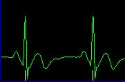
13 Cardiac Gaiting There are several forms of cardiac gating: Electrocardiogram (ECG, EKG) Peripheral gating Pseudo gating
 A T wave that represents ventricular diastole (ventricular")
14 ECG waves: A P wave that represents atrial systole (atrial depolarization) A QRS complex that represents ventricular systole (ventricular depolarization) A T wave that represents ventricular diastole (ventricular repolarization)
15

16 Cardiac Gaiting Cardiac gating monitors cardiac motion by coordinating the excitation pulse with R wave of the cardiac cycle. This achieved by using an electrical signal generated by the cardiac motion to trigger each excitation pulse.
17 R to R Interval To calculate the R to R interval we can use the following formula: R to R = ms / heart beat There are milliseconds in 1 minute If the heart beat is 80 beats per minute: R to R = ms / 80 R to R = 750ms
18 The peak of R wave is used to trigger each pulse sequence, because electrically, it has the greatest amplitude. The TR, depends entirely on the time interval between each R wave. This is called the R to R interval and is controlled by the patient s heart rate. If the patient has a rapid heart rate, the RR interval is shorter than if the patient has slow heart rate. R-R interval R R P T P T Q S Q S Cardiac cycle
19 R to R Interval If the patient has a rapid heart rate, the RR interval decreases. If the heart rate is 120 bpm R to R = ms / 120 R to R = 500ms R 500 ms 500 ms R R P T P T P T Q S Q S Q S
20 R to R Interval If the patient has a slow heart rate, the RR interval increases. If the heart rate is 60 bpm R to R = ms / 60 R to R = 1000ms R 1000 ms 1000 ms R R P T P T P T Q S Q S Q S
21 ECG gating Electrocardiogram gating uses electrodes and lead wires that are attached to the patient chest to produce an ECG. This is use to determine the timing of the application of each excitation pulse.

22 No Cardiac Gaiting

23 Cardiac Gaiting

24 R R R R P T P T P T P T Q S Q S Q S Q S R 500 ms 500 ms 500 ms R R R P T P T P T P T Q S Q S Q S Q S
25 If the rate changes at all, data is obtained at different times during the cardiac cycle, and the images contain a great deal of artifact. 500 ms 500 ms 500 ms 700 ms 800 ms 800 ms P R T P R T P R T P R T P R T P R T P R T Q S Q S Q S Q S Q S Q S Q S The safeguards are waiting periods before and after each R wave. They are named: Trigger window Trigger delay
26 ECG Triggering The trigger window: which is the period before each R wave, usually expressed as a percentage of the RR interval, where the system stops scanning and waits for the next R wave, it is about the 10 to 20% of the RR interval. R R P T P T Q S Q S Trigger window
27 ECG Triggering Trigger delay is the waiting period after each R waive. There is always a slight hardware delay between the system detecting the R wave and transmitting the RF to excite the first slice (few ms). R R P T P T Q S Q S Trigger delay
28 ECG Triggering The available imaging time is the actual time available to acquire the slices. It is defined as the effective TR minus the trigger window and the trigger delay. R R P T P T Q Q S Available imaging time S Trigger delay Trigger window
29 B A C D
30 ECG Triggering Available imaging time = R to R interval (trigger window + trigger delay) If the R to R interval is 1000 ms, trigger window 10% and trigger delay 100 ms, the time available to acquire the data is: 1000 ms 100 ms 100 ms = 800 ms
31 The available scan time is not the effective TR. The effective TR is the time between the excitation of slice 1 in the first R to R interval, to its excitation in the second R to R interval. The available imaging time is purely the time allowed to collect data, and governs the number of slices that can be obtained. Effective TR R R R R P T P T P T P T Q S Q S Q S Q S Available Imaging Time
32 Heart Rate & TR If HR is slow (bradicardia) the effective TR will be longer. Longer TR will: Increase scan time Increases maximum number of slices per TR Decrease T1 Effects on the image

33 Peripheral Gating Peripheral gating works exactly the same way as ECG gaiting. This method uses a light sensor attached to the patient finger to detect pulsation of blood through the capillaries. It is estimated that the R wave of the ECG occurs approximately 250 ms before blood reach the fingers capillaries.

34 Peripheral Gaiting Not a very accurate method because factors such as age, weight, health can alter this estimated time. But useful for procedures that don t required exact timing. R 250 ms P T Q S 250 ms R R R P T P T P T Q S Q S Q S

35 Pseudo Gating This method calculates the R to R interval and set the Repetition Time (TR) based on the RR. If hart rate changes motion will result on the image. TR 1000 ms TR 1000 ms R 1000 ms R R P T P T P T Q S Q S Q S


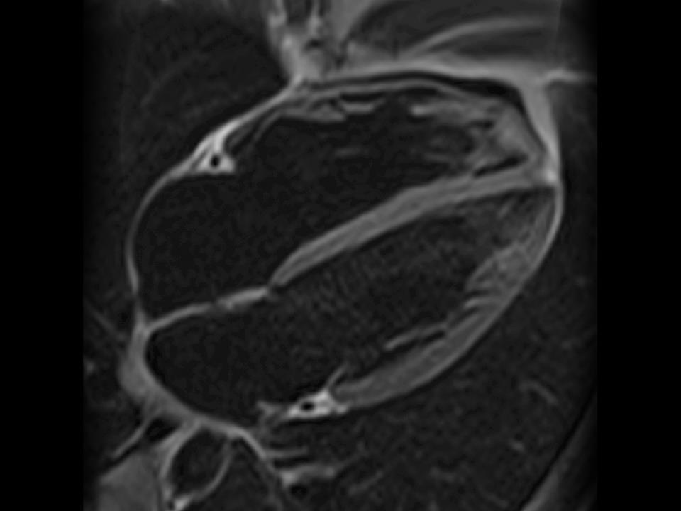
36 Multiphase Cardiac Imaging In this technique a spin echo pulse sequence is used with slices acquired at precise phases of the cardiac cycle.

37 Cine If 18 phases are collected each slice must demonstrate 18 different positions of the heart in one cardiac cycle. This is referred to the number of phases per cardiac cycle.
38 Cine Cardiac cine acquisition are acquired with gradient echo sequences with retrospective gaiting technique Retrospective gaiting uses a method of collecting data continuously throughout the cardiac cycle. Data from each slice location can be acquired at different phases during the cardiac cycle.


39 The Uses of Cine Cine is useful for dynamic imaging of the vessels and CSF. For example evaluate aortic dissection and cardiac function. In the brain, it may be useful to demonstrate dynamically the flow of CSF in patient with hydrocephalus.

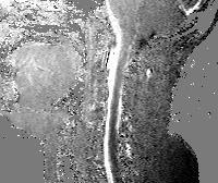

40 PC-MRA Systole Diastole Subtraction - =
41 Respiratory Compensation When imaging the chest and abdomen, respiratory motion along the phase axis produces phase mismapping.

42 Respiratory Compensation Breathing Motion compensation techniques: Breath hold technique Respiratory gaiting Multi-average imaging
43 Breath Hold The best way of reducing breathing motion is: Use gradient echo pulse sequences to be able to scan faster Ask the patient to hold his breath during image acquisition (breath hold).
44 Motion Compensation Breath hold technique: helps to minimize motion form breathing. Tips: Explain the patient before start examination Always follow same instructions Aloud time for the patient to recover
45 Respiratory Gaiting Respiratory gating or respiratory compensation is achieved by monitoring the patient breathing cycle.
 is connected to the scanner and it will advise the scanner")
46 Respiratory Gaiting This is accomplished by placing a breading detection device on the patient. This breading detection device (belt or couching) is connected to the scanner and it will advise the scanner about breathing cycle.

47 Respiratory Gaiting A more sophisticated option is the use of a detection voxel on top of the liver to detect the liver motion during breathing activity.
48 Respiratory Compensation The image acquisition will always be at the same point during the respiration cycle. This technique is very effective but scan time is significantly increase.

")
49 This technique is very effective but scan time is significantly increase. No respiratory gaiting (20 s) Respiratory gaiting (4 min)
50 T1 and Respiratory Gating The breathing cycle is slower then the cardiac cycle, this will result in longer effective TR s. Longer TR will significantly reduce the T1 effects on T1 weighted images, resulting in PD weighted images. Example: Respiratory rate: 20 breath p/m Effective TR = 60,0000 ms / 20 Effective TR = 3000 ms
51 T1 and Respiratory Gating 3000 ms 3000 ms 3000 ms
52 Multi Average Acquisition Increasing the number of excitations may also help, as this increases the number of times the signal is averaged. Motion is averaged out of the image as it is more random in nature than the signal itself.


53 NSA & Motion Acquisition 1 Acquisition 2 Acquisition 3 Average of the 3 Acquisition


54 NEX & Motion Since moving tissues change position during different acquisitions the motion tend to disappear when several acquisitions are average out. Acquisition 12 Average of the 3 Acquisition = 54
55 Navigation System The navigation system is a combination of cardiac and respiratory gaiting at the same time to obtain a image free of respiratory and cardiac image. This application will increase imaging time.

56 Flow Related Artifacts Flow Phenomena Time of Flight Entry Slice phenomena Intra-voxel dephasing Flow Artifacts Flow Void Entry Slice Phenomenon Pulsatile Flow Artifact


57 Maximize Flood Void Increase TE Increase flow velocity Decrease slice thickness


58 Minimize Flood Void Decrease TE Decrease flow velocity Increase slice thickness

59 Entry Slice Phenomenon Maximize Increase TR Increase flow velocity Decrease slice thickness Counter current acquisition Minimize Decrease TR Decrease flow velocity Increase slice thickness
 Pre-Sat bands")
60 Pulsatile Flow Maximize Increase TE Increase flow velocity Increase voxel volume Minimize Decrease TE Decrease flow velocity Decrease voxel volume Flow Compensation (BBI) Pre-Sat bands (DBI)

61 Gradient Moment Rephasing 1 ms 2 ms 3 ms
62 Pre-saturation band Pre-saturation band
63 Blood Bleeding, technically known as hemorrhaging or is blood escaping from the circulatory system. Blood Composition Plasma Blood cells Red White Platelets
64 Red Blood Cells Red blood cells (RBCs), also called erythrocytes, are the most common type of blood cell. Its principal function is the deliver of oxygen (O 2 ). The cytoplasm of erythrocytes is rich in hemoglobin, an iron-containing biomolecule that can bind oxygen and is responsible for the red color of the cells.
65 Hemorrhage Phase Time Hemoglobin Location Magnetic Susceptibility Water Soluble Hyperacute < 24 h Oxyhemoglobin, intracellular Diamagnetic (Fe 1 unpaired e) + Acute 1-3 d Deoxyhemoglobin, intracellular Paramagnetic (Fe 4 unpaired e) + Early subacute >3 d Methemoglobin, intracellular Paramagnetic (Fe 5 unpaired e) + Late subacute >7 d Methemoglobin, extracellular Paramagnetic (Fe 5 unpaired e) + Chronic >14 d Ferritin (Extracellular) Hemosiderin (Extracellular-Intralysosomal) (Macrophages) Superparamagnetic (Fe 10,000 unpaired e) + -
66 Blood Time T1 T2 OxyHg (Hyper acute) DeOxyHg (Acute) Intracellular MetHg (Early subacute) Extracellular MetHg (late subacute) Hemorrhage <24 hrs Iso Hyper 1-3d Iso Hypo 3-7d Hyper Hypo 1-2 wks Hyper Hyper Hemosiderin & Ferritin (Chronic) >2 wks Rim (Iso) Center (Iso) Rim (Hypo) Center (Hyper) Edema Hypo Hyper
 imaging and with susceptibility appearing as low signal intensity due to blood on gradient-echo (GRE) images.")
67 Hyperacute Hematoma Hyperacute hematoma in T1-weighted image (T1W) shows isointense to hypointense lesion in the right temporoparietal region that is hyperintense on T2-weighted (T2W) imaging and with susceptibility appearing as low signal intensity due to blood on gradient-echo (GRE) images. A small rim of vasogenic edema surrounds the hematoma T1 T2 GRE
 and T2-weighted (T2W) images show hypointensity due to the hematoma.")
68 Acute Hematoma MR images show an acute hematoma in the left frontal region. Axial T1-weighted (T1W) and T2-weighted (T2W) images show hypointensity due to the hematoma. A small rim of vasogenic edema surrounds the hematoma seen on T2W imaging. T1 T2
 imaging.")
69 Early Subacute Hemorrage MR images show early subacute hematoma in the left occipital region. The lesion is seen as hyperintensity on T1WI and hypointense on T2WI with marked susceptibility due to hematoma on gradient-echo (GRE) imaging. The intraventricular hematoma also is well visualized as low signal on GRE imaging. T1 T2 GRE
 MR images show high signal intensity suggestive of a late subacute")
70 Late Subacute Hematoma Late Subacute subdural hematoma Both T1-weighted (T1W) and T2- weighted (T2W) MR images show high signal intensity suggestive of a late subacute hemorrhage.
 is hypointense on both T1- weighted and T2-weighted imaging.")
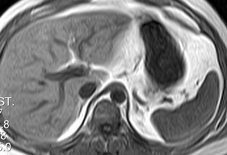
71 Chronic Hematoma MR imaging shows a late subacute to chronic hematoma as a spaceoccupying lesion in the right posterior fossa. The hematoma shows a large medial subacute component and a small lateral chronic component. The chronic component (arrow) is hypointense on both T1- weighted and T2-weighted imaging. This hypointensity is enhanced due to the blooming effect of blood on the gradient-echo (GRE) image. T2 T1 GRE
 imaging.")
72 Various Ages Hematoma Hemorrhages of various ages are seen in the left cerebellar hemisphere with blood-fluid levels in a patient on anticoagulation therapy for chronic venous sinus thrombosis. The hematoma is seen as a mixed signal on T2- and T1-weighted MRI with marked susceptibility on gradient-echo (GRE) imaging. T2 T1 GRE

73 SWI Susceptibility weighted image (SWI) technique uses a very susceptible GRE pulse sequence to make sure of detecting the artifact coming form iron content in hemorrhage. it is so sensitive that is even affected by the susceptibility of intravascular blood.

74 SWI SWI is more susceptible than regular GRE-T2*.

, T1, T2, and T2*.")
75 SWI Susceptibility-weighted imaging (SWI) is a neuroimaging technique, which uses tissues magnetic susceptibility differences to generate a unique contrast, different from that of proton density (PD), T1, T2, and T2*. T1 SE T2 FSE SWI (T2*)
 gradient recalled echo (GRE) scan.")
76 SWI SWI uses a fully flow/velocity compensated, RF spoiled, high-resolution, three-dimensional (3D) gradient recalled echo (GRE) scan. A magnitude and a phase images are obtained
.")
77 SWI The magnitude image is combined with the HP filtered phase image to create an enhanced contrast magnitude image referred to as the susceptibility weighted image (SWI). It is also common to create minimum intensity projections (minip) over 8 to 10 mm to better visualize vein connectivity.
78 SWI SWI can be used better at higher field strengths. First of all, magnetic susceptibility increases accordingly to the square of the magnetic field strength. Moreover, the high signal-to-noise (SNR) ratio available at higher magnetic fields allows higher resolution scans. Finally, stronger magnetic fields allow shorter echo times (TE) without a loss of contrast which can reduce scan time and motion related artifacts.
79 Clinical Applications Improved detection of hemorrhage, microbleeding (diffuse axonal injury) and hemorrhagic transformation (stroke). Tumor characterization. Ability to detect tumor vasculature and micro-hemorrhages. Detection of occult vascular disease (cavernomas, angiomas, telangiectasias). Identification of iron and other mineral deposition. Helpful in MR diagnosis of neurodegenerative diseases (Alzheimer s, multiple sclerosis, etc.)

80 Comparison of diffuse axonal injury (DAI) imaged with conventional GRE (left) and SWI (right) at 1.5 Tesla. SWI is 3 to 6 times more sensitive than GRE T2*- weighted imaging for detection of hemorrhagic DAI.

81 Patient with multiple sclerosis (MS). Iron deposition bilaterally in globus pallidus interna is seen significantly better with SWI.
MR Advance Techniques. Cardiac Imaging. Class IV
 MR Advance Techniques Cardiac Imaging Class IV Heart The heart is a muscular organ responsible for pumping blood through the blood vessels by repeated, rhythmic contractions. Layers of the heart Endocardium
MR Advance Techniques. Vascular Imaging. Class II
 MR Advance Techniques Vascular Imaging Class II 1 Vascular Imaging There are several methods that can be used to evaluate the cardiovascular systems with the use of MRI. MRI will aloud to evaluate morphology
Magnetic Resonance Angiography
 Magnetic Resonance Angiography 1 Magnetic Resonance Angiography exploits flow enhancement of GR sequences saturation of venous flow allows arterial visualization saturation of arterial flow allows venous
Clinical Applications
 C H A P T E R 16 Clinical Applications In selecting pulse sequences and measurement parameters for a specific application, MRI allows the user tremendous flexibility to produce variations in contrast between
Magnetic Resonance Imaging. Basics of MRI in practice. Generation of MR signal. Generation of MR signal. Spin echo imaging. Generation of MR signal
 Magnetic Resonance Imaging Protons aligned with B0 magnetic filed Longitudinal magnetization - T1 relaxation Transverse magnetization - T2 relaxation Signal measured in the transverse plane Basics of MRI
Magnetization Preparation Sequences
 Magnetization Preparation Sequences Acquisition method may not give desired contrast Prep block adds contrast (and/or encoding) MP-RAGE = Magnetization prepared rapid acquisition with gradient echo (Mugler,
Essentials of Clinical MR, 2 nd edition. 99. MRA Principles and Carotid MRA
 99. MRA Principles and Carotid MRA As described in Chapter 12, time of flight (TOF) magnetic resonance angiography (MRA) is commonly utilized in the evaluation of the circle of Willis. TOF MRA allows depiction
MR Imaging with the CCSVI or Haacke protocol
 MR Imaging with the CCSVI or Haacke protocol Reports from the Haacke protocol are often made available to the patients. The report consists of four major components: 1. anatomical images of major neck
Non Contrast MRA. Mayil Krishnam. Director, Cardiovascular and Thoracic Imaging University of California, Irvine
 Non Contrast MRA Mayil Krishnam Director, Cardiovascular and Thoracic Imaging University of California, Irvine No disclosures Non contrast MRA-Why? Limitations of CTA Radiation exposure Iodinated contrast
CARDIAC MRI. Cardiovascular Disease. Cardiovascular Disease. Cardiovascular Disease. Overview
 CARDIAC MRI Dr Yang Faridah A. Aziz Department of Biomedical Imaging University of Malaya Medical Centre Cardiovascular Disease Diseases of the circulatory system, also called cardiovascular disease (CVD),
Susceptibility-weighted MRI ups contrast, offers minute detail 9/15/04 By: Shalmali Pal
 Susceptibility-weighted MRI ups contrast, offers minute detail 9/15/04 By: Shalmali Pal With its flashy sequences and high-speed protocols, there's no shortage of razzle-dazzle in MRI. But learning to
1Pulse sequences for non CE MRA
 MRI: Principles and Applications, Friday, 8.30 9.20 am Pulse sequences for non CE MRA S. I. Gonçalves, PhD Radiology Department University Hospital Coimbra Autumn Semester, 2011 1 Magnetic resonance angiography
Lab #3: Electrocardiogram (ECG / EKG)
") Lab #3: Electrocardiogram (ECG / EKG) An introduction to the recording and analysis of cardiac activity Introduction The beating of the heart is triggered by an electrical signal from the pacemaker. The
Lab #3: Electrocardiogram (ECG / EKG) An introduction to the recording and analysis of cardiac activity Introduction The beating of the heart is triggered by an electrical signal from the pacemaker. The
High Field MR of the Spine
 Department of Radiology University of California San Diego 3T for MR Applications Advantages High Field MR of the Spine Increased signal-to-noise Better fat suppression Increased enhancement with gadolinium
Why Talk About Technique? MRI of the Knee:
 Why Talk About Technique? MRI of the Knee: Part 1 - Imaging Techniques Mark Anderson, M.D. University of Virginia Health Sciences Center Charlottesville, Virginia Always had an interest teach our fellows
Functional Chest MRI in Children Hyun Woo Goo
 Functional Chest MRI in Children Hyun Woo Goo Department of Radiology and Research Institute of Radiology Asan Medical Center, University of Ulsan College of Medicine, Seoul, Korea No ionizing radiation
Variable Appearances of Subacute Intracranial Hematomas on High-Field Spin-Echo MR
 1019 Variable Appearances of Subacute Intracranial Hematomas on High-Field Spin-Echo MR John M. Gomori' 2 Robert I. Grossman' David B. Hackney' Herbert I. Goldberg' Robert A. Zimmerman' Larissa T. Bilaniuk'
MR Flow Imaging in Vascular Malformations Using Gradient Recalled Acquisition
 637 MR Flow Imaging in Vascular Malformations Using Gradient Recalled Acquisition William M. Needell 1 Kenneth R. Maravilla Twenty patients with known or suspected intracranial vascular lesions were evaluated
Essentials of Clinical MR, 2 nd edition. 14. Ischemia and Infarction II
 14. Ischemia and Infarction II Lacunar infarcts are small deep parenchymal lesions involving the basal ganglia, internal capsule, thalamus, and brainstem. The vascular supply of these areas includes the
Perfusion MRI. Youngkyoo Jung, PhD Associate Professor Radiology, Biomedical Engineering, and Clinical & Translational Science Institute
 Perfusion MRI Youngkyoo Jung, PhD Associate Professor Radiology, Biomedical Engineering, and Clinical & Translational Science Institute Perfusion The delivery of blood to a capillary bed in tissue Perfusion
6/23/2009. Inversion Recovery (IR) Techniques and Applications. Variations of IR Technique. STIR, FLAIR, TI and TI Null. Applications of IR
 Techniques and Applications. Variations of IR Technique. STIR, FLAIR, TI and TI Null. Applications of IR") The Anatomy of Basic R Pulse Sequences Inversion Recovery () Techniques and Applications Chen Lin, PhD Indiana University School of edicine & Clarian Health Partners agnetization Preparation Section Chemical
The Anatomy of Basic R Pulse Sequences Inversion Recovery () Techniques and Applications Chen Lin, PhD Indiana University School of edicine & Clarian Health Partners agnetization Preparation Section Chemical
Adaptive RR Prediction for Cardiac MRI
 Adaptive RR Prediction for Cardiac MRI Julien Oster, Olivier Pietquin, Gilles Bosser, Jacques Felblinger To cite this version: Julien Oster, Olivier Pietquin, Gilles Bosser, Jacques Felblinger. Adaptive
Objectives 8/17/2011. Challenges in Cardiac Imaging. Challenges in Cardiac Imaging. Basic Cardiac MRI Sequences
 8/17/2011 Traditional Protocol Model for Tomographic Imaging Cardiac MRI Sequences and Protocols Frandics Chan, M.D., Ph.D. Stanford University Medical Center Interpretation Lucile Packard Children s Hospital
How I do it: Non Contrast-Enhanced MR Angiography (syngo NATIVE)
") Clinical How-I-do-it Cardiovascular How I do it: Non Contrast-Enhanced MR Angiography (syngo NATIVE) Manuela Rick, Nina Kaarmann, Peter Weale, Peter Schmitt Siemens Healthcare, Erlangen, Germany Introduction
Clinical How-I-do-it Cardiovascular How I do it: Non Contrast-Enhanced MR Angiography (syngo NATIVE) Manuela Rick, Nina Kaarmann, Peter Weale, Peter Schmitt Siemens Healthcare, Erlangen, Germany Introduction
Initial Clinical Experience of TOSHIBA 3T MRI
 The 21st Conference of the Japanese Society of Cardiovascular Imaging & Dynamics Sponsored Seminar The Leading Edge of CT/MRI Diagnosis for the Cardiovascular System Initial Clinical Experience of TOSHIBA
8/4/2016. Optimizing Pediatric Cardiovascular MRI. Disclosure. Outline. Jie Deng, PhD, DABMP, Cynthia Rigsby, MD
 Optimizing Pediatric Cardiovascular MRI Jie Deng, PhD, DABMP, Cynthia Rigsby, MD Department of Medical Imaging Radiology, Feinberg School of Medicine, Northwestern University Aug 4 th, 2016 Disclosure
JAMES R. KNIGHT
 JAMES R. KNIGHT knightjr@ah.org James is currently the supervisor of clinical engineering at Sonora Regional Medical Center. He is the chairman of the CMIA Training & Education Committee. He also develops
Non-Contrast MRA. How and When 1996! Why Non-Contrast MRA? Angiography: What are our goals? Inflow Techniques Differences in excitation hx
 A major teaching hospital of Harvard Medical School Angiography: What are our goals? Non-Contrast MRA: How and When Neil M. Rofsky, M.D. Professor of Radiology, Harvard Medical School Director of MRI &
Cerebral cavernous malformation (CCM) comprise
 comprise") ORIGINAL RESEARCH J.M. de Souza R.C. Domingues L.C.H. Cruz, Jr. F.S. Domingues T. Iasbeck E.L. Gasparetto Susceptibility-Weighted Imaging for the Evaluation of Patients with Familial Cerebral Cavernous
ORIGINAL RESEARCH J.M. de Souza R.C. Domingues L.C.H. Cruz, Jr. F.S. Domingues T. Iasbeck E.L. Gasparetto Susceptibility-Weighted Imaging for the Evaluation of Patients with Familial Cerebral Cavernous
Pearls and Pitfalls in Neuroradiology of Cerebrovascular Disease The Essentials with MR and CT
 Pearls and Pitfalls in Neuroradiology of Cerebrovascular Disease The Essentials with MR and CT Val M. Runge, MD Wendy R. K. Smoker, MD Anton Valavanis, MD Control # 823 Purpose The focus of this educational
Department of Radiology University of California San Diego. MR Angiography. Techniques & Applications. John R. Hesselink, M.D.
 Department of Radiology University of California San Diego MR Angiography Techniques & Applications John R. Hesselink, M.D. Vascular Imaging Arterial flow void Flow enhancement Gadolinium enhancement Vascular
Fat Suppression in the Abdomen
 Clinical How I do it? Fat Suppression in the Abdomen Wilhelm Horger Siemens Medical Solutions, Erlangen, Germany Introduction Due to the different chemical environment, hydrogen nuclei in - and in -tissue
The Circulatory System. The Heart, Blood Vessels, Blood Types
 The Circulatory System The Heart, Blood Vessels, Blood Types The Closed Circulatory System Humans have a closed circulatory system, typical of all vertebrates, in which blood is confined to vessels and
How to Learn MRI An Illustrated Workbook
 How to Learn MRI An Illustrated Workbook Exercise 8: Cine Imaging of the Heart Teaching Points: How to do cardiac gating? What is Steady State Free Precession (SSFP)? What are the basic cardiac views and
Biology 13A Lab #10: Cardiovascular System II ECG & Heart Disease
 Biology 13A Lab #10: Cardiovascular System II ECG & Heart Disease Lab #10 Table of Contents: Expected Learning Outcomes...... 83 Introduction....... 84 Activity 1: Collecting ECG Data..... 85 Activity
Cardiovascular MR Imaging at 3 T: Opportunities, Challenges, and Solutions 1
 TECHNICAL ADVANCEMENTS IN CARDIAC MR IMAGING 1612 Cardiovascular MR Imaging at 3 T: Opportunities, Challenges, and Solutions 1 Prabhakar Rajiah, MD, FRCR Michael A. Bolen, MD Abbreviations: BOLD = blood
Normal and Abnormal Cerebrospinal Fluid Dynamics Evaluated by Optimized Cine Phase-contrast MR Imaging
 Chin J Radiol 2000; 25: 191-195 191 ORIGINAL ARTICLE Normal and Abnormal Cerebrospinal Fluid Dynamics Evaluated by Optimized Cine Phase-contrast MR Imaging LUNG-HUI GIIANG 1,3 CHENG-YU CHEN 1 MING-YEN
Cardiovascular System Notes: Physiology of the Heart
 Cardiovascular System Notes: Physiology of the Heart Interesting Heart Fact Capillaries are so small it takes ten of them to equal the thickness of a human hair. Review What are the 3 parts of the cardiovascular
Introduction. Cardiac Imaging Modalities MRI. Overview. MRI (Continued) MRI (Continued) Arnaud Bistoquet 12/19/03
 MRI (Continued) Arnaud Bistoquet 12/19/03") Introduction Cardiac Imaging Modalities Arnaud Bistoquet 12/19/03 Coronary heart disease: the vessels that supply oxygen-carrying blood to the heart, become narrowed and unable to carry a normal amount
Introduction Cardiac Imaging Modalities Arnaud Bistoquet 12/19/03 Coronary heart disease: the vessels that supply oxygen-carrying blood to the heart, become narrowed and unable to carry a normal amount
Noncontrast CT scan is currently the imaging modality
 Original Contributions MRI Features of Intracerebral Hemorrhage Within 2 Hours From Symptom Onset Italo Linfante, MD; Rafael H. Llinas, MD; Louis R. Caplan, MD; Steven Warach, MD, PhD Background and Purpose
Cerebro-vascular stroke
 Cerebro-vascular stroke CT Terminology Hypodense lesion = lesion of lower density than the normal brain tissue Hyperdense lesion = lesion of higher density than normal brain tissue Isodense lesion = lesion
Neuroradiology MR Protocols
 Neuroradiology MR Protocols Brain protocols N 1: Brain MRI without contrast N 2: Pre- and post-contrast brain MRI N 3 is deleted N 4: Brain MRI without or pre-/post-contrast (seizure protocol) N 5: Pre-
The Cardiac Cycle Clive M. Baumgarten, Ph.D.
 The Cardiac Cycle Clive M. Baumgarten, Ph.D. OBJECTIVES: 1. Describe periods comprising cardiac cycle and events within each period 2. Describe the temporal relationships between pressure, blood flow,
General Cardiovascular Magnetic Resonance Imaging
 2 General Cardiovascular Magnetic Resonance Imaging 19 Peter G. Danias, Cardiovascular MRI: 150 Multiple-Choice Questions and Answers Humana Press 2008 20 Cardiovascular MRI: 150 Multiple-Choice Questions
Benign brain lesions
 Benign brain lesions Diagnostic and Interventional Radiology Hung-Wen Kao Department of Radiology, Tri-Service General Hospital, National Defense Medical Center Computed tomography Hounsfield unit (HU)
非對比劑與對比劑增強 MRA. 血管攝影與對比劑 A Course of MRI. 本週課程內容 -MR Angiography (MRA) Unenhanced MRA
 Unenhanced MRA") 本週課程內容 -MR Angiography (MRA) 血管攝影與對比劑 A Course of MRI 盧家鋒助理教授國立陽明大學物理治療暨輔助科技學系 alvin4016@ym.edu.tw 非對比劑增強 MRA(Unenhanced MRA) Time-of-flight (TOF) angiography Phase-contrast (PC) angiography 對比劑增強 MRA(Contrast-enhanced
本週課程內容 -MR Angiography (MRA) 血管攝影與對比劑 A Course of MRI 盧家鋒助理教授國立陽明大學物理治療暨輔助科技學系 alvin4016@ym.edu.tw 非對比劑增強 MRA(Unenhanced MRA) Time-of-flight (TOF) angiography Phase-contrast (PC) angiography 對比劑增強 MRA(Contrast-enhanced
Cardiac Imaging Tests
 Cardiac Imaging Tests http://www.medpagetoday.com/upload/2010/11/15/23347.jpg Standard imaging tests include echocardiography, chest x-ray, CT, MRI, and various radionuclide techniques. Standard CT and
Speed, Comfort and Quality with NeuroDrive
 Speed, Comfort and Quality with NeuroDrive Echelon Oval provides a broad range of capabilities supporting fast, accurate diagnosis of brain conditions and injuries. From anatomical depiction to vascular
Journal of Radiology Case Reports
 Pediatric Holohemispheric Developmental Venous Anomaly: Definitive characterization by 3D Susceptibility Weighted Magnetic Resonance Angiography Michael A. Casey 1, Sourabh Lahoti 2, Ajeet Gordhan 2* 1.
Cardiovascular magnetic resonance artefacts
 Ferreira et al. Journal of Cardiovascular Magnetic Resonance 2013, 15:41 REVIEW Open Access Cardiovascular magnetic resonance artefacts Pedro F Ferreira 1,2*, Peter D Gatehouse 1,2, Raad H Mohiaddin 1,2
Fulfilling the Promise
 Fulfilling the Promise of Cardiac MR Non-contrast, free-breathing technique generates comprehensive evaluation of the coronary arteries By Maggie Fung, MR Cardiovascular Clinical Development Manager; Wei
A New Trend in Vascular Imaging: the Arterial Spin Labeling (ASL) Sequence
 Sequence") A New Trend in Vascular Imaging: the Arterial Spin Labeling (ASL) Sequence Poster No.: C-1347 Congress: ECR 2013 Type: Educational Exhibit Authors: J. Hodel, A. GUILLONNET, M. Rodallec, S. GERBER, R. 1
A New Trend in Vascular Imaging: the Arterial Spin Labeling (ASL) Sequence Poster No.: C-1347 Congress: ECR 2013 Type: Educational Exhibit Authors: J. Hodel, A. GUILLONNET, M. Rodallec, S. GERBER, R. 1
CRITICAL THINKING QUESTIONS AND ANSWERS AND CYCLE 2 LAB EXAM TEMPLATE. There are two main mechanisms that work in conjunction to return the blood
 CRITICAL THINKING QUESTIONS AND ANSWERS AND CYCLE 2 LAB EXAM TEMPLATE There are two main mechanisms that work in conjunction to return the blood THE CARDIAC PUMP 1) The forward pull(vis a fronte) This
#6 - Cardiovascular III Heart Sounds, Pulse Rate, Hemoglobin Saturation, and Blood Pressure
 #6 - Cardiovascular III Heart Sounds, Pulse Rate, Hemoglobin Saturation, and Blood Pressure Objectives: Observe slide of artery and vein cross-section Auscultate heart sounds using a stethoscope Measure
Daniel Bulte. Centre for Functional Magnetic Resonance Imaging of the Brain. University of Oxford
 Daniel Bulte Centre for Functional Magnetic Resonance Imaging of the Brain University of Oxford Overview Signal Sources BOLD Contrast Mechanism of MR signal change FMRI Modelling Scan design details Factors
醫用磁振學 MRM 肌肉骨骼磁振造影簡介 肌肉骨骼磁振造影. 本週課程內容 General Technical Considerations 肌肉骨骼磁振造影簡介 盧家鋒助理教授國立陽明大學生物醫學影像暨放射科學系
 本週課程內容 http://www.ym.edu.tw/~cflu 肌肉骨骼磁振造影簡介 醫用磁振學 MRM 肌肉骨骼磁振造影 盧家鋒助理教授國立陽明大學生物醫學影像暨放射科學系 alvin4016@ym.edu.tw MRI of the musculoskeletal system (5th/6th edition) Editor: Thomas H. Berquist MD 2 General
Outline. Electrical Activity of the Human Heart. What is the Heart? The Heart as a Pump. Anatomy of the Heart. The Hard Work
 Electrical Activity of the Human Heart Oguz Poroy, PhD Assistant Professor Department of Biomedical Engineering The University of Iowa Outline Basic Facts about the Heart Heart Chambers and Heart s The
Investigations in Resting State Connectivity. Overview
 Investigations in Resting State Connectivity Scott FMRI Laboratory Overview Introduction Functional connectivity explorations Dynamic change (motor fatigue) Neurological change (Asperger s Disorder, depression)
PHYSICS OF MRI ACQUISITION. Alternatives to BOLD for fmri
 PHYSICS OF MRI ACQUISITION Quick Review for fmri HST-583, Fall 2002 HST.583: Functional Magnetic Resonance Imaging: Data Acquisition and Analysis Harvard-MIT Division of Health Sciences and Technology
Topic 6: Human Physiology
 Topic 6: Human Physiology 6.2 The Blood System D.4 The Heart Essential Questions: 6.2 The blood system continuously transports substances to cells and simultaneously collects waste products. D.3 The chemical
Tools for cardiovascular magnetic resonance imaging
 Review Article Tools for cardiovascular magnetic resonance imaging Ramkumar Krishnamurthy, Benjamin Cheong, Raja Muthupillai Department of Diagnostic and Interventional Radiology, CHI St. Luke s Health,
Announcements. Final Exam will be a take-home exam. Format similar to the short assignment (no multiple choice, etc.)
") Announcements Final Exam will be a take-home exam Format similar to the short assignment (no multiple choice, etc.) Will be handed out at end of last class period (Thursday June 5 th ) Due by 6 pm June
Announcements Final Exam will be a take-home exam Format similar to the short assignment (no multiple choice, etc.) Will be handed out at end of last class period (Thursday June 5 th ) Due by 6 pm June
Methods for assessing the brain basis of developmental disorders
 Announcements LIGN171: Child Language Acquisition http://ling.ucsd.edu/courses/lign171 Final Exam will be a take-home exam Format similar to the short assignment (no multiple choice, etc.) Will be handed
CT & MRI of Benign Liver Neoplasms Srinivasa R Prasad
 CT & MRI of Benign Liver Neoplasms Srinivasa R Prasad No financial disclosures Acknowledgements Many thanks to Drs. Heiken, Narra & Menias (MIR) Dr. Sahani (MGH) for sharing images Benign Liver Tumors:
BIPN100 F15 Human Physiology I (Kristan) Problem set #5 p. 1
 Problem set #5 p. 1") BIPN100 F15 Human Physiology I (Kristan) Problem set #5 p. 1 1. Dantrolene has the same effect on smooth muscles as it has on skeletal muscle: it relaxes them by blocking the release of Ca ++ from the
BIPN100 F15 Human Physiology I (Kristan) Problem set #5 p. 1 1. Dantrolene has the same effect on smooth muscles as it has on skeletal muscle: it relaxes them by blocking the release of Ca ++ from the
Development of Ultrasound Based Techniques for Measuring Skeletal Muscle Motion
 Development of Ultrasound Based Techniques for Measuring Skeletal Muscle Motion Jason Silver August 26, 2009 Presentation Outline Introduction Thesis Objectives Mathematical Model and Principles Methods
HST.583 Functional Magnetic Resonance Imaging: Data Acquisition and Analysis Fall 2008
 MIT OpenCourseWare http://ocw.mit.edu HST.583 Functional Magnetic Resonance Imaging: Data Acquisition and Analysis Fall 2008 For information about citing these materials or our Terms of Use, visit: http://ocw.mit.edu/terms.
UK Biobank. Imaging modality Cardiovascular Magnetic Resonance (CMR) Version th Oct 2015
 Version th Oct 2015") Imaging modality Cardiovascular Magnetic Resonance (CMR) Version 1.0 http://www.ukbiobank.ac.uk/ 30 th Oct 2015 This document details the procedure for the CMR scan performed at an Imaging assessment centre
Imaging modality Cardiovascular Magnetic Resonance (CMR) Version 1.0 http://www.ukbiobank.ac.uk/ 30 th Oct 2015 This document details the procedure for the CMR scan performed at an Imaging assessment centre
Previous talks. Clinical applications for spiral flow imaging. Clinical applications. Clinical applications. Coronary flow: Motivation
 for spiral flow imaging Joao L. A. Carvalho Previous talks Non-Cartesian reconstruction (2005) Spiral FVE (Spring 2006) Aortic flow Carotid flow Accelerated spiral FVE (Fall 2006) 2007? Department of Electrical
A. Incorrect! The left ventricle receives oxygenated blood from the lungs via the left atrium.
 Anatomy and Physiology - Problem Drill 16: The Cardiovascular System No. 1 of 10 Instruction: (1) Read the problem statement and answer choices carefully (2) Work the problems on paper as needed (3) Pick
Tips and Tricks of State of the art MRA
 Tips and Tricks of State of the art MRA Mayil Krishnam, MD,MBA, MRCP,FRCR(UK) Professor of Radiology Director, Cardiovascular and Thoracic Imaging University of California, Irvine Objectives Technical
Abdominal MRI Techniques in Pediatric Oncology
 Abdominal MRI Techniques in Pediatric Oncology Jonathan R. Dillman, M.D. Assistant Professor Departments of Radiology & Urology Section of Pediatric Radiology C.S. Mott Children s Hospital Disclosures
37 1 The Circulatory System
 H T H E E A R T 37 1 The Circulatory System The circulatory system and respiratory system work together to supply cells with the nutrients and oxygen they need to stay alive. a) The respiratory system:
Cardiovascular Physiology
 Cardiovascular Physiology The mammalian heart is a pump that pushes blood around the body and is made of four chambers: right and left atria and right and left ventricles. The two atria act as collecting
MRI Physics: Basic to Advanced
 Annual Meeting of the American Society of Neuroimaging MRI Physics: Basic to Advanced Mike Moseley, PhD Department of Radiology Stanford University CA 94305-5488 USA http://www-radiology.stanford.edu moseley@stanford.edu
MR imaging techniques
 Eur Radiol (2002) 12:2866 2882 DOI 10.1007/s00330-002-1428-9 MAGNETIC RESONANCE W. R. Nitz Fast and ultrafast non-echo-planar MR imaging techniques Received: 21 August 2001 Revised: 21 February 2002 Accepted:
INTRO TO BOLD FMRI FRANZ JOSEPH GALL ( ) OUTLINE. MRI & Fast MRI Observations Models Statistical Detection
 OUTLINE. MRI & Fast MRI Observations Models Statistical Detection") INTRO TO BOLD FMRI 2014 M.S. Cohen all rights reserved mscohen@g.ucla.edu OUTLINE FRANZ JOSEPH GALL (1758-1828) MRI & Fast MRI Observations Models Statistical Detection PAUL BROCA (1824-1880) WILLIAM JAMES
INTRO TO BOLD FMRI 2014 M.S. Cohen all rights reserved mscohen@g.ucla.edu OUTLINE FRANZ JOSEPH GALL (1758-1828) MRI & Fast MRI Observations Models Statistical Detection PAUL BROCA (1824-1880) WILLIAM JAMES
How Much Tesla Is Too Much?
 How Much Tesla Is Too Much? Johnny U. V. Monu, MB, BS; MSc Professor of Radiology and Orthopedics University of Rochester School of Medicine Rochester, New York Historical Timeline Clinical Imaging 1970
Fully Refocused Gradient Recalled Echo (FRGRE): Factors Affecting Flow and Motion Sensitivity in Cardiac MRI
: Factors Affecting Flow and Motion Sensitivity in Cardiac MRI") Journal of Cardiovascular Magnetic Resonance w, 4(2), 211 222 (2002) METHODS Fully Refocused Gradient Recalled Echo (FRGRE): Factors Affecting Flow and Motion Sensitivity in Cardiac MRI Laurie B. Hildebrand
Journal of Cardiovascular Magnetic Resonance w, 4(2), 211 222 (2002) METHODS Fully Refocused Gradient Recalled Echo (FRGRE): Factors Affecting Flow and Motion Sensitivity in Cardiac MRI Laurie B. Hildebrand
Cardiovascular Effects of Exercise. Background Cardiac function
 Cardiovascular Effects of Exercise In this experiment, you will record an electrocardiogram, or ECG, and finger pulse from a healthy volunteer. You will then compare the ECG and pulse recordings when the
3.4. The Circulatory System
 The Circulatory System The human circulatory system is made up of the blood, the heart, and the blood vessels. The function of the circulatory system is to transport substances around the body. It moves
Chapter 42: Circulation / Gas Exchange. d = t 2
 Chapter 42: Circulation / Gas Exchange Transport systems connect organs of exchange with body cells Diffusion Lung Blood 100 m 1 s 1 mm 100 s 1 cm 10000 s d = t 2 Bulk Flow (Pressure) Blood Cells Methods
P2 Visual - Perception
 P2 Visual - Perception 2014 SOSE Neuroimaging of high-level visual functions gyula.kovacs@uni-jena.de 11/09/06 Functional magnetic resonance imaging (fmri) The very basics What is fmri? What is MRI? The
: Biomedical Signal Processing
 : Biomedical Signal Processing 0. Introduction: Biomedical signal processing refers to the applications of signal processing methods, such as Fourier transform, spectral estimation and wavelet transform,
SWI including phase and magnitude images
 On-line Table: MRI imaging recommendation and summary of key features Sequence Pathologies Visible Key Features T1 volumetric high-resolution whole-brain reformatted in axial, coronal, and sagittal planes
Collin County Community College
 Collin County Community College BIOL. 2402 Anatomy & Physiology WEEK 5 The Heart 1 The Heart Beat and the EKG 2 1 The Heart Beat and the EKG P-wave = Atrial depolarization QRS-wave = Ventricular depolarization
Orthopedic Hardware Imaging Part II: MRI v. Metal
 Orthopedic Hardware Imaging Trent Roth, MD And Lauren Ladd, MD Indiana University School of Medicine IU Health Physicians-Radiology Recap: Imaging Techniques Radiography Standard for initial and surveillance
Cardiovascular system
 BIO 301 Human Physiology Cardiovascular system The Cardiovascular System: consists of the heart plus all the blood vessels transports blood to all parts of the body in two 'circulations': pulmonary (lungs)
Chapter 9, Part 2. Cardiocirculatory Adjustments to Exercise
 Chapter 9, Part 2 Cardiocirculatory Adjustments to Exercise Electrical Activity of the Heart Contraction of the heart depends on electrical stimulation of the myocardium Impulse is initiated in the right
A Magnetic Resonance Imaging Method for
 Journal of Cardiovascular Magnetic Resonance, 1(1), 59-64 (1999) INVITED PAPER Use of MRI in ASD Asessment A Magnetic Resonance Imaging Method for Evaluating Atrial Septa1 Defects Godtfred Holmvang Cardiac
Essentials of Clinical MR, 2 nd edition. 65. Benign Hepatic Masses
 65. Benign Hepatic Masses Pulse sequences acquired for abdominal MRI typically consist of fast acquisition schemes such as single-shot turbo spin echo (i.e. HASTE) and gradient echo schemes such as FLASH
Imaging Acute Stroke and Cerebral Ischemia
 Department of Radiology University of California San Diego Imaging Acute Stroke and Cerebral Ischemia John R. Hesselink, M.D. Causes of Stroke Arterial stenosis Thrombosis Embolism Dissection Hypotension
EI2311 BIOMEDICAL INSTRUMENTATION
 66 EI2311 BIOMEDICAL INSTRUMENTATION 1. What is meant by cell? UNIT I PHYSIOLOGY AND TRANSDUCERS The basic living unit of the body is cell. The function of organs and other structure of the body is understood
Cardiac CT - Coronary Calcium Basics Workshop II (Basic)
") Cardiac CT - Coronary Calcium Basics Workshop II (Basic) J. Jeffrey Carr, MD, MSCE Dept. of Radiology & Public Health Sciences Wake Forest University School of Medicine Winston-Salem, NC USA No significant
Cardiac CT - Coronary Calcium Basics Workshop II (Basic) J. Jeffrey Carr, MD, MSCE Dept. of Radiology & Public Health Sciences Wake Forest University School of Medicine Winston-Salem, NC USA No significant
Biology 236 Spring 2002 Campos/Wurdak/Fahey Laboratory 4. Cardiovascular and Respiratory Adjustments to Stationary Bicycle Exercise.
 BACKGROUND: Cardiovascular and Respiratory Adjustments to Stationary Bicycle Exercise. The integration of cardiovascular and respiratory adjustments occurring in response to varying levels of metabolic
Anyone can get breast cancer BREAST MRI BREAST CANCER. The incidence of getting breast cancer is 1:19 in Malaysia
 Anyone can get breast cancer BREAST MRI KATE Datin Dr Fatimah Moosa Sunway Medical Centre DATIN SERI ENDON KYLIE SIZE DOES NOT MAKE A DIFFERENCE BREAST CANCER The incidence of getting breast cancer is
P215 SPRING 2019: CIRCULATORY SYSTEM Chaps 13, 14 & 15: pp , , , I. Major Functions of the Circulatory System
 P215 SPRING 2019: CIRCULATORY SYSTEM Chaps 13, 14 & 15: pp 360-390, 395-404, 410-428 433-438, 441-445 I. Major Functions of the Circulatory System 1. 2. 3. 4. II. Structure of the Heart 1. atria 2. ventricles
ECG and Cardiac Electrophysiology
 ECG and Cardiac Electrophysiology Simon Some very basic electrophysiology Intracellular fluid: 10 mm Na, 140 mm K, etc. K Na-K ATPase Extracellular fluid: 140mM Na, 4mM K, etc. Na Ion gradient plus selective
Blood Flow, Blood Pressure, Cardiac Output. Blood Vessels
 Blood Flow, Blood Pressure, Cardiac Output Blood Vessels Blood Vessels Made of smooth muscle, elastic and fibrous connective tissue Cells are not electrically coupled Blood Vessels Arteries arterioles
Extraction of the magnetohydrodynamic blood flow potential from the surface electrocardiogram in magnetic resonance imaging
 Med Biol Eng Comput (2008) 46:729 733 DOI 10.1007/s11517-008-0307-1 TECHNICAL NOTE Extraction of the magnetohydrodynamic blood flow potential from the surface electrocardiogram in magnetic resonance imaging