醫用磁振學 MRM 肌肉骨骼磁振造影簡介 肌肉骨骼磁振造影. 本週課程內容 General Technical Considerations 肌肉骨骼磁振造影簡介 盧家鋒助理教授國立陽明大學生物醫學影像暨放射科學系
|
|
|
- Godfrey Chase
- 5 years ago
- Views:
Transcription


1 本週課程內容 肌肉骨骼磁振造影簡介 醫用磁振學 MRM 肌肉骨骼磁振造影 盧家鋒助理教授國立陽明大學生物醫學影像暨放射科學系 MRI of the musculoskeletal system (5th/6th edition) Editor: Thomas H. Berquist MD 2 General Technical Considerations Patient screening Positioning Coil selection 肌肉骨骼磁振造影簡介 Principles of Musculoskeletal (MSK) MRI Pulse sequences Use of intravenous/intraarticular contrast agents 3 4


 surgical clip artifacts (white arrowheads) in")


 to achieve the maximum SNR and the")
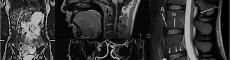
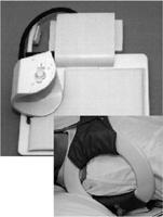
2 Patient Screening MRI safety issues Surgical/hemostasis clips mostly nonferromagnetic or contain minimal ferromagnetic material usually not a significant problem in imaging cerebral aneurysm clips or pacemakers ferromagnetic and may twist or turn in a magnetic environment Titanium and tantalum clips showed the least attractive force and minimal image distortion 石墨鈦鋁不鏽鋼 MR artifacts of Hemostasis Clips The focal areas of no signal with small bright halos (black arrowheads) surgical clip artifacts (white arrowheads) in the right calf medially 5 6 Metal Artifacts bipolar implant on the right and three cannulated hip screws on the left axial Coronal Coronal Positioning & Coil Selection Positioning considerations body part or anatomic region to be examined expected examination time Pathology is suspected in the posterior soft tissues prone position; to prevent compression of soft tissues and anatomic distortion Coil Selection closely coupled coil (smallest coil that covers the anatomy) to achieve the maximum SNR and the best spatial resolution 7 8

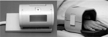


3 Positioning & Coil Selection Positioning & slicing - Shoulder 9 axial imaging coronal imaging sagittal imaging 10 Positioning & slicing - Elbow Positioning & slicing - Wrist Remember to pronate the wrist! axial imaging coronal imaging sagittal imaging 11 axial imaging coronal imaging sagittal imaging 12


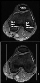


4 Positioning & slicing - Finger Distal Phalanx Mid Phalanx Extensor Tendon Positioning & slicing Hip joint Proximal Phalanx coronal imaging Metacarpal coronal imaging axial imaging sagittal imaging axial imaging sagittal imaging Positioning & slicing Knee Positioning & slicing Ankle axial imaging coronal imaging sagittal imaging 15 axial imaging coronal imaging sagittal imaging 16
 sequences Additional parameters Fat suppression or water excitation Other sequences Diffusion imaging (for fracture) https://mrimaster.")
 Additional considerations Intravenous/ intraarticular gadolinium injection MR angiography Relaxation Times of MSK Tissues Fat has a bright signal on T1W images e.g. (yellow) bone marrow.")
5 Positioning & slicing L-Spine Pulse Sequences axial imaging sagittal imaging Common sequences Spin echo (SE) Fast spin echo (FSE) Inversion recovery (IR) Short tau inversion recovery (STIR) gradient-recalled echo (GRE) sequences Additional parameters Fat suppression or water excitation Other sequences Diffusion imaging (for fracture) Routine Protocols For diagnostic purpose T1-weighted images (bone marrow/fat) T2-weighted images (lesions with long T2 relaxation time) STIR sequence/ fat suppression (bone metastasis/post Gd) Additional considerations Intravenous/ intraarticular gadolinium injection MR angiography Relaxation Times of MSK Tissues Fat has a bright signal on T1W images e.g. (yellow) bone marrow. Tissues with little fat or water e.g. cortical bone, tendons, ligaments are dark in both T1W & T2W

 Increased signal intensity in T2-weighted images")


6 Effects of pathology on MSK Fibrosis: The mobile spin density of predominantly fibrous tissue is low providing little MR signal. Fatty Infiltration: little exchange between the protons in fat and those in the host tissue biexponential nature. Hematoma: prolongation of both T1 and T2 relaxation times, probably due to the presence of inflammation and edema. Lesion Characteristics Abnormal tissues (long T2 relaxation time) Increased signal intensity in T2-weighted images Differentiation from muscle, cortical bone, and fibrous structures (ligaments, tendon, and scar tissues) Soft Tissue Sarcoma SE 450/20 image shows excellent soft tissue anatomy, but the mass is isointense to muscle and only visible due to the differences in size (arrows). T1-weighted image Interpretation of MRI Pathologic processes of the musculoskeletal system are identified at MRI by abnormal morphology, abnormal signal characteristics, or the combination of both. SE 2000/80 image clearly demonstrates the high signal intensity tumor. T2-weighted image acute disruption of the anterior cruciate ligament chronic tear of a ligament Postgadolinium, fat-suppressed T1- weighted image shows tumor enhancement except the central necrotic area (arrows). Fat suppressed postgd T1-weighted 23 24
 2000/20 SE T2 (TR/TE) 2000/80 Fat High (white) Intermediate")
")

 Fibrocartilage Low (black) Low (black)")
 Low (black) Low or high Joint fluid vs.")

 intercondylar")
 would change the direction of the artifacts ;")
7 Tissue Contrasts in Spin Echo SE T1 (TR/TE) /12-17 SE PD (TR/TE) 2000/20 SE T2 (TR/TE) 2000/80 Fat High (white) Intermediate Low intensity Tissue Contrasts T1W, SE 500/20 PD, SE 2000/20 T2W, FSE 4900/98 Fat sat T2W, SE 2500/80 Marrow High (white) Intermediate/white Low intensity Hyaline cartilage Intermediate Intermediate High Muscle Intermediate Intermediate Intermediate Nerves Intermediate (slightly<muscle) Intermediate (slightly<muscle) Intermediate (slightly<muscle) Fibrocartilage Low (black) Low (black) Low (black) Ligaments tendons Low (black) Low (black) Low (black) Blood vessels Low (black) Low (black) Low or high Joint fluid vs. meniscus meniscal tear (arrow) intercondylar erosions (arrowheads) Tissue Contrasts Flow-related artifacts GRE 700/12, 25 o GRE 700/31, 25 o Linear artifact created, usually caused by arteries. Proton density T2 weighted meniscal tear (arrow) intercondylar erosions (arrowheads) Flow artifact (arrowheads); swapping the phase direction (open arrows) would change the direction of the artifacts ; spatial presaturation may be an alternative


8 Scan Time SE T1-weighted sequences TR/TE: 568/15, , one acquisition = 4 min, 55 s. FSE T1-weighted sequences TR/TE: 663/12, ET 3, , one acquisition = 1 min, 57 s. SE Double echo (PD and T2) T2-weighted sequences TR/TE: 2,000/20, 80, , 1 acquisition = 8 min, 53 s. FSE sequences using proton density or T2-weighting with fat suppression TR/TE: 4,000/92, ETL 8, , 1 acquisition = 3 min, 39 s. Intravenous Gd Injections Fat-suppression techniques are commonly employed when T1-weighted sequences are used after contrast injection. Pre- and post-gadolinium images Fat-sat T1W Intraarticular Gadolinium The technique is most commonly used for shoulder, elbow, wrist, hip, and knee MR arthrography. Normal saline, iodinated contrast, and lidocaine could be safely mixed with gadolinium without concern for dissociation that could result in free gadolinium. Intraarticular injection was safe and efficacious at a concentration of 2 mmol/l. Intraarticular Gadolinium Images should be obtained within 30 minutes of injection to optimize contrast benefit. Fat-suppressed, T1-weighted images are optimal for intraarticular detail. Shoulder arthrogram Fat sat Sagittal T1W Fat sat Axial T2W 31 32
 Saturation Artifact (cross talk) Aliasing Artifacts (small FOV) Nonuniform Fat Suppression (field uniformity )")
9 MR Angiography Two-dimensional TOF sequences maximize flow signal using flow-related enhancement combined with suppression of signal intensity from stationary tissue. Common Artifacts Motion Artifacts Chemical Shift (high field-strength magnets ) Saturation Artifact (cross talk) Aliasing Artifacts (small FOV) Nonuniform Fat Suppression (field uniformity ) pulsatile artifacts (arrowheads) artifactual flow voids in collaterals (arrows) THE END alvin4016@ym.edu.tw 35
Why Talk About Technique? MRI of the Knee:
 Why Talk About Technique? MRI of the Knee: Part 1 - Imaging Techniques Mark Anderson, M.D. University of Virginia Health Sciences Center Charlottesville, Virginia Always had an interest teach our fellows
非對比劑與對比劑增強 MRA. 血管攝影與對比劑 A Course of MRI. 本週課程內容 -MR Angiography (MRA) Unenhanced MRA
 Unenhanced MRA") 本週課程內容 -MR Angiography (MRA) 血管攝影與對比劑 A Course of MRI 盧家鋒助理教授國立陽明大學物理治療暨輔助科技學系 alvin4016@ym.edu.tw 非對比劑增強 MRA(Unenhanced MRA) Time-of-flight (TOF) angiography Phase-contrast (PC) angiography 對比劑增強 MRA(Contrast-enhanced
本週課程內容 -MR Angiography (MRA) 血管攝影與對比劑 A Course of MRI 盧家鋒助理教授國立陽明大學物理治療暨輔助科技學系 alvin4016@ym.edu.tw 非對比劑增強 MRA(Unenhanced MRA) Time-of-flight (TOF) angiography Phase-contrast (PC) angiography 對比劑增強 MRA(Contrast-enhanced
FieldStrength. Achieva 3.0T enables cutting-edge applications, best-in-class MSK images
 FieldStrength Publication for the Philips MRI Community Issue 33 December 2007 Achieva 3.0T enables cutting-edge applications, best-in-class MSK images Palo Alto Medical Clinic Sports Medicine Center employs
This presentation is the intellectual property of the author. Contact them for permission to reprint and/or distribute.
 MRI of the Knee Jennifer Swart, M.D. Musculoskeletal Radiology South Texas Radiology Group Outline Coils, Patient Positioning Acquisition Parameters, Planes and Pulse Sequences Knee Arthrography Normal
This presentation is the intellectual property of the author. Contact them at for permission to reprint and/or distribute.
 MRI of the Knee Jennifer Swart, M.D. Musculoskeletal Radiology South Texas Radiology Group Financial Disclosure Dr. Jennifer Swart has no relevant financial relationships with commercial interests to disclose.
MR Advance Techniques. Vascular Imaging. Class II
 MR Advance Techniques Vascular Imaging Class II 1 Vascular Imaging There are several methods that can be used to evaluate the cardiovascular systems with the use of MRI. MRI will aloud to evaluate morphology
Echelon Oval provides a robust suite of leading musculoskeletal imaging capabilities for detailed assessment of all anatomy for your most challenging
 Echelon Oval provides a robust suite of leading musculoskeletal imaging capabilities for detailed assessment of all anatomy for your most challenging cases. Hitachi Medical Systems America, Inc. 1959 Summit
Orthopedic Hardware Imaging Part II: MRI v. Metal
 Orthopedic Hardware Imaging Trent Roth, MD And Lauren Ladd, MD Indiana University School of Medicine IU Health Physicians-Radiology Recap: Imaging Techniques Radiography Standard for initial and surveillance
Clinical Applications
 C H A P T E R 16 Clinical Applications In selecting pulse sequences and measurement parameters for a specific application, MRI allows the user tremendous flexibility to produce variations in contrast between
Musculoskeletal MR Protocols
 Musculoskeletal MR Protocols Joint-based protocols MSK 1: Shoulder MRI MSK 1A: Shoulder MR arthrogram MSK 1AB: Shoulder MR arthrogram (instability protocol) MSK 2: Elbow MRI MSK 2A: Elbow MR arthrogram
Magnetic Resonance Angiography
 Magnetic Resonance Angiography 1 Magnetic Resonance Angiography exploits flow enhancement of GR sequences saturation of venous flow allows arterial visualization saturation of arterial flow allows venous
1Pulse sequences for non CE MRA
 MRI: Principles and Applications, Friday, 8.30 9.20 am Pulse sequences for non CE MRA S. I. Gonçalves, PhD Radiology Department University Hospital Coimbra Autumn Semester, 2011 1 Magnetic resonance angiography
Non Contrast MRA. Mayil Krishnam. Director, Cardiovascular and Thoracic Imaging University of California, Irvine
 Non Contrast MRA Mayil Krishnam Director, Cardiovascular and Thoracic Imaging University of California, Irvine No disclosures Non contrast MRA-Why? Limitations of CTA Radiation exposure Iodinated contrast
RECENT ADVANCES IN CLINICAL MR OF ARTICULAR CARTILAGE
 In Practice RECENT ADVANCES IN CLINICAL MR OF ARTICULAR CARTILAGE By Atsuya Watanabe, MD, PhD, Director, Advanced Diagnostic Imaging Center and Associate Professor, Department of Orthopedic Surgery, Teikyo
MRI of Cartilage. D. BENDAHAN (PhD)
") MRI of Cartilage D. BENDAHAN (PhD) Centre de Résonance Magnétique Biologique et Médicale UMR CNRS 7339 Faculté de Médecine de la Timone 27, Bd J. Moulin 13005 Marseille France david.bendahan@univ-amu.fr
MRI of Cartilage D. BENDAHAN (PhD) Centre de Résonance Magnétique Biologique et Médicale UMR CNRS 7339 Faculté de Médecine de la Timone 27, Bd J. Moulin 13005 Marseille France david.bendahan@univ-amu.fr
Rad Tech 4643 MRI Torso and Extremities
 Rad Tech 4643 MRI Torso and Extremities Prostate Cancer Leiomyoma Retroverted Anteverted Ovarian Cyst Gone Wrong Fibroid (Leiomyoma) IUD Ovary Hysterectomy? What are we to see when imaging a female pelvis
Publication for the Philips MRI Community
 FieldStrength Publication for the Philips MRI Community Issue 38 Summer 2009 Pediatric MSK imaging benefits from tailored scan protocols Vanderbilt University Children s Hospital builds dedicated scans
JMSCR Vol 05 Issue 01 Page January
 www.jmscr.igmpublication.org Impact Factor 5.244 Index Copernicus Value: 83.27 ISSN (e)-2347-176x ISSN (p) 2455-0450 DOI: https://dx.doi.org/10.18535/jmscr/v5i1.28 Diagnostic Accuracy of Magnetic Resonance
Musculoskeletal Imaging What to order? Brian Cole, MD
 Musculoskeletal Imaging What to order? Brian Cole, MD my background: 1994 University of Illinois 1998 MD University of Illinois College of Medicine 1999-2003 Diagnostic Radiology Mayo Clinic 2004 Fellowship
Musculoskeletal Imaging at 3T with Simultaneous Use of Multipurpose Loop Coils
 Clinical Orthopedic Imaging Musculoskeletal Imaging at 3T with Simultaneous Use of Multipurpose Loop Coils Elena Ferrer 1 ; Rafael Coronado Santos 2 1 Radiology Department, Clínica Creu Blanca, Barcelona,
Basics of MR Imaging. Dynamic MRI. MRI Closed. The bed rotates from Upright to Recumbent, stopping at any angle in between.
 Basics of MR Imaging Dynamic MRI MRI Closed The bed rotates from Upright to Recumbent, stopping at any angle in between MRI Open Patient with Low Back Pain After Surgery Extremity MRI Sagittal T2 WI of
MRI KNEE WHAT TO SEE. Dr. SHEKHAR SRIVASTAV. Sr.Consultant KNEE & SHOULDER ARTHROSCOPY
 MRI KNEE WHAT TO SEE Dr. SHEKHAR SRIVASTAV Sr.Consultant KNEE & SHOULDER ARTHROSCOPY MRI KNEE - WHAT TO SEE MRI is the most accurate and frequently used diagnostic tool for evaluation of internal derangement
SPINAL MAGNETIC RESONANCE IMAGING INTERPRETATION
 CLINICAL VIGNETTE 2017; 3:2 SPINAL MAGNETIC RESONANCE IMAGING INTERPRETATION Editor-in-Chief: Idowu, Olufemi E. Neurological surgery Division, Department of Surgery, LASUCOM/LASUTH, Ikeja, Lagos, Nigeria.
Available online at
 Original Research Article Evaluation of knee joint by MRI in 65 patients Gulamus sibtain asad *, Himanshu Singla, Ankit vasoya, P. J. Jhala 2 2 nd year Resident, 2 Professor Radiology Department, SBKS
Magnetization Preparation Sequences
 Magnetization Preparation Sequences Acquisition method may not give desired contrast Prep block adds contrast (and/or encoding) MP-RAGE = Magnetization prepared rapid acquisition with gradient echo (Mugler,
Magnetic Resonance Imaging. Basics of MRI in practice. Generation of MR signal. Generation of MR signal. Spin echo imaging. Generation of MR signal
 Magnetic Resonance Imaging Protons aligned with B0 magnetic filed Longitudinal magnetization - T1 relaxation Transverse magnetization - T2 relaxation Signal measured in the transverse plane Basics of MRI
High Field MR of the Spine
 Department of Radiology University of California San Diego 3T for MR Applications Advantages High Field MR of the Spine Increased signal-to-noise Better fat suppression Increased enhancement with gadolinium
FOR CMS (MEDICARE) MEMBERS ONLY NATIONAL COVERAGE DETERMINATION (NCD) FOR MAGNETIC RESONANCE IMAGING:
 MEMBERS ONLY NATIONAL COVERAGE DETERMINATION (NCD) FOR MAGNETIC RESONANCE IMAGING:") National Imaging Associates, Inc. Clinical guidelines BONE MARROW MRI Original Date: July 2008 Page 1 of 5 CPT Codes: 77084 Last Review Date: September 2014 NCD 220.2 MRI Last Effective Date: July 2011
National Imaging Associates, Inc. Clinical guidelines BONE MARROW MRI Original Date: July 2008 Page 1 of 5 CPT Codes: 77084 Last Review Date: September 2014 NCD 220.2 MRI Last Effective Date: July 2011
Anyone can get breast cancer BREAST MRI BREAST CANCER. The incidence of getting breast cancer is 1:19 in Malaysia
 Anyone can get breast cancer BREAST MRI KATE Datin Dr Fatimah Moosa Sunway Medical Centre DATIN SERI ENDON KYLIE SIZE DOES NOT MAKE A DIFFERENCE BREAST CANCER The incidence of getting breast cancer is
JMSCR Vol 05 Issue 07 Page July 2017
 www.jmscr.igmpublication.org Impact Factor 5.84 Index Copernicus Value: 83.27 ISSN (e)-2347-176x ISSN (p) 2455-0450 DOI: https://dx.doi.org/10.18535/jmscr/v5i7.84 Anatomical Differences Between T2 WI FSE
CARDIAC MRI. Cardiovascular Disease. Cardiovascular Disease. Cardiovascular Disease. Overview
 CARDIAC MRI Dr Yang Faridah A. Aziz Department of Biomedical Imaging University of Malaya Medical Centre Cardiovascular Disease Diseases of the circulatory system, also called cardiovascular disease (CVD),
How Much Tesla Is Too Much?
 How Much Tesla Is Too Much? Johnny U. V. Monu, MB, BS; MSc Professor of Radiology and Orthopedics University of Rochester School of Medicine Rochester, New York Historical Timeline Clinical Imaging 1970
Essentials of Clinical MR, 2 nd edition. 99. MRA Principles and Carotid MRA
 99. MRA Principles and Carotid MRA As described in Chapter 12, time of flight (TOF) magnetic resonance angiography (MRA) is commonly utilized in the evaluation of the circle of Willis. TOF MRA allows depiction
Message of the Month for GPs June 2013
 Message of the Month for GPs June 2013 Dr Winn : Consultant Musculoskeletal Radiologist, Manchester Royal Infirmary Imaging of the musculoskeletal system Musculoskeletal pain is a common problem in the
Imaging the Athlete s Knee. Peter Lowry, MD Musculoskeletal Radiology University of Colorado
 Imaging the Athlete s Knee Peter Lowry, MD Musculoskeletal Radiology University of Colorado None Disclosures Knee Imaging: Radiographs Can be performed weight-bearing or non-weight-bearing View options
MR IMAGING OF THE WRIST
 MR IMAGING OF THE WRIST Wrist Instability Dissociative Pattern apparent on routine radiographs Non-dissociative Stress / positional radiographs Dynamic fluoroscopy during stress Arthrography MRI / MR arthrography
Revised Dec Spine MR Protocols
 Spine MR Protocols Sp 1: Cervical spine MRI without contrast Sp 2: Pre- and post-contrast cervical spine MRI Sp 3: Pre- and post-contrast cervical spine MRI (multiple sclerosis protocol) Sp 4: Thoracic
Sensitivity and Specificity in Detection of Labral Tears with 3.0-T MRI of the Shoulder
 Magee and Williams MRI for Detection of Labral Tears Musculoskeletal Imaging Clinical Observations C M E D E N T U R I C L I M G I N G JR 2006; 187:1448 1452 0361 803X/06/1876 1448 merican Roentgen Ray
Role of Magnetic Resonance Imaging in Patients with Knee Trauma
 Original Research Article Role of Magnetic Resonance Imaging in Patients with Knee Trauma Bhautik Kapadia 1, Bhumika Suthar 2* 1 Associate Professor, 2 Assistant Professor, Department of Radiodiagnosis,
MRI PEDIATRIC PROTOCOLS (Updated 6/19/2018)
") MRI PEDIATRIC PROTOCOLS (Updated 6/19/2018) *Please get or let us know where radiologist can review plain films. *For Texas Orthopedics and other Docs requesting only MSK section read for their pediatric
MRI PEDIATRIC PROTOCOLS (Updated 6/19/2018) *Please get or let us know where radiologist can review plain films. *For Texas Orthopedics and other Docs requesting only MSK section read for their pediatric
Imaging the musculoskeletal system. An Introduction
 Imaging the musculoskeletal system An Introduction Objectives Discuss: commonly used imaging modalities in the musculoskeletal system normal imaging anatomy in the extremities fracture description Imaging
Upper Extremity Page Lower Extremity Special Cases
 MSK MRI PROTOCOLS Contents Upper Extremity Page Shoulder Elbow Wrist Finger Thumb Lower Extremity Hip Pelvis Thigh Knee Lower Extremity/Shin Ankle Foot Special Cases Soft Tissue Mass Metal Protocol MSK
6/23/2009. Inversion Recovery (IR) Techniques and Applications. Variations of IR Technique. STIR, FLAIR, TI and TI Null. Applications of IR
 Techniques and Applications. Variations of IR Technique. STIR, FLAIR, TI and TI Null. Applications of IR") The Anatomy of Basic R Pulse Sequences Inversion Recovery () Techniques and Applications Chen Lin, PhD Indiana University School of edicine & Clarian Health Partners agnetization Preparation Section Chemical
The Anatomy of Basic R Pulse Sequences Inversion Recovery () Techniques and Applications Chen Lin, PhD Indiana University School of edicine & Clarian Health Partners agnetization Preparation Section Chemical
UPPER EXTREMITY
 UPPER EXTREMITY 11-24-17 MSK TIPS: Ensure extremity of interest is as isocenter as possible SHIM all Fat sat scans!! Only use 4ch wrist if additional coverage needed If Contrast needed: Multihance.1mmol/kg
Upper Extremity Page Lower Extremity Special Cases
 MSK MRI PROTOCOLS Contents Upper Extremity Shoulder Elbow Wrist Finger Thumb Lower Extremity Hip Pelvis Thigh Knee Lower Extremity/Shin Ankle Foot Special Cases Soft Tissue Mass Metal Protocol Page MSK
Imaging of Articular Cartilage
 Clinical Imaging of Articular Cartilage Imaging of Articular Cartilage Prof. Dr. K. Verstraete Ghent University Introduction : Articular Cartilage Histology and biochemical composition Review of Imaging
Neuroradiology MR Protocols
 Neuroradiology MR Protocols Brain protocols N 1: Brain MRI without contrast N 2: Pre- and post-contrast brain MRI N 3 is deleted N 4: Brain MRI without or pre-/post-contrast (seizure protocol) N 5: Pre-
ORIGINAL ARTICLE. ROLE OF MRI IN EVALUATION OF TRAUMATIC KNEE INJURIES Saurabh Chaudhuri, Priscilla Joshi, Mohit Goel
 ROLE OF MRI IN EVALUATION OF TRAUMATIC KNEE INJURIES Saurabh Chaudhuri, Priscilla Joshi, Mohit Goel 1. Associate Professor, Department of Radiodiagnosis & imaging, Bharati Vidyapeeth Medical College and
CT ARTHROGRAPHY It s not Always About the
 CT ARTHROGRAPHY It s not Always About the Magnet Kirkland W. Davis, M.D. University of Wisconsin Department of Radiology Disclosures Financial FDA IA Gd! What Is CT Arthrography? (CTR) Arthrogram: imaging
Knee: Cruciate Ligaments
 72 Knee: Cruciate Ligaments R. Kent Sanders Sagittal oblique 2.5-mm sequences along the plane of the anterior cruciate ligament (ACL) typically yield three to four images of the ACL, with the first medial
Take Pride in Performance
 2017 Take Pride in Performance Knee: Meniscal Tear FSE PD - Sagittal FSE PD - Coronal FSTIR - Coronal Knee: ACL Tibial Avulsion 3D SHARC ISO - Sagittal FSE PD - Sagittal FSTIR - Coronal Knee: Subchondral
Ultrasound Evaluation of Masses
 Ultrasound Evaluation of Masses Jon A. Jacobson, M.D. Professor of Radiology Director, Division of Musculoskeletal Radiology University of Michigan Disclosures: Consultant: Bioclinica Advisory Panel: GE,
KNEE ALIGNMENT SYSTEM (KAS) MRI Protocol
 MRI Protocol") KNEE ALIGNMENT SYSTEM (KAS) MRI Protocol Sample referral sticker Referral Sticker Insert here Corin 17 Bridge Street Pymble NSW Australia 2073 P: +61 (0)2 9497 7400 F: +61 (0)2 9497 7498 E: KAS.customerservice@coringroup.com
KNEE ALIGNMENT SYSTEM (KAS) MRI Protocol Sample referral sticker Referral Sticker Insert here Corin 17 Bridge Street Pymble NSW Australia 2073 P: +61 (0)2 9497 7400 F: +61 (0)2 9497 7498 E: KAS.customerservice@coringroup.com
Personal use only. MRI Metal Artifact Reduction: Shoulder Implants and Arthroplasty. Reto Sutter, MD
 MRI Metal Artifact Reduction: Shoulder Implants and Arthroplasty Reto Sutter, MD University Hospital Balgrist Zurich University of Zurich Cor PD fat sat 56-year old male patient with positive lift-off
STIR Sequence With Increased Receiver Bandwidth of the Inversion Pulse for Reduction of Metallic Artifacts
 Musculoskeletal Imaging Technical Innovation Ulbrich et al. Artifact Reduction in MRI Musculoskeletal Imaging Technical Innovation Erika J. Ulbrich 1,2 Reto Sutter 1 Ranieri F. Aguiar 3 Mathias Nittka
IMAGING TECHNIQUES CHAPTER 4. Imaging techniques
 IMAGING TECHNIQUES Imaging techniques 23 4.1. Conventional radiographic findings Conventional radiography, tomography, arthrography and stress views have traditionally been used for imaging the ankle and
MR imaging of the knee in marathon runners before and after competition
 Skeletal Radiol (2001) 30:72 76 International Skeletal Society 2001 ARTICLE W. Krampla R. Mayrhofer J. Malcher K.H. Kristen M. Urban W. Hruby MR imaging of the knee in marathon runners before and after
大體老師無語良師 大體解剖學實驗 HUMAN DISSECTION ANTERIOR ABDOMINAL WALL & INGUINAL REGION 盧家鋒助理教授 臺北醫學大學醫學系解剖學暨細胞生物學科 臺北醫學大學醫學院轉譯影像研究中心.
 大體老師無語良師 大體解剖學實驗 HUMAN DISSECTION ANTERIOR ABDOMINAL WALL & INGUINAL REGION 盧家鋒助理教授 臺北醫學大學醫學系解剖學暨細胞生物學科 臺北醫學大學醫學院轉譯影像研究中心 http://www.ym.edu.tw/~cflu REFERENCES Dissector s guide [1] Dissection Guide for
1 Normal Anatomy and Variants
 1 Normal Anatomy and Variants 1.1 Normal Anatomy MR Technique. e standard MR protocol for a routine evaluation of the spine always comprises imaging in sagittal and axial planes, while coronal images are
Meniscal Tears: Role of Axial MRI Alone and in Combination with Other Imaging Planes
 Nefise Cagla Tarhan 1,2 Christine. Chung 1 urea Valeria Rosa Mohana-orges 1 Tudor Hughes 1 Donald Resnick 1 Received September 30, 2003; accepted after revision February 2, 2004. 1 Department of Radiology,
MRI Imaging of GP Medicare Eligible Conditions
 MRI Imaging of GP Medicare Eligible Conditions By Dr. Andrew Stuart Radiologist Sydney Adventist Hospital 0562/SAH/1112/SAH Learning Objectives Indications for GP referred Medicare eligible MRI scans MRI
Anatomical and Functional MRI of the Pancreas
 Anatomical and Functional MRI of the Pancreas MA Bali, MD, T Metens, PhD Erasme Hospital Free University of Brussels Belgium mbali@ulb.ac.be Introduction The use of MRI to investigate the pancreas has
The Role of IDEAL and DTI in Peripheral Nerve MR Imaging
 In Practice The Role of IDEAL and DTI in Peripheral Nerve MR Imaging y Darryl. Sneag, MD, Assistant Attending Radiologist, and Hollis G. Potter, MD, Chairman and The Coleman Chair, MRI Research, Department
Consortium of MS Centres Guidelines Revised Standardized MRI Protocol. for the Diagnosis and Follow-up of MS. David K.B.
 Consortium of MS Centres Guidelines Revised Standardized MRI Protocol for the Diagnosis and Follow-up of MS David K.B. Li MD FRCPC Indianapolis, Indiana May 27, 2015 Disclosure I have received research
Can SCMR CMR protocol recommendations
 Can SCMR CMR protocol recommendations V1.3 - April 2009 CanSCMR CMR Protocol and SOP Recommendation 2009 (15 minutes) 2 Planning of LV fct. real time multiple axes Realtime 3 cine long axis 6 long axes
Attenuation value in HU From -500 To HU From -10 To HU From 60 To 90 HU. From 200 HU and above
 Brain Imaging Common CT attenuation values Structure Air Fat Water Brain tissue Recent hematoma Calcifications Bone Brain edema and infarction Normal liver parenchyma Attenuation value in HU From -500
Tibial stress injury: MRI findings
 Tibial stress injury: MRI findings Poster No.: P-0047 Congress: ESSR 2013 Type: Scientific Exhibit Authors: A. Castrillo 1, J. J. Fondevila 2, B. Canteli 3, A. Urresola Olabarrieta 1, A. I. Ezquerro 2,
MR imaging the post operative spine - What to expect!
 MR imaging the post operative spine - What to expect! Poster No.: C-2334 Congress: ECR 2012 Type: Educational Exhibit Authors: A. Jain, M. Paravasthu, M. Bhojak, K. Das ; Warrington/UK, 1 1 1 2 1 2 Liverpool/UK
MRI of LEFT KNEE. There is a fluid collection seen anterior to and inferior to the superiorly displaced patella.
 MRI of LEFT KNEE Protocol: Multiplanar MRI of the left knee joint performed in the sagittal, coronal and transverse planes using T1 weighted spin echo, T2 and proton-density weighted fast spin echo, fatsaturated
Stability of Post Traumatic Osteochondritis Dissecans of the Knee: MR Imaging Findings
 Chin J Radiol 2005; 30: 199-204 199 Stability of Post Traumatic Osteochondritis Dissecans of the Knee: MR Imaging Findings YU-CHUNG HUNG 1 JON-KWAY HUANG 1,2 Department of Radiology 1, Mackay Memorial
Comparative study of high resolusion ultrasonography and magnetic resonance imaging in diagnosing traumatic knee injuries & pathologies
 Original article: Comparative study of high resolusion ultrasonography and magnetic resonance imaging in diagnosing traumatic knee injuries & pathologies Dr. Rakesh Gujjar*, Dr. R. P. Bansal, Dr. Sandeep
Oak foundation for donating the 3T Siemens Verio scanner. Board of directors BBH and Frh Hospitals for supporting the
 Knee pain and inflammation in the infrapatellar fat pad estimated by conventional and dynamic contrast-enhanced magnetic resonance imaging in obese patients with osteoarthritis: a crosssectional study
FOR CMS (MEDICARE) MEMBERS ONLY NATIONAL COVERAGE DETERMINATION (NCD) FOR MAGNETIC RESONANCE IMAGING:
 MEMBERS ONLY NATIONAL COVERAGE DETERMINATION (NCD) FOR MAGNETIC RESONANCE IMAGING:") National Imaging Associates, Inc. Clinical guidelines SINUS MRI Original Date: November 2007 Page 1 of 5 CPT Codes: 70540, 70542, 70543 Last Review Date: July 2014 NCD 220.2 MRI Last Effective Date: July
National Imaging Associates, Inc. Clinical guidelines SINUS MRI Original Date: November 2007 Page 1 of 5 CPT Codes: 70540, 70542, 70543 Last Review Date: July 2014 NCD 220.2 MRI Last Effective Date: July
Role of magnetic resonance imaging in the evaluation of traumatic knee joint injuries
 Original Research Article Role of magnetic resonance imaging in the evaluation of traumatic knee joint injuries Dudhe Mahesh 1*, Rathi Varsha 2 1 Resident, 2 Professor, Department of Radio-Diagnosis, Grant
RADIOLOGY TEACHING CONFERENCE
 RADIOLOGY TEACHING CONFERENCE John Athas, MD Monica Tadros, MD Columbia University, College of Physicians & Surgeons Department of Otolaryngology- Head & Neck Surgery September 27, 2007 CT SCAN IMAGING
MSK CT Extremities: Positioning and Reformations
 MSK CT Extremities: Positioning and Reformations Hand: Patient lying in prone position, with affected arm extended above head. Place body off centered in effort to set affected hand in isocenter. Hand
The Elbow Scanning Protocol
 The Elbow Scanning Protocol Diagnostic Imaging of the Elbow: Introduction The elbow maybe considered as consisting of four quadrants, anterior, medial, lateral and posterior. Ultrasound would normally
Sport Specific MRI. The symptoms of the majority, if not all sports injuries are experienced when upright, and weight-bearing
 Sport Specific MRI The symptoms of the majority, if not all sports injuries are experienced when upright, and weight-bearing A complete, accurate MRI assessment can only be made when in the position of
Evaluation and Management of Knee Pain. Michael Cassat, MD University of Arkansas for Medical Sciences
 Evaluation and Management of Knee Pain Michael Cassat, MD University of Arkansas for Medical Sciences Disclosure I have no actual or potential conflict of interest in relation to this program/presentation.
MR Advance Techniques. Cardiac Imaging. Class III
 MR Advance Techniques Cardiac Imaging Class III Black Blood Imaging & IR Blue= O2 poor blood Red=O2 rich blood Inversion pulses can produce black blood imaging in GRE pulse sequences. Specially on the
MRI of the Shoulder What to look for and how to find it? Dr. Eric Handley Musculoskeletal Radiologist Cherry Creek Imaging
 MRI of the Shoulder What to look for and how to find it? Dr. Eric Handley Musculoskeletal Radiologist Cherry Creek Imaging MRI of the Shoulder Benefits of Ultrasound: * Dynamic * Interactive real time
NEURO PROTOCOLS MRI NEURO PROTOCOLS (SIEMENS SCANNERS)
") Page 1 NEURO PROTOCOLS Brain Stroke Brain Brain with contrast Brain for seizures Brain for MS Brain for Pineal gland Sella FAST Scan for hydrocephalus MRA/MRV Brain MRA carotids 8 th nerve Cranial nerves
Page 1 NEURO PROTOCOLS Brain Stroke Brain Brain with contrast Brain for seizures Brain for MS Brain for Pineal gland Sella FAST Scan for hydrocephalus MRA/MRV Brain MRA carotids 8 th nerve Cranial nerves
ACR MRI Accreditation Program. ACR MRI Accreditation Program Update. Educational Objectives. ACR accreditation. History. New Modular Program
 ACR MRI Accreditation Program Update Donna M. Reeve, MS, DABR, DABMP Department of Imaging Physics University of Texas M.D. Anderson Cancer Center Educational Objectives Present requirements of the new
Functional Orthopedic Imaging Capturing Motion, Flow and Perfusion. Case Study Brochure Centre University Hospital Nancy.
 Capturing Motion, Flow and Perfusion dynamic volume CT Case Study Brochure Centre University Hospital Nancy http://www.toshibamedicalsystems.com Toshiba Medical Systems Corporation 2013. All rights reserved.
Tips and Tricks of State of the art MRA
 Tips and Tricks of State of the art MRA Mayil Krishnam, MD,MBA, MRCP,FRCR(UK) Professor of Radiology Director, Cardiovascular and Thoracic Imaging University of California, Irvine Objectives Technical
Case Report: Knee MR Imaging of Haemarthrosis in a Case of Haemophilia A
 Clinical > Pediatric Imaging Case Report: Knee MR Imaging of Haemarthrosis in a Case of Haemophilia A M. A. Weber, J. K. Kloth University Hospital Heidelberg, Department of Diagnostic and Interventional
Ankle impingement syndromes - pictorial review.
 Ankle impingement syndromes - pictorial review. Poster No.: P-0148 Congress: ESSR 2015 Type: Educational Poster Authors: R. D. T. Mesquita, J. Pinto, J. L. Rosas, A. Vieira ; Porto/PT, 1 2 2 3 1 1 3 Matosinhos/PT,
Ankle impingement syndromes - pictorial review.
 Ankle impingement syndromes - pictorial review. Poster No.: P-0148 Congress: ESSR 2015 Type: Educational Poster Authors: R. D. T. Mesquita, J. Pinto, J. L. Rosas, A. Vieira ; Porto/PT, 1 2 2 3 1 1 3 Matosinhos/PT,
The posterolateral corner of the knee: the normal and the pathological
 The posterolateral corner of the knee: the normal and the pathological Poster No.: P-0104 Congress: ESSR 2014 Type: Educational Poster Authors: M. Bartocci 1, C. Dell'atti 2, E. Federici 1, V. Martinelli
Pictorial Essay. Jang Gyu Cha, MD, Hyun Sook Hong, MD, Jai Soung Park, MD, Sang Hyun Paik, MD, Hae Kyung Lee, MD INTRODUCTION
 Pictorial Essay http://dx.doi.org/10.3348/kjr.2012.13.3.332 pissn 1229-6929 eissn 2005-8330 Korean J Radiol 2012;13(3):332-341 Practical pplication of Iterative Decomposition of Water and Fat with Echo
Diagnostic Imaging
 www.fisiokinesiterapia.biz Diagnostic Imaging Diagnostic Imaging is no longer limited to radiography. Major technological advancements have lead to the use of new and improved imaging technologies. The
Multi-ligamentous knee injuries - MRI injury patterns at a glance
 Multi-ligamentous knee injuries - MRI injury patterns at a glance Poster No.: P-0068 Congress: ESSR 2015 Type: Educational Poster Authors: A. Rastogi, D. Whelan, R. Martin, W. Mak, D. Pearce ; 1 1 1 2
Original Report. The Reverse Segond Fracture: Association with a Tear of the Posterior Cruciate Ligament and Medial Meniscus
 Eva M. Escobedo 1 William J. Mills 2 John. Hunter 1 Received July 10, 2001; accepted after revision October 1, 2001. 1 Department of Radiology, University of Washington Harborview Medical enter, 325 Ninth
Chapter 30 - Musculoskeletal_Trauma
 Introduction to Emergency Medical Care 1 OBJECTIVES 30.1 Define key terms introduced in this chapter. Slides 11 12, 19 20, 22 23, 37 30.2 Describe the anatomy of elements of the musculoskeletal system.
Basics of MRI Part I
 Basics of MRI Part I Mathew J. Dixon, D.O. Chairman Department of Radiology Memorial Health University Medical Center Savannah, GA Objectives Brief History Concept of MRI Creation of a Magnetic Field Concepts
Topics. Musculoskeletal Infection Extremities. Detection of Infection. Role of Imaging in Extremity Infection. Detection of Infection
 Topics Musculoskeletal Infection Extremities Nuttaya Pattamapaspong M.D. Department of Radiology, Faculty of Medicine, Chiang Mai University, Chiang Mai, Thailand Role of imaging in extremity infection
Magnetic Resonance Imaging of the Knee: An Overview and Update of Conventional and State of the Art Imaging
 CME ARTICLE Magnetic Resonance Imaging of the Knee: An Overview and Update of Conventional and State of the Art Imaging Nicholas C. Nacey, MD, Matthew G. Geeslin, MD, Grady Wilson Miller, PhD, and Jennifer
Comparative study of imaging at 3.0 T versus 1.5 T of the knee
 Skeletal Radiol (2009) 38:761 769 DOI 10.1007/s00256-009-0683-0 SCIENTIFIC ARTICLE Comparative study of imaging at 3.0 T versus 1.5 T of the knee Scott Wong & Lynne Steinbach & Jian Zhao & Christoph Stehling
Usefulness of Unenhanced MRI and MR Arthrography of the Shoulder in Detection of Unstable Labral Tears
 Musculoskeletal Imaging Original Research Unenhanced MRI and MR rthrography for Unstable Labral Tears Musculoskeletal Imaging Original Research Thomas 1,2 T Keywords: labral tear, MRI, shoulder DOI:10.2214/JR.14.14262