MDCT and Pulmonary Embolism. Heber MacMahon The University of Chicago Department of Radiology
|
|
|
- Eugene Pierce
- 6 years ago
- Views:
Transcription
1 MDCT and Pulmonary Embolism Heber MacMahon The University of Chicago Department of Radiology
2 Disclosures Consultant for Riverain Medical Minor stockholder in Hologic, Inc. Consultant for GE Healthcare Research Support from Philips Healthcare License and royalty fees from University of Chicago (UC Tech)
3 Pulmonary Embolism Risk Factors & pre-test probability CXR findings CT technique Dx on contrast and non-contrast CT scans Acute vs Chronic PE Non thrombotic PE
4 Pulmonary Embolism 500,000 episodes of PE / yr in USA 12 64% ICU patients at autopsy Contributing cause of death in 10-15% ICU pts Clinical diagnosis difficult
5 Risk Factors for PE Strong Risk Factors Lower limb Fx, Hip or Knee Replacement Major Trauma MI in previous 3 mos. Spinal Cord Injury Previous VTE 2014 ESC Guidelines on the diagnosis and management of acute pulmonary embolism; Task Force for the Diagnosis and Management of Acute Pulmonary Embolism of the European Society of Cardiology (ESC) European Heart Journal (2014) 35,
6 Risk Factors for PE Moderate Risk Factors CHF Infection Cancer Postpartum Oral contraceptives Autoimmune disease Blood transfusions Weak Risk Factors Bed rest > 3 days Travel of 4 hr or more Pregnancy Obesity Advanced age
7 Risk Factors for PE Weak Risk Factors Bed rest > 3 days Travel of 4 hr or more in the past month Pregnancy Obesity Advanced age
8 Most Common Symptoms of PE Unexplained: Dyspnea (50%) Esp. sudden onset Pleuritic pain (39%) Substernal (15%) Hemoptysis (8%) Syncope/near syncope (6%) More specific
9 Clinical Signs in PE Hypoxemia (70%) Tachypnea/dyspnea (90%) Tachycardia (40%) Arrythmia Wheezing Hemoptysis Fever (but < 39.5C) Signs of DVT
10 Simplified Wells Criteria (>2: PE likely) History of DVT or PE 1 point Tachycardia (heart rate > 100) 1 point Immobilization ( 3d)/surgery in previous four weeks 1 point Hemoptysis 1 point Malignancy (with treatment within 6 months) or palliative 1 point Clinically suspected DVT 1 point Alternative diagnosis is less likely than PE 3.0 points
11 D-dimer for PE Cut-off value: 500 ug/l Negative predictive value: 98-99% Positive predictive value: 27-29% False positives associated with: female sex; increasing age; black (vs. white) race; cocaine use; immobility; hemoptysis; hemodialysis; active malignancy; rheumatoid arthritis; lupus; sickle cell disease; prior venous thromboembolism (VTE; not under treatment); pregnancy and postpartum state; and abdominal, chest, orthopedic, or other surgery.
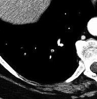
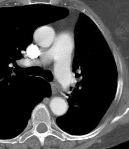
12 80 y/o female with seizures and pneumonia

13 Multiple PEs with infarcts


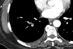
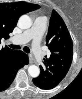
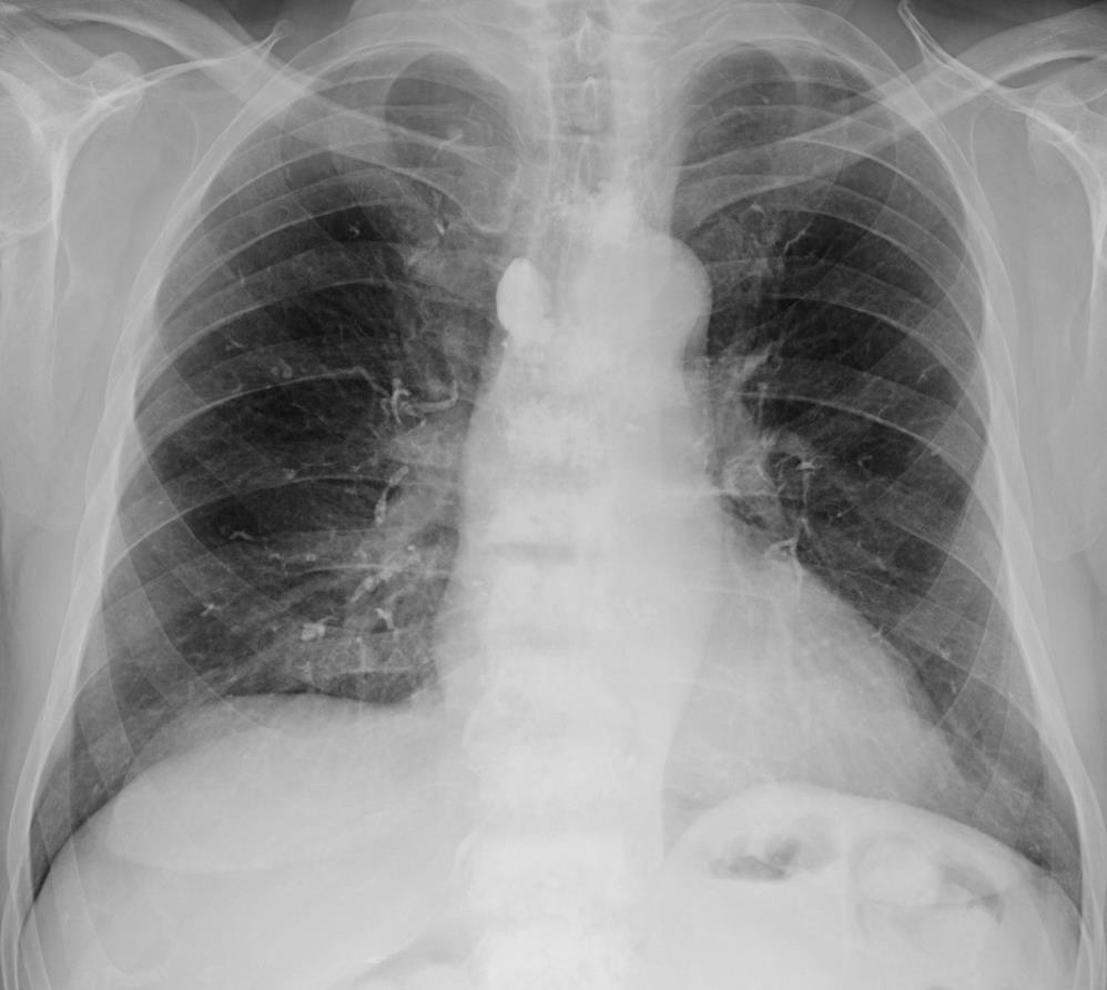
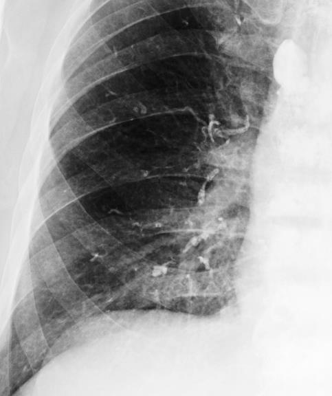
14 76 y/o with pleuritic chest pain


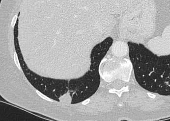
15 Multiple Emboli with Small Infarcts
16 CXR in PE CXR often normal Focal subpleural rounded opacity, esp. CP angles Westermark Sign : Peripheral oligemia, caused by chronic embolism, but often with superimposed new acute episode
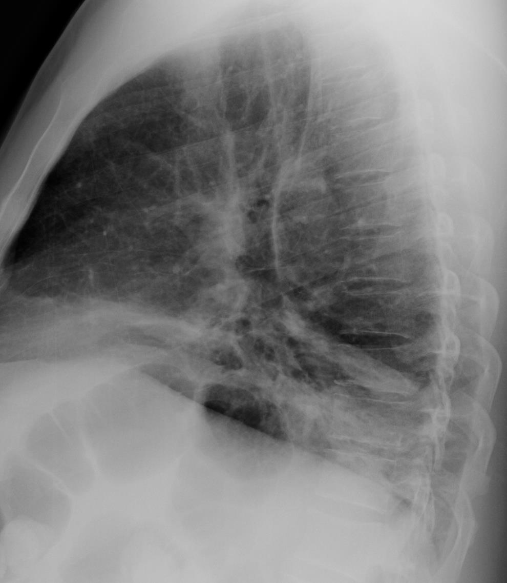
17 Westermark Sign

18 72 y/o with acute hypoxia

19 Large PE causing asymmetric pulmonary edema
20 CXR in PE CXR often normal Focal subpleural rounded opacity, esp. CP angles Westermark Sign : Peripheral oligemia, caused by chronic embolism, but often with superimposed new acute episode Fleischner Sign : Enlargement and sharp definition of central PA
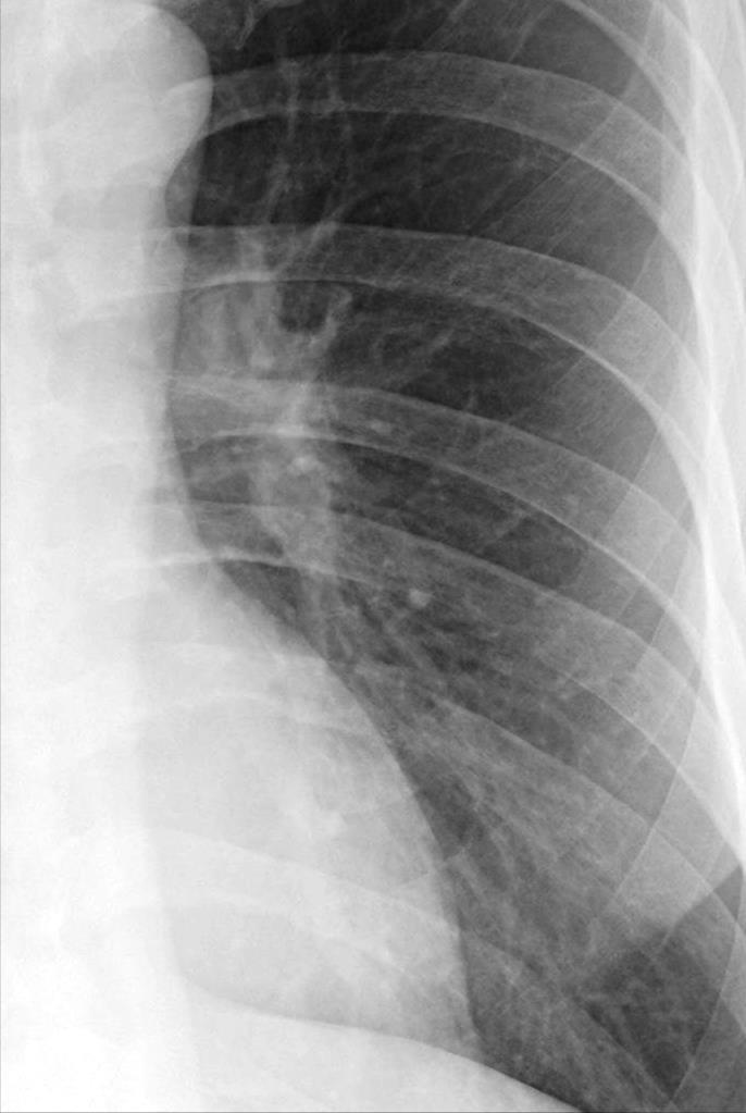
21 Fleischner Sign
22 CXR in PE CXR often normal Focal subpleural rounded opacity, esp. CP angles Westermark Sign : Peripheral oligemia, caused by chronic embolism, but often with superimposed new acute episode Fleischner Sign : Enlargement and sharp definition of central PA Bibasilar atelectasis, subpleural opacity, effusions, suggestive in previously healthy individual

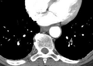
23 Unexplained Basilar Subsegmental Atelectasis

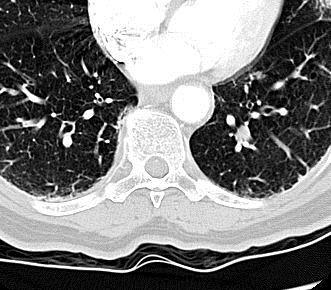
24 V/Q Scan accuracy for PE V/Q Scan Results PE Prevalence Normal or low probability + low clinical suspicion 4% High probability + high clinical suspicion 96% Still a reasonable test for young women with low PE probability and normal CXR. or Patients with contraindication to IV contrast
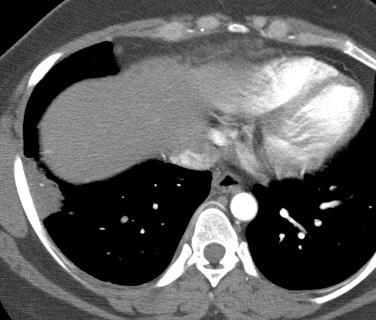
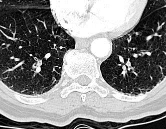
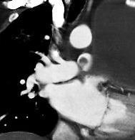
25 Pulmonary C.T. Angiography Rapid contrast infusion (3-6 cc/sec) Antecubital fossa vein mm collimation Shallow breath hold Fixed delay, bolus tracking, timing bolus
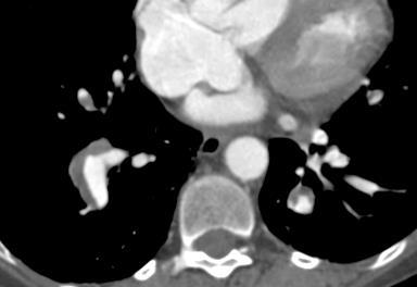
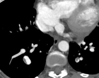
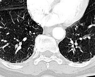
26 Test bolus vs Bolus Tracking Test bolus: Visual estimation by tech of optimal contrast timing. Makes cursor placement less critical. Subjective judgement Bolus Tracking: Automatic trigger for scan based on bolus arrival Cursor placement critical Adds several seconds to scan delay

27 Test Bolus. Cursor on PA
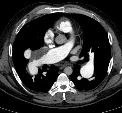
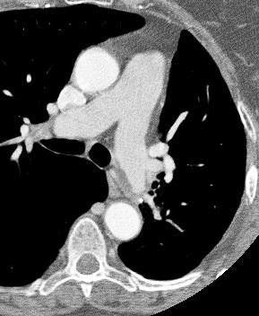
28 Normal Circulation Time


29 Abnormal Test Bolus Slow Circulation


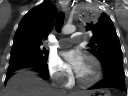
30 Main Contrast Bolus Slow Circulation

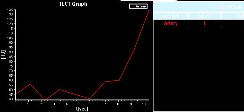
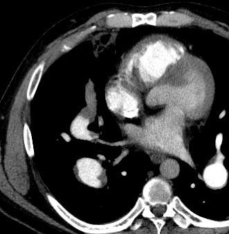
31 Test Bolus using Descending Aorta
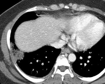
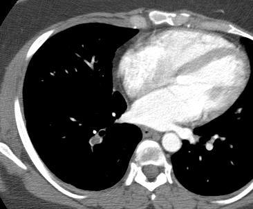
32 Non-Diagnostic CTA Bolus Timing Motion artifact Poor contrast enhancement - Flow obstruction - Deep inspiration > dilution - Contrast timing error Beam hardening and noise obesity


33 Dilution by unopacified IVC inflow Relax, take in a small breath and hold it

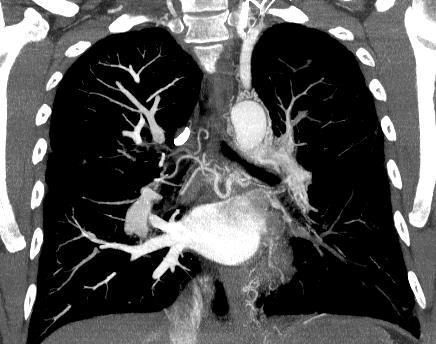
34 PE CT Results over 12 months at U Chicago

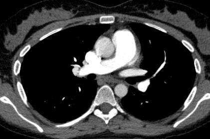
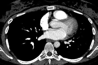
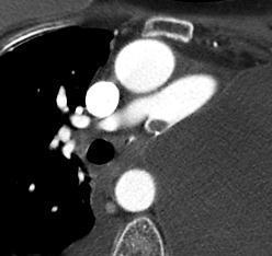
35 Saddle and Embolus Subsegmental Emboli

36 Very small PE of uncertain chronicity and clinical significance


37 Clinical Significance and Prognosis in Acute PE PA diameter Septal deviation
38 PE-related mortality predictors Age above 60 years RV/LV area >1 (odds ratio 8.6) RV/LV diameter >1.5 (odds ratio 48.8,P<0.001) Timing bolus upslope time > 6 seconds (odds ratio 23.3), 50% downslope time >6 seconds (odds ratio 20) Embolus load score >15 (odds ratio 25) Li C, Lin CT, Kligerman SJ, Hong SN, White CS. JTI 2014

39 Pulmonary Infarct - Hampton s Hump

40 Pulmonary Infarct Only 15% cause true infarction Most in lower lobes Usually multiple


41 Pulmonary Infarct Sharply defined consolidation Central lucency Absence of air bronchogram
 0.2 for air bronchograms Revel et al.")
42 Pulmonary Infarcts Likelihood Ratios: 23.0 for central lucencies 2.9 for vessel sign (enlarged vessel at apex) 0.2 for air bronchograms Revel et al. Radiology: Volume 244: Number 3 September 2007
43 R flank PE pain. with R/O Hemorrhage kidney stone

44 R PE flank with pain. Hemorrhage Acute PE


45 Breast cancer patient with lung nodule

46 Acute PE Resolving Infarct

47 Evolving Infarct Melting Ice cube Sign Incidental RUL Nodule


48 Evolving Infarct: Melting Ice Cube Sign Baseline Baseline 3 weeks months 6 months

49 60 y/o male with hemoptysis
50 60 y/o male with hemoptysis

51 60 y/o male with hemoptysis
52 Non-Contrast Scan

53 Non-Contrast Scan


54 Saddle Embolus on Non- Enhanced CT Scan

55 Chronic PE

56 Chronic PE
57 Chronic PE Recanalized, organized or calcified thrombus

58 Arterial Web

59 12-02 Embolus > Web 1-03


60 2 yrs later- arterial web 12-04
61 Chronic PE Recanalized, organized or calcified thrombus Intraluminal webs or bands Pulmonary hypertension

62 Chronic PE

63 Chronic and Acute PE

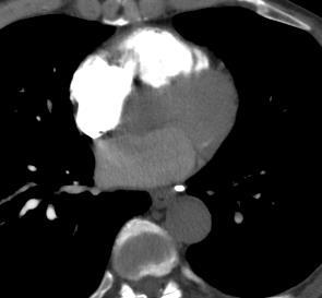
64 Chronic PE Mosaic Perfusion

65 Chronic PE Decreased perfusion & bronchiectasis

66 Enlarged Bronchial Collaterals in Chronic PE
67 Chronic PE Recanalized, organized, calcified thrombus Intraluminal webs or bands Pulmonary hypertension Mosaic perfusion Collateral vascularization
68 CTA for PE: Common Pitfalls Failure to use proper window & level

69 Standard ST Window Narrow window setting
70 CTA for PE: Common Pitfalls Failure to use correct window &level Use of lung (sharp) algorithm


71 Effect of lung filter Lung Standard

72 ECG Gated Cardiac Scan
73 CTA for PE: Common Pitfalls Failure to optimize window level/width Use of lung reconstruction filter Vascular bifurcations (false positives)
74
75 5 mm 1mm
76 CTA for PE: Common Pitfalls Failure to optimize window level/width Use of lung reconstruction algorithm Vascular bifurcations (false positives) Motion/averaging artifact (false positive or negative)

77 Motion with Density Averaging


78 Density Averaging : Crossing Bronchus

79 Decreased flow with Incomplete Opacification
80 CTA for PE: Common Pitfalls Failure to use workstation (window/level) Use of lung reconstruction algorithm Vascular bifurcations (false positives) Motion/averaging artifact (false positive or negative) Bronchiectasis

81
82 Bronchiectasis
83 Postpneumonectomy. R/O PE

84 Arterial Stump Thrombus Occurs in up to 12% of cases Likelihood related to length of arterial stump May propagate or resolve Benign course without treatment
85 Non- embolic thrombus & Non-thrombotic emboli Post-op Lobectomy or Pneumonectomy thrombosis in situ


86 51 y/o woman with severe SOB
87 Uterine Leiomyosarcoma
88 Non- embolic thrombus & Non-thrombotic emboli Post-op Lobectomy or Pneumonectomy thrombosis in situ Tumor Embolism Tumor embolism from remote solid tumors (breast, lung, stomach) Propagation of tumor via IVC (renal, hepatic, uterine) Primary arterial sarcomas
89 Asymptomatic Patient
90 Asymptomatic Patient
91 Polymethymethacrylate Cement Embolism


92 Polymethymethacrylate Cement Embolism Polymethymethacrylate cement embolism 4-23% of procedures <1% symptomatic
93 Non- embolic thrombus & Non-thrombotic emboli Post-op Lobectomy or Pneumonectomy thrombosis in situ Tumor Embolism Tumor embolism from remote solid tumors (breast, lung, stomach) Propagation of tumor via IVC (renal, hepatic, uterine) Primary arterial sarcomas Foreign material (Surgical cement, filter fragments, Needle fragments)
94 Take Home Points CTA is the preferred diagnostic test for PE except in young patients with low clinical probability. PE can often be recognized on CXRs and non contrast CT scans Technique is important; use high injection rate with saline chaser, thin sections, and small inspiration. Use bolus time to aorta to estimate timing Significance of isolated subsegmental emboli is uncertain

95
Pulmonary Embolism. Thoracic radiologist Helena Lauri
 Pulmonary Embolism Thoracic radiologist Helena Lauri 8.5.2017 Statistics 1-2 out of 1000 adults annually are diagnosed with deep vein thrombosis (DVT) and/or pulmonary embolism (PE) About half of patients
Epidermiology Early pulmonary embolism
 Epidermiology Early pulmonary embolism Sitang Nirattisaikul Faculty of Medicine, Prince of Songkla University 3 rd most common cause of cardiovascular death in the United States, following ischemic heart
Radiation Exposure in Pregnancy. John R. Mayo UNIVERSITY OF BRITISH COLUMBIA
 Radiation Exposure in Pregnancy John R. Mayo UNIVERSITY OF BRITISH COLUMBIA Illustrative Clinical Scenario 32 year old female 34 weeks pregnant with recent onset shortness of breath and central chest pain
Proper Diagnosis of Venous Thromboembolism (VTE)
") Proper Diagnosis of Venous Thromboembolism (VTE) Whal Lee, M.D. Seoul National University Hospital Department of Radiology 2 nd EFORT Asia Symposium, 3 rd November 2010, Taipei DVT - Risk Factors Previous
Proper Diagnosis of Venous Thromboembolism (VTE) Whal Lee, M.D. Seoul National University Hospital Department of Radiology 2 nd EFORT Asia Symposium, 3 rd November 2010, Taipei DVT - Risk Factors Previous
Dr. Rami M. Adil Al-Hayali Assistant Professor in Medicine
 Dr. Rami M. Adil Al-Hayali Assistant Professor in Medicine Venous thromboembolism: pulmonary embolism (PE) deep vein thrombosis (DVT) 1% of all patients admitted to hospital 5% of in-hospital mortality
Single Center 4 year series of 114 consecutive patients treated for massive and submassive PE. Mark Goodwin, MD
 Single Center 4 year series of 114 consecutive patients treated for massive and submassive PE Mark Goodwin, MD Disclosure Speaker name:... I have the following potential conflicts of interest to report:
October 2017 Pulmonary Embolism
 October 2017 Pulmonary Embolism Prof. Ahmed BaHammam, FRCP, FCCP Professor of Medicine College of Medicine King Saud University 1 Objectives Epidemiology Pathophysiology Diagnosis Massive PE Treatment
Pulmonary Thromboembolism
 Pulmonary Thromboembolism James Allen, MD Epidemiology of Pulmonary Embolism 1,500,000 new cases per year in the United States Often asymptomatic 300,000 deaths per year DVT or PE present in 10% of ICU
Radiologic Features of The Pulmonary Embolus
 January 2003 Radiologic Features of The Pulmonary Embolus Travis McGlothin HMSIII Mr. J is a 51 y.o. male who presented to the BIDMC ED w/ acute onset of: Lft. Hemiparesis slurred speech mild dyspnea mild
Pathology of pulmonary vascular disease. Dr.Ashraf Abdelfatah Deyab. Assistant Professor of Pathology Faculty of Medicine Almajma ah University
 Pathology of pulmonary vascular disease Dr.Ashraf Abdelfatah Deyab Assistant Professor of Pathology Faculty of Medicine Almajma ah University Pulmonary vascular disease Type of pulmonary circulation: Types
Imaging Pulmonary Embolism
 May 2001 Imaging Pulmonary Embolism New ways to look at a diagnostic dilemma Emily Willner,, HMS III Core Radiology Clerkship, BIDMC New approaches to imaging PE: Agenda 1. Review two patients who had
Clinical Guide - Suspected PE (Reviewed 2006)
") Clinical Guide - Suspected (Reviewed 2006) Principal Developer: B. Geerts Secondary Developers: C. Demers, C. Kearon Background Investigation of patients with suspected pulmonary emboli () remains problematic
Clinical Guide - Suspected (Reviewed 2006) Principal Developer: B. Geerts Secondary Developers: C. Demers, C. Kearon Background Investigation of patients with suspected pulmonary emboli () remains problematic
Chronic Thromboembolic Pulmonary Hypertension (CTEPH): A Primer
: A Primer") Chronic Thromboembolic Pulmonary Hypertension (CTEPH): A Primer H. Page McAdams, MD Duke University Medical Center Durham, NC 27710 page.mcadams@duke.edu Question Which of the following imaging tests is
Chronic Thromboembolic Pulmonary Hypertension (CTEPH): A Primer H. Page McAdams, MD Duke University Medical Center Durham, NC 27710 page.mcadams@duke.edu Question Which of the following imaging tests is
Disclosures. CTA of Acute and Chronic Pulmonary Embolism. Background. Imaging. Which imaging test should be used to evaluate VTE? Objectives.
 CTA of Acute and Chronic Pulmonary Embolism None Disclosures Smita Patel, M.B.B.S., M.R.C.P., F.R.C.R. Professor, Cardiothoracic Radiology Department of Radiology University of Michigan Objectives To assess
Pulmonary Embolism. Pulmonary Embolism. Pulmonary Embolism. PE - Clinical
 Pulmonary embolus - a practical approach to investigation and treatment Sam Janes Wellcome Senior Fellow and Respiratory Physician, University College London Background Diagnosis Treatment Common: 50 cases
PE and DVT. Dr Anzo William Adiga WatsApp or Call Medical Officer/RHEMA MEDICAL GROUP
 PE and DVT Dr Anzo William Adiga WatsApp or Call +256777363201 Medical Officer/RHEMA MEDICAL GROUP OBJECTIVES DEFINE DVT AND P.E PATHOPHYSIOLOGY OF DVT CLINICAL PRESENTATION OF DVT/PE INVESTIGATE DVT MANAGEMENT
Thrombolysis in PE. Outline. Disclosure. Overview on Pulmonary Embolism. Hot Topics in Emergency Medicine 2012 Midyear Clinical Meeting
 Disclosure Thrombolysis in PE Daniel P. Hays, PharmD, BCPS, FASHP reports no relevant financial relationships. Daniel P. Hays, PharmD, BCPS, FASHP Outline 55 YOF presents to ED with SOB PMH of DVT + noncompliance
PULMONARY NODULES AND MASSES : DIAGNOSTIC APPROACH AND NEW MANAGEMENT GUIDELINES. https://tinyurl.com/hmpn2018
 PULMONARY NODULES AND MASSES : DIAGNOSTIC APPROACH AND NEW MANAGEMENT GUIDELINES Heber MacMahon MB, BCh Department of Radiology The University of Chicago https://tinyurl.com/hmpn2018 Disclosures Consultant
MATERIALS AND METHODS
 RETROSPECTIVE STUDY OF OPTIMISING THE USE OF COMPUTED TOMOGRAPHY PULMONARY ANGIOGRAPHY (CTPA) FOR THE DIAGNOSIS OF PULMONARY EMBOLISM IN PLACES WITH LIMITED RESOURCES P. V. Kalyan Kumar 1, Ramakrishna
Interesting Cases. Pulmonary
 Interesting Cases Pulmonary 54M with prior history of COPD, hep B/C, and possible history of TB presented with acute on chronic dyspnea, and productive cough Hazy opacity overlying the left hemithorax
GUIDELINES FOR PULMONARY NODULE MANAGEMENT : RECENT CHANGES AND UPDATES
 Venice 2017 GUIDELINES FOR PULMONARY NODULE MANAGEMENT : RECENT CHANGES AND UPDATES Heber MacMahon MB, BCh Department of Radiology The University of Chicago Disclosures Consultant for Riverain Medical
Surgical Management in Chronic Thromboembolic Pulmonary Hypertension. Michael Bates, MD, FACS Ochsner Health System, New Orleans, LA
 Surgical Management in Chronic Thromboembolic Pulmonary Hypertension Michael Bates, MD, FACS Ochsner Health System, New Orleans, LA Disclosures No industry conflicts I am a surgeon and always disclose
Cover Page. The handle holds various files of this Leiden University dissertation.
 Cover Page The handle http://hdl.handle.net/1887/21764 holds various files of this Leiden University dissertation. Author: Mos, Inge Christina Maria Title: A more granular view on pulmonary embolism Issue
Risk factors for DVT. Venous thrombosis & pulmonary embolism. Anticoagulation (cont d) Diagnosis 1/5/2018. Ahmed Mahmoud, MD
 Diagnosis 1/5/2018. Ahmed Mahmoud, MD") Risk factors for DVT Venous thrombosis & pulmonary embolism Ahmed Mahmoud, MD Surgery ; post op especially for long cases, pelvic operations (THR), Trauma ; long bone fractures, pelvic fractures (posterior
Risk factors for DVT Venous thrombosis & pulmonary embolism Ahmed Mahmoud, MD Surgery ; post op especially for long cases, pelvic operations (THR), Trauma ; long bone fractures, pelvic fractures (posterior
Venous thrombosis & pulmonary embolism. Ahmed Mahmoud, MD
 Venous thrombosis & pulmonary embolism Ahmed Mahmoud, MD Risk factors for DVT Surgery ; post op especially for long cases, pelvic operations (THR), Trauma ; long bone fractures, pelvic fractures (posterior
Too much medicine and venous thromboembolism: How can we make things Well again?
 Too much medicine and venous thromboembolism: How can we make things Well again? Emily G McDonald MD MSc; Assistant professor of medicine; McGill University Health Centre Canadian Society of Internal Medicine;
Reporting SPECT-VQ. Alp Notghi
 Reporting SPECT-VQ Alp Notghi 20 year old female 24 weeks pregnant Clinical History : SOB and chest pain for past 3 days.?pe Doppler USS excluded DVT Case 4413041 Normal Case 4413041 CXR report: The heart
CT angiography techniques. Boot camp
 CT angiography techniques Boot camp Overview Basic concepts Contrast administration arterial opacification Time scan acquisition during the arterial phase Protocol examples Helical non-gated CTA Pulmonary
Systemic lupus erythematosus (SLE): Pleuropulmonary Manifestations
: Pleuropulmonary Manifestations") 08/30/10 09/26/10 Systemic lupus erythematosus (SLE): Pleuropulmonary Manifestations Camila Downey S. Universidad de Chile, School of Medicine, Year VII Harvard University, School of Medicine Sept 17,
08/30/10 09/26/10 Systemic lupus erythematosus (SLE): Pleuropulmonary Manifestations Camila Downey S. Universidad de Chile, School of Medicine, Year VII Harvard University, School of Medicine Sept 17,
Real life management of CTEPH: patient case
 2 nd International Congress on cardiovascular imaging in clinical practice k Real life management of CTEPH: patient case Anastasia Anthi Pulmonary Hypertension Clinic, Attikon University Hospital, Athens
PULMONARY EMBOLISM (PE): DIAGNOSIS AND TREATMENT
: DIAGNOSIS AND TREATMENT") PULMONARY EMBOLISM (PE): DIAGNOSIS AND TREATMENT OBJECTIVE: To provide a diagnostic algorithm and treatment options for patients with acute pulmonary embolism (PE). BACKGROUND: Venous thromboembolism (VTE)
PULMONARY EMBOLISM (PE): DIAGNOSIS AND TREATMENT OBJECTIVE: To provide a diagnostic algorithm and treatment options for patients with acute pulmonary embolism (PE). BACKGROUND: Venous thromboembolism (VTE)
CURRENT & FUTURE THERAPEUTIC MANAGEMENT OF VENOUS THROMBOEMBOLISM. Gordon Lowe Professor of Vascular Medicine University of Glasgow
 CURRENT & FUTURE THERAPEUTIC MANAGEMENT OF VENOUS THROMBOEMBOLISM Gordon Lowe Professor of Vascular Medicine University of Glasgow VENOUS THROMBOEMBOLISM Common cause of death and disability 50% hospital-acquired
Deep Vein Thrombosis and Pulmonary Embolism: Patient Information
 Deep Vein Thrombosis and Pulmonary Embolism: Patient Information A Deep Vein Thrombosis (DVT) and a Pulmonary Embolism (PE) are both disorders of unwanted blood clotting. Unwanted blood clots can occur
Interventional Treatment VTE: Radiologic Approach
 Interventional Treatment VTE: Radiologic Approach Hae Giu Lee, MD Professor, Dept of Radiology Seoul St. Mary s Hospital The Catholic University of Korea Introduction Incidence High incidence: 250,000-1,000,000/year
Signs in Chest Radiology
 Signs in Chest Radiology Jonathan H. Chung, MD Disclosures No pertinent disclosures Jonathan H. Chung, MD Assistant Professor Institute t of fadvanced d Biomedical Imaging National Jewish Health Denver,
The Location and Size of Pulmonary Embolism in Antineoplastic Chemotherapy Patients 1
 The Location and Size of Pulmonary Embolism in Antineoplastic Chemotherapy Patients 1 Yun Joo Park, M.D., Woocheol Kwon, M.D., Won-Yeon Lee, M.D. 2, Sang Baek Koh, M.D. 3, Seong Ah Kim, M.D., Myung Soon
Avoiding Pitfalls In PE
 UC SF Avoiding Pitfalls In PE Jeffrey Tabas, MD Professor UCSF School of Medicine Emergency Department San Francisco General Hospital sf g h Disclosure No Financial Relationships to Disclose No significant
Fundamentals, Techniques, Pitfalls, and Limitations of MDCT Interpretation and Measurement
 Fundamentals, Techniques, Pitfalls, and Limitations of MDCT Interpretation and Measurement 3 rd Annual Imaging & Physiology Summit November 20-21, 21, 2009 Seoul, Korea Wm. Guy Weigold, MD, FACC Cardiovascular
CT Pulmonary Angiography 2010
 2010 Annual Course CT Pulmonary Angiography 2010 Vassilios Raptopoulos, MD Beth Israel Deaconess Medical Center Harvard Medical School CTPA 2010 Utilization Technique DVT Radiation In Pregnancy Large Small
A A U
 PVD Venous AUC Rating Sheet 2nd Round 1 2 3 4 5 6 7 8 9 10 11 12 13 14 15 Median I NI MADM Rating Agree Disagree Upper Extremity Venous Evaluation Table 1. Venous Duplex of the Upper Extremities for Patency
SCINTIGRAPHY OF THE LUNGS THE VQ SCAN
 SCINTIGRAPHY OF THE LUNGS THE VQ SCAN By George N. Sfakianakis, M.D. Professor of Radiology and Pediatrics October 2009 PULMONARY EMBOLISM 94,000 cases annually in US. Not a disease by itself. A potentially
Pulmonary Embolism..Diagnostic Approach and Algorithm. Tolulope Adesiyun Harvard Medical School, Year III Gillian Lieberman, MD
 Pulmonary Embolism..Diagnostic Approach and Algorithm Tolulope Adesiyun Harvard Medical School, Year III Gillian Lieberman, MD Epidemiology of Pulmonary Embolism Pulmonary Embolus (PE): Thrombus originating
How to Analyse Difficult Chest CT
 How to Analyse Difficult Chest CT Complex diseases are:- - Large lesion - Unusual or atypical pattern - Multiple discordant findings Diffuse diseases are:- - Numerous findings in both sides 3 basic steps
Jeffrey Tabas, MD. sf g h. Risk Assessment Do we understand risk stratification? Are we limiting radiation /contrast with the PERC rule and D-Dimers?
 Pulmonary Embolism Update Jeffrey Tabas, MD Professor UCSF School of Medicine Emergency Department San Francisco General Hospital Disclosure No Financial Relationships to Disclose No significant investments
Imaging in Pulmonary Embolism. Gamal Rabie Agmy, MD,FCCP Professor of Chest Diseases, Assiut university
 Imaging in Pulmonary Embolism Gamal Rabie Agmy, MD,FCCP Professor of Chest Diseases, Assiut university Background Information Pulmonary embolism is a life-threatening condition that occurs when a clot
Pulmonary Nodules & Masses
 Pulmonary Nodules & Masses A Diagnostic Approach Heber MacMahon The University of Chicago Department of Radiology Disclosure Information Consultant for Riverain Technology Minor equity in Hologic Royalties
Acute Pulmonary Embolism and Deep Vein Thrombosis. Barbara LeVarge MD Beth Israel Deaconess Medical Center Pulmonary Hypertension Center COPYRIGHT
 Acute Pulmonary Embolism and Deep Vein Thrombosis Barbara LeVarge MD Beth Israel Deaconess Medical Center Pulmonary Hypertension Center Acute PE and DVT No disclosures. Acute PE and DVT Learning objectives
Updates in Medical Management of Pulmonary Embolism and Deep Vein Thrombosis. By: Justin Youtsey, Elliott Reiff, William Montgomery, Grant Finlan
 Updates in Medical Management of Pulmonary Embolism and Deep Vein Thrombosis By: Justin Youtsey, Elliott Reiff, William Montgomery, Grant Finlan Objectives Describe the prevalence of PE and DVT as it relates
Difficulties of timely diagnosis of the Pulmonary Embolism of patients with chronic obstructive lung disease: possibility MSCT.
 Difficulties of timely diagnosis of the Pulmonary Embolism of patients with chronic obstructive lung disease: possibility MSCT. Poster No.: C-2618 Congress: ECR 2012 Type: Scientific Exhibit Authors: I.
Inferior Vena Cava Filters
 Inferior Vena Cava Filters and the American Society of Hematology Choosing Wisely Campaign Kevin P. Hubbard, DO, HMDC MACOI Chief - Division of Specialty Medicine Professor and Chair - Section of Internal
Venous thrombosis is common and often occurs spontaneously, but it also frequently accompanies medical and surgical conditions, both in the community
 Venous Thrombosis Venous Thrombosis It occurs mainly in the deep veins of the leg (deep vein thrombosis, DVT), from which parts of the clot frequently embolize to the lungs (pulmonary embolism, PE). Fewer
Results of Ischemic Heart Disease
 Ischemic Heart Disease: Angina and Myocardial Infarction Ischemic heart disease; syndromes causing an imbalance between myocardial oxygen demand and supply (inadequate myocardial blood flow) related to
CTPA for Pulmonary Emboli: 2016 update
 2016 Annual Course CTPA for Pulmonary Emboli: 2016 update Olga R Brook, MD, FSCBTMR Beth Israel Deaconess Medical Center, Boston Beth Israel Deaconess Medical Center HarvardMedical School Contents Technique
Venous Thromboembolism (VTE)
") Venous Thromboembolism (VTE) Nursing A guide for patients and carers Contents Why do blood clots form in veins?... 1 How common is a deep vein thrombosis (DVT) or pulmonary embolus (PE)?... 2 How are DVTs/
Venous Thromboembolism (VTE) Nursing A guide for patients and carers Contents Why do blood clots form in veins?... 1 How common is a deep vein thrombosis (DVT) or pulmonary embolus (PE)?... 2 How are DVTs/
Case 1. Technegas Case Studies. Prostate cancer. Finished treatment recently. Smoker. Angina. Presents sudden dyspnea and poorly defined chest pains.
 Case 1 Michel Leblanc MD; RCPSC; ABNM Head of the Nuclear Medicine Department, Centre Hospitalier Régional de Trois-Rivières. Clinical Professor, Centre Hospitalier Universitaire de Sherbrooke, Cannada.
Imaging of acute pulmonary thromboembolism*
 Silva, Isabela et al. Imaging of acute pulmonary thromboembolism Imaging of acute pulmonary thromboembolism* C. ISABELA S. SILVA, NESTOR L. MÜLLER The diagnosis of acute pulmonary thromboembolism is based
Inferior Venacaval Filters Valuable vs. Dangerous Valuable Annie Kulungowski. Department of Surgery Grand Rounds March 24, 2008
 Inferior Venacaval Filters Valuable vs. Dangerous Valuable Annie Kulungowski Department of Surgery Grand Rounds March 24, 2008 History of Vena Cava Filters Virchow-1846-Proposes PE originate from veins
Disclosures. DVT: Diagnosis and Treatment. Questions To Ask. Dr. Susanna Shin - DVT: Diagnosis and Treatment. Acute Venous Thromboembolism (VTE) None
 None") Disclosures DVT: Diagnosis and Treatment None Susanna Shin, MD, FACS Assistant Professor University of Washington Acute Venous Thromboembolism (VTE) Deep Venous Thrombosis (DVT) Pulmonary Embolism (PE)
Disclosures DVT: Diagnosis and Treatment None Susanna Shin, MD, FACS Assistant Professor University of Washington Acute Venous Thromboembolism (VTE) Deep Venous Thrombosis (DVT) Pulmonary Embolism (PE)
DVT and Pulmonary Embolus. Dr Piers Blombery BSc(Biomed), MBBS (Hons), FRACP, FRCPA Consultant Haematologist Peter MacCallum Cancer Centre
, MBBS (Hons), FRACP, FRCPA Consultant Haematologist Peter MacCallum Cancer Centre") DVT and Pulmonary Embolus Dr Piers Blombery BSc(Biomed), MBBS (Hons), FRACP, FRCPA Consultant Haematologist Peter MacCallum Cancer Centre Overview Structure of deep and superficial venous system of upper
DVT and Pulmonary Embolus Dr Piers Blombery BSc(Biomed), MBBS (Hons), FRACP, FRCPA Consultant Haematologist Peter MacCallum Cancer Centre Overview Structure of deep and superficial venous system of upper
PULMONARY EMBOLISM ANGIOCT (CTA) ASSESSMENT OF VASCULAR OCCLUSION EXTENT AND LOCALIZATION OF EMBOLI 1. BACKGROUND
 ASSESSMENT OF VASCULAR OCCLUSION EXTENT AND LOCALIZATION OF EMBOLI 1. BACKGROUND") JOURNAL OF MEDICAL INFORMATICS & TECHNOLOGIES Vol. 11/2007, ISSN 1642-6037 Damian PTAK * pulmonary embolism, AngioCT, postprocessing techniques, Mastora score PULMONARY EMBOLISM ANGIOCT (CTA) ASSESSMENT
JOURNAL OF MEDICAL INFORMATICS & TECHNOLOGIES Vol. 11/2007, ISSN 1642-6037 Damian PTAK * pulmonary embolism, AngioCT, postprocessing techniques, Mastora score PULMONARY EMBOLISM ANGIOCT (CTA) ASSESSMENT
Pulmonary Embolism through the CT Angiography looking glass
 Pulmonary Embolism through the CT Angiography looking glass Poster No.: C-1467 Congress: ECR 2015 Type: Educational Exhibit Authors: M. Tapp, A. Mannava, F. C. Lyall ; Torquay/UK, Torbay 1 2 3 1 2 3 Hospital,Torquay,
Canadian Society of Internal Medicine Annual Meeting 2016 Montreal, QC
 Canadian Society of Internal Medicine Annual Meeting 2016 Montreal, QC 1 st workshop: update to VTE guidelines in 2016 2 nd workshop: VTE controversies + new horizons André Roussin MD, FRCP, CSPQ CHUM
Diagnosis and Treatment of Pulmonary Embolism. Farzin Ghiasi, MD Pulmonologist August, 2016
 Diagnosis and Treatment of Pulmonary Embolism Farzin Ghiasi, MD Pulmonologist August, 2016 DVT & PE Hypercoagulable state is characteristic of pregnancy, and DVT occurs in about 1 in 500 pregnancies. In
Audit of CT Pulmonary Angiogram in suspected pulmonary embolism patients
 Audit of CT Pulmonary Angiogram in suspected pulmonary embolism patients Poster No.: C-2511 Congress: ECR 2012 Type: Scientific Exhibit Authors: N. D. Gupta, M. K. Heir, P. Bradding; Leicester/UK Keywords:
Cover Page. The handle holds various files of this Leiden University dissertation
 Cover Page The handle http://hdl.handle.net/1887/40114 holds various files of this Leiden University dissertation Author: Exter, Paul L. den Title: Diagnosis, management and prognosis of symptomatic and
Pulmonary Thromboembolism
 Pulmonary Thromboembolism Jing ZHANG ( 张静 ), MD, PhD zhang.jing@zs-hospital.sh.cn Department of Pulmonary Medicine Zhongshan Hospital Fudan University OUTLINE Understand the historical context of pulmonary
High density thrombi of pulmonary embolism on precontrast CT scan: Is it dangerous?
 High density thrombi of pulmonary embolism on precontrast CT scan: Is it dangerous? Poster No.: C-1753 Congress: ECR 2013 Type: Authors: Keywords: DOI: Scientific Exhibit B. Y. Lee, H. R. KIM, J. I. Jung,
Lung diseases of Vascular Origin. By: Shefaa Qa qqa
 Lung diseases of Vascular Origin By: Shefaa Qa qqa Pulmonary Hypertension Pulmonary hypertension is defined as a mean pulmonary artery pressure greater than or equal to 25 mm Hg at rest. Based on underlying
Understanding thrombosis in venous thromboembolism. João Morais Head of Cardiology Division and Research Centre Leiria Hospital Centre Portugal
 Understanding thrombosis in venous thromboembolism João Morais Head of Cardiology Division and Research Centre Leiria Hospital Centre Portugal Disclosures João Morais On the last year JM received honoraria
Pediatric Lung Ultrasound (PLUS) In Diagnosis of Community Acquired Pneumonia (CAP)
 In Diagnosis of Community Acquired Pneumonia (CAP)") Pediatric Lung Ultrasound (PLUS) In Diagnosis of Community Acquired Pneumonia (CAP) Dr Neetu Talwar Senior Consultant, Pediatric Pulmonology Fortis Memorial Research Institute, Gurugram Study To compare
Pediatric Lung Ultrasound (PLUS) In Diagnosis of Community Acquired Pneumonia (CAP) Dr Neetu Talwar Senior Consultant, Pediatric Pulmonology Fortis Memorial Research Institute, Gurugram Study To compare
Audit of CT Pulmonary Angiogram in suspected pulmonary embolism patients
 Audit of CT Pulmonary Angiogram in suspected pulmonary embolism patients Poster No.: C-2511 Congress: ECR 2012 Type: Scientific Exhibit Authors: N. D. Gupta, M. K. Heir, P. Bradding; Leicester/UK Keywords:
Amanda Phillips, RVT
 Amanda Phillips, RVT Cockett Syndrome Iliac vein compression syndrome http://www.ardms.org/volunteer-now/clearly-connected/pages/may-thurner-syndrome-what-sonographers-should-know.aspx Compression of Lt
Simplified approach to investigation of suspected VTE
 Simplified approach to investigation of suspected VTE Diagnosis of DVT and PE THSNA 2016, Chicago 15 April 2016 Clive Kearon, McMaster University, Canada Relevant Disclosures Research Support/P.I. Employee
Cover Page. The handle holds various files of this Leiden University dissertation
 Cover Page The handle http://hdl.handle.net/1887/40114 holds various files of this Leiden University dissertation Author: Exter, Paul L. den Title: Diagnosis, management and prognosis of symptomatic and
Missed Pulmonary Embolism on Abdominal CT
 Cardiopulmonary Imaging Original Research Lim et al. Pulmonary Emboli Missed on CT of the Abdomen Cardiopulmonary Imaging Original Research Kun Young Lim 1 Seth J. Kligerman 2 Cheng Ting Lin 2 Charles
Reducing the risk of venous thrombo-embolism (VTE) in hospital and after discharge
 in hospital and after discharge") Reducing the risk of venous thrombo-embolism (VTE) in hospital and after discharge What is a venous thromboembolism (VTE)? This is a medical term that describes a blood clot that develops in a deep vein
Reducing the risk of venous thrombo-embolism (VTE) in hospital and after discharge What is a venous thromboembolism (VTE)? This is a medical term that describes a blood clot that develops in a deep vein
Mabel Labrada, MD Miami VA Medical Center
 Mabel Labrada, MD Miami VA Medical Center *1-Treatment for acute DVT with underlying malignancy is for 3 months. *2-Treatment of provoked acute proximal DVT can be stopped after 3months of treatment and
Deep Vein Thrombosis
 Deep Vein Thrombosis from NHS (UK) guidelines Introduction Deep vein thrombosis (DVT) is a blood clot in one of the deep veins in the body. Blood clots that develop in a vein are also known as venous thrombosis.
REVIEW ON PULMONARY EMBOLISM
 REVIEW ON PULMONARY EMBOLISM * Shashi Kumar Yadav, Prof. Xiao Wei, Roshan Kumar Yadav, Sanjay Kumar Verma and Deepika Dhakal * Department of Medicine, Clinical College of Yangtze University, The first
Bronchiectasis: An Imaging Approach
 Bronchiectasis: An Imaging Approach Travis S Henry, MD Associate Professor of Clinical Radiology Cardiac and Pulmonary Imaging Section University of California, San Francisco Large Middle Small 1 Bronchiectasis
Shock, Hemorrhage and Thrombosis
 Shock, Hemorrhage and Thrombosis 1 Shock Systemic hypoperfusion due to: Reduction in cardiac output Reduction in effective circulating blood volume Hypotension Impaired tissue perfusion Cellular hypoxia
Lung Cancer - Suspected
 Lung Cancer - Suspected Shared Decision Making Lung Cancer: http://www.enhertsccg.nhs.uk/ Patient presents with abnormal CXR Lung cancer - clinical presentation History and Examination Incidental finding
University Journal of Medicine and Medical Specialities
 University Journal of Medicine and Medical Specialities Volume 1 Issue 1 2015 An unusual case of SADDLE Pulmonary Thrombo Embolism Boopathirajan P Jayanthi R Stanley Medical College Abstract: Pulmonary
Beyond Pulmonary Emboli: Acquired Abnormalities of the Pulmonary Arteries on CTA
 Beyond Pulmonary Emboli: Acquired Abnormalities of the Pulmonary Arteries on CTA Sean Cleary, MD and Katherine Kaproth-Joslin, MD/PhD University of Rochester Medical Center Rochester, NY Financial Disclosures
Anticoagulation Forum: Management of Tiny Clots
 Anticoagulation Forum: Management of Tiny Clots Casey O Connell, MD FACP Associate Professor Jane Anne Nohl Division of Hematology Keck School of Medicine USC DISCLOSURES None 4/11/2017 Objectives Define
NOTE: Deep Vein Thrombosis (DVT) Risk Factors
 Risk Factors") Deep Vein Thrombosis (DVT) Deep Vein Thrombosis (DVT) is the formation of a blood clot, known as a thrombus, in the deep leg vein. It is a very serious condition that can cause permanent damage to the
Deep Vein Thrombosis (DVT) Deep Vein Thrombosis (DVT) is the formation of a blood clot, known as a thrombus, in the deep leg vein. It is a very serious condition that can cause permanent damage to the
PE is a difficult diagnosis that may be missed because of non-specific clinical presentation.
 Pulmonary embolism (PE) is a relatively common cardiovascular emergency. By occluding the pulmonary arterial bed it may lead to acute life-threatening (3% early mortality rate), but potentially reversible
OBJECTIVES. Solitary Solid Spiculated Nodule. What would you do next? Case Based Discussion: State of the Art Management of Lung Nodules.
 Organ Imaging : September 25 2015 OBJECTIVES Case Based Discussion: State of the Art Management of Lung Nodules Dr. Elsie T. Nguyen Dr. Kazuhiro Yasufuku 1. To review guidelines for follow up and management
TB Intensive Houston, Texas
 TB Intensive Houston, Texas October 15-17, 17 2013 Diagnosis of TB: Radiology Rosa M Estrada-Y-Martin, MD MSc FCCP October 16, 2013 Rosa M Estrada-Y-Martin, MD MSc FCCP, has the following disclosures to
Provider Led Entity. CDI Quality Institute PLE Chest / Pulmonary Embolus AUC 07/31/2018
 Provider Led Entity CDI Quality Institute PLE Chest / Pulmonary Embolus AUC 07/31/2018 Appropriateness of advanced imaging procedures* in patients with suspected or known pulmonary embolus and the following
How long to continue anticoagulation after DVT?
 How long to continue anticoagulation after DVT? Dr. Nihar Ranjan Pradhan M.S., DNB (Vascular Surgery), FVES(UK) Consultant Vascular Surgeon Apollo Hospital, Jubilee Hills, Hyderabad (Formerly Faculty in
Evaluation of Chest Pain in the Primary Care Setting. Joseph Hackler, DO. Disclosures
 Evaluation of Chest Pain in the Primary Care Setting Joseph Hackler, DO Disclosures I have no relevant relationships with commercial interests to disclose. 1 Objectives 1. Discuss the different etiologies
Diagnosis and Treatment of Deep Venous Thrombosis and Pulmonary Embolism
 Agency for Healthcare Research and Quality Evidence Report/Technology Assessment Diagnosis and Treatment of Deep Venous Thrombosis and Pulmonary Embolism Summary Number 68 Overview Venous thromboembolism
Venous Thrombosis. Magnitude of the Problem. DVT 2 Million PE 600,000. Death 60,000. Estimated Cost of VTE Care $1.5 Billion/year.
 Venous Thrombosis Magnitude of the Problem DVT 2 Million Postthrombotic Syndrome 800,000 PE 600,000 Death 60,000 Silent PE 1 Million Pulmonary Hypertension 30,000 Estimated Cost of VTE Care $1.5 Billion/year
Jessica Bryan, Natalia Evans, Karlyn Henderson, & Whitney Parks
 Jessica Bryan, Natalia Evans, Karlyn Henderson, & Whitney Parks 1. What is the most common cause of death in hospitalized patients? 1. Hospital-acquired infection 2. Pulmonary embolism 3. Myocardial infarction
Exercise Test: Practice and Interpretation. Jidong Sung Division of Cardiology Samsung Medical Center Sungkyunkwan University School of Medicine
 Exercise Test: Practice and Interpretation Jidong Sung Division of Cardiology Samsung Medical Center Sungkyunkwan University School of Medicine 2 Aerobic capacity and survival Circulation 117:614, 2008
Pulmonary Embolism Is it the Greatest Danger in Deep Vein Thrombosis?
 Difficult issues in Deep Vein Thrombosis: Pulmonary Embolism Is it the Greatest Danger in Deep Vein Thrombosis? Raluca Dulgheru; C Gherghinescu; B Dorobat; H Muresan; R Darabont; M Cinteza; D Vinereanu
Pulmonary embolism: Strategies to optimize pulmonary MDCT angiography studies
 Pulmonary embolism: Strategies to optimize pulmonary MDCT angiography studies Poster No.: C-0152 Congress: ECR 2013 Type: Educational Exhibit Authors: G. Viteri, A. Garcia-lallana, I. Simon Yarza, A. Villanueva
Request Card Task ANSWERS
 Request Card Task ANSWERS Medical Student Workbook Author: Dr Sam Leach, SpR Case 1 What differential diagnoses are most likely? Which investigation is most appropriate? Case 1 The most likely diagnosis
Pulmonary Embolism Rule-out Criteria - Is it good enough?
 Pulmonary Embolism Rule-out Criteria - Is it good enough? DR. IMRON SUBHAN MBBS, FEM (CMC), MRCEM CONSULTANT & HEAD DEPARTMENT OF EMERGENCY MEDICINE APOLLO HOSPITALS HYDERABAD INDIA GENERAL SECRETARY SOCIETY