Major Veins of the Body
|
|
|
- Flora McKinney
- 5 years ago
- Views:
Transcription


1 Major Veins of the Body Please view our Editing File before studying this lecture to check for any changes. Color Code Important Doctors Notes Notes/Extra explanation
2 Objectives At the end of the lecture, the student should be able to: ü Define veins and understand the general principle of venous system. ü Describe the superior & inferior Vena Cava: formation and their tributaries ü List major veins and their tributaries in: head & neck thorax & abdomen upper & lower limbs ü Describe the Portal Vein: formation & tributaries. ü Describe the Portocaval Anastomosis: formation, sites and importance

 Their location (superficial/deep) (2) The circulation (systemic/portal) 1: are large veins that receive oxygenated blood")
3 Veins o Veins are blood vessels that bring blood back to the heart. o All veins carry deoxygenated blood except: o Pulmonary veins 1. o Umbilical veins 2. o There are two types of veins*: 1. Superficial veins: close to the surface of the body o o NO corresponding arteries 2. Deep veins: found deeper in the body With corresponding arteries (venae comitantes) Veins of the systemic circulation: Superior and inferior vena cava with their tributaries Veins of the portal circulation: Portal vein *Note: Vein can be classified in 2 ways based on: (1) Their location (superficial/deep) (2) The circulation (systemic/portal) 1: are large veins that receive oxygenated blood from the lung and drain into the left atrium. 2: The umbilical vein is a vein present during fetal development that carries oxygenated blood from the placenta into the growing fetus.
: a single layer of simple squamous endothelial cells. 2.")
 entirely made of connective tissue.")
4 Only on the boys slides The Histology Of Blood Vessels o The arteries and veins have three layers, but the middle layer is thicker in the arteries than it is in the veins: 1. Tunica Intima (the thinnest layer): a single layer of simple squamous endothelial cells. 2. Tunica Media (the thickest layer in arteries): is made up of smooth muscle cells and elastic tissue. 3. Tunica Adventitia: (the thickest layer in veins) entirely made of connective tissue. o Capillaries consist of little more than a layer of endothelium and occasional connective tissue.


5 Superior Vena Cava o Formed by the union of the right and left brachiocephalic veins. Brachiocephalic veins are formed by the union of internal jugular and subclavian veins. o Drains venous blood from: Head, neck, thoracic wall & upper limbs. o It Passes downward and enter the right atrium. o Receives azygos vein on the posterior aspect just before it enters the heart. Sup
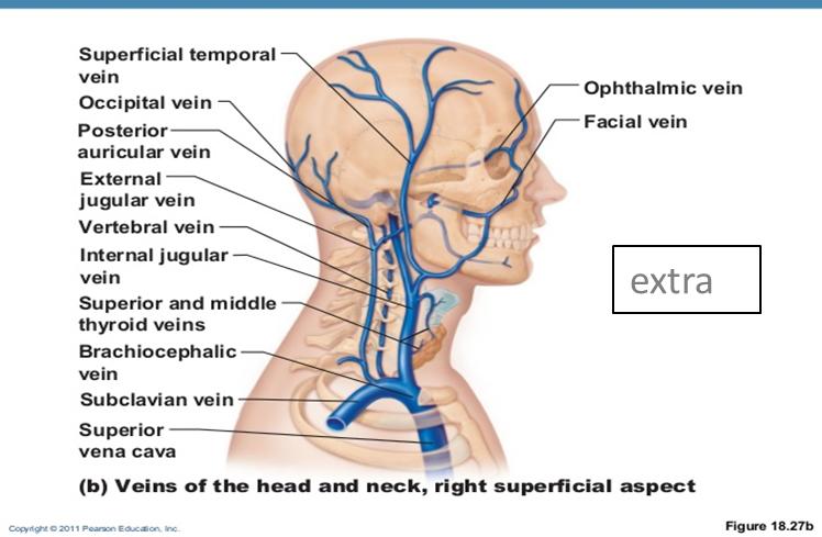
6 Veins Of Head & Neck Superficial Deep External Jugular Vein Anterior Jugular Vein Internal Jugular Vein Note: Union: when to veins join together they give rise to a vein with a new name. Tributary: when one vein drains into another, but no new vein is produced. Jugular means of the neck.


7 Veins of Head and Neck External Jugular Vein o Lies superficial to the sternomastoid (sternocleidomastoid) muscle. o It passes down the neck and it is the only tributary of the subclavian vein. o Begins just behind angle of mandible by union of: a. the posterior division of the retromandibular vein (temporomaxillary vein) b. with the posterior auricular vein. o It drains blood from: a. Outside of the skull b. Deep parts of the face. Tributaries: Posterior external jugular vein. Anterior jugular vein. Suprascapular vein. Transverse cervical vein. To remember: PAST


8 Veins of Head and Neck Anterior Jugular Vein o It begins in the upper part of the neck by the union of the submental veins (small veins found in an area known as the submental triangle). o It descends close to the median line of the neck, medial to the sternomastoid muscle. o At the lower part of the neck, it passes laterally beneath (deep to) sternomastoid to drain into the external jugular vein. o Just above the sternum the two anterior jugular veins communicate by a transverse vein to form the jugular arch. Extra

 Facial Occipital veins Pharyngeal veins Dural venous sinuses (Inferior petrosal sinus) Note: the occipital vein s drainage")
9 Veins of Face and Neck Internal Jugular Veins o Drains blood from the head, brain, face & neck. o It descends in the neck along with the internal and common carotid arteries and vagus nerve, within the carotid sheath. o Joins the subclavian vein to form the brachiocephalic vein. o Tributaries: Superior thyroid Lingual (in the tongue) Facial Occipital veins Pharyngeal veins Dural venous sinuses (Inferior petrosal sinus) Note: the occipital vein s drainage can vary. Dural venous sinuses: these are venous sinuses found between the periosteal and meningeal layer of the dura matter (check next slide). The inferior part of the internal jugular vein has a dilation known as the inferior bulb. Above the bulb there is a valve.

10 Extra pictures for understanding

11 Veins Of Upper Limbs 1- Superficial Veins A- Cephalic Vein o Ascends in the superficial fascia on the lateral side of the biceps. o Drains into the Axillary vein. B- Basilic Vein o Ascends in the superficial fascia on the medial side of the biceps. o Halfway up the arm, it pierces the deep fascia o At the lower border of the teres major it joins the venae comitantes of the brachial artery to form the Axillary vein. 2- Deep Veins A- Venae Commitantes o Which accompany all the large arteries and are usually in pairs. B- Axillary Vein o Formed by the union of basilic vein and the venae comitantes (brachial veins) of the brachial artery.


 10 Eggs At 12.")
12 Inferior Vena Cava o Drains most of the blood from the body below the diaphragm to the right atrium. o Formed by the union of the two common iliac veins behind the right common iliac artery at the level of the 5th lumbar vertebra. o Ascends on the right side of the aorta o Pierces the central tendon of diaphragm at the level of the 8th thoracic vertebra*. *Recall the descending aorta pierced the diaphragm at T12. To remember: Mnemonic of major openings of diaphragm: I ate (8) 10 Eggs At 12. (I 8= inferior vena cava pierce at T8, 10 Eggs= Esophagus pierces at T10, At 12 = Aorta pierces at T12)
 5. Paired renal veins 6.")

13 Inferior Vena Cava (Tributaries) From bottom to top 1. Median sacral vein 2. Two common iliac veins 3. Four paired lumbar veins 4. Right gonadal vein (the left vein drains into the left renal vein*) 5. Paired renal veins 6. Right suprarenal vein (the left vein drains into the left renal vein*) 7. Hepatic veins 8. Paired inferior phrenic vein *Note: The left gonadal/suprarenal veins drain into the IVC but INDIRECTLY. They first drain into the left renal vein which then drains into the IVC. That s why we didn t list them here as part of the tributaries. Median sacral vein
 saphenous vein Small (short) saphenous")
14 Veins Of Lower Limbs TWO DIVISIONS: SUPERFICIAL VEINS DEEP VEINS Form a network in the subcutaneous tissue Pattern is variable They are the tributaries of the: Great (long) saphenous vein Small (short) saphenous vein
 with the saphenous nerve.")

15 Veins Of Lower Limbs Great Saphenous Vein o The longest vein. o Beginning: from the medial end of the dorsal venous arch of the foot. Ascending : Passes upward in front of the medial malleolus (constant position) with the saphenous nerve. Then it ascends in accompany with the saphenous nerve in the superficial fascia over the medial side of the leg Ascends obliquely upwards, and lies behind the medial border of the patella. Passes behind the knee and curves forward around the medial side of the thigh. o Termination : Hooks through the lower part of the saphenous opening in the deep fascia to join the femoral vein about 1.5 inch (4 cm) below and lateral to the pubic tubercle.

.")

16 Veins Of Lower Limbs Great Saphenous Vein o It is connected to the small saphenous vein by one or two branches that pass behind the knee. o Numerous perforating veins connect the great saphenous vein with the deep veins (femoral vein). o The perforating veins have valves which allow blood flow from superficial to deep veins. o (التطعیم) The great saphenous vein is used in venous grafting and saphenous vein cutdown may be necessary for inserting the needle or canula (take care of the saphenous nerve) Only on the girls slides

17 Veins Of Lower Limbs Small Saphenous Vein o Beginning: from the lateral end of the dorsal venous arch of the foot. o Has numerous valves along its course. o Anastomosis freely with great saphenous vein. Ascending : Ascends behind the lateral malleolus in company with the sural nerve. Follows the lateral border of the tendocalcaneus and then runs up to the middle of the back of the leg. Pierces the deep fascia in the lower part of the popliteal fossa o Termination: Drains into the popliteal vein. Doctor s note: the small saphenous vein join the venae comitantes and forms the popliteal vein.


18 Veins Of Lower Limbs Deep Veins o Comprise the venae comitantes, which accompany all the large arteries, usually in pairs. o Venae comitantes unite to form the popliteal vein, which continues as the femoral vein. o Receive blood from superficial veins through perforating veins.
.")

19 Mechanism Of Venous Return From Lower Limb (For Your Information) o Much of the saphenous blood passes from superficial to deep veins through the perforating veins o The blood is pumped upwards in the deep veins by the contraction of the calfmuscles (calfpump). o This action of calf pump is assisted by the tight sleeve of deep fascia surrounding these muscles. Varicose Veins o If the valves in the perforating veins become incompetent, the direction of blood flow is reversed and the veins become varicosed. o Most common in posterior & medial parts of the lower limb, particularly in old people.
 promotes venous return by the muscle pump mechanism.")
20 Factors Aiming Blood Return Only on the boys slides o Muscle Contraction. Rhythmical contraction of limb muscles as occurs during normal locomotory activity (walking, running, swimming) promotes venous return by the muscle pump mechanism. o Respiratory Pump During respiratory inspiration, the venous return increases because of a decrease inright atrial pressure. o Decreased Venous Compliance Sympathetic activation of veins decreases venous compliance, increases central venous pressure and promotes venous return. o Gravity The effects of gravity on venous return seem paradoxical because when a person stands up hydrostatic forces cause the right atrial pressure to decrease and the venous pressure inthe dependent limbs toincrease


21 Portal Circulation o A portal venous system is a series of veins or venules that directly connect two capillary beds. o Examples of such systems include the hepatic portal vein and hypophseal portal system.

22 Portal Circulation Hepatic Portal Vein o Drains blood from the gastrointestinal tract and spleen to the liver. o It is formed by the union of the superior mesenteric and splenic veins behind the neck of pancreas. o Immediately before reaching the liver, the portal vein divides into right and left that enterthe liver. o Tributaries: Right and Left Gastric veins. Cystic vein from the gall bladder joins its right branch. Para-umbilical veins that drain veins from anterior abdominal wall to the hepatic portal vein. splenic
 is a specific type of anastomosis that")

23 Portal Circulation Portocaval Anastomosis o A portacaval anastomosis (also known as portal systemic anastomosis) is a specific type of anastomosis that occurs between the veins of portal circulation and those of systemic circulation. o The anastomotic channels become dilated (varicosed) in case of portal hypertension.
 Barearea of liver Portal Vein Left gastric vein Superior rectal vein Para umbilical veins Colic veins Umbilical vein + portal vein Portocaval Anastomosis")

24 Portal Circulation Sites Of Portocaval Anastomosis Site Lower end of esophagus Lower part of rectum (or upper part of anal canal) Para umbilical regoin Retroperitoneal Patent ductus venosus (intrahepatic) Barearea of liver Portal Vein Left gastric vein Superior rectal vein Para umbilical veins Colic veins Umbilical vein + portal vein Portocaval Anastomosis Systemic Vein Esophageal branch of azygos vein Middle and inferior rectal vein Superficial epigastric vein Veins of the posterior abdominal wall (retroperitoneal veins) Associated condition Esophageal Varices Hemorrhoids Caput Medusae Inferior Vena Cava -- There is some anastomosis between portal venous channels in the liver and azygous system of veins above the diaphragm. --
25 MCQs Which of the following is the thickest layer in the artery? A. Tunica Media. B. Tunica Intima. C. Tunica Adventitia. Answer: A Which of the following veins is the only tributary of the subclavian vein? A. External jugular vein B. Internal jugular vein C. Anterior jugular vein D. Occipital vein Answer: A which of the following is a tributary for the internal jugular vein? A. anterior jugular vein B. Transverse cervical vein C. facial vein D. Suprascapular vein Answer: C Which one of the following nerves accompany the great saphenous vein in the medial side of the leg? A. Sural nerve B. Sciatic nerve C. Saphenous nerve D. Tibial nerve Answer: C Which one of the following veins can be used in coronary artery bypass? a. Small saphenous vein b. Hepatic veins c. Renal veins d. Great saphenous vein Answer: D The anastomotic channels of portocaval become dilated (varicosed) in case of..? A. portal hypertension B. portal hypotension C. none of A&B D. both of A&B Answer : A

26 Leaders: Nawaf AlKhudairy Jawaher Members: Yazeed Alsuhaibani Abdulmalek Alhadlaq Hamad Alkhudairy Mohammed Habib Talal Alhuqail Majed Alzain Mohammed naser Abdulaziz Alsalman Abdulhakeem Alonaiq
The Cardiovascular System
 PowerPoint Lecture Slide Presentation by Patty Bostwick-Taylor, Florence-Darlington Technical College The Cardiovascular System 11PART B The Heart: Cardiac Output Cardiac output (CO) Amount of blood pumped
VESSELS: GROSS ANATOMY
 ACTIVITY 10: VESSELS AND CIRCULATION OBJECTIVES: 1) How to get ready: Read Chapter 23, McKinley et al., Human Anatomy, 4e. All text references are for this textbook. 2) Observe and sketch histology slide
3 Circulatory Pathways
 40 Chapter 3 Circulatory Pathways Systemic Arteries -Arteries carry blood away from the heart to the various organs of the body. -The aorta is the longest artery in the body; it branches to give rise to
YOU MUST BRING GLOVES FOR THIS ACTIVITY
 ACTIVITY 10: VESSELS AND CIRCULATION OBJECTIVES: 1) How to get ready: Read Chapter 23, McKinley et al., Human Anatomy, 5e. All text references are for this textbook. 2) Observe and sketch histology slide
Copy Right- Hongqi ZHANG-Department of Anatomy-Fudan University. Systematic Anatomy. Angiology Part 4. Veins. Dr.Hongqi Zhang ( 张红旗 )
") Systematic Anatomy Angiology Part 4 Veins Dr.Hongqi Zhang ( 张红旗 ) Email: zhanghq58@126.com 1 General introduction of the veins Vessel which return the blood back to atrium No pulsation,veneous blood, metabolic
Systematic Anatomy Angiology Part 4 Veins Dr.Hongqi Zhang ( 张红旗 ) Email: zhanghq58@126.com 1 General introduction of the veins Vessel which return the blood back to atrium No pulsation,veneous blood, metabolic
CARDIOVASCULAR DANIL HAMMOUDI.MD
 CARDIOVASCULAR DANIL HAMMOUDI.MD 18 Systemic Circulation Figure 19.19 Pulmonary Circulation Figure 19.18b 1. Thyroid gland 2. Trachea 3. Brachiocephalic 4. Common carotid 5. Internal jugular 6. Superior
Misc Anatomy. Upper Limb! 2. Lower Limb! 5. Venous Drainage! Head & neck! 8
 Misc Anatomy Upper Limb! 2 Arteries!... 2 Veins!... 2 Spaces!... 4 Lower Limb! 5 Arteries!... 5 Venous Drainage!... 6 Spaces!... 7 Head & neck! 8 Artery!... 8 Ultrasound View for IJ CVL!... 8 Arteries
HUMAN HEART. Learn the following structures on the heart models.
 HUMAN HEART Learn the following structures on the heart models. The human heart has four chambers that consist of the right atrium, left atrium, right ventricle, and left ventricle. The atria are smaller
Lab Activity 25. Blood Vessels & Circulation. Portland Community College BI 232
 Lab Activity 25 Blood Vessels & Circulation Portland Community College BI 232 Artery and Vein Histology Walls have 3 layers: Tunica intima Tunica media Tunica externa 2 Tunica Intima Is the innermost layer
Venous drainage of the lower limb
 Venous drainage of the lower limb INTRODUCTION It is of immense clinical and surgical importance. The venous blood against gravity. FACTORS HELPING THE VENOUS DRAINAGE OF THE LOWER LIMB The contraction
Large veins of the thorax Brachiocephalic veins
 Large veins of the thorax Brachiocephalic veins Right brachiocephalic vein: formed at the root of the neck by the union of the right subclavian & the right internal jugular veins. Left brachiocephalic
REVIEW SHEET Anatomy of Blood Vessels
 REVIEW SHEET Anatomy of Blood Vessels Name LabTime/Date Microscopic Structure of the Blood Vessels 1. Cross-sectional views of an aftery of a vein are shown here. ldentify each; on the lines to the sides,
Figure ) The specific chamber of the heart that is indicated by letter A is called the. Diff: 1 Page Ref: 364
 The specific chamber of the heart that is indicated by letter A is called the. Diff: 1 Page Ref: 364") Essentials of Anatomy and Physiology, 9e (Marieb) Chapter 11 The Cardiovascular System Short Answer Figure 11.1 Using Figure 11.1, identify the following: 1) The Purkinje fibers are indicated by label.
Essentials of Anatomy and Physiology, 9e (Marieb) Chapter 11 The Cardiovascular System Short Answer Figure 11.1 Using Figure 11.1, identify the following: 1) The Purkinje fibers are indicated by label.
Day 5 Respiratory & Cardiovascular: Respiratory System
 Day 5 Respiratory & Cardiovascular: Respiratory System Be very careful not to damage the heart and lungs while separating the ribs! Analysis Questions-Respiratory & Cardiovascular Log into QUIA using your
Blood Vessels. Types of Blood Vessels Arteries carry blood away from the heart Capillaries smallest blood vessels. Veins carry blood toward the heart
 C H A P T E R Blood Vessels 20 Types of Blood Vessels Arteries carry blood away from the heart Capillaries smallest blood vessels The site of exchange of molecules between blood and tissue fluid Veins
Cardiovascular System. Chapter 22
 Cardiovascular System Blood Vessels Chapter 22 Cardiac Contractions Cardiac cycle consists of alternate periods of contraction and relaxation Contraction is the systolic pressure Blood is ejected into
Breathing. Heart Rate
 Breathing Heart Rate Inspiration Expiration (Pressos not Stretched) Heart Rate increases with inspiration (Pressos Stretched) Heart Rate decreases with expiration Upside Down (Pressos Stretched) HR Decreases
Anatomy of the Blood Vessels
 Biology 212: Anatomy and Physiology II Anatomy of the Blood Vessels References: Saladin, KS: Anatomy and Physiology, The Unity of Form and Function 8 th (2018). Required reading before beginning this lab:
Femoral Artery. Its entrance to the thigh Position Midway between ASIS and pubic symphysis
 Lower Limb Vessels Lecture Objectives Describe the major arteries of the lower limb. Describe the deep and superficial veins of the lower limb. Describe the topographical relationships of the arteries
The Thoracic wall including the diaphragm. Prof Oluwadiya KS
 The Thoracic wall including the diaphragm Prof Oluwadiya KS www.oluwadiya.com Components of the thoracic wall Skin Superficial fascia Chest wall muscles (see upper limb slides) Skeletal framework Intercostal
Thyroid and Parathyroid Glands
 Thyroid and Parathyroid Glands Please view our Editing File before studying this lecture to check for any changes. Color Code Important Doctors Notes Notes/ explanation Objectives: By the end of the lecture,
Human Anatomy, First Edition
 Human Anatomy, First Edition McKinley & O'Loughlin Chapter 23 : Vessels and Circulation 23-1 Blood Vessels An efficient style of transport for oxygen, nutrients, and waste products to and from body tissues.
Unit 11 - The Cardiovascular System 1
 Unit 11 - The Cardiovascular System 1 I. Unit 11: The Cardiovascular System A. The Cardiovascular System 1. A closed system of the heart and blood vessels a) The heart pumps blood b) Blood vessels allow
Veins of the Face and the Neck
 Veins of the Face and the Neck Facial Vein The facial vein is formed at the medial angle of the eye by the union of the supraorbital and supratrochlear veins. connected through the ophthalmic veins with
Pancreas and Biliary System
 Pancreas and Biliary System Please view our Editing File before studying this lecture to check for any changes. Color Code Important Doctors Notes Notes/Extra explanation Objectives At the end of the lecture,
Cardiovascular System
 Cardiovascular System I. Structure of the Heart A. Average adult heart is 14 cm long and 9 cm wide. B. Lies in the mediastinum. C. Enclosed in the pericardium. 1. Fibrous pericardium- Outer, tough connective
Which Artery am I? I am one of two smaller arteries that arise from the brachial. I supply blood to the medial aspect of the forearm.
 I am one of two smaller arteries that arise from the brachial. I supply blood to the medial aspect of the forearm. A. I supply blood to the head and neck. I am large and will branch into two smaller arteries.
Bio& 242, Unit 3/ Lab 4 Blood Vessels, Lymphatic System and Blood Pressure G. Blevins/ G. Brady Summer 2009
 Bio& 242, Unit 3/ Lab 4 Blood Vessels, Lymphatic System and Blood Pressure G. Blevins/ G. Brady Summer 2009 Major Arteries and for arteries and veins with common names your answer must include either artery
ANATOMY OF BLOOD VESSELS
 BIOLOGY 212: HUMAN ANATOMY & PHYSIOLOGY ****************************************************************************************************************** ANATOMY OF BLOOD VESSELS ********************************************************************************************************
THE AORTA AND IT S MAJOR BRANCHES
 1 THE AORTA AND IT S MAJOR BRANCHES The aorta commences at the aortic valve, above the vestible of the left ventricle and terminates at the level of the fourth lumbar vertebra (L4), where it bifurcates
Essentials of Anatony and Physiology, 5e (Martini/Nath) Chapter 13 The Cardiovascular System: Blood Vessels and Circulation
 Chapter 13 The Cardiovascular System: Blood Vessels and Circulation") Essentials of Anatony and Physiology, 5e (Martini/Nath) Chapter 13 The Cardiovascular System: Blood Vessels and Circulation Multiple-Choice Questions 1) The muscular layer of blood vessels is the A) tunica
Essentials of Anatony and Physiology, 5e (Martini/Nath) Chapter 13 The Cardiovascular System: Blood Vessels and Circulation Multiple-Choice Questions 1) The muscular layer of blood vessels is the A) tunica
CARDIOVASCULAR SYSTEM
 CARDIOVASCULAR SYSTEM CARDIAC SYSTEM TWO TYPES OF CIRCULATION Systemic system delivers blood to ALL body cells and carries away waste. The red blood cells use hemoglobin to carry oxygen to the cells Pulmonary
Unit 11: The Cardiovascular System
 Unit 11: The Cardiovascular System I. The Cardiovascular System A. A closed system of the heart and blood vessels 1. The heart pumps blood 2. Blood vessels allow blood to circulate to all parts of the
MODULE 2: CARDIOVASCULAR SYSTEM ANTOMY An Introduction to the Anatomy of the Heart and Blood vessels
 MODULE 2: CARDIOVASCULAR SYSTEM ANTOMY An Introduction to the Anatomy of the Heart and Blood vessels The cardiovascular system includes a pump (the heart) and the vessels that carry blood from the heart
THE VESSELS OF BLOOD CIRCULATION
 THE VESSELS OF BLOOD CIRCULATION scientistcindy.com /the-vessels-of-blood-circulation.html NOTE: You should familiarize yourself with the anatomy of the heart and have a good understanding of the flow
The posterior abdominal wall. Prof. Oluwadiya KS
 The posterior abdominal wall Prof. Oluwadiya KS www.oluwadiya.sitesled.com Posterior Abdominal Wall Lumbar vertebrae and discs. Muscles opsoas, quadratus lumborum, iliacus, transverse, abdominal wall
Omran Saeed. Mohammad Al-muhtaseb. 1 P a g e
 13 Omran Saeed Mohammad Al-muhtaseb 1 P a g e Posterior abdominal wall - The diaphragm separates between thoracic cavity and abdominal cavity. Structures of posterior abdominal wall: (below diaphragm)
Chapter 5: Other mediastinal structures. The Large Arteries. The Aorta. Ascending aorta
 Chapter 5: Other mediastinal structures The Large Arteries The Aorta The aorta is the main arterial trunk of the systemic circulation and in the healthy state its wall contain a large amount of yellow
Lab 6: Blood. BIO104 Laboratory Handouts 147. Unit 12: Blood and Lymphatics. 1. Blood Characteristics Volume Functions Composition -
 147 Lab 6: Blood Unit 12: Blood and Lymphatics Ex. 12-1: Formed Elements (Cells) of Blood, p. 313-316 1. Blood Characteristics Volume Functions Composition - 2. Leukocytes (WBCs) a. WBC count normal b.
2. capillaries - allow exchange of materials between blood and tissue fluid
 Chapter 19 - Vascular System A. categories and general functions: 1. arteries - carry blood away from heart 2. capillaries - allow exchange of materials between blood and tissue fluid 3. veins - return
Diaphragm and intercostal muscles. Dr. Heba Kalbouneh Associate Professor of Anatomy and Histology
 Diaphragm and intercostal muscles Dr. Heba Kalbouneh Associate Professor of Anatomy and Histology Skeletal System Adult Human contains 206 Bones 2 parts: Axial skeleton (axis): Skull, Vertebral column,
c) What is the name of RBC (erythrocyte) formation? Where do blood cells form?
 What is the name of RBC (erythrocyte) formation? Where do blood cells form?") UNIT 6: CARDIOVASCULAR SYSTEM 1) List the three general functions of BLOOD. REVIEW QUESTIONS Blood 2) a) What are the three formed elements /cellular elements in blood? b) Describe the composition of the
UNIT 6: CARDIOVASCULAR SYSTEM 1) List the three general functions of BLOOD. REVIEW QUESTIONS Blood 2) a) What are the three formed elements /cellular elements in blood? b) Describe the composition of the
The Cardiovascular System
 PowerPoint Lecture Slide Presentation by Patty Bostwick-Taylor, Florence-Darlington Technical College The Cardiovascular System 11PART A The Cardiovascular System A closed system of the heart and blood
GI module Lecture: 9 د. عصام طارق. Objectives:
 GI module Lecture: 9 د. عصام طارق Objectives: To list structures forming posterior abdominal wall. To follow aorta & its main branches. To describe IVC & its main tributaries. To list nerves of posterior
Anatomy and Physiology, Spring 2015 Exam II: Form A April 9, Name Student Number
 Anatomy and Physiology, Spring 2015 Exam II: Form A April 9, 2015 Name Student Number For Questions 1 2 refer to the following table. 1 Ventricular pressure is greater than aortic 6 AV valve is open 2
ANATYOMY OF The thigh
 ANATYOMY OF The thigh 1- Lateral cutaneous nerve of the thigh Ι) Skin of the thigh Anterior view 2- Femoral branch of the genitofemoral nerve 5- Intermediate cutaneous nerve of the thigh 1, 2 and 3 are
Lab Photo Review Sheet
 9 8 0. Posterior Median Sulcus. Central Canal. Dorsal (Posterior) Horn. Ventral (Anterior) Horn. Grey Matter. White Matter. Anterior Median Fissure 8. Ventral (Anterior) Root (ramus) 9. Dorsal (Posterior)
Artery 1 Head and Thoracic Arteries. Arrange the parts in the order blood flows through them.
 Artery 1 Head and Thoracic Arteries 1. Given the following parts of the aorta: 1. abdominal aorta 2. aortic arch 3. ascending aorta 4. thoracic aorta Arrange the parts in the order blood flows through
ANATOMY - II. FOR 2 MARKS QUESTIONS(for Anatomy II Q.NO 1 ) THORAX
 THORAX") ANATOMY - II FOR 2 MARKS QUESTIONS(for Anatomy II Q.NO 1 ) THORAX 1. Total no. of true ribs. 2. Total no. of false ribs. 3. Total no. of floating ribs. 4. Total no. of typical ribs. 5. Total no. of vertebra.
ANATOMY - II FOR 2 MARKS QUESTIONS(for Anatomy II Q.NO 1 ) THORAX 1. Total no. of true ribs. 2. Total no. of false ribs. 3. Total no. of floating ribs. 4. Total no. of typical ribs. 5. Total no. of vertebra.
Chapter 21: Cardiovascular System: Peripheral Circulation and Regulation
 Chapter 21: Cardiovascular System: Peripheral Circulation and Regulation I. General Features of Blood Vessel Structure A. General Pattern of Circulation 1. Ventricles pump blood into 2. These arteries
Mediastinum It is a thick movable partition between the two pleural sacs & lungs. It contains all the structures which lie
 Dr Jamila EL medany OBJECTIVES At the end of the lecture, students should be able to: Define the Mediastinum. Differentiate between the divisions of the mediastinum. List the boundaries and contents of
Arteries. Lecture #2
 Arteries Lecture #2 The essential components of the human cardiovascular system: Heart Blood Blood vessels Arteries - blood vessels that conduct arterial blood from heart ventricle to organs and tissues
The Neck the lower margin of the mandible above the suprasternal notch and the upper border of the clavicle
 The Neck is the region of the body that lies between the lower margin of the mandible above and the suprasternal notch and the upper border of the clavicle below Nerves of the neck Cervical Plexus Is formed
Cardiovascular system:
 Cardiovascular system: Mediastinum: The mediastinum: lies between the right and left pleura and lungs. It extends from the sternum in front to the vertebral column behind, and from the root of the neck
ANATYOMY OF The thigh
 ANATYOMY OF The thigh 1- Lateral cutaneous nerve of the thigh Ι) Skin of the thigh Anterior view 2- Femoral branch of the genitofemoral nerve 1, 2 and 3 are From the lumber plexus 5- Intermediate cutaneous
Dr. Weyrich G07: Superior and Posterior Mediastina. Reading: 1. Gray s Anatomy for Students, chapter 3
 Dr. Weyrich G07: Superior and Posterior Mediastina Reading: 1. Gray s Anatomy for Students, chapter 3 Objectives: 1. Subdivisions of mediastinum 2. Structures in Superior mediastinum 3. Structures in Posterior
slide 23 The lobes in the right and left lungs are divided into segments,which called bronchopulmonary segments
 Done By : Rahmeh Alsukkar Date : 26 /10/2017 slide 23 The lobes in the right and left lungs are divided into segments,which called bronchopulmonary segments Each segmental bronchus passes to a structurally
ANATYOMY OF The thigh
 ANATYOMY OF The thigh 1- Lateral cutaneous nerve of the thigh Ι) Skin of the thigh Anterior view 2- Femoral branch of the genitofemoral nerve 5- Intermediate cutaneous nerve of the thigh 1, 2 and 3 are
Overview of Anatomy and Physioloy II Second Year Students
 University of Baghdad College of Nursing Department of Basic Medical Sciences Overview of Anatomy and Physioloy II Second Year Students Asaad Ismail Ahmad, Ph.D. Asaad Ismail Ahmad, Ph.D. Electrolyte and
The Cardiovascular System (Part II)
") The Cardiovascular System (Part II) 黃敏銓 mchuang@ntu.edu.tw 解剖學暨細胞生物學研究所 1 Development of veins Three paired veins drain into the tubular heart of a 4-week embryo Vitelline veins: poorly oxygenated blood
The Cardiovascular System (Part II) 黃敏銓 mchuang@ntu.edu.tw 解剖學暨細胞生物學研究所 1 Development of veins Three paired veins drain into the tubular heart of a 4-week embryo Vitelline veins: poorly oxygenated blood
Posterior Triangle of the Neck By Prof. Dr. Muhammad Imran Qureshi
 Posterior Triangle of the Neck By Prof. Dr. Muhammad Imran Qureshi For the purpose of anatomical description the neck is sub divided into two major triangles, the Anterior and the Posterior by muscle bellies
The Cardiovascular System
 C H A P T E R 1 4 The Cardiovascular System OBJECTIVES After studying this chapter, you should be able to: 1. Describe how the heart is positioned in the thoracic cavity. 2. List and describe the layers
Cardiovascular System Blood Vessels
 Cardiovascular System Blood Vessels Structure of Blood Vessels The three layers (tunics) Tunica intima composed of simple squamous epithelium Tunica media sheets of smooth muscle Contraction vasoconstriction
10/14/2018 Dr. Shatarat
 2018 Objectives To discuss mediastina and its boundaries To discuss and explain the contents of the superior mediastinum To describe the great veins of the superior mediastinum To describe the Arch of
Mediastinum and pericardium
 Mediastinum and pericardium Prof. Abdulameer Al-Nuaimi E-mail: a.al-nuaimi@sheffield.ac.uk E. mail: abdulameerh@yahoo.com The mediastinum: is the central compartment of the thoracic cavity surrounded by
Cat Dissection. Muscular Labs
 Cat Dissection Muscular Labs Tibialis anterior External oblique Pectroalis minor Gastrocnemius Sartorius Pectoralis major Gastrocnemius Semitendinosis Sartorius External oblique Trapezius Latissimus dorsi
1. Distinguish among the types of blood vessels on the basis of their structure and function.
 Blood Vessels and Circulation Objectives This chapter describes the structure and functions of the blood vessels Additional subjects contained in Chapter 13 include cardiovascular physiology, regulation,
Circulatory system. Lecture #2
 Circulatory system Lecture #2 The essential components of the human cardiovascular system: Heart Blood Blood vessels Arteries - blood vessels that conduct arterial blood from heart ventricle to organs
The arm: *For images refer back to the slides
 The arm: *For images refer back to the slides Muscles of the arm: deltoid, triceps (which is located at the back of the arm), biceps and brachialis (it lies under the biceps), brachioradialis (it lies
The Cardiovascular System: Blood Vessels Outline PART 1: BLOOD VESSEL STRUCTURE AND FUNCTION
 The Cardiovascular System: Blood s Outline PART 1: BLOOD VESSEL STRUCTURE AND FUNCTION A. The three major types of blood vessels are, capillaries, and. (p. 699; Fig. 19.1) 1. Arteries carry blood away
NBME Anatomy Review. Sylvia Nelsen, Ph.D. March 19, 2015
 NBME Anatomy Review Sylvia Nelsen, Ph.D. March 19, 2015 UPPER & LOWER LIMBS 1. What is the most likely diagnosis in this case? A. Rotator cuff tendinitis: pain w/o weakness B. Adhesive capsulitis: absolute
The Human Body. Lesson Goal. Lesson Objectives 9/10/2012. Provide a brief overview of body systems, anatomy, physiology, and topographic anatomy
 The Human Body Lesson Goal Provide a brief overview of body systems, anatomy, physiology, and topographic anatomy Medial Lateral Proximal Distal Superior Inferior Anterior Lesson Objectives Explain the
OBJECTIVE: To obtain a fundamental knowledge of the root of the neck with respect to structure and function
 The root of the neck Jeff Dupree, Ph.D. e mail: jldupree@vcu.edu OBJECTIVE: To obtain a fundamental knowledge of the root of the neck with respect to structure and function READING ASSIGNMENT: Moore and
Right lung. -fissures:
 -Right lung is shorter and wider because it is compressed by the right copula of the diaphragm by the live.. 2 fissure, 3 lobes.. hilum : 2 bronchi ( ep-arterial, hyp-arterial ), one artery mediastinal
Chapter 14. The Cardiovascular System
 Chapter 14 The Cardiovascular System Introduction Cardiovascular system - heart, blood and blood vessels Cardiac muscle makes up bulk of heart provides force to pump blood Function - transports blood 2
TRACE A DROP OF BLOOD FROM RIGHT EAR TO LEFT OCULOMOTOR NERVE
 TRACE A DROP OF BLOOD FROM RIGHT EAR TO LEFT OCULOMOTOR NERVE KEY: TRACE A DROP OF BLOOD FROM RIGHT EAR TO LEFT OCULOMOTOR NERVE RIGHT EAR RIGHT ATRIUM LEFT SUBCLAVIAN ARTERY RIGHT EXTERNAL JUGULAR VEIN
Neck-2. Dr. Heba Kalbouneh Associate Professor of Anatomy and Histology
 Neck-2 ` Dr. Heba Kalbouneh Associate Professor of Anatomy and Histology Triangles of the neck Side of the neck Midline Lower border of mandible Line between angle of mandible and mastoid Superior nuchal
Vascular System Part One
 Vascular System Part One Objectives Trace the route taken by blood as it leaves, and then returns to the heart. Describe the structure of the walls of arteries and veins. Discuss the structure and function
Benha University. Faculty of Medicine. Anatomy Department Course code (MED 0701) Model answer of Anatomy examination. (Abdomen,Pelvis and Thorax)
 Model answer of Anatomy examination. (Abdomen,Pelvis and Thorax)") 1 Benha University Faculty of Medicine Anatomy Department Course code (MED 0701) Model answer of Anatomy examination (Abdomen,Pelvis and Thorax) 1 st year 2 nd term Date :18 /5 /2013 2 I-Short account
1 Benha University Faculty of Medicine Anatomy Department Course code (MED 0701) Model answer of Anatomy examination (Abdomen,Pelvis and Thorax) 1 st year 2 nd term Date :18 /5 /2013 2 I-Short account
Chapter 13. Cardiovascular System
 Chapter 13 Cardiovascular System 1 Introduction A. The cardiovascular system consists of the heart and vessels (arteries, capillaries and veins.) B. A functional cardiovascular system is vital for supplying
VENOUS DRAINAGE OF THE LOWER LIMB
 Anatomy of the lower limb Superficial veins & nerve injuries Dr. Hayder VENOUS DRAINAGE OF THE LOWER LIMB The venous drainage of the lower limb is of huge clinical & surgical importance. Since the venous
The abdominal Esophagus, Stomach and the Duodenum. Prof. Oluwadiya KS
 The abdominal Esophagus, Stomach and the Duodenum Prof. Oluwadiya KS www.oluwadiya.com Viscera of the abdomen Abdominal esophagus: Terminal part of the esophagus The stomach Intestines: Small and Large
Mediastinum. Respiratory block-anatomy-lecture 6. Editing file
 Mediastinum Respiratory block-anatomy-lecture 6 Editing file Objectives At the end of the lecture, students should be able to: Define the Mediastinum. Differentiate between the divisions of the mediastinum.
Anatomy of thoracic wall
 Anatomy of thoracic wall Topographic Anatomy of the Thorax 1 Bones of Thoracic wall ribs 1-7"true" ribs -those which attach directly to the sternum true ribs actually attach to the sternum by means of
CRITICAL THINKING QUESTIONS AND ANSWERS AND CYCLE 2 LAB EXAM TEMPLATE. There are two main mechanisms that work in conjunction to return the blood
 CRITICAL THINKING QUESTIONS AND ANSWERS AND CYCLE 2 LAB EXAM TEMPLATE There are two main mechanisms that work in conjunction to return the blood THE CARDIAC PUMP 1) The forward pull(vis a fronte) This
musculoskeletal system <lower limb vessle> <1> done by:renad abu ruman &rama alawamleh
 musculoskeletal system done by:renad abu ruman &rama alawamleh The entrance to the anterior compartment of the leg is through lingual lig. & superior ramus, & to the posterior compartment
MUSCULOSKELETAL LOWER LIMB
 MUSCULOSKELETAL LOWER LIMB Spinal Cord Lumbar and Sacral Regions Spinal cord Dorsal root ganglion Conus medullaris Cauda equina Dorsal root ganglion of the fifth lumbar nerve End of subarachnoid space
Biology 323 Human Anatomy for Biology Majors Lecture 11 Dr. Stuart S. Sumida. Peripheral Circulation
 Biology 323 Human Anatomy for Biology Majors Lecture 11 Dr. Stuart S. Sumida Peripheral Circulation Structures of the Splanchnopleure: receive unpaired vessels of the abdominal aorta. Structures of the
#5 Cardiovascular II Blood Vessels
 Page1 #5 Cardiovascular II Blood Vessels Objectives: Observe slide of artery and vein cross-section Identify a list of human arteries and veins using a virtual human dissection Dissect and identify a list
Introduction to the Human Cardiovascular System. Dr. Rakesh Kumar Verma Assistant Professor Department of Anatomy KGMU UP Lucknow
 Introduction to the Human Cardiovascular System Dr. Rakesh Kumar Verma Assistant Professor Department of Anatomy KGMU UP Lucknow INTRODUCTION The cardiovascular system is transport system of body It comprises
Pancreas & Biliary System. Dr. Vohra & Dr. Jamila
 Pancreas & Biliary System Dr. Vohra & Dr. Jamila 1 Objectives At the end of the lecture, the student should be able to describe the: Location, surface anatomy, parts, relations & peritoneal reflection
VENOUS DRAINAGE O US F UPPER UPPER LIM B BY dr.fahad Ullah
 VENOUS DRAINAGE OF UPPER LIMB BY dr.fahad Ullah Venous drainage of the supper limb The venous system of the upper limb drains deoxygenated blood from the arm, forearm and hand It can anatomically be divided
The sinus venosus represent the venous end of the heart It receives 3 veins: 1- Common cardinal vein body wall 2- Umbilical vein from placenta 3-
 1 2 The sinus venosus represent the venous end of the heart It receives 3 veins: 1- Common cardinal vein body wall 2- Umbilical vein from placenta 3- Vitelline vein from yolk sac 3 However!!!!! The left
#5 Cardiovascular II Blood Vessels
 #5 Cardiovascular II Blood Vessels Objectives: Identify a list of human arteries and veins using a virtual human dissection and a human model Dissect and identify a list of arteries and veins in the cat
STERNUM. Lies in the midline of the anterior chest wall It is a flat bone Divides into three parts:
 STERNUM Lies in the midline of the anterior chest wall It is a flat bone Divides into three parts: 1-Manubrium sterni 2-Body of the sternum 3- Xiphoid process The body of the sternum articulates above
Identify the lines used in anatomical surface descriptions of the thorax. median line mid-axillary line mid-clavicular line
 L 14 A B O R A T O R Y Thorax THORACIC WALL Identify the lines used in anatomical surface descriptions of the thorax. median line mid-axillary line mid-clavicular line Identify the surface landmarks of
Adductor canal (Subsartorial) or Hunter s canal
 or Hunter s canal") Adductor canal (Subsartorial) or Hunter s canal John Hunter described the exposure and ligation of the femoral artery in this canal for aneurysm of the popliteal artery; this method has the advantage that
Adductor canal (Subsartorial) or Hunter s canal John Hunter described the exposure and ligation of the femoral artery in this canal for aneurysm of the popliteal artery; this method has the advantage that
Perineum. done by : zaid al-ghnaneem
 Perineum done by : zaid al-ghnaneem Hello everyone, this sheet will talk about 2 nd Lecture which is Perineum but there are some slides and info from 1 st Lecture. Everything included Slides + Pics Let
AN ATOMY OF THE CARDIOVASCULAR SYSTEM
 Student Name CHAPTER 18 AN ATOMY OF THE CARDIOVASCULAR SYSTEM T he heart is actually two pumps one moves blood to the lungs, the other pushes it out into the body. These two functions seem rather elementary