THE VESSELS OF BLOOD CIRCULATION
|
|
|
- Stuart Kelley
- 6 years ago
- Views:
Transcription
1 THE VESSELS OF BLOOD CIRCULATION scientistcindy.com /the-vessels-of-blood-circulation.html NOTE: You should familiarize yourself with the anatomy of the heart and have a good understanding of the flow of blood through the heart before reading this page. You may want to go to the "Anatomy of the Heart" page to review (link is provided above). The circulatory system functions to circulate blood through the body. The blood carries vital nutrients, oxygen and hormones to the cells, and also picks up unwanted toxins, waste and carbon dioxide from the cells. The circulatory system circulates blood around two loops. 1) THE PULMONARY CIRCUIT - Functions to bring blood from the heart to the lungs and back to the heart 2) THE SYSTEMIC CIRCUIT - Functions to bring blood from the heart to all the parts of the body (except the lungs) and then back to the heart The Components of the Circulatory System are... 1) The Heart - The function of the heart is to pump the blood around the body. It does this by creating a pressure gradient that forces the movement of the blood. 2) The Blood - The function of the blood is to deliver oxygen and nutrients to the cells of the body and to take away the unwanted waste products and carbon dioxide from the cells. 3) The Blood Vessels - The function of the blood vessels is to provide the network of passage ways that the blood can travel in. In short, the blood vessels allow the blood to travel through the body. via GIPHY 1/45
 The Aorta 2) The Pulmonary Trunk 3) The Pulmonary Arteries ARTERIES = Arteries are the blood vessels that are carrying blood AWAY FROM the heart.")
2 The circulatory system has specialized vessels that transport blood around the body. These vessels fall into one of two broad categories; arteries and veins. The Arteries The arteries of the heart include 1) The Aorta 2) The Pulmonary Trunk 3) The Pulmonary Arteries ARTERIES = Arteries are the blood vessels that are carrying blood AWAY FROM the heart. The arteries have high pressure. VEINS = Veins are the blood vessels that are carrying blood TOWARDS the heart. The veins have low pressure since they are further away from the heart. Veins have one-way valves that prevent the blood from flowing backward. Overview of the Vessels of the Thoracic Cavity 2/45


3 The Aorta We begin our discussion on blood vessels with the AORTA. The aorta is the largest and strongest artery in the body and it marks the beginning of the systemic circuit (HEART --> BODY --> HEART). Oxygenated blood gets pumped out of the left ventricle through the aortic semilunar valve, into the aorta. The aorta is separated into 3 segments 3/45

 and travel to the surface of the heart.")
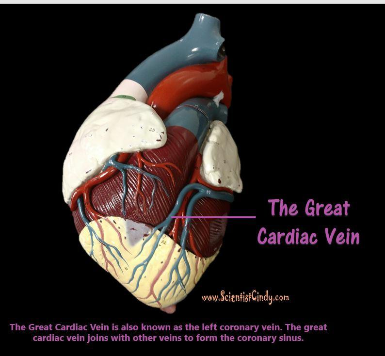
4 1. The ascending aorta 2. The aortic arch 3. The descending aorta The Ascending Aorta and its Branches The heart itself needs a supply of blood. There are two arteries that begin at the base of the aorta called the right and left coronary arteries. The right coronary artery supplies oxygenated blood to the cardiac muscles on the right side of the heart and the left coronary artery supplies oxygenated blood to the cardiac muscles on the left side of the heart. The right and left coronary arteries begin at the base of the aorta (the ascending aorta) and travel to the surface of the heart. The right coronary artery runs along the surface of the right atrium. The left coronary artery reaches the surface of the heart and quickly branches off to form two different arteries (see next image). The left coronary artery branches off to form the circumflex artery and the anterior interventricular artery. The circumflex artery circles around the inferior border of the left atrium. The anterior interventricular artery continues downward lying in between the right and left ventricles. 4/45

5 via GIPHY 5/45
6 The Aortic Arch and its Branches 6/45

 the brachiocephalic trunk 2) the left common carotid artery and 3) the left subclavian artery.")

7 The ascending aorta brings oxygenated blood from the left ventricle (the blood passes through the aortic semilunar valve) to the portions of the aorta called the AORTIC ARCH and the DESCENDING AORTA. The aortic arch contains branches that send blood to the upper body. The descending aorta sends blood to the lower regions of the body. The aortic arch functions to send blood to the upper body via 3 branches. The 3 of the aortic arch are the 1) the brachiocephalic trunk 2) the left common carotid artery and 3) the left subclavian artery. The first branch is the brachiocephalic trunk (brachiocephalic artery). The brachiocephalic trunk sends blood to the right arm, and the head and neck. The brachiocephalic trunk branches off to form the right subclavian artery and the right common carotid artery. The right subclavian artery supplies blood to the right arm and the right common carotid artery supplies blood to the right side of the head and neck. The second branch is the left common carotid artery and it supplies blood to the left side of the head and neck. The third branch is the left subclavian artery and it supplies blood to the left arm. Arteries of the Thoracic Region 7/45


8 The descending aorta supplies blood to the lower body and has branches that travel to the abdomen, pelvis, perineum and the lower limbs. The descending aorta is the portion of the aorta that comes just after the aortic arch. It turns downward and descends to deliver oxygenated blood to the lower portions of the body. The portion of the descending aorta that goes through the thoracic cavity is the THORACIC AORTA. The portion of the descending aorta that travels into the abdominal cavity is the ABDOMINAL AORTA. 8/45


9 The Pulmonary Trunk and Its Branches The next largest artery you see in the heart is colored BLUE because it is carrying deoxygenated blood from the right ventricle to the lungs. This artery is called the pulmonary trunk. The pulmonary trunk branches off to form the right and left pulmonary arteries. The right pulmonary artery carries the blood to the right lung and the left pulmonary artery carries blood to the left lung. THE VEINS 9/45

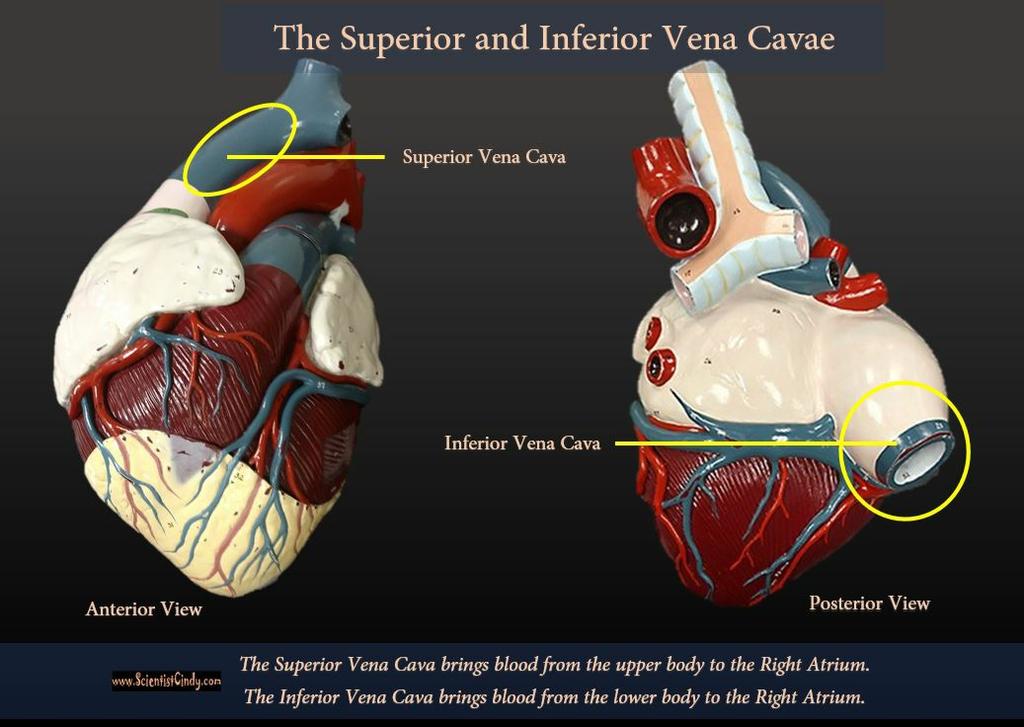
10 The largest veins of the body are the superior and inferior vena cava. The superior vena cava brings deoxygenated blood from the upper body to the right atrium. The inferior vena cava brings deoxygenated blood from the lower body to the right atrium. 10/45
11 11/45

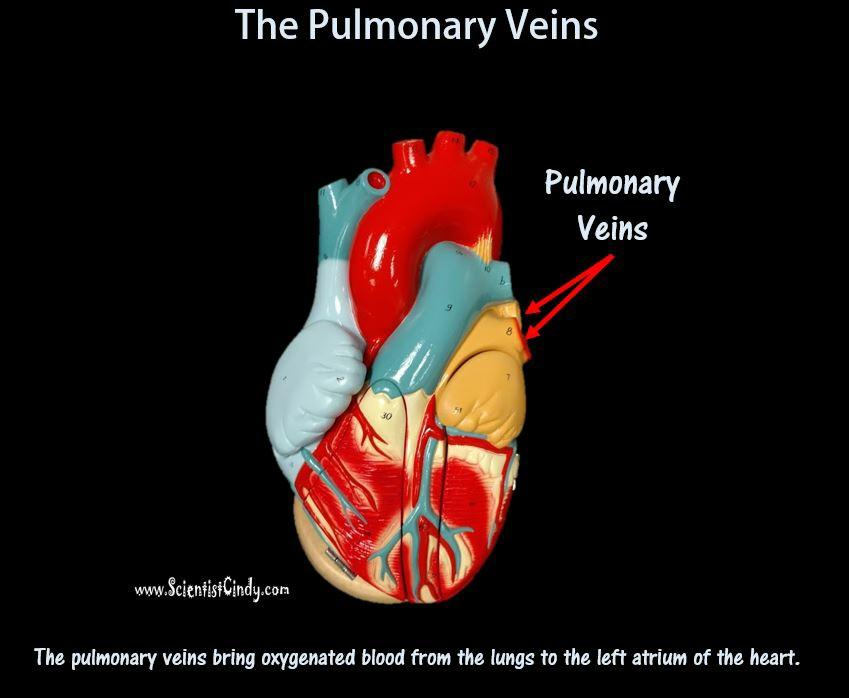
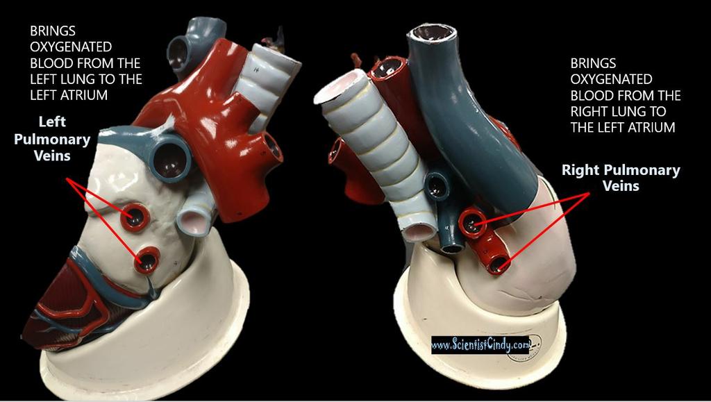
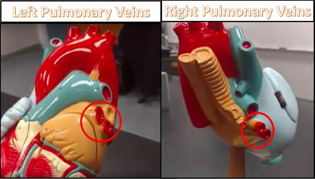
12 The right and left brachiocephalic veins come together to form the superior vena cava. The right brachiocephalic vein brings deoxygenated blood from the right side of the head and neck and the right arm, to the superior vena cava. The left brachiocephalic vein brings deoxygenated blood from the left side of the head and neck and the left arm, to the superior vena cava. Oxygenated blood coming from the lungs returns to the heart through the right and left pulmonary veins. The right pulmonary veins bring oxygenated blood from the right lung to the left atrium of the heart. The left pulmonary veins bring oxygenated blood from the left lung to the left atrium of the heart. 12/45
13 13/45
14 14/45
15 15/45
16 16/45
 THE PULMONARY CIRCUIT - Functions to bring blood from the heart to the lungs and back to the heart 2) THE SYSTEMIC CIRCUIT - Functions to bring blood from the heart to")

17 THE CIRCUITS OF THE CIRCULATORY SYSTEM The circulatory system circulates blood around two major loops. 1. 1) THE PULMONARY CIRCUIT - Functions to bring blood from the heart to the lungs and back to the heart 2) THE SYSTEMIC CIRCUIT - Functions to bring blood from the heart to all the parts of the body except the lungs and then back to the heart In pulmonary circulation, deoxygenated blood travels from the heart to the lungs to gain oxygen and get rid of carbon dioxide before returning to the heart. In contrast, in systemic circulation, oxygenated blood is pumped from the heart to the body and deoxygenated blood is returned back to the heart. The Arteries of the Head and Neck 17/45
18 Your brain uses a lot of blood for its size. About 20% of your blood supply is directed toward your brain to keep you reading this page! Let's look at the basics of how the blood gets there. Arterial Pathways to the Brain 18/45
19 19/45
20 20/45
21 21/45
, the left common carotid artery, and the left subclavian artery.")
22 Oxygenated blood from the heart travels through he aorta. The aorta contains 3 branches; the brachiocephalic trunk (or artery), the left common carotid artery, and the left subclavian artery. All 3 of these branches carry blood from the aorta to the brain, head and neck. 22/45
. 1.")
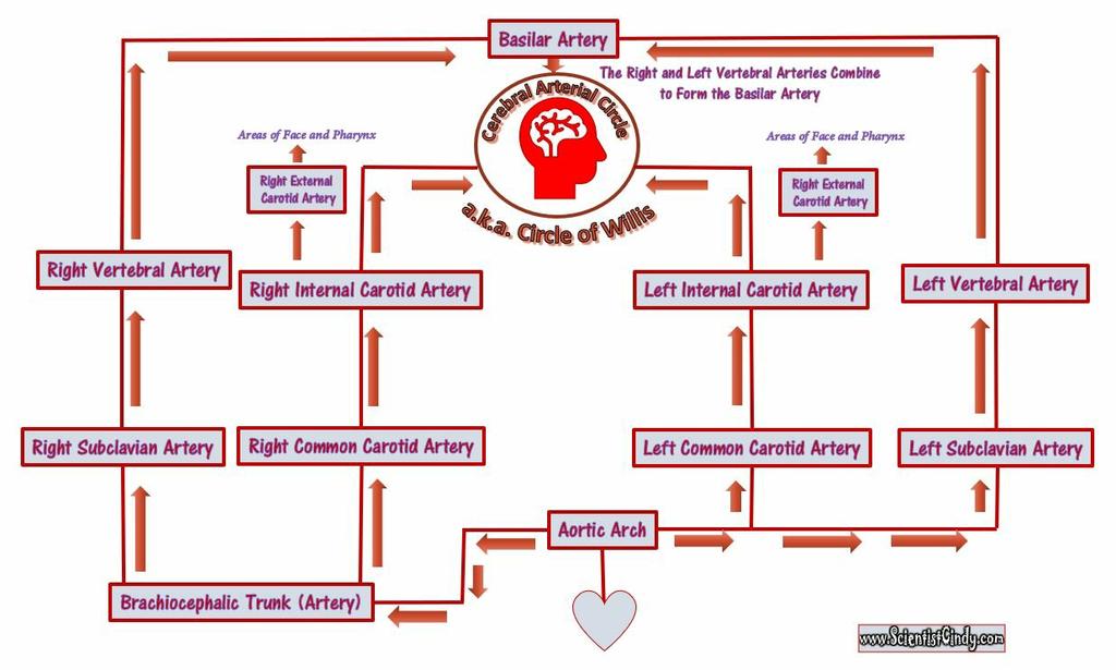
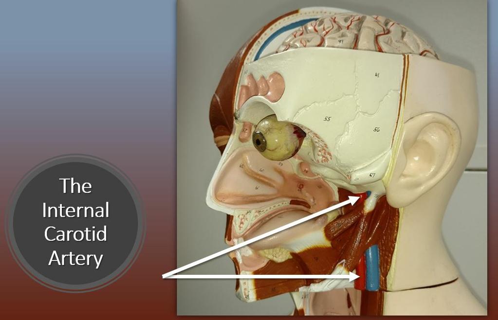
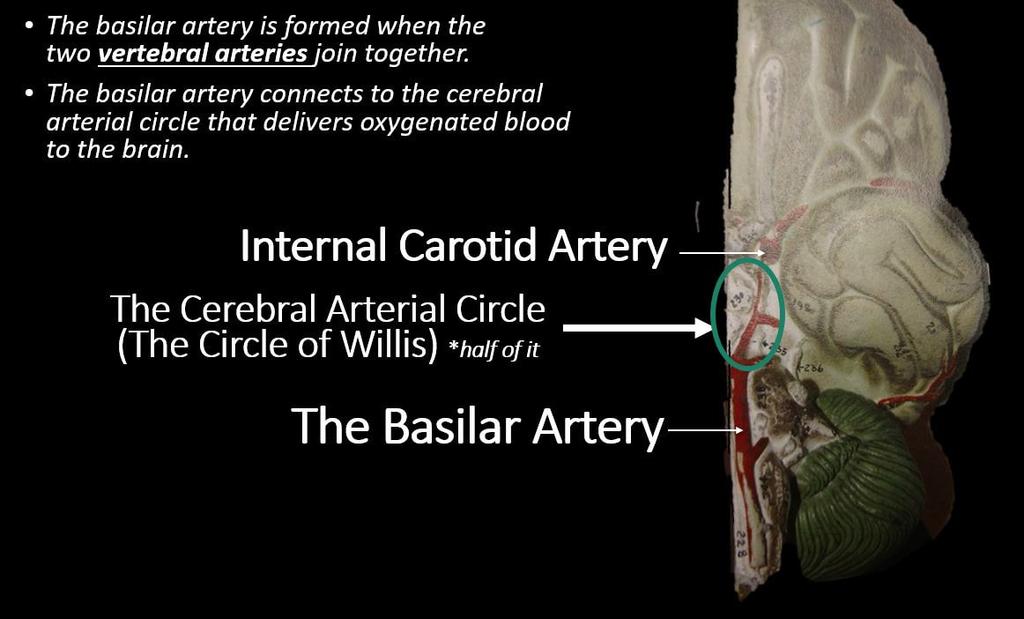
23 Oxygenated blood from the left ventricle leave the heart through the aorta. This blood travels through the ascending aorta to the aortic arch. The aortic arch has 3 branches that send blood to the upper portions of the body. Each of the 3 branches off the aorta have pathways that lead to the brain (the arterial cerebral circle). 1. The first branch off of the aortic arch is the brachiocephalic trunk. The brachiocephalic trunk (or artery) branches off to form the right subclavian artery and the right common carotid artery. The right subclavian artery brings blood to the right vertebral artery which joins with the basilar artery which lies at the base of the brain. The basilar artery connects to the cerebral arterial circle (a.k.a. the circle of Willis) which delivers oxygenated blood to the brain. 2. The brachiocephalic trunk (the first branch off of the aortic arch) has another branch called the right common carotid artery. The right common carotid artery branches to form the right internal carotid artery which leads directly to the cerebral arterial circle (a.k.a. the circle of Willis) of the brain. 3. The second branch coming off of the aortic arch is the left common carotid artery. The left common carotid artery ascends to form the internal carotid artery which leads directly to the cerebral arterial circle (a.k.a. the circle of Willis) of the brain. 4. The third branch coming off of the aortic arch is the left subclavian artery. The left subclavian artery brings blood to the right vertebral artery which joins with the basilar artery which lies at the base of the brain. The basilar artery connects to the cerebral arterial circle (a.k.a. the circle of Willis) which delivers oxygenated blood to the brain. The Veins from the Brain, Head and Neck Areas to the Heart. 23/45
24 24/45
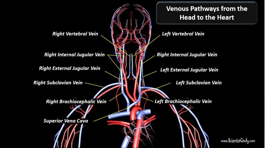
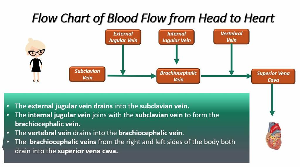
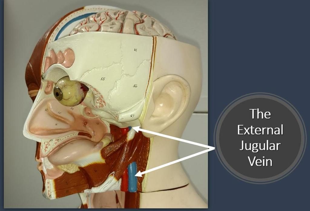
25 What goes up, must come down. Deoxygenated blood from the brain, head and neck to the heart. There are 6 major pathways that this blood from the brain, head and neck travels to the heart. These pathways begin at the right and left external jugular veins, the right and left vertebral veins and the right and left internal jugular veins. The right and left external jugular veins travel to the right and left subclavian vein. From there the blood travels through the right and left brachiocephalic veins to the superior vena cava. The right and left vertebral veins connect to the right and left brachiocephalic veins which then travels to the superior vena cava. The right and left internal jugular veins also travel to the brachiocephalic veins which connects with the superior vena cava. After the deoxygenated blood reaches the superior vena cava, it flows into the right atrium of the heart. 25/45
26 26/45
27 The Blood Vessels of the Arm 27/45


28 Oxygenated blood travels down the arms as well. Oxygenated blood leaves the left ventricle of the heart through the aortic semilunar valve and enters the ascending aorta. The blood travels superiorly to the aortic arch. Blood destined for the right arm enters the right brachiocephalic trunk (the first branch of the aortic arch) and then enters the right subclavian artery. Blood destined for the left arm travels though the 2nd branch of the aortic arch, which is the left subclavian artery. From this point, the arterial pathways from the right and left subclavian arteries to the palms (palmar arches) are identical. The subclavian artery continues to travel laterally toward the shoulder, then turns inferiorly at the axillary region (area of the arm pit) to become the axillary artery. The axillary artery then becomes the brachial artery at the brachial region (the region of the upper arm). The brachial artery branches off inferiorly to form the radial and ulnar arteries. The radial and ulnar arteries travel downward through the forearm (antebrachial region) and then rejoin at the palmar region (region of the palm of the hend), forming the palmar arches. 28/45

29 THE MAJOR ARTERIAL PATHWAYS FROM THE HEART TO THE HANDS. 29/45

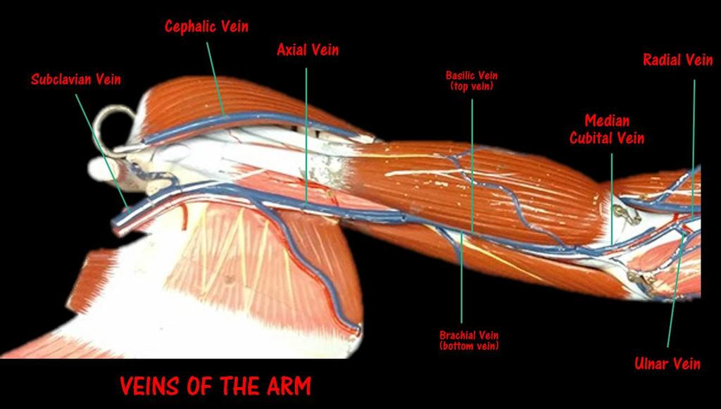
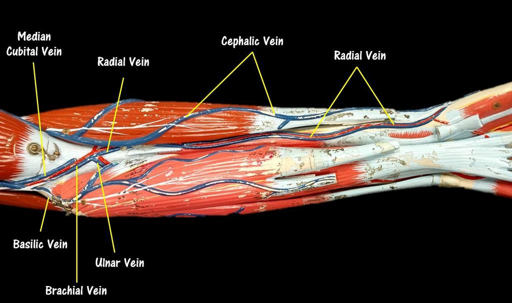
30 Veins of the Upper Limbs Once the blood has circulated through the tissues of the arm, it will become "deoxygenated" and will need to return to the heart to get another "push" before picking up more oxygen at the lungs. There are few pathways that this blood return to the heart from the hands. The deep veins mirror the pathway of the arteries and may be a bit easier to learn, given this fact. The deep pathway begins as the ulnar and radial veins collect deoxygenated blood from the hands. The ulnar and radial veins travel superiorly through the lower arm, then merge to form the brachial vein in the brachial region (area of the upper arm). The blood travels superiorly through the brachial vein which turns into axillary vein at the axillary region (area of the arm pit). This vein becomes the subclavian vein as it passes the clavicle bone. 30/45
31 31/45


32 Deoxygenated blood from the hand can also travel superiorly through the cephalic vein which runs the length of the arm on the radial side (the side of the thumb) and directly connects superiorly with the subclavian vein. A similar pathway exists on the ulnar side of the arm in which deoxygenated blood from the hand travels through the basilic vein. The basilic vein runs the length of the arm on the ulnar side (the side of the pinky) and connects superiorly to the axillary vein, which connects with the subclavian vein. There is another vein of interest that can play a role in sending deoxygenated blood from the hand to the heart, called the medial cubital vein which branches off of the brachial vein at the antecubital region (area 32/45

33 of the inner elbow). The medial cubital vein has branches that merge with the cephalic vein and the basilic vein, so that blood can essentially jump over to one of the other pathways to the subclavian vein! All of these pathways merge at the level of the subclavian vein located around the shoulder area. The subclavian vein turns medially and travels through the thoracic region towards the heart. The subclavian vein connects with the brachiocephalic vein which empties into the superior vena cava. All of the deoxygenated blood coming from the upper body will enter the heart through the superior vena cava. The superior vena cava connects directly to the right atrium of the heart. 33/45

34 Blood Vessels of the Abdominal Cavity Arterial and Venous Pathways Between the Heart and Kidneys 34/45


.")
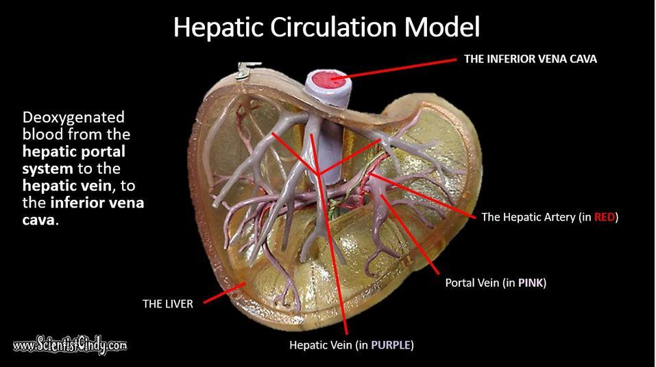
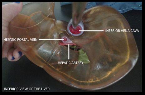
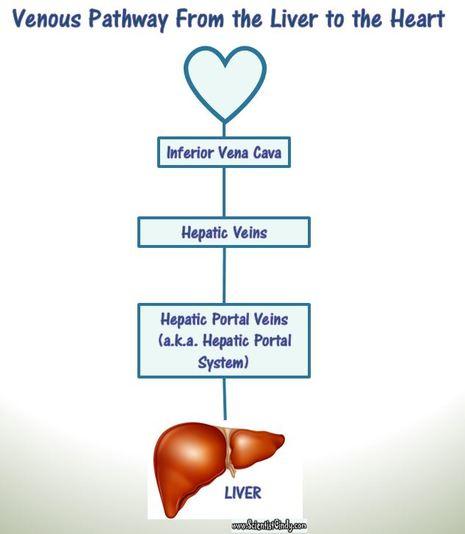
35 The pathways of blood to the kidneys is very straight-forward. Oxygenated blood travels from the left ventricle of the heart to the aorta. The blood travels through the ascending aorta, the aortic arch and the thoracic aorta (part of the descending aorta) and enters the abdominal aorta (part of the descending aorta). Venous Pathway From the Liver to the Heart 35/45

36 36/45

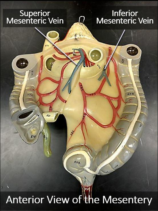
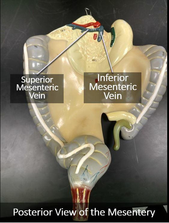
37 The Superior and Inferior Mesenteric Veins The abdominal aorta branches off to form the right and left renal arteries which connect with the right and the left kidneys. The abdominal aorta also branches off to form the superior and inferior mesenteric arteries which connect to the upper and lower mesentery. 37/45
38 38/45


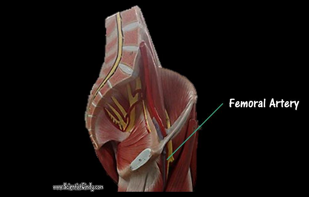
39 Arterial Pathway From the Heart to the Mesentery The Pelvic Arteries and Veins Arteries of the Lower Limbs 39/45
40 40/45


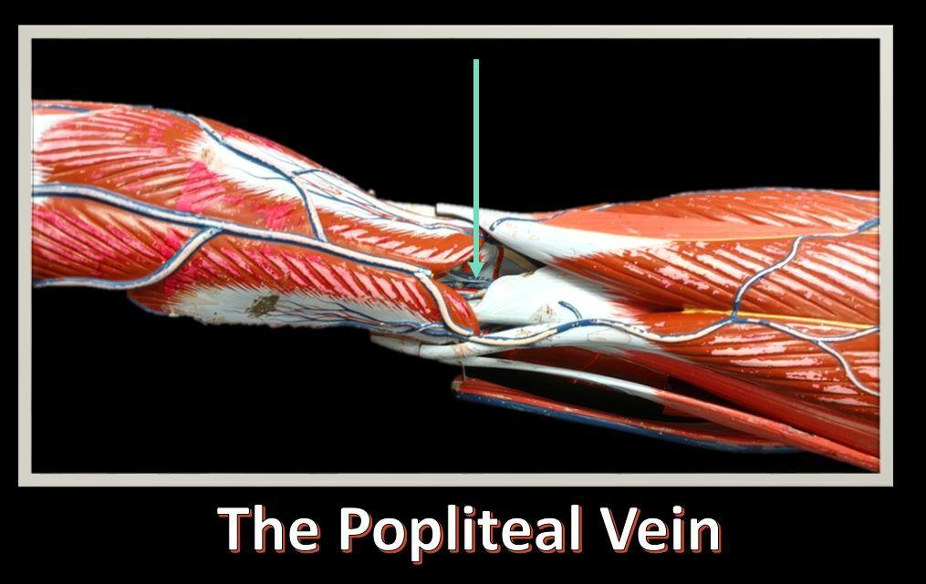
41 Veins of the Lower Limbs The Great Saphenous Vein branches off from the Femoral Vein and continues to travel inferiorly on the medial side of the leg. 41/45
")
42 The popliteal vein is located deep inside the popliteal region (at the back of the knee) 42/45
43 43/45
44 44/45
45 45/45
3 Circulatory Pathways
 40 Chapter 3 Circulatory Pathways Systemic Arteries -Arteries carry blood away from the heart to the various organs of the body. -The aorta is the longest artery in the body; it branches to give rise to
VESSELS: GROSS ANATOMY
 ACTIVITY 10: VESSELS AND CIRCULATION OBJECTIVES: 1) How to get ready: Read Chapter 23, McKinley et al., Human Anatomy, 4e. All text references are for this textbook. 2) Observe and sketch histology slide
YOU MUST BRING GLOVES FOR THIS ACTIVITY
 ACTIVITY 10: VESSELS AND CIRCULATION OBJECTIVES: 1) How to get ready: Read Chapter 23, McKinley et al., Human Anatomy, 5e. All text references are for this textbook. 2) Observe and sketch histology slide
HUMAN HEART. Learn the following structures on the heart models.
 HUMAN HEART Learn the following structures on the heart models. The human heart has four chambers that consist of the right atrium, left atrium, right ventricle, and left ventricle. The atria are smaller
TRACE A DROP OF BLOOD FROM RIGHT EAR TO LEFT OCULOMOTOR NERVE
 TRACE A DROP OF BLOOD FROM RIGHT EAR TO LEFT OCULOMOTOR NERVE KEY: TRACE A DROP OF BLOOD FROM RIGHT EAR TO LEFT OCULOMOTOR NERVE RIGHT EAR RIGHT ATRIUM LEFT SUBCLAVIAN ARTERY RIGHT EXTERNAL JUGULAR VEIN
Artery 1 Head and Thoracic Arteries. Arrange the parts in the order blood flows through them.
 Artery 1 Head and Thoracic Arteries 1. Given the following parts of the aorta: 1. abdominal aorta 2. aortic arch 3. ascending aorta 4. thoracic aorta Arrange the parts in the order blood flows through
Which Artery am I? I am one of two smaller arteries that arise from the brachial. I supply blood to the medial aspect of the forearm.
 I am one of two smaller arteries that arise from the brachial. I supply blood to the medial aspect of the forearm. A. I supply blood to the head and neck. I am large and will branch into two smaller arteries.
MODULE 2: CARDIOVASCULAR SYSTEM ANTOMY An Introduction to the Anatomy of the Heart and Blood vessels
 MODULE 2: CARDIOVASCULAR SYSTEM ANTOMY An Introduction to the Anatomy of the Heart and Blood vessels The cardiovascular system includes a pump (the heart) and the vessels that carry blood from the heart
The Cardiovascular System
 PowerPoint Lecture Slide Presentation by Patty Bostwick-Taylor, Florence-Darlington Technical College The Cardiovascular System 11PART B The Heart: Cardiac Output Cardiac output (CO) Amount of blood pumped
Day 5 Respiratory & Cardiovascular: Respiratory System
 Day 5 Respiratory & Cardiovascular: Respiratory System Be very careful not to damage the heart and lungs while separating the ribs! Analysis Questions-Respiratory & Cardiovascular Log into QUIA using your
Lab Activity 25. Blood Vessels & Circulation. Portland Community College BI 232
 Lab Activity 25 Blood Vessels & Circulation Portland Community College BI 232 Artery and Vein Histology Walls have 3 layers: Tunica intima Tunica media Tunica externa 2 Tunica Intima Is the innermost layer
THE HEART. Unit 3: Transportation and Respiration
 THE HEART Unit 3: Transportation and Respiration The Circulatory System Also called the Cardiovascular System Circulates blood in the body Transports nutrients, oxygen, carbon dioxide, hormones, and blood
Figure ) The specific chamber of the heart that is indicated by letter A is called the. Diff: 1 Page Ref: 364
 The specific chamber of the heart that is indicated by letter A is called the. Diff: 1 Page Ref: 364") Essentials of Anatomy and Physiology, 9e (Marieb) Chapter 11 The Cardiovascular System Short Answer Figure 11.1 Using Figure 11.1, identify the following: 1) The Purkinje fibers are indicated by label.
Essentials of Anatomy and Physiology, 9e (Marieb) Chapter 11 The Cardiovascular System Short Answer Figure 11.1 Using Figure 11.1, identify the following: 1) The Purkinje fibers are indicated by label.
CARDIOVASCULAR DANIL HAMMOUDI.MD
 CARDIOVASCULAR DANIL HAMMOUDI.MD 18 Systemic Circulation Figure 19.19 Pulmonary Circulation Figure 19.18b 1. Thyroid gland 2. Trachea 3. Brachiocephalic 4. Common carotid 5. Internal jugular 6. Superior
CRITICAL THINKING QUESTIONS AND ANSWERS AND CYCLE 2 LAB EXAM TEMPLATE. There are two main mechanisms that work in conjunction to return the blood
 CRITICAL THINKING QUESTIONS AND ANSWERS AND CYCLE 2 LAB EXAM TEMPLATE There are two main mechanisms that work in conjunction to return the blood THE CARDIAC PUMP 1) The forward pull(vis a fronte) This
Anatomy of the Blood Vessels
 Biology 212: Anatomy and Physiology II Anatomy of the Blood Vessels References: Saladin, KS: Anatomy and Physiology, The Unity of Form and Function 8 th (2018). Required reading before beginning this lab:
Cardiovascular system:
 Cardiovascular system: Mediastinum: The mediastinum: lies between the right and left pleura and lungs. It extends from the sternum in front to the vertebral column behind, and from the root of the neck
#5 Cardiovascular II Blood Vessels
 #5 Cardiovascular II Blood Vessels Objectives: Identify a list of human arteries and veins using a virtual human dissection and a human model Dissect and identify a list of arteries and veins in the cat
Anatomy and Physiology, Spring 2015 Exam II: Form A April 9, Name Student Number
 Anatomy and Physiology, Spring 2015 Exam II: Form A April 9, 2015 Name Student Number For Questions 1 2 refer to the following table. 1 Ventricular pressure is greater than aortic 6 AV valve is open 2
#5 Cardiovascular II Blood Vessels
 Page1 #5 Cardiovascular II Blood Vessels Objectives: Observe slide of artery and vein cross-section Identify a list of human arteries and veins using a virtual human dissection Dissect and identify a list
Chapter 14. The Cardiovascular System
 Chapter 14 The Cardiovascular System Introduction Cardiovascular system - heart, blood and blood vessels Cardiac muscle makes up bulk of heart provides force to pump blood Function - transports blood 2
Lab 6: Blood. BIO104 Laboratory Handouts 147. Unit 12: Blood and Lymphatics. 1. Blood Characteristics Volume Functions Composition -
 147 Lab 6: Blood Unit 12: Blood and Lymphatics Ex. 12-1: Formed Elements (Cells) of Blood, p. 313-316 1. Blood Characteristics Volume Functions Composition - 2. Leukocytes (WBCs) a. WBC count normal b.
This lab activity is aligned with Visible Body s A&P app. Learn more at visiblebody.com/professors
 1 This lab activity is aligned with Visible Body s A&P app. Learn more at visiblebody.com/professors 2 PRE-LAB EXERCISES: A. Watch the video 29.1 Heart Overview and make the following observations: 1.
Breathing. Heart Rate
 Breathing Heart Rate Inspiration Expiration (Pressos not Stretched) Heart Rate increases with inspiration (Pressos Stretched) Heart Rate decreases with expiration Upside Down (Pressos Stretched) HR Decreases
Chapter 13. Cardiovascular System
 Chapter 13 Cardiovascular System 1 Introduction A. The cardiovascular system consists of the heart and vessels (arteries, capillaries and veins.) B. A functional cardiovascular system is vital for supplying
REVIEW SHEET Anatomy of Blood Vessels
 REVIEW SHEET Anatomy of Blood Vessels Name LabTime/Date Microscopic Structure of the Blood Vessels 1. Cross-sectional views of an aftery of a vein are shown here. ldentify each; on the lines to the sides,
Unit 11 - The Cardiovascular System 1
 Unit 11 - The Cardiovascular System 1 I. Unit 11: The Cardiovascular System A. The Cardiovascular System 1. A closed system of the heart and blood vessels a) The heart pumps blood b) Blood vessels allow
Cardiovascular System
 Cardiovascular System I. Structure of the Heart A. Average adult heart is 14 cm long and 9 cm wide. B. Lies in the mediastinum. C. Enclosed in the pericardium. 1. Fibrous pericardium- Outer, tough connective
CARDIOVASCULAR SYSTEM
 CARDIOVASCULAR SYSTEM CARDIAC SYSTEM TWO TYPES OF CIRCULATION Systemic system delivers blood to ALL body cells and carries away waste. The red blood cells use hemoglobin to carry oxygen to the cells Pulmonary
The Function. To carry nutrients and oxygen to and remove waste from the cells of the body.
 The Function To carry nutrients and oxygen to and remove waste from the cells of the body. What makes up the circulatory system? 1. Heart 2. Blood 3. Blood vessels Blood travels from the heart to the body
Copy Right- Hongqi ZHANG-Department of Anatomy-Fudan University. Systematic Anatomy. Angiology Part 4. Veins. Dr.Hongqi Zhang ( 张红旗 )
") Systematic Anatomy Angiology Part 4 Veins Dr.Hongqi Zhang ( 张红旗 ) Email: zhanghq58@126.com 1 General introduction of the veins Vessel which return the blood back to atrium No pulsation,veneous blood, metabolic
Systematic Anatomy Angiology Part 4 Veins Dr.Hongqi Zhang ( 张红旗 ) Email: zhanghq58@126.com 1 General introduction of the veins Vessel which return the blood back to atrium No pulsation,veneous blood, metabolic
Bio& 242, Unit 3/ Lab 4 Blood Vessels, Lymphatic System and Blood Pressure G. Blevins/ G. Brady Summer 2009
 Bio& 242, Unit 3/ Lab 4 Blood Vessels, Lymphatic System and Blood Pressure G. Blevins/ G. Brady Summer 2009 Major Arteries and for arteries and veins with common names your answer must include either artery
Vascular System Part One
 Vascular System Part One Objectives Trace the route taken by blood as it leaves, and then returns to the heart. Describe the structure of the walls of arteries and veins. Discuss the structure and function
The Cardiovascular System
 C H A P T E R 1 4 The Cardiovascular System OBJECTIVES After studying this chapter, you should be able to: 1. Describe how the heart is positioned in the thoracic cavity. 2. List and describe the layers
Contents. Page 1. Homework 11 Chapter Blood Vessels Due: Week 6 Lec 11
 Page 1 Homework 11 Chapter 18-19 Blood Vessels Due: Week 6 Lec 11 Contents When printing, make sure that you specify the page range that you want to print out! Learning objectives for Lecture 11:...pg
Cardiovascular System. Heart Anatomy
 Cardiovascular System Heart Anatomy 1 The Heart Location & general description: Atria vs. ventricles Pulmonary vs. systemic circulation Coverings Walls The heart is found in the mediastinum, the medial
Chapter 2 The Human Cardiovascular System
 Chapter 2 The Human Cardiovascular System 2.1 Introduction Before delving into the computational methods of CHD, this chapter provides a preliminary understanding of the circulatory system from a physiological
Cardiovascular System
 Hole s Essentials of Human Anatomy & Physiology David Shier Jackie Butler Ricki Lewis Created by Dr. Melissa Eisenhauer Head Athletic Trainer/Assistant Professor Trevecca Nazarene University Chapter 13
Unit 11: The Cardiovascular System
 Unit 11: The Cardiovascular System I. The Cardiovascular System A. A closed system of the heart and blood vessels 1. The heart pumps blood 2. Blood vessels allow blood to circulate to all parts of the
Biology 105 Human Biology QZM CVS. Cardiovascular System. Name these Vessels, Valves and Chambers
 Cardiovascular System Name these Vessels, Valves and Chambers 1. The heart and pericardium are located within the and medial to the A) pleural cavities B) peritoneal cavity C) cardiac skeleton D) mediastinum
Pearson's Comprehensive Medical Assisting Administrative and Clinical Competencies
 Pearson's Comprehensive Medical Assisting Administrative and Clinical Competencies THIRD EDITION CHAPTER 27 The Cardiovascular System Lesson 1: Overview of the Cardiovascular System Lesson Objectives Upon
c) What is the name of RBC (erythrocyte) formation? Where do blood cells form?
 What is the name of RBC (erythrocyte) formation? Where do blood cells form?") UNIT 6: CARDIOVASCULAR SYSTEM 1) List the three general functions of BLOOD. REVIEW QUESTIONS Blood 2) a) What are the three formed elements /cellular elements in blood? b) Describe the composition of the
UNIT 6: CARDIOVASCULAR SYSTEM 1) List the three general functions of BLOOD. REVIEW QUESTIONS Blood 2) a) What are the three formed elements /cellular elements in blood? b) Describe the composition of the
THE CIRCULATORY SYSTEM
 Biology 30S THE CIRCULATORY SYSTEM Name: This module adapted from bblearn.merlin.mb.ca 1 Introduction to Circulation The first organ to form, and the last organ to die. The heart is the pump of life. The
The Cardiovascular System
 PowerPoint Lecture Slide Presentation by Patty Bostwick-Taylor, Florence-Darlington Technical College The Cardiovascular System 11PART A The Cardiovascular System A closed system of the heart and blood
Chapter 21: Cardiovascular System: Peripheral Circulation and Regulation
 Chapter 21: Cardiovascular System: Peripheral Circulation and Regulation I. General Features of Blood Vessel Structure A. General Pattern of Circulation 1. Ventricles pump blood into 2. These arteries
TOPIC 6: HUMAN HEALTH AND PHYSIOLOGY
 TOPIC 6: HUMAN HEALTH AND PHYSIOLOGY 6.2 Transport System/Circulatory Draw and label a diagram of the heart showing the four chambers, associated blood vessels, valves and the route of blood through the
Blood Vessels. Types of Blood Vessels Arteries carry blood away from the heart Capillaries smallest blood vessels. Veins carry blood toward the heart
 C H A P T E R Blood Vessels 20 Types of Blood Vessels Arteries carry blood away from the heart Capillaries smallest blood vessels The site of exchange of molecules between blood and tissue fluid Veins
The Human Body. Lesson Goal. Lesson Objectives 9/10/2012. Provide a brief overview of body systems, anatomy, physiology, and topographic anatomy
 The Human Body Lesson Goal Provide a brief overview of body systems, anatomy, physiology, and topographic anatomy Medial Lateral Proximal Distal Superior Inferior Anterior Lesson Objectives Explain the
ANATOMY OF BLOOD VESSELS
 BIOLOGY 212: HUMAN ANATOMY & PHYSIOLOGY ****************************************************************************************************************** ANATOMY OF BLOOD VESSELS ********************************************************************************************************
Health Science 20 Circulatory System Notes
 Health Science 20 Circulatory System Notes Functions of the Circulatory System The circulatory system functions mainly as the body s transport system. It transports: o Oxygen o Nutrients o Cell waste o
Large veins of the thorax Brachiocephalic veins
 Large veins of the thorax Brachiocephalic veins Right brachiocephalic vein: formed at the root of the neck by the union of the right subclavian & the right internal jugular veins. Left brachiocephalic
Ch.15 Cardiovascular System Pgs {15-12} {15-13}
 Ch.15 Cardiovascular System Pgs {15-12} {15-13} E. Skeleton of the Heart 1. The skeleton of the heart is composed of rings of dense connective tissue and other masses of connective tissue in the interventricular
The Heart. Made up of 3 different tissue: cardiac muscle tissue, nerve tissue, and connective tissue.
 The Heart The Heart Made up of 3 different tissue: cardiac muscle tissue, nerve tissue, and connective tissue. Your heart pumps with a regular beat (Heart Rate) Your heart rate can change depending on
Misc Anatomy. Upper Limb! 2. Lower Limb! 5. Venous Drainage! Head & neck! 8
 Misc Anatomy Upper Limb! 2 Arteries!... 2 Veins!... 2 Spaces!... 4 Lower Limb! 5 Arteries!... 5 Venous Drainage!... 6 Spaces!... 7 Head & neck! 8 Artery!... 8 Ultrasound View for IJ CVL!... 8 Arteries
This is not a required assignment but it is recommended.
 SU 12 Name: This is not a required assignment but it is recommended. BIO 116 - Anatomy & Physiology II Practice Assignment 2 - The Respiratory and Cardiovascular Systems 1. The exchange of oxygen and carbon
Lesson 10 Circulatory System (Nelson p.88-93)
") Name: Date: Lesson 10 Circulatory System (Nelson p.88-93) Learning Goals: A. I can explain the primary functions of the circulatory system in animals. B. I can identify and explain all the parts of the
Name: Date: Lesson 10 Circulatory System (Nelson p.88-93) Learning Goals: A. I can explain the primary functions of the circulatory system in animals. B. I can identify and explain all the parts of the
Human Anatomy, First Edition
 Human Anatomy, First Edition McKinley & O'Loughlin Chapter 23 : Vessels and Circulation 23-1 Blood Vessels An efficient style of transport for oxygen, nutrients, and waste products to and from body tissues.
THE HEART OBJECTIVES: LOCATION OF THE HEART IN THE THORACIC CAVITY CARDIOVASCULAR SYSTEM
 BIOLOGY II CARDIOVASCULAR SYSTEM ACTIVITY #3 NAME DATE HOUR THE HEART OBJECTIVES: Describe the anatomy of the heart and identify and give the functions of all parts. (pp. 356 363) Trace the flow of blood
A closed system of the heart/blood. Function: The heart pumps blood. Blood vessels allow blood to circulate throughout the body
 A closed system of the heart/blood The heart pumps blood It is no more than a transportation pump Blood vessels allow blood to circulate throughout the body MILES of blood vessels intricate network At
Cardiovascular System
 Cardiovascular System angio BELLWORK Day One: Define using technology hemo/hema cardio Medical Therapeutics Standards 11) Outline the gross normal structure and function of all body systems and summarize
Today s objectives:! - Learn BASICS of circulatory system (Heart, different veins and arteries)! - Appreciate effects and treatment for
! - Appreciate effects and treatment for") Today s objectives:! - Learn BASICS of circulatory system (Heart, different veins and arteries)! - Appreciate effects and treatment for hyperlipidemia! Agenda! - Review objectives for 6.2! - Video of circulatory
Today s objectives:! - Learn BASICS of circulatory system (Heart, different veins and arteries)! - Appreciate effects and treatment for hyperlipidemia! Agenda! - Review objectives for 6.2! - Video of circulatory
Cardiovascular System
 Cardiovascular System 1 The Heart Major organ of the cardiovascular system 2 What is the mediastinum? The mediastinum is the space between the lungs, where the heart is located. The heart is divided into
1. Distinguish among the types of blood vessels on the basis of their structure and function.
 Blood Vessels and Circulation Objectives This chapter describes the structure and functions of the blood vessels Additional subjects contained in Chapter 13 include cardiovascular physiology, regulation,
(2) (1) (3) (4) BLOOD PATHWAY ASSESSMENT RUBRIC
 (1) (3) (4) BLOOD PATHWAY ASSESSMENT RUBRIC") BLOODPATHWAYASSESSMENT(4) BLOOD%PATHWAY%ASSESSMENT%(3)% BLOODPATHWAYASSESSMENT(3) (4) (3) (2) (1) Using a completely blank diagram of the heart, all valves, chambers, great vessels, and direction of blood
BLOODPATHWAYASSESSMENT(4) BLOOD%PATHWAY%ASSESSMENT%(3)% BLOODPATHWAYASSESSMENT(3) (4) (3) (2) (1) Using a completely blank diagram of the heart, all valves, chambers, great vessels, and direction of blood
The Cardiovascular System (Heart)
") The Cardiovascular System The Cardiovascular System (Heart) A closed system of the heart and blood vessels The heart pumps blood Blood vessels allow blood to circulate to all parts of the body The function
The Cardiovascular System The Cardiovascular System (Heart) A closed system of the heart and blood vessels The heart pumps blood Blood vessels allow blood to circulate to all parts of the body The function
Circulatory Systems. All cells need to take in nutrients and expel metabolic wastes.
 Circulatory Systems All cells need to take in nutrients and expel metabolic wastes. Single celled organisms: nutrients from the environment can diffuse (or be actively transported) directly in to the cell
Chapter 14. Circulatory System Images. VT-122 Anatomy & Physiology II
 Chapter 14 Circulatory System Images VT-122 Anatomy & Physiology II The mediastinum Dog heart Dog heart Cat heart Dog heart ultrasound Can see pericardium as distinct bright line Pericardial effusion Fluid
Heart Facts. The average adult heart beats 72 times a min 100,000 times a day 3,600,000 times a year 2.5 billion times during a lifetime.
 Circulatory System Heart Facts The average adult heart beats 72 times a min 100,000 times a day 3,600,000 times a year 2.5 billion times during a lifetime. Heart Facts Weighs 11 oz A healthy heart pumps
Human Anatomy, First Edition
 Human Anatomy, First Edition McKinley & O'Loughlin Chapter 22 : Heart 1 Functions of the Heart Center of the cardiovascular system, the heart. Connects to blood vessels that transport blood between the
1. Which of the following blood vessels has a thin elastic layer? A. Aorta. B. Pulmonary artery. C. Posterior vena cava. D. Mesenteric capillary.
 CIRCULATORY SYSTEM 1. Which of the following blood vessels has a thin elastic layer? A. Aorta. B. Pulmonary artery. C. Posterior vena cava. D. Mesenteric capillary. 2. Capillary beds are equipped with
C3, 4, 5, 6, & 7 Worksheet. C3 Describe the inter-relationships of the structures of the heart
 Name: Date: C3, 4, 5, 6, & 7 Worksheet C3 Describe the inter-relationships of the structures of the heart 1. Label and give the functions of the following: a. left and right atrium: b. left and right ventricle:
CV Anatomy Quiz. Dr Ella Kim Dr Pip Green
 CV Anatomy Quiz Dr Ella Kim Dr Pip Green Q1 The location of the heart is correctly described as A) lateral to the lungs. B) medial to the sternum. C) superior to the diaphragm. D) posterior to the spinal
Anatomy of the Heart
 Biology 212: Anatomy and Physiology II Anatomy of the Heart References: Saladin, KS: Anatomy and Physiology, The Unity of Form and Function 8 th (2018). Required reading before beginning this lab: Chapter
37 1 The Circulatory System
 H T H E E A R T 37 1 The Circulatory System The circulatory system and respiratory system work together to supply cells with the nutrients and oxygen they need to stay alive. a) The respiratory system:
The Cardiovascular System. Preview of Heart Action. The CV system provides oxygen & nutrients to tissues-removes wastes.
 The Cardiovascular System BIO 250 Human Anatomy & Physiology Preview of Heart Action http://www.youtube.com/watch?v=d3zdj gfddk0&nr=1 The CV system provides oxygen & nutrients to tissues-removes wastes.
ACTIVITY 9: BLOOD AND HEART BLOOD
 ACTIVITY 9: BLOOD AND HEART OBJECTIVES: 1) How to get ready: Read Chapters 21 & 22, McKinley et al., Human Anatomy, 4e. All text references are for this textbook. Read dissection instructions BEFORE YOU
The Cardiovascular System. Chapter 15. Cardiovascular System FYI. Cardiology Closed systemof the heart & blood vessels. Functions
 Chapter 15 Cardiovascular System FYI The heart pumps 7,000 liters (4000 gallons) of blood through the body each day The heart contracts 2.5 billion times in an avg. lifetime The heart & all blood vessels
Cardiovascular System. Chapter 22
 Cardiovascular System Blood Vessels Chapter 22 Cardiac Contractions Cardiac cycle consists of alternate periods of contraction and relaxation Contraction is the systolic pressure Blood is ejected into
Lab Photo Review Sheet
 9 8 0. Posterior Median Sulcus. Central Canal. Dorsal (Posterior) Horn. Ventral (Anterior) Horn. Grey Matter. White Matter. Anterior Median Fissure 8. Ventral (Anterior) Root (ramus) 9. Dorsal (Posterior)
Chapter 11. The Cardiovascular System. Clicker Questions Pearson Education, Inc.
 Chapter 11 The Cardiovascular System Clicker Questions Oxygen-poor blood is pumped through the venae cavae to the right side of the heart, and then through the pulmonary arteries to the lungs and back
The Cardiovascular System
 11 The Cardiovascular System Yong Jeong, MD, PhD Department of Bio and Brain Engineering The Cardiovascular System A closed system of the heart and blood vessels The heart pumps blood Blood vessels allow
Latin. VASA REGIONIS PELVINAE 72 A. et V. iliaca communis 73 A. et V. iliaca externa 74 A. sacralis mediana 75 A. et V.
 G30 Latin VASA CAPITIS et CERVICIS 1 V. frontalis 2 V. temporalis superficialis 3 A. temporalis superficialis 3 a A. maxillaris 4 A. occipitalis 5 A. supratrochlearis 6 A. et V. angularis 7 A. et V. facialis
An Illustrated 1. Dissection Guide. To The... Mammalian. rr= Heart. Right ventricle+---, by David H. Hall
 An Illustrated 1. Dissection Guide. To The... Mammalian rr= Heart ventricle+---, by David H. Hall The Mam.malian Heart Because mammals are warm blooded (endothermic) and generally very active animals,
Functions of the Circulatory System
 Functions of the Circulatory System Carry oxygen from lungs to tissues Carry carbon dioxide from tissues to lungs thereby aiding in the exchange of gases Helps carry nutrients to cells Helps to fight against
Essentials of Anatony and Physiology, 5e (Martini/Nath) Chapter 13 The Cardiovascular System: Blood Vessels and Circulation
 Chapter 13 The Cardiovascular System: Blood Vessels and Circulation") Essentials of Anatony and Physiology, 5e (Martini/Nath) Chapter 13 The Cardiovascular System: Blood Vessels and Circulation Multiple-Choice Questions 1) The muscular layer of blood vessels is the A) tunica
Essentials of Anatony and Physiology, 5e (Martini/Nath) Chapter 13 The Cardiovascular System: Blood Vessels and Circulation Multiple-Choice Questions 1) The muscular layer of blood vessels is the A) tunica
Your heart is a muscular pump about the size of your fist, located
 How Your Heart Works Your heart is a muscular pump about the size of your fist, located slightly to the left and behind your breastbone. Its function is to pump blood throughout your body. As your heart
THE DIFINITIVE GUIDE TO HUMAN ANATOMY & PHYSIOLOGY (HAP 2).
.") THE DIFINITIVE GUIDE TO HUMAN ANATOMY & PHYSIOLOGY (HAP 2). Pages 2-49 Lecture 1 notes: Cardiovascular 1. Pages 50-97 Lecture 2 notes: Cardiovascular 2. Pages 98-128 Lecture 3 notes: Respiratory 1. Pages
THE DIFINITIVE GUIDE TO HUMAN ANATOMY & PHYSIOLOGY (HAP 2). Pages 2-49 Lecture 1 notes: Cardiovascular 1. Pages 50-97 Lecture 2 notes: Cardiovascular 2. Pages 98-128 Lecture 3 notes: Respiratory 1. Pages
VENOUS DRAINAGE O US F UPPER UPPER LIM B BY dr.fahad Ullah
 VENOUS DRAINAGE OF UPPER LIMB BY dr.fahad Ullah Venous drainage of the supper limb The venous system of the upper limb drains deoxygenated blood from the arm, forearm and hand It can anatomically be divided
AP2 Lab 1 - Blood & Heart
 AP2 Lab 1 - Blood & Heart Project 1 - Formed Elements Identification & Recognition See fig. 17.10 and Table 17.2. Instructor may also provide other images. Note: See Fig. 17.11 All formed elements are
THE CARDIOVASCULAR SYSTEM. Part 1
 THE CARDIOVASCULAR SYSTEM Part 1 CARDIOVASCULAR SYSTEM Blood Heart Blood vessels What is the function of this system? What other systems does it affect? CARDIOVASCULAR SYSTEM Functions Transport gases,
Dr. Weyrich G07: Superior and Posterior Mediastina. Reading: 1. Gray s Anatomy for Students, chapter 3
 Dr. Weyrich G07: Superior and Posterior Mediastina Reading: 1. Gray s Anatomy for Students, chapter 3 Objectives: 1. Subdivisions of mediastinum 2. Structures in Superior mediastinum 3. Structures in Posterior
Read Chapters 21 & 22, McKinley et al
 ACTIVITY 9: BLOOD AND HEART OBJECTIVES: 1) How to get ready: Read Chapters 21 & 22, McKinley et al., Human Anatomy, 5e. All text references are for this textbook. Read dissection instructions BEFORE YOU
10/14/2018 Dr. Shatarat
 2018 Objectives To discuss mediastina and its boundaries To discuss and explain the contents of the superior mediastinum To describe the great veins of the superior mediastinum To describe the Arch of
Heart Dissection. 5. Locate the tip of the heart or the apex. Only the left ventricle extends all the way to the apex.
 Heart Dissection Page 1 of 6 Background: The heart is a four-chambered, hollow organ composed primarily of cardiac muscle tissue. It is located in the center of the chest in between the lungs. It is the
Mr. Epithelium s Anatomy and Physiology Test SSSS
 Mr. Epithelium s Anatomy and Physiology Test SSSS You have 50 minutes to complete this test packet. One 8.5 x 11 cheat sheet is allowed, along with 1 non-programmable calculator dedicated to computation.
Monday 14 th May The Body Anatomy and Physiology Lesson 10 Cardio-Respiratory System
 Monday 14 th May 2018 The Body Anatomy and Physiology Lesson 10 Cardio-Respiratory System Homework 1. What is the function of the cardiovascular system? 2. List the main components of blood and describe
Learning Objectives; 1. Identify the key features of the cardiovascular system. 2. Describe the function of the cardiovascular system.
 Learning Objectives; 1. Identify the key features of the cardiovascular system. 2. Describe the function of the cardiovascular system. https://www.youtube.com/watch?v=yaxna8lmoiy The Circulatory System
Bellwork Define: hemostasis anticoagulation hemophilia (Then write the underline portion of the two state standards in your notes).
.") Bellwork Define: hemostasis anticoagulation hemophilia (Then write the underline portion of the two state standards in your notes). A&P Standards 31) Identify the liquid and cellular components of blood
Bellwork Define: hemostasis anticoagulation hemophilia (Then write the underline portion of the two state standards in your notes). A&P Standards 31) Identify the liquid and cellular components of blood
LAB: Sheep or Pig Heart Dissection
 Biology 12 Name: Circulatory System Per: Date: Observation: External Anatomy LAB: Sheep or Pig Heart Dissection 1. Line a dissecting tray with paper towel for easy clean up as the heart is fatty and will
The HEART. What is it???? Pericardium. Heart Facts. This muscle never stops working It works when you are asleep
 This muscle never stops working It works when you are asleep The HEART It works when you eat It really works when you exercise. What is it???? Located between the lungs in the mid thoracic region Apex