Cardiac Imaging Transesophageal Echocardiogram Cardic MRI Cardiac CT
|
|
|
- Buddy Barnett
- 5 years ago
- Views:
Transcription
1 Cardiac Imaging Transesophageal Echocardiogram Cardic MRI Cardiac CT David Stultz, MD Cardiology Fellow, PGY 6 May 2, 2006
2 Ways to visualize the heart Xray Chest film Computed tomography Calcium evaluation Coronary arteriography Angiography/Fluorscopy Ultrasound Transthoracic echocardiogram (TTE) Transesophageal echocardiogram (TEE) Intravascular ultrasound (IVUS) Intracardiac ultrasound (ICE) Magnetic Resonance (CMR) Too many types of sequences to list Angioscopy
3 Transesophageal Echo Internal Medicine Boards perspective TEE/Cardioversion for atrial fibrillation Endocarditis The Stroke evaluation Patent Foramen Ovale Atrial Septal Defect Cardiac source of embolus These will also be the situations most commonly faced by primary care physicians
4 Transesophageal Echo Procedural description NPO overnight Oropharynx anesthetized Conscious sedation 2mg versed +/- 25mg demerol or 25 mcg fentanyl TEE probe advanced gently into the esophagus Exam duration typically 10 minutes of probe activity Risks include esophageal perforation Especially with zenkers diverticulum, webs, strictures Overall low risk of complecations
5 TEE/Cardioversion for atrial fibrillation General recommendations for atrial fibrillation with planned cardioversion INR goal weeks before cardioversion Unless onset of Afib <48 hours before cardioversion 4 weeks after cardioversion Patients less tolerant of atrial fibrillation Heparin/LWMH at diagnosis/admission TEE to evaluate for Left Atrial appendage thrombus Safe to cardiovert if no thrombus seen?

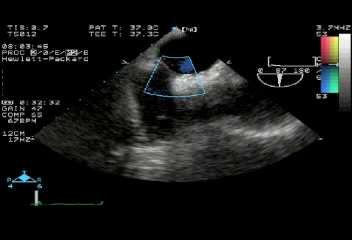
6 Feigenbaum, 6 th ed

7 Feigenbaum, 6 th ed
8 TEE/Cardioversion ACUTE trial Randomized, prospective Patients with atrial fibrillation Compared 3 weeks on anticoagulation prior to cardioversion vs TEE guided 70 sites, 1222 patients 8 weeks followup Primary outcome: Embolic events 0.8%TEE, 0.5% conventional (p=0.5) Minor and Major bleeding 2.9% TEE, 5.5% conventional (p=0.03) Asher CR, Klein AL; ACUTE trial: Transesophael echocardiography to guide cardioversion in patients with atrial fibrillation: ACUTE trial update. Card Electrophysiol Rev 7(4): , 2003
9 Endocarditis Most often affects left sided valves Ideally suited for TEE Exception is IV drug users Typically vegetations occur on low pressure side of valves TEE more sensitive than TTE to see Vegetations Regurgitation Leaflet damage/perforation Abscess formation Unstable prosthesis

10 Feigenbaum, 6 th ed
11 Feigenbaum, 6 th ed
12 Feigenbaum, 6 th ed
13 The stroke evaluation Cardiogenic etiologies of stroke Left atrial thrombus Left ventricular thrombus Patent foramen ovale (PFO) or atrial septal defect (ASD) Allows paradoxical emoblization Venous thrombus passes through interatrial septum Cardiac masses Endocarditis/vegetations Tumors
14 Feigenbaum, 6 th ed

15 Feigenbaum, 6 th ed

16 Feigenbaum, 6 th ed

17 Feigenbaum, 6 th ed
18 Which TIA/stroke patients need a TEE? Which patients need a TTE? Almost everyone Who needs a TEE? Patients younger than 45 Older patients without identified cause of CVA However Controversial whether findings change management Anticoagulation indicated for thrombus Treatment/Surgery indicated for masses/vegetations PFO/ASD Closure may or may not be helpful Anticoagulation usually not indicated for primary/secondary prevention About 10-20% of general population have a PFO!
19 Other situations where TEE is useful When TTE is suboptimal to answer a clinical question Prosthetic valve evaluation Cardiac masses Pulmonary vein stenosis Native valve stenosis/regurgitation Cardiac Anuerysm, pseudoaneurysm Aortic dissection Evaluation of ejection fraction alone is challenging with TEE

20 Feigenbaum, 6 th ed
21 Coronary arteriography Cardiac tissue evaluation Cardiac masses Cardiac perfusion Infarct/viability assessment Quantification Valve stenosis/regurgitation Ejection fraction Great vessels Stress testing/wall motion Cardiac MRI Some modalities
22 How does MRI/CMR work? A brief overview Our body is mostly water Water has H+ atoms A magnetic field is used to align all H+ atoms A radiofrequency pulse hits these atoms and knocks them out of alignment As the atoms relax back toward alignment, they send out another radiofrequency signal which is detected
23 H+ atoms Magnet RF pulse Relaxation RF signal
24 CMR procedure NPO Specifics depend on protocols run CMR studies are pre-specified Cannot shotgun everything Gadolinium used if contrast is indicated Non-nephrotoxic But it is a heavy metal, which limits total amount given Duration of scan variable: 15 minutes 1+ hours

25 CMR Coronary Arteriography 3 Tesla CMR of coronary arteries Braunwald 7 th ed
26 CMR tissue evaluation Right Ventricular dysplasia Infiltrative cardiomypathies Amyloid Hemochromatosis Sarcoidosis Cardiac Masses Malignant/benign
27 CMR Cardiac Viability and perfusion Cardiac perfusion can be visualized by using gadolinium contrast Can quantify perfusion Infarcted segments perfuse slowly or not at all Cardiac Syndrome X patients show slow perfusion An area that is infarcted is not viable, and will not benefit from revascularization Delayed enhancement Gadolinium contrast injected, then imaging performed minutes later

28 CMR Perfusion Braunwald 7 th ed Imaging after Gadolinium injection demonstrates lack of perfusion in the inferoseptal wall. Cath shows RCA and LAD stenoses

29 CMR viability Braunwald 7 th ed Subendocardial infarct produced by ligation of a dog coronary artery Infarcted area appears bright
30 CMR Quantification 3D imaging allows volume calcuations Gold standard? Right and Left ventricular ejection fractions Stenotic valve areas (planimetry) Valvular regurgitation volumes
31 Great vessel imaging High sensitivity for aortic detection Often can identify the site of intimal tear However Limited monitoring available in MRI scan Patient needs to be hemodynamically stable Contraindicated with ICD, pacemaker

32 CMR of Aortic Dissection Braunwald 7 th ed
33 CMR stress testing Performed with dobutamine Analysis of wall motion Very uncommon in routine practice
34 Cardiac Computed Tomography Coronary artery calcium scoring Coronary arteriography Atherosclerosis Bypass grafts Anomaly Pericardial imaging Constriction/calcification Left atrial imaging Appendage thrombus Pulmonary vein anatomy Functional imaging Chamber size, volumes, EF Perfusion/Viability?
35 Cardiac Computed Tomography Calcium scoring Non-contrast study Multidetector CT, at least 16 slice Most common, scanner can be used for any other application Electron beam CT (EBCT) Less common, exclusively used for calcium scoring Scan time about seconds
 Braunwald, 7 th")
36 Beam focused by electromagnetic coil Very rapid imaging (50-100ms) Braunwald, 7 th ed

37 Noncontrast CT Braunwald, 7 th ed

38 CT calcium scoring Braunwald, 7 th ed
39 Calcium scoring Scoring is a combination of manual and automated calcium identification Scores generally reported as a number and an age-matched percentile Current guidelines use only the absolute number <100 low risk/normal low-moderate risk; modify risk factors >400 high risk; consider stress test
40 Interpreting Calcium Score asymptomatic patients Low risk Mild risk Moderate risk High risk Calcium Score or 399 >=300 or 400 Annual coronary event rate 0.12% 0.37% 0.71% 1.56% Shaw LJ, Taylor A, Raggi P, Berman DS. Role of noninvasive imaging in asymptomatic high-risk patients. J Nucl Cardiol Mar-Apr;13(2):
41 Shaw LJ, Taylor A, Raggi P, Berman DS. Role of noninvasive imaging in asymptomatic high-risk patients. J Nucl Cardiol Mar-Apr;13(2):
42 Cardiac CT coronary angiogram a brief history Is it possible to get a coronary angiogram without the risk of a heart catheterization? Initial CT s had a single detector slice multidetector CT 1 st attempts at CT coronary angiograms slice multidetector CT slice MDCT
43 CT angiogram procedure NPO except fluids Give beta blockers to control heart rate Target rate <65 Slower is better Before contrast injection, give SL NTG Dilates coronary vessels for better imaging No viagra, cialis, levitra use! Contrast injected Breath hold Scan time is approximately 15 seconds (64 slice)
44 Factors involved in coronary imaging Heart is a moving object It beats It moves with respiration Temporal resolution and number of slices Coronary vessels are small Smallest stent is 2.0 mm diameter Spatial resolution


45 Sequential vs Helical CT scan Braunwald 7 th ed Temporal resolution depends on how quickly the scanner can image a slice; this correlates with speed of scanner revolution
46 Inside a 64 slice CT scanner
47 Effect of Slices 4 Slice
48 16 Slice
49 64 Slice

50 Effect of Spatial Resolution

51 Fast temporal resolution

52 Medium temporal resolution

53 Slow Temporal Resolution
54 Comparing the technology Coronary Calcium Coronay Arteriography 64 slice Fluoroscopy (Cath) CMR Radiation 1.5 msv 8-22 msv (avg 10-12) 3-5 msv 0 Spatial resolution 1.5mm 0.6mm (0.4mm) 0.2mm IV contrast no cc cc no Breath Hold 10s 10s no Temporal Resolution ms 165ms (83ms) Fast (<30ms) Braunwald 7 th ed
55 Limitations of CTA Temporal resolution Currently tube rotates at 330ms Only need ½ rotation to obtain imaging, so temporal resolution is around165ms This is limiting factor for temporal resolution Dual source CT may improve this Why not just rotate the tube faster? Tube components weigh about 2000 pounds At current speeds (180 rpm), about 18 G s generated To improve rotation speed to 200ms, 75 G s would be generated

56 LAD stenosis Raggi P, Thomas GS. Role of computed tomography and perfusion imaging in patients with known or suspected coronary artery disease. J Nucl Cardiol Mar-Apr;13(2):170-5.

57 RCA stenosis Braunwald 7 th ed

58 CTA of stent Braunwald 7 th ed
:2318 15):2318-23. 23.")
59 , How David good Stultz, MD is 64 slice coronary MDCT? Mollet NR, Cademartiri F, van Mieghem CA, Runza G, McFadden EP, Baks T, Serruys PW, Krestin GP, de Feyter PJ. High-resolution spiral computed tomography coronary angiography in patients referred for diagnostic conventional coronary angiography. Circulation Oct 11;112(15): ): Epub 2005 Oct 3.

60 CT of Pericardium Braunwald 7 th ed

61 CT of LV thrombus Braunwald 7 th ed

62 CT of LA Myxoma Braunwald 7 th ed

63 CT pulmonary veins Braunwald 7 th ed
64 Now for some real world cases
65 Obese patient

66

67 Misregistration patient did not hold breath

68

69 How to analyze Cardiac CT

70 , David LAD Stultz, MD Aneurysm and stenosis axial cuts

71 LAD aneurysm MIP
72 Summary of Technologies TEE Atrial fibrillation/cardioversion Endocarditis Aortic dissection ASD/PFO CMR Not widely available Wide range of imaging techniques No contrast, no radiation
73 Summary of Cardiac CT Calcium scoring Low radiation, noncontrast Risk stratification (>=400 is high risk) Low/intermediate risk patients? Not very useful over age of 65
74 Tips for the Boards CMR not likely to be on boards TEE Atrial fibrillation If pt is hemodynamically unstable, cardiovert without waiting for a TEE Endocarditis Should be 1 st test in patients with hx of prosthetic valve or prior endocarditis Use for any patient with hemodynamic or electrical complication of endocarditis CVA CT Calcium score >400 is high risk Unlikely that CTA will be on boards
75 Patient selection for Cardiac CT Angiography Coronary arteriography Higher radiation, contrast exposure Need slow, regular heart rhythm Factors limiting interpretation Calcification (? Calcium score >700 uninterpretable) Stents Inability to hold breath for 10-15s Obesity Grafts can be identified and interpreted, but distal vessel evaluation can be challenging
76 Who should have CTA? No guidelines yet In my humble opinion Lower risk patients with atypical/noncardiac chest pain Patients <65 years of age No prior known CAD/stent Cooperative patient Sinus rhythm
77 Interpreting a CTA A negative CTA almost certainly means that the patient does not have any coronary stenosis >50% 99% Negative predictive value on per-segment basis A positive CTA has a 75% positive predictive value on a per-segment basis Calcium usually does not mean stenosis, but exceptions are possible
78 As you go into practice Be cautious with CTA Do not use CTA when a stress test would be better Payers will not pay for tests that do not produce quality results We should not convert the current 2 step paradigm (stress test cath) to a 3 step paradigm (CT stress test cath) Studies will hopefully guide our practice
, David Stultz, MD. Cardiac CT. David Stultz, MD Cardiology Fellow, PGY 6 March 28, 2006
 Cardiac CT David Stultz, MD Cardiology Fellow, PGY 6 March 28, 2006 Courtesy Tom Kracus Courtesy Kettering Tom Medical Kracus Cente Kettering Medical Center 2003-2006, David Stultz, MD Courtesy Tom Kracus
General Cardiovascular Magnetic Resonance Imaging
 2 General Cardiovascular Magnetic Resonance Imaging 19 Peter G. Danias, Cardiovascular MRI: 150 Multiple-Choice Questions and Answers Humana Press 2008 20 Cardiovascular MRI: 150 Multiple-Choice Questions
ADVANCED CARDIOVASCULAR IMAGING. Medical Knowledge. Goals and Objectives PF EF MF LF Aspirational
 Medical Knowledge Goals and Objectives PF EF MF LF Aspirational Know the basic principles of magnetic resonance imaging (MRI) including the role of the magnetic fields and gradient coil systems, generation
Covered Indications. Evaluation of chest pain syndrome uninterpretable or equivocal stress test (exercise, perfusion, or stress echo)
") BCBS Plans Covered Indications Policy No. 230, Cardiac Computed Tomography, Cardiac Computed Tomography Angiography (CPT 75574, 75573,75572) Last reviewed January 2017 Cardiac Computed Tomography (CCT),
BCBS Plans Covered Indications Policy No. 230, Cardiac Computed Tomography, Cardiac Computed Tomography Angiography (CPT 75574, 75573,75572) Last reviewed January 2017 Cardiac Computed Tomography (CCT),
Detailed Order Request Checklists for Cardiology
 Next Generation Solutions Detailed Order Request Checklists for Cardiology 8600 West Bryn Mawr Avenue South Tower Suite 800 Chicago, IL 60631 www.aimspecialtyhealth.com Appropriate.Safe.Affordable 2018
Imaging of the Heart Todd Tessendorf MD FACC
 Imaging of the Heart Todd Tessendorf MD FACC Outline Imaging Modalities for Structural Heart Disease ECHO, MRI Imaging Modalities for Ischemic Heart Disease SPECT, PET, CCTA Show lots of pretty pictures
Cardiac Imaging Tests
 Cardiac Imaging Tests http://www.medpagetoday.com/upload/2010/11/15/23347.jpg Standard imaging tests include echocardiography, chest x-ray, CT, MRI, and various radionuclide techniques. Standard CT and
7. Echocardiography Appropriate Use Criteria (by Indication)
") Criteria for Echocardiography 1133 7. Echocardiography Criteria (by ) Table 1. TTE for General Evaluation of Cardiac Structure and Function Suspected Cardiac Etiology General With TTE 1. Symptoms or conditions
Criteria for Echocardiography 1133 7. Echocardiography Criteria (by ) Table 1. TTE for General Evaluation of Cardiac Structure and Function Suspected Cardiac Etiology General With TTE 1. Symptoms or conditions
EAE RECOMMENDATIONS FOR TRANSESOPHAGEAL ECHO. Cardiac Sources of Embolism. Luigi P. Badano, MD, FESC
 EAE RECOMMENDATIONS FOR TRANSESOPHAGEAL ECHO. Cardiac Sources of Embolism Luigi P. Badano, MD, FESC Background Stroke is the 3 cause of death in several industrial countries; Embolism accounts for 15-30%
Use of Nuclear Cardiology in Myocardial Viability Assessment and Introduction to PET and PET/CT for Advanced Users
 Use of Nuclear Cardiology in Myocardial Viability Assessment and Introduction to PET and PET/CT for Advanced Users February 1 5, 2011 University of Santo Tomas Hospital Angelo King A-V Auditorium Manila,
Adult Echocardiography Examination Content Outline
 Adult Echocardiography Examination Content Outline (Outline Summary) # Domain Subdomain Percentage 1 2 3 4 5 Anatomy and Physiology Pathology Clinical Care and Safety Measurement Techniques, Maneuvers,
Chapter 5 Section 1.1. Diagnostic Radiology (Diagnostic Imaging)
") Radiology Chapter 5 Section 1.1 Issue Date: March 7, 1986 Authority: 32 CFR 199.4(a), (b)(2)(x), (c)(2)(viii), (e)(14) and 32 CFR 199.6(d)(2) 1.0 CPT 1 PROCEDURE CODES 70010-72292, 73000-76499, 77071-77084,
Radiology Chapter 5 Section 1.1 Issue Date: March 7, 1986 Authority: 32 CFR 199.4(a), (b)(2)(x), (c)(2)(viii), (e)(14) and 32 CFR 199.6(d)(2) 1.0 CPT 1 PROCEDURE CODES 70010-72292, 73000-76499, 77071-77084,
When Should I Order a Stress Test or an Echocardiogram
 When Should I Order a Stress Test or an Echocardiogram Updates in Cardiology 2015 March 7, 2015 Donald L. Lappé, MD, FAHA, FACC Chairman, Cardiovascular Department Medical Director, Intermountain Cardiovascular
Cardiac Computed Tomography
 Cardiac Computed Tomography Authored and approved by Koen Nieman Stephan Achenbach Francesca Pugliese Bernard Cosyns Patrizio Lancellotti Anastasia Kitsiou Contents CARDIAC COMPUTED TOMOGRAPHY Page 1.
ASE 2011 Appropriate Use Criteria for Echocardiography
 ASE 2011 Appropriate Use Criteria for Echocardiography Table 1. TTE for General Evaluation of Cardiac Structure and Function 1 2 Suspected Cardiac Etiology General With TTE Symptoms or conditions potentially
Index. Note: Page numbers of article titles are in boldface type.
 Index Note: Page numbers of article titles are in boldface type. A Acute coronary syndrome(s), anticoagulant therapy in, 706, 707 antiplatelet therapy in, 702 ß-blockers in, 703 cardiac biomarkers in,
Cardiac Imaging. Kimberly Delcour, DO, FACC. Mahi Ashwath, MD, FACC, FASE. Director, Cardiac CT. Director, Cardiac MRI
 Cardiac Imaging Kimberly Delcour, DO, FACC Director, Cardiac CT Mahi Ashwath, MD, FACC, FASE Director, Cardiac MRI Cardiac Imaging Discuss the clinical applications of and indications for: Cardiac CT Nuclear
Chapter 5 Section 1.1
 Radiology Chapter 5 Section 1.1 Issue Date: March 7, 1986 Authority: 32 CFR 199.4(a), (b)(2)(x), (c)(2)(viii), (e)(14) and 32 CFR 199.6(d)(2) Copyright: CPT only 2006 American Medical Association (or such
Cardiology for the Practitioner Advanced Cardiac Imaging: Worth the pretty pictures?
 Keenan Research Centre Li Ka Shing Knowledge Institute Cardiology for the Practitioner Advanced Cardiac Imaging: Worth the pretty pictures? Howard Leong-Poi, MD, FRCPC Associate Professor of Medicine St.
MEDICAL POLICY. Proprietary Information of Excellus Health Plan, Inc. A nonprofit independent licensee of the BlueCross BlueShield Association
 MEDICAL POLICY SUBJECT: CARDIAC COMPUTED TOMOGRAPHIC PAGE: 1 OF: 7 If the member's subscriber contract excludes coverage for a specific service it is not covered under that contract. In such cases, medical
b. To facilitate the management decision of a patient with an equivocal stress test.
 National Imaging Associates, Inc. Clinical guidelines EBCT HEART CT & HEART CT CONGENITAL CCTA CPT4 Codes: 75571 EBCT 75572, 75573 Heart CT & Heart CT Congenital 75574 - CCTA LCD ID Number: L33559 J K
I have no financial disclosures
 Manpreet Singh MD I have no financial disclosures Exercise Treadmill Bicycle Functional capacity assessment Well validated prognostic value Ischemic assessment ECG changes ST segments Arrhythmias Hemodynamic
Coronary Artery Imaging. Suvipaporn Siripornpitak, MD Inter-hospital Conference : Rajavithi Hospital
 Coronary Artery Imaging Suvipaporn Siripornpitak, MD Inter-hospital Conference : Rajavithi Hospital Larger array : cover scan area Detector size : spatial resolution Rotation speed : scan time Retrospective
Introduction. Cardiac Imaging Modalities MRI. Overview. MRI (Continued) MRI (Continued) Arnaud Bistoquet 12/19/03
 MRI (Continued) Arnaud Bistoquet 12/19/03") Introduction Cardiac Imaging Modalities Arnaud Bistoquet 12/19/03 Coronary heart disease: the vessels that supply oxygen-carrying blood to the heart, become narrowed and unable to carry a normal amount
Introduction Cardiac Imaging Modalities Arnaud Bistoquet 12/19/03 Coronary heart disease: the vessels that supply oxygen-carrying blood to the heart, become narrowed and unable to carry a normal amount
Disclosure Information
 Coronary CTA Pearls and Pitfalls Ricardo C. Cury, MD, FSCCT, FAHA, FACC Chairman of Radiology Radiology Associates of South Florida Director of Cardiac Imaging Miami Cardiac and Vascular Institute Past-President
, David Stultz, MD. Journal Club. David Stultz, MD Cardiology Fellow, PGY 6 November 3, 2005
 Journal Club David Stultz, MD Cardiology Fellow, PGY 6 November 3, 2005 COPE Imazio M, Bobbio M, Cecchi E, Demarie D, Demichelis B, Pomari F, Moratti M, Gaschino G, Giammaria M, Ghisio A, Belli R, Trinchero
Index. radiologic.theclinics.com. Note: Page numbers of article titles are in boldface type.
 Index Note: Page numbers of article titles are in boldface type. A ALCAPA. See Anomalous left coronary artery from the pulmonary artery. Angiosarcoma computed tomographic assessment of, 809 811 Anomalous
Horizon Scanning Technology Summary. Magnetic resonance angiography (MRA) imaging for the detection of coronary artery disease
 imaging for the detection of coronary artery disease") Horizon Scanning Technology Summary National Horizon Scanning Centre Magnetic resonance angiography (MRA) imaging for the detection of coronary artery disease April 2007 This technology summary is based
Horizon Scanning Technology Summary National Horizon Scanning Centre Magnetic resonance angiography (MRA) imaging for the detection of coronary artery disease April 2007 This technology summary is based
SPECT-CT: Τι πρέπει να γνωρίζει ο Καρδιολόγος
 SPECT-CT: Τι πρέπει να γνωρίζει ο Καρδιολόγος Δρ Αναστασία Κίτσιου Διευθύντρια, Καρδιολογική Κλινική, Σισμανόγλειο ΓΝΑ Chair, Education Committee, Section on Nuclear Cardiology & Cardiac CT, EACVI, ESC
ROLE OF MULTISLICE COMPUTED TOMOGRAPHY IN CARDIAC IMAGING
 ROLE OF MULTISLICE COMPUTED TOMOGRAPHY IN CARDIAC IMAGING Non-invasive coronary angiography along with multidetector computed tomography or magnetic resonance imaging is attracting increasing interest
Calcium Scoring and Cardiac CT
 Calcium Scoring and Cardiac CT John C. Finley, MD, FACC, FASE Medical Director, CT Department; Alaska Heart and Vascular Institute February 9, 2018 1. Calcium Scoring 2. CT Coronary Angiography 3. Use
Cardiovascular Nursing Practice: A Comprehensive Resource Manual and Study Guide for Clinical Nurses 2 nd Edition
 Cardiovascular Nursing Practice: A Comprehensive Resource Manual and Study Guide for Clinical Nurses 2 nd Edition Table of Contents Volume 1 Chapter 1: Cardiovascular Anatomy and Physiology Basic Cardiac
Cardiac MRI in ACHD What We. ACHD Patients
 Cardiac MRI in ACHD What We Have Learned to Apply to ACHD Patients Faris Al Mousily, MBChB, FAAC, FACC Consultant, Pediatric Cardiology, KFSH&RC/Jeddah Adjunct Faculty, Division of Pediatric Cardiology
SYMPOSIA. Coronary CTA. Indications, Patient Selection, and Clinical Implications
 SYMPOSIA Indications, Patient Selection, and Clinical Implications Christian Thilo, MD,* Mark Auler, MD,* Peter Zwerner, MD,w Philip Costello, MD,* and U. Joseph Schoepf, MD* Abstract: Recent technical
MEDICAL POLICY. Proprietary Information of YourCare Health Plan
 TOMOGRAPHIC ANGIOGRAPHY (CARDIAC CTA): CONTRAST- MEDICAL POLICY PAGE: 1 OF: 7 If the member's subscriber contract excludes coverage for a specific service it is not covered under that contract. In such
Cardiac CT Angiography
 Cardiac CT Angiography Dr James Chafey, Radiologist Why do we need a better test for C.A.D? 1. CAD is the leading cause of death in the US CAD 31% Cancer 23% Stroke 7% 2. The prevalence of atherosclerosis
LVHN Cardiac Diagnostic Testing PCP/PCP Office Testing Cheat Sheet. September 2017
 LVHN Cardiac Diagnostic Testing PCP/PCP Office Testing Cheat Sheet September 2017 1. ECHOCARDIOGRAM A (transthoracic) echocardiogram (2D Echo) is a 2-dimensional graphic of the heart s movement, valves
Cardiac Diagnostic Testing Reference Guide January 2018
 STAT Cardiac Testing is available for inpatients only. ECHOCARDIOGRAM Cardiac Diagnostic Testing A (transthoracic) echocardiogram (2D Echo) is a 2-dimensional graphic of the heart's movement, valves and
New Cardiovascular Devices and Interventions: Non-Contrast MRI for TAVR Abhishek Chaturvedi Assistant Professor. Cardiothoracic Radiology
 New Cardiovascular Devices and Interventions: Non-Contrast MRI for TAVR Abhishek Chaturvedi Assistant Professor Cardiothoracic Radiology Disclosure I have no disclosure pertinent to this presentation.
Echocardiography Conference
 Echocardiography Conference David Stultz, MD Cardiology Fellow, PGY-6 September 20, 2005 Atrial Septal Aneurysm Bulging of Fossa Ovalis Associated commonly with Atrial septal defect or small perforations
PROSTHETIC VALVE BOARD REVIEW
 PROSTHETIC VALVE BOARD REVIEW The correct answer D This two chamber view shows a porcine mitral prosthesis with the typical appearance of the struts although the leaflets are not well seen. The valve
ROUVIERE Héloïse, DE MEESTER Antoine, DESCAMPS Olivier, NICAISE Grégory, MARCOVITCH Olivier,BADOT Damien, TUTUS Caroline, BENAHMED Ahmed
 Left atrial appendage thrombus detection in patients with atrial fibrillation: Comparison between multidetector computed tomography and transesophageal echocardiography ROUVIERE Héloïse, DE MEESTER Antoine,
9/2/2016 CARDIOLOGY TESTING WHAT TO ORDER WHEN REFERENCE OBJECTIVES
 CARDIOLOGY TESTING WHAT TO ORDER WHEN A J W A D F A R A H, M S, P A - C A S S O C I A T E D I R E C T O R O F M E D I C A L O P E R A T I O N S O F A D V A N C E D P R A C T I C E P R O V I D E R S W I
Advanced Imaging MRI and CTA
 Advanced Imaging MRI and CTA Who and why may benefit. Matthew W. Martinez, M.D. FACC Lehigh Valley Health Network Director, Cardiovascular Imaging Learning Objectives Review basics of CMR and CTA Review
Structural Heart Disease Patient Guide. Guide contents: 2 What Is Structural Heart Disease? 2 What Happens During Structural Heart Disease?
 Guide contents: 2 What Is Structural Heart Disease? 2 What Happens During Structural Heart Disease? 3 Diagnosis 3 Treatment Options 4 Managing the Condition 5 Surgery and Insurance 6 Symptom Tracker Form
Original Date: October 2009 TRANSESOPHAGEAL (TEE) ECHO Page 1 of 6
 ECHO Page 1 of 6") National Imaging Associates, Inc. Clinical guideline Original Date: October 2009 TRANSESOPHAGEAL (TEE) ECHO Page 1 of 6 CPT codes: 93312, 93313, 93314, 93315, Last Review Date: September 2017 93316, 93317,
National Imaging Associates, Inc. Clinical guideline Original Date: October 2009 TRANSESOPHAGEAL (TEE) ECHO Page 1 of 6 CPT codes: 93312, 93313, 93314, 93315, Last Review Date: September 2017 93316, 93317,
Rotation: Echocardiography: Transthoracic Echocardiography (TTE)
") Rotation: Echocardiography: Transthoracic Echocardiography (TTE) Rotation Format and Responsibilities: Fellows rotate in the echocardiography laboratory in each clinical year. Rotations during the first
Rotation: Echocardiography: Transthoracic Echocardiography (TTE) Rotation Format and Responsibilities: Fellows rotate in the echocardiography laboratory in each clinical year. Rotations during the first
What s New in Cardiac MRI
 What s New in Cardiac MRI Katie M. Hawthorne, MD Director, Cardiac MRI Main Line Health Philadelphia Cardiovascular Summit November 18, 2017 Cardiac MRI: Disclosure 2 Disclosures No financial disclosures
2019 Qualified Clinical Data Registry (QCDR) Performance Measures
 Performance Measures") 2019 Qualified Clinical Data Registry (QCDR) Performance Measures Description: This document contains the 18 performance measures approved by CMS for inclusion in the 2019 Qualified Clinical Data Registry
2019 Qualified Clinical Data Registry (QCDR) Performance Measures Description: This document contains the 18 performance measures approved by CMS for inclusion in the 2019 Qualified Clinical Data Registry
You Won t Believe What I Saw on. Disclosures. Goals. Dimensions 2013 October 18 th Michael Pfeiffer, MD. No Financial Disclosures
 You Won t Believe What I Saw on that ECHO! Dimensions 2013 October 18 th Michael Pfeiffer, MD Disclosures No Financial Disclosures Goals Review unusual and unique echocardiographic images. Briefly present
Indications of Coronary Angiography Dr. Shaheer K. George, M.D Faculty of Medicine, Mansoura University 2014
 Indications of Coronary Angiography Dr. Shaheer K. George, M.D Faculty of Medicine, Mansoura University 2014 Indications for cardiac catheterization Before a decision to perform an invasive procedure such
MEDICAL POLICY. Proprietary Information of Excellus Health Plan, Inc. A nonprofit independent licensee of the BlueCross BlueShield Association
 MEDICAL POLICY SUBJECT: CARDIAC/CORONARY COMPUTED TOMOGRAPHIC ANGIOGRAPHY PAGE: 1 OF: 6 If a product excludes coverage for a service, it is not covered, and medical policy criteria do not apply. If a commercial
Case Report Sinus Venosus Atrial Septal Defect as a Cause of Palpitations and Dyspnea in an Adult: A Diagnostic Imaging Challenge
 Case Reports in Medicine Volume 2015, Article ID 128462, 4 pages http://dx.doi.org/10.1155/2015/128462 Case Report Sinus Venosus Atrial Septal Defect as a Cause of Palpitations and Dyspnea in an Adult:
Pushing the limits of cardiac CT. Steven Dymarkowski Radiology / Medical Imaging Research Centre
 Pushing the limits of cardiac CT Steven Dymarkowski Radiology / Medical Imaging Research Centre 5 X 2013 Introduction Rapid technological advances and new clinical applications in cardiovascular imaging
Recent developments in cardiac CT
 REVIEW Recent developments in cardiac CT With the introduction of 64-multidetector row CT, coronary CT angiography has become a clinical tool, owing to improved image quality and reduced breath-hold time,
Clinical Indications for Echocardiography
 Clinical Indications for Echocardiography Echocardiography is widely utilised and potential applications are increasing with advances in technology. The aim of this document is two-fold: 1) To define clinical
Case 47 Clinical Presentation
 93 Case 47 C Clinical Presentation 45-year-old man presents with chest pain and new onset of a murmur. Echocardiography shows severe aortic insufficiency. 94 RadCases Cardiac Imaging Imaging Findings C
Why Cardiac MRI? Presented by:
 Why Cardiac MRI? Presented by: Lisa G. Carkner, MD, FACC 1 Disclosures I have no financial disclosures Objectives Review basic principles of Cardiac MRI. What patient characteristics do I need to consider
Diagnostic and Prognostic Value of Coronary Ca Score
 Diagnostic and Prognostic Value of Coronary Ca Score Dr. Ghormallah Alzahrani Cardiac imaging division, Adult Cardiology department Prince Sultan Cardiac Center ( PSCC) Madina, June 2 Coronary Calcium
Specific Basic Standards for Osteopathic Fellowship Training in Cardiology
 Specific Basic Standards for Osteopathic Fellowship Training in Cardiology American Osteopathic Association and American College of Osteopathic Internists BOT 07/2006 Rev. BOT 03/2009 Rev. BOT 07/2011
MICHIGAN CORONARY CTA PRIOR-AUTHORIZATION INFORMATIONAL GUIDE
 Payer BlueCross BlueShield Michigan Priority Health Health Alliance Plan of Michigan Policy Name Clinical Appropriateness Guidelines: Computerized Tomographic Angiography Coronary evicore Cardiac Imaging
The Value of Stress MRI in Evaluation of Myocardial Ischemia
 The Value of Stress MRI in Evaluation of Myocardial Ischemia Dr. Saeed Al Sayari, MBBS, EBCR, MBA Department of Radiology and Nuclear Medicine Mafraq Hospital, Abu Dhabi United Arab Emirates Introduction
Left atrial function. Aliakbar Arvandi MD
 In the clinic Left atrial function Abstract The left atrium (LA) is a left posterior cardiac chamber which is located adjacent to the esophagus. It is separated from the right atrium by the inter-atrial
Echocardiography as a diagnostic and management tool in medical emergencies
 Echocardiography as a diagnostic and management tool in medical emergencies Frank van der Heusen MD Department of Anesthesia and perioperative Care UCSF Medical Center Objective of this presentation Indications
Disclosures. GETTING TO THE HEART OF THE MATTER WITH MULTIMODALITY CARDIAC IMAGING Organ Review Meeting 25 September. Overview
 GETTING TO THE HEART OF THE MATTER WITH MULTIMODALITY CARDIAC IMAGING Organ Review Meeting 25 September Disclosures None relevant to this presentation Mini Pakkal Assistant Professor of Radiology University
Multisclice CT in combination with functional imaging for CAD. Temporal Resolution. Spatial Resolution. Temporal resolution = ½ of the rotation time
 Multisclice CT in combination with functional imaging for CAD Prof. Juhani Knuuti, MD, FESC Turku University Hospital and University of Turku Turku, Finland MSCT and functional imaging for CAD Practical
The Final 10-Year Follow-up Results from the Bari Randomized Trial J Am Coll Cardiol (2007) 49;1600-6
 49;1600-6") The Final 10-Year Follow-up Results from the Bari Randomized Trial J Am Coll Cardiol (2007) 49;1600-6 n&list_uids=17433949 64-Multislice Detector Computed Tomography Coronary Angiography as Potential Alternative
The Final 10-Year Follow-up Results from the Bari Randomized Trial J Am Coll Cardiol (2007) 49;1600-6 n&list_uids=17433949 64-Multislice Detector Computed Tomography Coronary Angiography as Potential Alternative
Conflict Disclosures. Vermont Cardiac Network. Outline. Series Learning Objectives 4/27/2016. Scott E. Friedman April 28, 2016
 Conflict Disclosures Vermont Cardiac Network The Speaker has reported no significant financial relationship with any companies whose product may be germane to the content of their presentations or who
ECHOCARDIOGRAPHY. Patient Care. Goals and Objectives PF EF MF LF Aspirational
 Patient Care Be able to: Perform and interpret basic TTE and X cardiac Doppler examinations Perform and interpret a comprehensive X TTE and cardiac Doppler examination Perform and interpret a comprehensive
Responsibilities. Teaching Methods
 Cognitive and Procedural Skills for Cardiothoracic Surgery Residents in Diagnostic and Therapeutic Cardiovascular Disease: Echocardiography, and Diagnostic and Therapeutic Catheterization The length of
Current Indications for Cardiac MRI: What You See is What You Get?
 Current Indications for Cardiac MRI: What You See is What You Get? Javier Ganame, MD, PhD, FASE No disclosures Cardiology Update, Niagara, Sept 24th, 2016 The Ideal Diagnostic Technique Easy to apply Accurate
2014 CPT Radiology Codes Requiring Review
 CT Head 1 70480 CT orbit, sella or posterior fossa; w/o contrast 1 CT Head 1 70481 CT orbit, sella or posterior fossa; with CT orbit, sella or posterior fossa; w/o contrast CT Head 1 70482 followed by
Copyright 2017 American College of Emergency Physicians. All rights reserved.
 POLICY Approved April 2017 Guidelines for the Use of Transesophageal Echocardiography (TEE) in the ED for Cardiac Arrest Approved by the ACEP Board of Directors April 2017 1. Introduction The American
Aortic Valve Stenosis and TAVR: Putting it all together.
 Aortic Valve Stenosis and TAVR: Putting it all together. Maria L. Held, MSN CNS Valve Clinic Coordinator at The Cleveland Clinic Alliance of Cardiovascular Professionals April 14 th, 2018 Brief Anatomy
Review of Cardiac Imaging Modalities in the Renal Patient. George Youssef
 Review of Cardiac Imaging Modalities in the Renal Patient George Youssef ECHO Left ventricular hypertrophy (LVH) assessment Diastolic dysfunction Stress ECHO Cardiac CT angiography Echocardiography - positives
IAEA. Department of Technical Cooperation. And. Nuclear Medicine Section RAS 6/063
 IAEA Department of Technical Cooperation And Nuclear Medicine Section RAS 6/063 Strengthening the Application of Nuclear Medicine in the Management of Cardiovascular Diseases Cardiac Imaging CT and MR
Cardiology Fellowship Manual. Goals & Objectives -Cardiac Imaging- 1 P a g e
 Cardiology Fellowship Manual Goals & Objectives -Cardiac Imaging- 1 P a g e UNIV. OF NEBRASKA CHILDREN S HOSPITAL & MEDICAL CENTER DIVISION OF CARDIOLOGY FELLOWSHIP PROGRAM CARDIAC IMAGING ROTATION GOALS
Ultrasound. Computed tomography. Case studies. Utility of IQon Spectral CT in. cardiac imaging
 Ultrasound Computed tomography Case studies Utility of IQon Spectral CT in cardiac imaging Cardiac imaging is a challenging procedure where it is necessary to image a motion-free heart. This requires a
Cigna - Prior Authorization Procedure List Cardiology
 Cigna - Prior Authorization Procedure List Cardiology Category CPT Code CPT Code Description 33206 Insertion of new or replacement of permanent pacemaker with transvenous electrode(s); atrial 33207 Insertion
Case # 1. Page: 8. DUKE: Adams
 Case # 1 Page: 8 1. The cardiac output in this patient is reduced because of: O a) tamponade physiology O b) restrictive physiology O c) coronary artery disease O d) left bundle branch block Page: 8 1.
CMS Limitations Guide - Radiology Services
 CMS Limitations Guide - Radiology Services Starting October 1, 2015, CMS will update their existing medical necessity limitations on tests and procedures to correspond to ICD-10 codes. This limitations
A Light in the Dark: Cardiac MRI and Risk Mitigation. J. Ronald Mikolich MD Professor of Internal Medicine Northeast Ohio Medical University (NEOMED)
") A Light in the Dark: Cardiac MRI and Risk Mitigation J. Ronald Mikolich MD Professor of Internal Medicine Northeast Ohio Medical University (NEOMED) Dr. Mikolich has NO financial disclosures relative to
A Light in the Dark: Cardiac MRI and Risk Mitigation J. Ronald Mikolich MD Professor of Internal Medicine Northeast Ohio Medical University (NEOMED) Dr. Mikolich has NO financial disclosures relative to
Ve V rmont rmon Card Car iac d Netw Ne ork tw Scott E. Friedman April 28, 2016
 Vermont Cardiac Network Scott E. Friedman April 28, 2016 Conflict Disclosures Th S k h d i ifi fi i l l i hi ih The Speaker has reported no significant financial relationship with any companies whose product
Bogdan A. Popescu. University of Medicine and Pharmacy Bucharest, Romania. EAE Course, Bucharest, April 2010
 Bogdan A. Popescu University of Medicine and Pharmacy Bucharest, Romania EAE Course, Bucharest, April 2010 This is how it started Mitral stenosis at a glance 2D echo narrow diastolic opening of MV leaflets
Non Invasive Diagnostic Modalities for Coronary Artery Disease. Dr. Amitesh Aggarwal
 Non Invasive Diagnostic Modalities for Coronary Artery Disease Dr. Amitesh Aggarwal Ebers papyrus, ca. 1555 BCE If thou examine a man for illness in his cardia, and he has pains in his arms, in his breasts
Mitral Regurgitation
 UW MEDICINE PATIENT EDUCATION Mitral Regurgitation Causes, symptoms, diagnosis, and treatment This handout describes mitral regurgitation, a disease of the mitral valve. It explains how this disease is
Cigna - Prior Authorization Procedure List: Radiology & Cardiology
 Cigna - Prior Authorization Procedure List: Radiology & Cardiology Product Category CPT Code CPT Code Description Radiology MR 70336 MRI Temporomandibular Joint(s), (TMJ) Radiology CT 70450 CT Head or
ACUTE CENTRAL PERIFERALEMBOLISM
 EAE TEACHING COURSE 2010 Belgrade, Serbia October 22-23, 2010 ACUTE CENTRAL and PERIFERALEMBOLISM Maria João Andrade Lisbon, PT BACKGROUND Stroke is a leading cause of mortality and long-term disability
Fundamentals, Techniques, Pitfalls, and Limitations of MDCT Interpretation and Measurement
 Fundamentals, Techniques, Pitfalls, and Limitations of MDCT Interpretation and Measurement 3 rd Annual Imaging & Physiology Summit November 20-21, 21, 2009 Seoul, Korea Wm. Guy Weigold, MD, FACC Cardiovascular
9/8/2009 < 1 1,2 3,4 5,6 7,8 9,10 11,12 13,14 15,16 17,18 > 18. Tetralogy of Fallot. Complex Congenital Heart Disease.
 Current Indications for Pediatric CTA S Bruce Greenberg Professor of Radiology Arkansas Children s Hospital University of Arkansas for Medical Sciences greenbergsbruce@uams.edu 45 40 35 30 25 20 15 10
A Noninvasive Assessment of CAD
 : A Noninvasive Assessment of CAD In this article, Dr. Heilbron and Dr. Forster look at the noninvasive assessment of coronary artery disease (CAD), by means of coronary computed tomography angiography
Optimal Imaging Technique Prior to TAVI -Echocardiography-
 2014 KSC meeting Optimal Imaging Technique Prior to TAVI -Echocardiography- Geu-Ru Hong, M.D. Ph D Associate Professor of Medicine Division of Cardiology, Severance Cardiovascular Hospital Yonsei University
TEE Outside of the Cardiac OR
 TEE Outside of the Cardiac OR STEVE GIBSON MD PHD OU DEPARTMENT OF ANESTHEIOLOGY I have no financial relationships or conflicts of interest to disclose TRANSESOPHAGEAL ECHOCARDIOGRAPHY Basic principles
Cardiac Imaging in abnormal rhythm Role of MDCT
 Cardiac Imaging in abnormal rhythm Role of MDCT Cardiac Imaging in abnormal rhythm Role of MDCT Scope of the problem CT in Atrial Fibrillation CT and pacing Ventricular arrhythmia Other applications 1
Cigna - Prior Authorization Procedure List: Radiology & Cardiology
 Cigna - Prior Authorization Procedure List: Radiology & Cardiology Category CPT Code CPT Code Description 93451 Right heart catheterization 93452 Left heart catheterization 93453 Combined right and left
Looking Outside the Box: Incidental Extracardiac Finding in Echo
 Looking Outside the Box: Incidental Extracardiac Finding in Echo Dr. Aijaz Shah Head of Division, Adult Echocardiography Laboratory Prince Sultan Cardiac Centre Riyadh Case 1 17 year old boy presented
Improved Noninvasive Assessment of Coronary Artery Bypass Grafts With 64-Slice Computed Tomographic Angiography in an Unselected Patient Population
 Journal of the American College of Cardiology Vol. 49, No. 9, 2007 2007 by the American College of Cardiology Foundation ISSN 0735-1097/07/$32.00 Published by Elsevier Inc. doi:10.1016/j.jacc.2006.10.066
My Patient Needs a Stress Test
 My Patient Needs a Stress Test Amy S. Burhanna,, MD, FACC Coastal Cardiology Cape May Court House, New Jersey Absolute and relative contraindications to exercise testing Absolute Acute myocardial infarction
IMAGING the AORTA. Mirvat Alasnag FACP, FSCAI, FSCCT, FASE June 1 st, 2011
 IMAGING the AORTA Mirvat Alasnag FACP, FSCAI, FSCCT, FASE June 1 st, 2011 September 11, 2003 Family is asking $67 million in damages from two doctors Is it an aneurysm? Is it a dissection? What type of
Index. interventional.theclinics.com. Note: Page numbers of article titles are in boldface type.
 Index Note: Page numbers of article titles are in boldface type. A Alagille syndrome, pulmonary artery stenosis in, 143 145, 148 149 Amplatz devices for atrial septal defect closure, 42 46 for coronary