Concepts in Small Animal Thoracic Radiology Thoracic Radiology
|
|
|
- Samson Harmon
- 6 years ago
- Views:
Transcription
1 Concepts in Small Animal Thoracic Radiology + Radiology of the Pleural Space VMB 960 2/21/2011 Optimizing Image Quality Inherent subject contrast Thorax has high inherent subject contrast c/f abdomen Primarily air and soft tissue Looking for subtle changes in opacity Need a long gray scale = wide latitude Patient motion Optimizing Image Quality film / screen systems Motion and contrast High detail not critical (except cats) System speed and latitude more important Use a detail system when table top Use a grid when patient thicker than 4 with a 250 speed or higher system 1
 Decrease mas 50% ( 1 / 2 ) Increase kvp 20% Increase mas 100% (2)")
2 Optimizing Image Quality Machine Settings Maximize kvp Maximize ma setting Minimize exposure time Use variable kvp technique charts Consider film type High kvp affords latitude and reduces the total mas required for optimal film blackness Optimizing Image Quality film Machine Settings kvp / mas relationship (same film blackness) Decrease mas 50% ( 1 / 2 ) Increase kvp 20% Increase mas 100% (2) Decrease kvp 16% 2
 Wide exposure latitude Digital Processing both processing")

3 Optimizing Image Quality Digital Systems Digital Systems can result in significantly increased lesion conspicuity.. due to Extended image latitude (available on well configured digital systems) Wide exposure latitude Digital Processing both processing and post processing These advantages outweigh reduced resolution of digital systems compared to film/screen systems (for the most part ) Thoracic Radiography Std exam comprises 3 views DV or VD Left and Right Lateral Aeration is critical Minimize sedation Peak inspiration Minimize prolonged recumbency 3


4 L L R 4


5 R Effect of Recumbency Impact of vertical beam Trade off Patient handling Accuracy of positioning Radiation safety issues vs Impact of recumbency on compression / aeration / atelectasis 5

 Most pronounced when patients are in lateral")


6 Effect of Recumbency Impact of vertical beam Recumbency results in reduced aeration of the down lung. This reduces lesion conspicuity in the dependent hemithorax (silhouette sign) Most pronounced when patients are in lateral recumbency Also occurs when in dorsal recumbency Effect of Recumbency Impact of vertical beam Recumbent atelectasis is exacerbated by sedation, anesthesia, and patient size Solution Take radiographs at full inspiration Minimize sedation PPV when anesthetized Make both lateral radiographs and if necessary both a VD and DV projections 6


7 L R R L 7


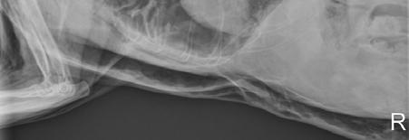
8 L R Caudodorsal lung mass 8


9 Cranial Lobe Vessels Vessel/bronchus triad more easily seen in the left lateral projection Right cranial lobe vessels are ventral Vein is always ventral Vessel size = width of 4 th rib at most narrow point 9


10 R L L R Caudal Lobe Vessels 9 VD DV Approx same size as 9 th rib at the point at which they cross on DV view 10


11 Other Effects of Recumbency Down (recumbent) lung anatomy rises relative to up lung Can be used to help differentiate side of lesion = reason why the right cranial lobe vessels often cross the left cranial lobe vessels in the right lateral projection L R Effect of Sedation on Aeration 11


12 Aeration and Positioning Reduced lung aeration has advantages Confirm pneumothorax Better assess airway collapse Cavitated masses, bullae, blebs will be more conspicuous if the pathology is down - increased opacity surrounding pathology Other Important Technique Related Issues Snapshot in time Assess dynamic problems with static images? Hiatal hernia Dynamic airway disease Disclose a hidden dynamic lesion Vary posture/position Inspiration vs expiration 12


13 Complete intrathoracic tracheal collapse Principles of Interpretation Evaluate Radiographic Study Is there a complete set of radiographs? Thorax 3 or 4 views Technical quality Positioning / phase of respiration Collimation Contrast / film blackness Safety issues Artifacts The Systematic Approach. Consider false positive findings Right heart enlargement Dorsocaudal rotation of the apex in left lateral view Breed variations Alveolar disease Pneumothorax Esophageal dilation Pleural effusion - atelectasis - skin folds - sedation - thickening 13


14 Principles of Interpretation Silhouette sign When 2 structures of the same opacity are in contact it is impossible to distinguish a border between them Summation When parts of an object in different planes are superimposed, the resultant film opacity is a summation of x ray absorption of each structure 14


15 A structure not readily visible can become more apparent when there is a change in opacity adjacent to it Roentgen signs of Pleural Fluid widened interlobar fissures- fissures of soft tissue opacity retraction of lung from thoracic wall space between lung and chest wall soft tissue opacity increased opacity dorsal to sternum on lateral radiographs- scalloped margins blunting of costophrenic sulci in VD view decreased visualization of the heart in DV view obscured diaphragmatic outline in DV and lateral view VD is best view to detect small amounts of pleural fluid 15


16 Pleural Fluid Pleural fluid Pneumothorax Trauma, Lung rupture, Chest wall rent Extension of pneumomediastinum Rupture of cavitary lung mass Air within the pleural space results in collapse of lung and lack of the heart cushion There is a resulting mediastinal shift to the side of greatest lung collapse 16


17 Roentgen signs of pneumothorax Retraction of lung from chest wall Space between lung and chest wall is radiolucent Lung markings do not extend to thoracic wall Lung atelectasis with increased opacity Apparent dorsal displacement of the heart on lateral views 17


18 Tension Pneumothorax 18


19 Conditions mimicking pneumothorax Deep chested dog Skin fold Pleural Space 19


20 Pleural Fibrosis Inflammatory pleural fluids can result in fibrosis of the visceral and rounding of lung margins 20
21 Introduction to Thoracic Radiology and Pleural Space Case Discussions 2/22/2011 Why do we use a vertical beam in veterinary radiography? What effect does this have on the appearance of the thorax? What factors exacerbate the disadvantages of a vertical beam configuration On what side is the dog most likely lying? Why? 1


22 Case 1 3 year old Labrador Hit by car 6 weeks ago Now suddenly distressed 2


23 3


24 Case 1 Findings Dorsal displacement of the heart from the sternum on the lateral view. Gas opacity between heart and sternum Retraction of caudodorsal lung lobes Space between chest wall and lung gas opacity Increased lung opacity, likely secondary to atelectasis Chest wall trauma right T12 Sternal lymphomegaly 4


25 Case 2 10 year old Pekingese Male castrate Sudden onset of seizures 5


26 Case 2 Findings Pleural fissure lines are present on the right and left, with lung lobe retraction, consistent with pleural fluid. The cardiac silhouette and pulmonary vasculature appear normal. There is a slightly increased interstitial pulmonary pattern, probably secondary to atelectasis due to pleural fluid. No rib masses or thoracic wall masses are seen, so differential diagnoses for the pleural fluid include hypoproteinemia, hemorrhage, chylous effusion, inflammatory or non inflammatory effusions/transudates. Labrador Retriever 6 months Male Case 3 Breathing hard for 10 days 6


27 7


28 Case 3 There is retraction of the lung lobes on the right, with interposed soft tissue opacity, consistent with pleural fluid accumulation. This fluid appears unilateral, suggestive of viscous effusion. There are rounded gas opacities within the fluid in the pleural space, most likely representing iatrogenic pneumothorax. There is a smooth, bulbous periosteal reaction on the right 5th rib, consistent with a healing rib fracture. Case 3 continued There is a mild alveolar pattern in the ventral aspects of the right cranial and right caudal lung lobes, likely due to fluid atelectasis. The right middle bronchus is not definitively identified. There is a metallic transponder chip in the dorsal soft tissues. The cardiovascular structures appear within normal limits, although silhouetting with pleural and pulmonary disease complicates interpretation. 8


29 Case 3 Assessment Right sided pleural fluid; consider pyothorax Mild right pneumothorax; possibly iatrogenic Healing fracture, right 5th rib Suspect fluid atelectasis, right cranial and caudal lung lobes Case 4 9 year old Rottweiler Male castrate Respiratory distress and collapse 9
 characterized by marked osteolysis and irregular osteoproliferation.")


30 Case 4 Pleural fissure lines and lung lobe retraction indicate the presence of pleural fluid, which is more severe on the left. Poorly defined, expansile rib lesion (left 7th) characterized by marked osteolysis and irregular osteoproliferation. A rounded soft tissue opacity is superimposed over a 4th rib on the left lateral projection and likely represents a cutaneous nodule present in the right axillary region on the DV projection. An ovoid soft tissue opacity is seen on the right lateral projection at the 6th intercostal space; this opacity may be due to a pulmonary nodule. A focal region of increased soft tissue opacity is seen in the right cranial lung lobe, possibly representing fluid related atelectasis; however, a pulmonary nodule cannot be ruled out. Causes of Pleural Fluid Heart Disease Effusions Septic -pyothorax Non septic lung torsion Malignant Chylothorax Coagulopathies Hypoproteinemia Diaphragmatic hernia Vasculitis 10


31 Case 5 5 year old Labrador Male castrate Fell off an ATV and hit a tree Paraplegic suspect broken back Horizontal beam view done because of neurological status 11


32 Case 5 There is probable subluxation at T12-T13. T13. Retraction of the right caudal lung margins is present, consistent with mild pneumothorax. A left sided alveolar pattern and leftward mediastinal shift are consistent with recumbent atelectasis. 12


33 Case 6 13 year old Golden Retriever Nasal carcinoma Here for thoracic metastasis check 13


34 Case 6 Normal thorax The heart and pulmonary vessels are smaller than normal suggesting hypovolemia. 14


35 Case 7 6 year old Bassett Hound Lethargy and pitting edema 15


36 Case 7 Essentially Normal Thorax The increased opacity along the thoracic wall is common in chondrodystrophoid breeds The cardiac silhouette is mildly enlarged. Extra 2 year old DSH Acc Female Sp Had routine dental prophylaxis 10 days ago puffy and breathing hard 16


37 17
38 Extra A left tension pneumothorax is present. The diaphragm is flattened, the left cranial and caudal lung lobes markedly collapsed and the mediastinum is shifted to the right. Pneumomediastinum is present. Extensive subcutaneous gas is present throughout the neck, thorax, abdomen and forelimbs. 18
A Cardiologist s Approach to Thoracic Radiology. Outline. Technique. Technique. Principles of interpretation. Case Examples. Optimize image quality
 A Cardiologist s Approach to Thoracic Radiology Kacie Schmitt Felber, DVM, DACVIM Cardiology Thursday, May 17 th, 2018 Mid Atlantic States Veterinary Clinic Conference Outline Technique Principles of interpretation
Chest X rays and Case Studies. No disclosures. Outline 5/31/2018. Carlo Manalo, M.D. Department of Radiology Loma Linda University Children s Hospital
 Chest X rays and Case Studies Carlo Manalo, M.D. Department of Radiology Loma Linda University Children s Hospital No disclosures. Outline Importance of history Densities delineated on radiography An approach
Corso Base di Cardiologia Unisvet 2012
 Principi di Radiologia del torace Dr Luca Ferasin DVM PhD CertVC PGCert(HE) DipECVIM-CA (Cardiology) MRCVS European and RCVS Recognised Specialist in Veterinary Cardiology Introduction Thoracic radiography
The Thorax Excluding the Heart and Pulmonary Patterns
 The Thorax Excluding the Heart and Pulmonary Patterns Lisa G. Britt, DVM, MS, Diplomate American College of Veterinary Radiology, Clinical Assistant Professor @ University of Missouri s College of Veterinary
Chest X-ray Interpretation
 Chest X-ray Interpretation Introduction Routinely obtained Pulmonary specialist consultation Inherent physical exam limitations Chest x-ray limitations Physical exam and chest x-ray provide compliment
4/16/2017. Learning Objectives. Interpretation of the Chest Radiograph. Components. Production of the Radiograph. Density & Appearance
 Interpretation of the Arthur Jones, EdD, RRT Learning Objectives Identify technical defects in chest radiographs Identify common radiographic abnormalities This Presentation is Approved for 1 CRCE Credit
1/13/2014. Proper Radiographs. Proper Radiographs. A Review of Pulmonary Patterns
 Live Webinar A Review of Pulmonary Patterns Sofija R. Liles, DVM, DACVR Proper Radiographs Which views? One lateral plus ventrodorsal (at least) Left lateral is best for thorax Three views for full metastatic
Small Animal Teaching Hospital, Leahurst Campus, University of Liverpool, Chester High Road, Neston, Wirral, CH64 7TE
 Thoracic Imaging: taking and reading a great X-ray J. Fraser McConnell BVM&S CertSAM DVR DipECVDI MRCVS Small Animal Teaching Hospital, Leahurst Campus, University of Liverpool, Chester High Road, Neston,
Shedding Light on Neonatal X-rays. Objectives. Indications for X-Rays 5/14/2018
 Shedding Light on Neonatal X-rays Barbara C. Mordue, MSN, NNP-BC Neonatal Nurse Practitioner LLUH Children s Hospital, NICU Objectives Utilize a systematic approach to neonatal x-ray interpretation Identify
UERMMMC Department of Radiology. Basic Chest Radiology
 UERMMMC Department of Radiology Basic Chest Radiology PHYSICS DENSITIES BONE SOFT TISSUES WATER FAT AIR TELEROENTGENOGRAM Criteria for an Ideal Chest Radiograph 1. Upright 2. Posteroanterior View 3. Full
8/14/2017. Objective: correlate radiographic findings of common lung diseases to actual lung pathologic features
 What is that lung disease? Pulmonary Patterns & Correlated Pathology Dr. Russell Tucker, DACVR Objective: correlate radiographic findings of common lung diseases to actual lung pathologic features Improved
10/17/2016. Nuts and Bolts of Thoracic Radiology. Objectives. Techniques
 Nuts and Bolts of Thoracic Radiology October 20, 2016 Carleen Risaliti Objectives Understand the basics of chest radiograph Develop a system for interpreting chest radiographs Correctly identify thoracic
Pulmonary Patterns & Correlated Pathology
 Pulmonary Patterns & Correlated Pathology Russell Tucker, DVM, DACVR Washington State University College of Veterinary Medicine Objective: correlate radiographic findings of common lung diseases to actual
Chest X-ray (CXR) Interpretation Brent Burbridge, MD, FRCPC
 Interpretation Brent Burbridge, MD, FRCPC") Chest X-ray (CXR) Interpretation Brent Burbridge, MD, FRCPC An approach to reviewing a chest x-ray will create a foundation that will facilitate the detection of abnormalities. You should create your own
Chest X-ray (CXR) Interpretation Brent Burbridge, MD, FRCPC An approach to reviewing a chest x-ray will create a foundation that will facilitate the detection of abnormalities. You should create your own
Radiological conference. Left upper lobe collapse. Citation Hong Kong Practitioner, 1998, v. 20 n. 9, p
 Title Radiological conference. Left upper lobe collapse Author(s) Wong, LLS; Peh, WCG Citation Hong Kong Practitioner, 1998, v. 20 n. 9, p. 513-517 Issued Date 1998 URL http://hdl.handle.net/10722/44672
Radiological Anatomy of Thorax. Dr. Jamila Elmedany & Prof. Saeed Abuel Makarem
 Radiological Anatomy of Thorax Dr. Jamila Elmedany & Prof. Saeed Abuel Makarem Indications for Chest x - A chest x-ray may be used to diagnose and plan treatment for various conditions, including: Diseases/Fractures
Calvin 9 year old NM DLH. Dr. Norman Ackerman Memorial Radiography Case Challenge
 September 2014 Dr. Norman Ackerman served the University of Florida, College of Veterinary Medicine with distinction as Professor of Radiology from 1979 to 1994. A concerned teacher of veterinary students
Pneumothorax. Defined as air in the pleural space which can occur through a number of mechanisms
 Pneumothorax Defined as air in the pleural space which can occur through a number of mechanisms Traumatic pneumothorax Penetrating chest trauma Common secondary to bullet or knife penetration Chest tube
Signs in Chest Radiology
 Signs in Chest Radiology Jonathan H. Chung, MD Disclosures No pertinent disclosures Jonathan H. Chung, MD Assistant Professor Institute t of fadvanced d Biomedical Imaging National Jewish Health Denver,
GENERAL ABDOMINAL IMAGING PERITONEAL SPACE, PANCREAS, & SPLEEN. VMB 960 March 25, 2013
 GENERAL ABDOMINAL IMAGING PERITONEAL SPACE, PANCREAS, & SPLEEN VMB 960 March 25, 2013 REFERENCE Chapters 35-36 Pages 650-678 Chapter 37 Pages 694-701 Chapter 3 Pages 38-49 OBJECTIVES Radiography and Ultrasound
Children are not small adults Children are Not Small Adults Anatomic considerations Pliable bony & cartilagenous structures - Significant thoracic inj
 PEDIATRIC CHEST TRAUMA Children are not small adults Role of imaging Spectrum of injury Children are not small adults Children are Not Small Adults Anatomic considerations Pliable bony & cartilagenous
EUROPEAN ASSOCIATION OF VETERINARY DIAGNOSTIC IMAGING EUROPEAN COLLEGE OF VETERINARY DIAGNOSTIC IMAGING
 EISAGOGIKO EUROPEAN ASSOCIATION OF VETERINARY DIAGNOSTIC IMAGING EUROPEAN COLLEGE OF VETERINARY DIAGNOSTIC IMAGING ARISTOTLE UNIVERSITY OF THESSALONIKI SCHOOL OF VETERINARY MEDICINE SECTION OF RADIOLOGY
Imaging of Thoracic Trauma: Tips and Traps. Arun C. Nachiappan, MD Associate Professor of Clinical Radiology University of Pennsylvania
 Imaging of Thoracic Trauma: Tips and Traps Arun C. Nachiappan, MD Associate Professor of Clinical Radiology University of Pennsylvania None Disclosures Objectives Describe blunt and penetrating traumatic
Disclosure. Clinical Chest Radiography Interpretation Part I
 Clinical Chest Radiography Interpretation Part I Anthony M. Angelow, PhD(c), MSN, ACNPC, AGACNP-BC, CEN Associate Lecturer, Fitzgerald Health Education Associates Clinical practice Division of Trauma Surgery
Chest XRay interpretation INTERPRETATIONS Identifications: Name & Date Technical evaluation Basic Interpretations
 Chest XRay interpretation INTERPRETATIONS Identifications: Name & Date Technical evaluation Basic Interpretations TECHNICAL EVALUATION 1. Projection: AP/PA view To differentiate between AP & PA films,
Proceedings of the World Small Animal Veterinary Association Sydney, Australia 2007
 Proceedings of the World Small Animal Sydney, Australia 2007 Hosted by: Next WSAVA Congress THE LAST GASP II: LUNGS AND THORAX David Holt, BVSc, Diplomate ACVS University of Pennsylvania School of Veterinary
What s Your Diagnosis? Jessica Eisenbarth. Signalment: Jazz is a female intact 2 year old German Shorthaired Pointer.
 What s Your Diagnosis? Jessica Eisenbarth Signalment: Jazz is a female intact 2 year old German Shorthaired Pointer. Presenting complaint: Jazz was presented to the K-State emergency service on August
Manage TB Dr. A. Chitrakumar Madras Medical College and RGGGH Institute of Thoracic Medicine, Chennai
 Manage TB Dr. A. Chitrakumar Madras Medical College and RGGGH Institute of Thoracic Medicine, Chennai Lecture 16 Radiology in diagnosis of Tuberculosis Session 01 So, welcome to the session Radiology in
X-rays. Dr Will Dooley
 X-rays Dr Will Dooley Plan Chest X-Rays Abdominal X-Rays Exam approach Presentation skills EMQ EMQ- answers Chest X-Ray - Systematic Approach D R Details RIP Image Quality +/- OBVIOUS ABNORMALITY A B C
B-I-2 CARDIAC AND VASCULAR RADIOLOGY
 (YEARS 1 3) CURRICULUM FOR RADIOLOGY 13 B-I-2 CARDIAC AND VASCULAR RADIOLOGY KNOWLEDGE To describe the normal anatomy of the heart and vessels including the lymphatic system as demonstrated by radiographs,
LUNG PATTERNS IN THE DOG NORMAL AND PATHOLOGICAL
 TRADITION AND MODERNITY IN VETERINARY MEDICINE, 2018, vol. 3, No 1(4): 7 14 LUNG PATTERNS IN THE DOG NORMAL AND PATHOLOGICAL Kalin Spasov 1, Michaela Kunovska 2, Dimo Dimov 3 1 University of Forestry,
What s Your Diagnosis? Signalment: Species: Ferret, Mustela putorius furo Sex: Female Spayed Date of Birth: 03/01/02 History of Adrenal Disease
 What s Your Diagnosis? Signalment: Species: Ferret, Mustela putorius furo Sex: Female Spayed Date of Birth: 03/01/02 History of Adrenal Disease Presenting Complaint: Diarrhea; Acute Dyspnea. For a couple
Interpreting thoracic x-ray of the supine immobile patient: Syllabus
 Interpreting thoracic x-ray of the supine immobile patient: Syllabus Johannes Godt Dep. of Radiology and Nuclear Medicine Oslo University Hospital Ullevål NORDTER 2017, Helsinki Content - Why bedside chest
International Congress of the Italian Association of Companion Animal Veterinarians
 Close this window to return to IVIS www.ivis.org International Congress of the Italian Association of Companion Animal Veterinarians 29-31 May, 2009 Rimini, Italy Next Congress : 65th SCIVAC International
Learning Radiology: Recognizing the Basics. Text with Student Consult Online Access Code
 Learning Radiology: Recognizing the Basics. Text with Student Consult Online Access Code Herring, W ISBN-13: 9780323074445 Table of Contents 1. Recognizing Anything The "colorful" world of radiology A
TB Radiology for Nurses Garold O. Minns, MD
 TB Nurse Case Management Salina, Kansas March 31-April 1, 2010 TB Radiology for Nurses Garold O. Minns, MD April 1, 2010 TB Radiology for Nurses Highway Patrol Training Center Salina, KS April 1, 2010
Objectives. What is a Chest X Ray? CXR Workshop. Definition (diagnostic tool/internal PE) Types. Cost
 Types. Cost") Objectives CAPA 2011 Christy Wilson, PA C Georgia Lung Associates Identify the radiographic landmarks on a chest radiograph Recognize identifiers of poor quality on the chest radiograph Outline an approach
Objectives CAPA 2011 Christy Wilson, PA C Georgia Lung Associates Identify the radiographic landmarks on a chest radiograph Recognize identifiers of poor quality on the chest radiograph Outline an approach
Undergraduate Teaching
 Prof. James F Meaney Undergraduate Teaching Chest X-Ray Understanding the normal anatomical by reference to cross sectional imaging Radiology? It s FUN! Cryptic puzzle Sudoku (Minecraft?) It s completely
!"#$%&'%()'*+,-%&&.'+('*/%)+%,#+0' 12/.,'3%)+"4#%52.
'*+,-%&&.'+('*/%)+%,#+0' 12/.,'3%)+4#%52.") !"#$%&'%()'*+,-%&&.'+('*/%)+%,#+0' 12/.,'3%)+"4#%52.!"#$%&'()$*+&,--#&$.//,0'1232'!-#0'45 *6 '7849!!"#$%&'"(&)*+),$-.*/*01) 2$34/&1)*+)5"-.3.(") 6%.(3")*+)7*(08/$)9(.:"%;.&1)) )?
!"#$%&'%()'*+,-%&&.'+('*/%)+%,#+0' 12/.,'3%)+"4#%52.!"#$%&'()$*+&,--#&$.//,0'1232'!-#0'45 *6 '7849!!"#$%&'"(&)*+),$-.*/*01) 2$34/&1)*+)5"-.3.(") 6%.(3")*+)7*(08/$)9(.:"%;.&1)) )?
Neonatal Chest X-Ray Interpretation
 CHAPTER 7 Neonatal Chest X-Ray Interpretation Prof. Praveen Kumar Neonatal unit, Department of Pediatrics, PGIMER, Chandigarh Learning Objectives At the end of this session, you should be able to: 1. Schematically
The Veteducation International Online Veterinary Conference 2011
 The Veteducation International Online Veterinary Conference 2011 Part of the Veteducation Live Online Web-Seminar Series The Artefacts of Life! With Dr Angela Hartman DVM Dipl. ACVR Massey University New
CHEST & ABDOMINAL X-RAYS MALIKA IBRAHIM CORE MEDICAL TRAINEE BLACKPOOL VICTORIA HOSPITAL DATA INTERPRETATION COURSE FEB 20, 2017
 CHEST & ABDOMINAL X-RAYS MALIKA IBRAHIM CORE MEDICAL TRAINEE BLACKPOOL VICTORIA HOSPITAL DATA INTERPRETATION COURSE FEB 20, 2017 1. Sample x-rays 2. Basic chest x-ray interpretation skills 3. Chest x-ray
Interpretation of the chest radiograph Elizabeth Puddy MB ChB FCARCSI Catherine Hill MB ChB MRCP FRCR
 Interpretation of the chest radiograph Elizabeth Puddy MB ChB FCARCSI Catherine Hill MB ChB MRCP FRCR The traditional technique used in the acquisition and development of a chest radiograph uses methods
Radiology of the respiratory disease
 Radiology of the respiratory disease [ Color index: Important Notes Extra ] [ Editing file Feedback Share your notes Shared notes ] Resources: - 435 Slides - 434 Team - 435 Notes Done by: - Mai Alageel
PLEURAE and PLEURAL RECESSES
 PLEURAE and PLEURAL RECESSES By Dr Farooq Aman Ullah Khan PMC 26 th April 2018 Introduction When sectioned transversely, it is apparent that the thoracic cavity is kidney shaped: a transversely ovoid space
Interactive Lecture. Lecture 7 - Interactive. Radiology of cardiorespiratory disease. Editing File. Done By. Color Coding Important Notes Extra
 Lecture 7 - Interactive 436 Teams Interactive Lecture Radiology of cardiorespiratory disease Done By Team Leaders: Khalid Alshehri Hanin Bashaikh Team Members: Ghaida Alsaeed Maha Alissa Nawwaf AlHarbi
like humans, have well-developed mediastinal separation between the left and right hemithorax, thus unilateral changes can occur. On the other hand,
 Tutorial Module 6 Thoracic Cavity and Tumors of Lung and Pleura Alfonso López Atlantic Veterinary College University of Prince Edward Island Canada 2009 Enero 3 Thoracic Cavity There are significant anatomical
Bony Thorax. Anatomy and Procedures of the Bony Thorax Edited by M. Rhodes
 Bony Thorax Anatomy and Procedures of the Bony Thorax 10-526-191 Edited by M. Rhodes Anatomy Review Bony Thorax Formed by Sternum 12 pairs of ribs 12 thoracic vertebrae Conical in shape Narrow at top Posterior
GENERAL DIAGNOSTIC IMAGING IN SMALL ANIMAL ONCOLOGY
 GENERAL DIAGNOSTIC IMAGING IN SMALL ANIMAL ONCOLOGY Jantra Ngosuwan Suran, DVM, Dipl. ACVR, Cert Clin Res University of Pennsylvania, School of Veterinary Medicine 3900 Delancey St, Philadelphia, PA 19104
Thorax Review and Revitalize
 Outline Thorax Review and Revitalize Anatomy Sarah Tibbs, BVetMed, DACVR April 2016 I will try to share little Tibbits along the way (Tibbs Tidbits) Patterns Review like students Lungs Pleura Heart Other
Discussing feline tracheal disease
 Vet Times The website for the veterinary profession https://www.vettimes.co.uk Discussing feline tracheal disease Author : ANDREW SPARKES Categories : Vets Date : March 24, 2008 ANDREW SPARKES aims to
Approach to CXR. Terminology. 1.Identification. Greg Blecher SCH Respir Fellow. Correct patient Correct date and time Correct examination
 Approach to CXR Greg Blecher SCH Respir Fellow From Rob Posteraro http://home.earthlink.net/~rhpos/cxr_interpret.txt.html ; http://home.earthlink.net/~rhpos/cxr_main.txt.html) Approach to viewing Chest
Disclosure. Clinical Chest Radiography Interpretation Part II
 Clinical Chest Radiography Interpretation Part II Anthony M. Angelow, PhD(c), MSN, ACNPC, AGACNP-BC, CEN Associate Lecturer, Fitzgerald Health Education Associates Clinical practice Division of Trauma
Thorax Lecture 2 Thoracic cavity.
 Thorax Lecture 2 Thoracic cavity. Spring 2016 Dr. Maher Hadidi, University of Jordan 1 Enclosed by the thoracic wall. Extends between (thoracic inlet) & (thoracic outlet). Thoracic inlet At root of the
General Abdominal Radiography
 General Abdominal Radiography Tony Pease, DVM, MS Assistant Professor of Radiology North Carolina State University Objectives Acquisition of radiographs Abdominal radiographic anatomy Radiographic patterns
X-Rays. Prepared by Prof.Dr. Magda Hassab Allah Assist.lecturer Marwa Al Hady
 X-Rays Prepared by Prof.Dr. Magda Hassab Allah Assist.lecturer Marwa Al Hady CHEST X-RAYS Normal Chest X-ray Comments on chest X ray includes examination of 1- Bony cage (ribs,clavicles &vertebral column
Close window to return to IVIS. in collaborazione con RICHIESTO ACCREDITAMENTO. organizzato da certificata ISO 9001:2000
 in collaborazione con Close window to return to IVIS RICHIESTO ACCREDITAMENTO SOCIETÀ CULTURALE ITALIANA VETERINARI PER ANIMALI DA COMPAGNIA SOCIETÀ FEDERATA ANMVI organizzato da certificata ISO 9001:2000
Don t Panic! Dr. Karau s Guide to Respiratory Emergencies November 4, 2018
 Don t Panic! Dr. Karau s Guide to Respiratory Emergencies November 4, 2018 Objectives Oxygen delivery methods Emergent diagnostic tests Differentiating between upper and lower respiratory disease Respiratory
X-Rays. Kunal D Patel Research Fellow IMM
 X-Rays Kunal D Patel Research Fellow IMM The 12-Steps } 1: Name 2: Date 3: Old films 4: What type of view(s) 5: Penetration } Pre-read 6: Inspiration 7: Rotation Quality Control 8: Angulation 9: Soft tissues
DETECTION OF PNEUMOTHORAX AND PLEURAL EFFUSION WITH HORIZONTAL BEAM RADIOGRAPHY
 DETECTION OF PNEUMOTHORAX AND PLEURAL EFFUSION WITH HORIZONTAL BEAM RADIOGRAPHY KATHERINE C. LYNCH, CINTIA R. OLIVEIRA, JODI S. MATHESON, MARK A. MITCHELL, ROBERT T. O BRIEN Forty-seven patients with a
Spinal radiographs are indicated for: THORACIC SPINE RADIOGRAPHY SMALL ANIMAL SPINAL RADIOGRAPHY SERIES. ImagIng EssEntIals
 PEER REVIEWED ImagIng EssEntIals SMLL NIML SPINL RDIOGRPHY SERIES THORCIC SPINE RDIOGRPHY Danielle Mauragis, CVT, and Clifford R. erry, DVM, Diplomate CVR Imaging Essentials provides comprehensive information
Medical NREMT-PTE. NREMT Paramedic Trauma Exam.
 Medical NREMT-PTE NREMT Paramedic Trauma Exam https://killexams.com/pass4sure/exam-detail/nremt-pte Question: 41 Which of the following most accurately describes the finding of jugular venous distension
Introduction to Chest Radiography
 Introduction to Chest Radiography RSTH 366: DIAGNOSTIC TECHNIQUES Alan Alipoon BS, RCP, RRT Instructor Department of Cardiopulmonary Sciences 1 Introduction Discovered in 1895 by Wilhelm Roentgen Terminology
The Thorax The Ever Challenging Pulmonary Patterns
 The Thorax The Ever Challenging Pulmonary Patterns Lisa G. Britt, DVM, MS, Diplomate American College of Veterinary Radiology, Clinical Assistant Professor @ University of Missouri s College of Veterinary
Thoracic Cavity and Tumors of Lung and Pleura
 Tutorial Module 6 Thoracic Cavity and Tumors of Lung and Pleura Alfonso López Atlantic Veterinary College University of Prince Edward Island Canada Sept 28, 2014 Thoracic Cavity There are anatomical differences
The External Anatomy of the Lungs. Prof Oluwadiya KS
 The External Anatomy of the Lungs Prof Oluwadiya KS www.oluwadiya.com Introduction The lungs are the vital organs of respiration Their main function is to oxygenate the blood by bringing inspired air into
Pediatric Lung Ultrasound (PLUS) In Diagnosis of Community Acquired Pneumonia (CAP)
 In Diagnosis of Community Acquired Pneumonia (CAP)") Pediatric Lung Ultrasound (PLUS) In Diagnosis of Community Acquired Pneumonia (CAP) Dr Neetu Talwar Senior Consultant, Pediatric Pulmonology Fortis Memorial Research Institute, Gurugram Study To compare
Pediatric Lung Ultrasound (PLUS) In Diagnosis of Community Acquired Pneumonia (CAP) Dr Neetu Talwar Senior Consultant, Pediatric Pulmonology Fortis Memorial Research Institute, Gurugram Study To compare
Advances in MDCT of Thoracic Trauma
 Baltic Congress of Radiology, Riga 2010 Advances in MDCT of Thoracic Trauma Robert A. Novelline, MD Professor of Radiology, Harvard Medical School Director of Emergency Radiology, Massachusetts General
What s Your Diagnosis? Allison Crow, Class of 2014
 What s Your Diagnosis? Allison Crow, Class of 2014 Signalment: 13 year old male castrated mixed breed dog History: The patient presented to the rdvm for pain in the hind end, weakness and neck stretching
Do you want to be an excellent Radiologist? - Focus on the thoracic aorta on lateral chest image!!!
 The lateral chest radiograph: Challenging area around the thoracic aorta!!! Do you want to be an excellent Radiologist? - Focus on the thoracic aorta on lateral chest image!!! Dong Yoon Han 1, So Youn
Spinal radiographs are indicated for: CerviCal Spine radiography. Small animal Spinal RadiogRaphy SeRieS. ImagIng EssEnTIals
 ImagIng EssEnTIals Peer reviewed Small animal Spinal RadiogRaphy SeRieS CerviCal Spine radiography Danielle Mauragis, CVT, and Clifford R. erry, DVM, Diplomate CVR Imaging Essentials provides comprehensive
Proceedings of the Southern European Veterinary Conference and Congreso Nacional de AVEPA
 www.ivis.org Proceedings of the Southern European Veterinary Conference and Congreso Nacional de AVEPA Oct. 18-21, 2012 - Barcelona, Spain Next Conference: Oct. 17-19, 2013 - Barcelona, Spain Reprinted
An Introduction to Radiology for TB Nurses
 An Introduction to Radiology for TB Nurses Garold O. Minns, MD September 14, 2017 TB Nurse Case Management September 12 14, 2017 EXCELLENCE EXPERTISE INNOVATION Garold O. Minns, MD has the following disclosures
Lecture 2: Clinical anatomy of thoracic cage and cavity II
 Lecture 2: Clinical anatomy of thoracic cage and cavity II Dr. Rehan Asad At the end of this session, the student should be able to: Identify and discuss clinical anatomy of mediastinum such as its deflection,
What s Your Diagnosis? Signalment: Species: Canine Breed: Golden Retriever Sex: Female (spayed) Date of Birth: 04/01/99
 Date of Birth: 04/01/99") What s Your Diagnosis? Signalment: Species: Canine Breed: Golden Retriever Sex: Female (spayed) Date of Birth: 04/01/99 Presenting Complaint: Acute onset of lethargy Vomited twice (partially digested food)
What s Your Diagnosis? Signalment: Species: Canine Breed: Golden Retriever Sex: Female (spayed) Date of Birth: 04/01/99 Presenting Complaint: Acute onset of lethargy Vomited twice (partially digested food)
GENERAL ABDOMINAL IMAGING PERITONEAL SPACE, PANCREAS, & SPLEEN
 GENERAL ABDOMINAL IMAGING PERITONEAL SPACE, PANCREAS, & SPLEEN VMB 960 March 25, 2013 REFERENCE Chapters 35-36 Pages 650-678 Chapter 37 Pages 694-701 Chapter 3 Pages 38-49 OBJECTIVES Radiography and Ultrasound
Instructions. Print out all three tabs of the guide to be used as a reference.
 Instructions For the Learner 1) The purpose of the observation checklist is to help evaluate the skills obtained from the course. It is also a reference and reminder for what was learned in this course.
Sectional Anatomy Quiz - III
 Sectional Anatomy - III Rashid Hashmi * Rural Clinical School, University of New South Wales (UNSW), Wagga Wagga, NSW, Australia A R T I C L E I N F O Article type: Article history: Received: 30 Jun 2018
PATIENT DATA EVALUATION AND RECOMMENDATION: IMAGING STUDIES
 PATIENT DATA EVALUATION AND RECOMMENDATION: IMAGING STUDIES Robert Harwood, MSA, RRT-NPS Objectives At the end of this presentation the student should be able to: Describe the indications of a chest radiograph.
ANATOMY OF THE PLEURA. Dr Oluwadiya KS
 ANATOMY OF THE PLEURA Dr Oluwadiya KS www.oluwadiya.sitesled.com Introduction The thoracic cavity is divided mainly into: Right pleural cavity Mediastinum Left Pleural cavity Pleural cavity The pleural
PULMONARY IMAGING: GETTING THE MOST INFORMATION FROM THORACIC RADIOGRAPHS
 PULMONARY IMAGING: GETTING THE MOST INFORMATION FROM THORACIC RADIOGRAPHS Peter Scrivani, DVM, DACVR Cornell University College of Veterinary Medicine, Ithaca, NY Outline Pulmonary Imaging Pulmonary anatomy
Elderly Man with Dyspnoea
 Asia Pacific Family Medicine, 2004; 3 (1-2): 46-50 RADIOLOGY SERIES Elderly Man with Dyspnoea Wei-Yang LIM Faculty of Medicine National University of Singapore, Singapore Wilfred CG PEH Singapore Health
Pulmonary Embolism. Thoracic radiologist Helena Lauri
 Pulmonary Embolism Thoracic radiologist Helena Lauri 8.5.2017 Statistics 1-2 out of 1000 adults annually are diagnosed with deep vein thrombosis (DVT) and/or pulmonary embolism (PE) About half of patients
Two Lesser Known but Useful Signs of Neonatal Pneumothorax
 Two Lesser Known but Useful Signs of Neonatal Pneumothorax LEONARD E. SWISCHUK1 Two lesser known signs of neonatal pneumothorax are presented : the large, hyperlucent hemithorax sign and the medial stripe
ARDS - a must know. Page 1 of 14
 ARDS - a must know Poster No.: C-1683 Congress: ECR 2016 Type: Authors: Keywords: DOI: Educational Exhibit M. Cristian; Turda/RO Education and training, Edema, Acute, Localisation, Education, Digital radiography,
Eun-Young Kang, M.D., Jae Wook Lee, M.D., Ji Yung Choo, M.D., Hwan Seok Yong, M.D., Ki Yeol Lee, M.D., Yu-Whan Oh, M.D.
 Eun-Young Kang, M.D., Jae Wook Lee, M.D., Ji Yung Choo, M.D., Hwan Seok Yong, M.D., Ki Yeol Lee, M.D., Yu-Whan Oh, M.D. Department of Radiology, Korea University Guro Hospital, College of Medicine, Korea
(SKILLS/HANDS-ON) Chest Tubes. Rebecca Carman, MSN, ACNP-BC. Amanda Shumway, PA-C. Thomas W. White, MD, FACS, CNSC
 Chest Tubes. Rebecca Carman, MSN, ACNP-BC. Amanda Shumway, PA-C. Thomas W. White, MD, FACS, CNSC") (SKILLS/HANDS-ON) Chest Tubes Rebecca Carman, MSN, ACNP-BC Nurse Practitioner, Trauma Services, Intermountain Medical Center, Intermountain Healthcare Amanda Shumway, PA-C APC Trauma and Critical Care
(SKILLS/HANDS-ON) Chest Tubes Rebecca Carman, MSN, ACNP-BC Nurse Practitioner, Trauma Services, Intermountain Medical Center, Intermountain Healthcare Amanda Shumway, PA-C APC Trauma and Critical Care
Chest X-Ray: the essentials
 Chest X-Ray: the essentials Poster No.: C-1264 Congress: ECR 2017 Type: Educational Exhibit Authors: J. J. Delgado Moraleda, A. ALEGRE DELGADO, R. M. Piqueras Olmeda, E. Chacón Avilés, J. F. Melo Villamarín,
Chest Radiology Interpretation: Findings of Tuberculosis
 Chest Radiology Interpretation: Findings of Tuberculosis Get out your laptops, smart phones or other devices pollev.com/chestradiology Case #1 1 Plombage Pneumonia Cancer 2 Reading the TB CXR Be systematic!
Proceedings of the World Small Animal Veterinary Association Sydney, Australia 2007
 Proceedings of the World Small Animal Veterinary Association Sydney, Australia 2007 Hosted by: Australian Small Animal Veterinary Association (ASAVA) Australian Small Animal Veterinary Association (ASAVA)
Lecturer: Ms DS Pillay ROOM 2P24 25 February 2013
 Lecturer: Ms DS Pillay ROOM 2P24 25 February 2013 Thoracic Wall Consists of thoracic cage Muscle Fascia Thoracic Cavity 3 Compartments of the Thorax (Great Vessels) (Heart) Superior thoracic aperture
Abdominal Ultrasound
 Abdominal Ultrasound Imaging Control Buttons Depth The organ imaged should take up 3/4 of the screen Frequency = Penetration Use high frequencies (harmonics) for fluid filled and superficial structures
Systemic lupus erythematosus (SLE): Pleuropulmonary Manifestations
: Pleuropulmonary Manifestations") 08/30/10 09/26/10 Systemic lupus erythematosus (SLE): Pleuropulmonary Manifestations Camila Downey S. Universidad de Chile, School of Medicine, Year VII Harvard University, School of Medicine Sept 17,
08/30/10 09/26/10 Systemic lupus erythematosus (SLE): Pleuropulmonary Manifestations Camila Downey S. Universidad de Chile, School of Medicine, Year VII Harvard University, School of Medicine Sept 17,
Techniques of examination of the thorax and lungs. Dr. Szathmári Miklós Semmelweis University First Department of Medicine 24. Sept
 Techniques of examination of the thorax and lungs Dr. Szathmári Miklós Semmelweis University First Department of Medicine 24. Sept. 2013. Inspection of the thorax Observe: the shape of chest Deformities
DENTAL RADIOGRAPH INTERPRETATION
 DENTAL RADIOGRAPH INTERPRETATION Brook A. Niemiec, DVM Diplomate, American Veterinary Dental College Fellow, Academy of Veterinary Dentistry www.vetdentaltraning.com www.vetdentalrad.com Interpreting dental
What s Your Diagnosis?
 What s Your Diagnosis? Courtney S. Wait Signalment: 11 year old FS Labrador Retriever Presenting Complaint/History: The patient presented to the referring DVM for inappetance, vomiting, lethargy, and anorexia.
An Image Repository for Chest CT
 An Image Repository for Chest CT Francesco Frajoli for the Chest CT in Antibody Deficiency Group An Image Repository for Chest CT he Chest CT in Antibody Deficiency Group is an international and interdisciplinary
Chylothorax Basics OVERVIEW GENETICS SIGNALMENT/DESCRIPTION OF PET
 Chylothorax Basics OVERVIEW Chylo- refers to chyle; thorax refers to the chest Chyle is a milky to slightly yellow fluid composed of lymph and fats (rich in triglycerides) taken up from the intestines
Tests Your Pulmonologist Might Order. Center For Cardiac Fitness Pulmonary Rehab Program The Miriam Hospital
 Tests Your Pulmonologist Might Order Center For Cardiac Fitness Pulmonary Rehab Program The Miriam Hospital BASIC ANATOMY OF THE LUNGS Lobes of Lung 3 lobes on the Right lung 2 lobes on the Left Blood