Intracardiac EchoCardiography (ICE) Common Views
|
|
|
- Emerald Cummings
- 5 years ago
- Views:
Transcription
1 Intracardiac EchoCardiography (ICE) Common Views

2 Introduction What is ICE? Catheter with microscopic ultrasound transducer tip and doppler capabilities inserted into the heart via the IVC (typically) or SVC. Why is it useful? Allows direct visualization of anatomical cardiac structures targeted in abalations Helping guide catheters; in detecting thromboemoboli, pericardial effusion, preventing esophageal damage and in evaluating hemodynamic consequences of ablation (eg increased velocities in pulmonary vein stenosis).
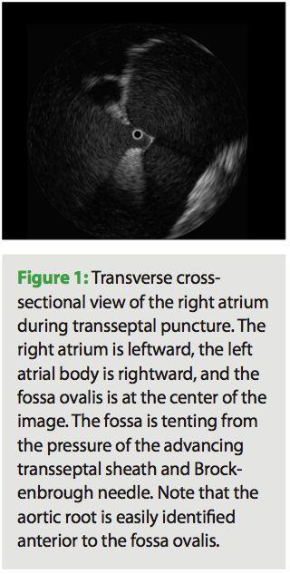
3 Introduction What types? There are two types of ICE - phased array and radial. Phased array: more common as it creates a view that is easier to interpret as it resembles a TTE or TEE. Radial ICE: greater resolution, but is more difficult to interpret and is best for discriminating near-field structures and therefore requires transseptal puncture to visualize left heart structures. Process: Insert catheter through the femoral vein up through the IVC to the right atrium (most commonly), the home base for the ICE catheter. The catheter can also be inserted via the right IJ or left subclavian and then through the SVC however this will give upsidedown images; this method is preferred by some who wish to avoid entanglement/interaction with other wires placed through the IVC. Since the pulmonary veins are of primary importance in ablation procedures, to gain access to to the LA where the PVs insert, a transseptal puncture of the interatrial septum is performed. Prior to performing this puncture, it is crucial to evaluate for the presence of an intracardiac thromboemboli in the left atrium that could then potentially pass through the puncture and embolize outside of the heart, for instance to the brain. Potential for thrombo-emboli would be seen as a spontaneous echographic contrast, also called smoke (1) and (2) Essentially, SCE appears as hyperdense substance where it should not be present (i.e. in the chamber of the left atrium which should appear hypodense). If SCE is detected, a trans-septal puncture should not be performed.
4 Phased Array

5 Home View Home view showing the right heart, including the interventricular septum: the slightly anteflexed probe is positioned in the mid-right atrium. IVS, interventricular septum; RA, right atrium; RV, right ventricle.
6 Septal View To obtain the view - While the catheter is in neutral position, rotate slightly posterior and slightly rightward, so that the transducer will face the interatrial septum. This is the septal view. In this view, the entire length of the atrial septum, the coronary sinus, and the pulmonary veins are well seen. Septal view without and with color Doppler demonstrating a large 22 mm defect (arrow).
7 Long axis/caval view To obtain the view - Advance more cephalad toward the SVC to obtain the SVC or long-axis view. In this plane, the transducer faces the interatrial septum, and the SVC can be seen as it relates to the right atrium. The interatrial septum is shown in a superior/inferior plane and corresponds to the TEE long-axis view. Greater portions of the SVC can be seen as the ICE catheter is further advanced in this flexed position toward the SVC with slightly more rightward flexion. Greater portions of the inferior septum can similarly be imaged by withdrawing the ICE catheter toward the IVC. A defect in the interatrial septum (ASD/PFO) can be well profiled, and the superior and inferior rims as well as the diameter of the defect can be measured. In this view, both the right and left pulmonary veins may also be imaged, depending on the exact angle of the imaging plane. The imaging angle can be manipulated with clockwise and counterclockwise rotation as well as flexion/anteflexion to achieve these views.
8 Short axis view To obtain the view - Lock the ICE catheter, then rotate the entire handle with the catheter shaft at the sheath hub clockwise until it sits with the transducer near the tricuspid valve annulus and inferior to the aorta. Minor adjustments of less posterior flexion and more leftward rotation can demonstrate the short-axis view. In this view, anatomic structures seen include the aortic valve in short axis and the interatrial septum. This view is very similar to the basal shortaxis view obtained by TEE. However, the right atrium is shown in the near field, and the left atrium is in the far field, which is opposite of that seen with TEE.
 Posterior view of a electroanatomic + ICE map of the left atrium with the ablation catheter tip (arrow) in the left inferior pulmonary vein (C) The circle, representing the ablation catheter tip")
9 Septal Perforation (A) AF ablation with desired atrial septal tenting (arrow) when the transseptal needle is about to cross from right atrium into left atrium. (B) Posterior view of a electroanatomic + ICE map of the left atrium with the ablation catheter tip (arrow) in the left inferior pulmonary vein (C) The circle, representing the ablation catheter tip in (B), confirms ICE location in the left inferior pulmonary vein.
10 Views of the left heart These NEXT 3 views taken from the RA. The catheter has a high resolution multiple frequency transducer that allows tissue penetration enhancement and thus depth control which allows visualization of the LA and PVs Border Between the LA and PVs can be determined by extrapolating the left atrial contour (see dotted line) thus revealing the atrial-pv junction LIPV and LSPV are the hypoechogenic structures entering the LA in this view.
11 Views of Left Pulmonary Veins Here we see a common ostium of the LIPV and LSPV (see double arrow). Using Long Axis view, the PVs can be visualized. From this view, the Superior PV is on the right and the Inferior on the left.
12 Views of Right Pulmonary Veins Here we see the RIPV and RSPV entering the LA without a common ostia Again, viewed by turning the catheter slightly in the RA, long axis view to view the right PVs. The Superior PV is on the right of the screen, the Inferior PV on the left
 The aortic valve is seen in short axis, with the mouth of the LAA visible adjacent to the left coronary cusp.")
13 Left Atrial Appendage view The imaging catheter is placed through the tricuspid valve and rotated 180 to obtain a superior view. Important for LAA thrombus monitoring (A) The aortic valve is seen in short axis, with the mouth of the LAA visible adjacent to the left coronary cusp. Note the proximity of the left main coronary artery to the LAA. The proximal segment of the left superior pulmonary vein also is visible in this view. (B) The imaging catheter is now deflected superiorly, inserted through the pulmonic valve and directed leftward in the pulmonary artery. The LAA is now seen in the near field with detailed visualization of the pectinate musculature.

 pectinate muscles of the LAA observed (arrowhead). (C) Short-axis view of the LAA.")
14 Left Atrial Appendage view ICE catheter is placed through the PA to visualize the LAA. LA left atrium, RV right ventricle, LV left ventricle, LPV left pulmonary vein, LAA left atrial appendage (A) Long-axis view of LAA and orifice. LSPV also observed. (B) pectinate muscles of the LAA observed (arrowhead). (C) Short-axis view of the LAA. (D) Apex of the LAA

15 Ablation views

16 Hemodynamic changes during ablation Increased flow velocity following ablation. Important in monitoring for pulmonary vein stenosis. Example: LIPV ostium Pre - 84 cm/sec Post cm/sec (increased turbulence) 1.6 months later cm/sec (improved flow)
17 Pathology Seen Using ICE

18 Aortic Dissection E = the Entry point through the tear in the intima. TL = the True Lumen of the aorta FL = the False Lumen between the intimal and medial layers of the aortic wall
19 Pericardial effusion

20 Left Atrial Appendage Thrombus As visualized by TEE (double click image to play video)
.")
21 Esophageal Mapping To prevent esophageal injury, including perforation and atrioesophageal fistula Mostly achieved with esophageal temperature monitoring. Also can be imaged with electroanatomic mapping and 2D ICE (3D has been suggested). To image and monitor the esophagus and the left atrial posterior wall contiguous to esophagus, place the ICE transducer in the mid/high RA and rotate clockwise to scan between the left and right mid/lower PV ostia.
22 Miscellaneous views

23 More views of the SVC Long axis/caval view
24 Radial ICE
25 Transseptal perforation LIPV LA LSPV RIPV

26 Radial ICE views
27
28 References 1. Haines, DE. Radial Intracardiac Echocardiographic Imaging for Atrial Fibrillation Ablation. EPLabDigest. May 2012, 12(5) Hijazi ZM, Shivkumar K, Sahn DJ. Intracardiac Echocardiography During Interventional and Electrophysiological Cardiac Catheterization. Circulation : DOI: /CIRCULATIONAHA ahajournals.org/content/119/4/587.full 3. Bartel T, Muller S, Biviano A, Hahn R. Why is intracardiac echocardiography helpful? Benefits, costs, and how to learn. European Heart Journal. Jan 2014, 35(2) DOI: /eurheartj/eht oxfordjournals.org/content/early/2013/10/14/eurheartj.eht411#ref Hutchinson M. et al. Imaging the left atrial appendage with intracardiac echocardiography: leveling the playing field. Circulation: Arrthythmia and electrophysiology Nishiyama et al. Visualization of the left atrial appendage by phased-array intracardiac echocardiography from the pulmonary artery in patients with atrial fibrillation. Europace Heart Rhythm Center. Part 1 Radial Intracardiac Echocardiography in the EP Lab: Right Atrial Procedures
29 References cont. 7. Ren J, Marchlinski FE, Callans DJ, Schwartzman D. Practical intracardiac echocardiography in Electrophysiology. Malden: Blackwell Futura, id=ohasw4pizfqc&pg=pa103&lpg=pa103&dq=intracardiac+echocardiography+phase+pulmonary+veins&source=bl&ots=mywt4m26f m&sig=ts-x28d145jzuiwyfcnxyqumuek&hl=en&sa=x&ved=0ahukewjstr75sdmahuj0gmkhey9b8oq6aeintaf#v=onepage&q=intracardiac%20echocardiography%20phase%20pulmonary% 20veins&f=false 8. Callans et al. Narrowing of the Superior Vena Cava Right Atrium Junction During Radiofrequency Catheter Ablation for Inappropriate Sinus Tachycardia: Analysis With Intracardiac Echocardiography. JACC Vol. 33, No. 6, May 1999: Ren J, Callans DJ, Marchlinski FE, Stiffler JA, Sadek MM, Supple FE. 3D Intracardiac Echocardiography/Cartosound Imaging Of Esophagus Guided Left Atrial Posterior Wall Ablation For Atrial Fibrillation. JAFib. Dec 2014-Jan Volume 7, Issue 4. jafib.com/published/webformat/fang_ren/fang_ren.pdf 10. Neelankavil J, Chua J, Howard-Quijano K, Mahajan A. Emerging Technology Review: Intracardiac Echocardiography. Journal of Cardiothoracic and Vascular Anesthesia. 29(2),
30 Cardiac Anatomy
31 Gross Anatomy of the Left Atrium (LA) Four Parts (+1): Venous component - receives the Pulmonary Veins (PV) Vestibule - between the body and the mitral valve (MV) Left Atrial Appendage (LAA) Interatrial Septum (IAS) +1 = The body of the LA. The body is the part of the LA between the PV components and the vestibule. Average size (A-P diameter) of LA: /- 4.9mm

32 LA Images Longitudinal section through the heart Floor - Mitral Valve Roof = superior wall ( atrial dome ) Posterior wall = adjacent to the LIPV. The coronary sinus can be seen on the inferior portion of the LA adjacent to the atrial infolding LLR = Left Lateral Ridge. The LLR is a useful landmark. On the supero-posterior side of the LLR are the ostia for the LIPV and LSPV. On the antero-inferior side of the LLR is the Left Atrial Appendage.
ViewFlex Xtra ICE Catheter. Positioning Reference Manual
 ViewFlex Xtra ICE Catheter Positioning Reference Manual ViewFlex Xtra ICE Catheter Index The ViewFlex Xtra ICE Catheter, which is compatible with the ViewMate Z and ViewMate II ultrasound consoles, provides
Normal TTE/TEE Examinations
 Normal TTE/TEE Examinations Geoffrey A. Rose, MD FACC FASE Sanger Heart & Vascular Institute Before you begin imaging... Obtain the patient s Height Weight BP PLAX View PLAX View Is apex @ 9-10 o clock?
Breakout Session: Transesophageal Echocardiography
 Breakout Session: Transesophageal Echocardiography Doris Ockert, MD Andrew Schroeder, MD University of Wisconsin School of Medicine and Public Health Jutta Novalija, MD, PhD Medical College of Wisconsin
Integration of CT and fluoroscopy images in the ablative treatment of atrial fibrillation
 Clinical applications Integration of CT and fluoroscopy images in the ablative treatment of atrial fibrillation C. Kriatselis M. Tang M. Roser J-H. erds-li E. leck Department of Internal Medicine/Cardiology,
Introduction to TEE using Heartworks Echocardiography Simulator
 Introduction to TEE using Heartworks Echocardiography Simulator Steven M. Ewer, MD Assistant Professor Division of Cardiovascular Medicine University of Wisconsin School of Medicine & Public Health Version
Comprehensive Transoesophageal Echocardiography Examination
 IJUTPC PICTORIAL REVIEW Comprehensive Transoesophageal Echocardiography Examination Comprehensive Transoesophageal Echocardiography Examination 1 Ravi Hebballi, 2 Ann Ngui 1 Consultant in Cardiothoracic
A case of mislabeled textbooks: misnomer of the traditional "bicaval" view.
 Providence St. Joseph Health Providence St. Joseph Health Digital Commons Journal Articles and Abstracts 8-1-2018 A case of mislabeled textbooks: misnomer of the traditional "bicaval" view. Laura Vaskelyte
The earliest application of intracardiac imaging
 Intracardiac Echocardiography for Structural Heart Defects A review of ICE innovations, devices, and techniques. BY ZAHID AMIN, MD; QI-LING CAO, MD; AND ZIYAD M. HIJAZI, MD The earliest application of
AF ablation Penn experience. Optimal approach to the ablation of PAF: Importance of identifying triggers 9/25/2009
 Optimal approach to the ablation of PAF: Importance of identifying triggers David J. Callans, MD University of Pennsylvania School of Medicine AF ablation Penn experience Antral (circumferential) PV ablation
Trans-septal Catheterization. December 8, Jonathan Tobis, MD Professor of Medicine Interventional Cardiology, UCLA
 Trans-septal Catheterization December 8, 2015 Jonathan Tobis, MD Professor of Medicine Interventional Cardiology, UCLA No conflicts of interest for this talk BRK = Brockenbrough needle BRK may be easier
JOINT MEETING 2 Tricuspid club Chairpersons: G. Athanassopoulos, A. Avgeropoulou, M. Khoury, G. Stavridis
 JOINT MEETING 2 Tricuspid club Chairpersons: G. Athanassopoulos, A. Avgeropoulou, M. Khoury, G. Stavridis Similarities and differences in Tricuspid vs. Mitral Valve Anatomy and Imaging. Echo evaluation
Since the introduction of transesophageal echocardiography
 ASE/SCA Guidelines for Performing a Comprehensive Intraoperative Multiplane Transesophageal Echocardiography Examination: Recommendations of the American Society of Echocardiography Council for Intraoperative
Atrial Septal Defects
 Supplementary ACHD Echo Acquisition Protocol for Atrial Septal Defects The following protocol for echo in adult patients with atrial septal defects (ASDs) is a guide for performing a comprehensive assessment
Revealing new insights. irotate electronic rotation and xplane adjustable biplane imaging. Ultrasound cardiology. irotate and xplane
 Ultrasound cardiology irotate and xplane Revealing new insights irotate electronic rotation and xplane adjustable biplane imaging Annemien van den Bosch and Jackie McGhie Department of Cardiology, Erasmus
Hybrid Ablation of AF in the Operating Room: Is There a Need? MAZE III Procedure. Spectrum of Atrial Fibrillation
 Hybrid Ablation of AF in the Operating Room: Is There a Need? MAZE III Procedure Paul J. Wang, MD Amin Al-Ahmad, MD Gan Dunnington, MD Stanford University Cox J, et al. Ann Thorac Surg. 1993;55:578-580.
Case # 1. Page: 8. DUKE: Adams
 Case # 1 Page: 8 1. The cardiac output in this patient is reduced because of: O a) tamponade physiology O b) restrictive physiology O c) coronary artery disease O d) left bundle branch block Page: 8 1.
The Normal Echocardiogram
 The Normal Echocardiogram Pravin V. Patil, MD FACC Lewis Katz School of Medicine at Temple University Acknowledgments Dr. Susan Wiegers Dr. Martin Keane Temple Cardiac Sonographers Disclosures No relevant
The radial procedure was developed as an outgrowth
 The Radial Procedure for Atrial Fibrillation Takashi Nitta, MD The radial procedure was developed as an outgrowth of an alternative to the maze procedure. The atrial incisions are designed to radiate from
ATRIAL SEPTAL CLOSURE AND LEFT ATRIAL APPENDAGE OCCLUSION: INDICATIONS AND GUIDANCE ECHOCARDIOGRAPHY IN INTERVENTIONAL CARDIOLOGY
 ATRIAL SEPTAL CLOSURE AND LEFT ATRIAL APPENDAGE OCCLUSION: INDICATIONS AND GUIDANCE Aristides G. Panlilio, MD, FPCP, FPCC,FPSE, FASE Philippine Heart Center Chinese General Hospital and Medical Center
Adult Echocardiography Examination Content Outline
 Adult Echocardiography Examination Content Outline (Outline Summary) # Domain Subdomain Percentage 1 2 3 4 5 Anatomy and Physiology Pathology Clinical Care and Safety Measurement Techniques, Maneuvers,
TEE Outside of the Cardiac OR
 TEE Outside of the Cardiac OR STEVE GIBSON MD PHD OU DEPARTMENT OF ANESTHEIOLOGY I have no financial relationships or conflicts of interest to disclose TRANSESOPHAGEAL ECHOCARDIOGRAPHY Basic principles
Copyright 2017 American College of Emergency Physicians. All rights reserved.
 POLICY Approved April 2017 Guidelines for the Use of Transesophageal Echocardiography (TEE) in the ED for Cardiac Arrest Approved by the ACEP Board of Directors April 2017 1. Introduction The American
Evidence for Longitudinal and Transverse Fiber Conduction in Human Pulmonary Veins
 Evidence for Longitudinal and Transverse Fiber Conduction in Human Pulmonary Veins Relevance for Catheter Ablation Javier E. Sanchez, MD; Vance J. Plumb, MD; Andrew E. Epstein, MD; G. Neal Kay, MD Background
Anatomy of left ventricular outflow tract'
 Anatomy of left ventricular outflow tract' ROBERT WALMSLEY British Heart Journal, 1979, 41, 263-267 From the Department of Anatomy and Experimental Pathology, The University, St Andrews, Scotland SUMMARY
PRACTICAL GUIDE TO FETAL ECHOCARDIOGRAPHY IC Huggon and LD Allan
 PRACTICAL GUIDE TO FETAL ECHOCARDIOGRAPHY IC Huggon and LD Allan Fetal Cardiology Unit, Harris Birthright Research Centre for Fetal Medicine, King's College Hospital, London, UK IMPORTANCE OF PRENATAL
Cardiac Radiography. Jared D. Christensen, M.D.
 Cardiac Radiography Jared D. Christensen, M.D. Cardiac radiography Jared D. Christensen, M.D. Overview Basic Concepts Technique Normal anatomy Cases Technique 3 Standard Views Posterior-Anterior (PA) Anterior-Posterior
Certificate in Clinician Performed Ultrasound (CCPU) Syllabus. Rapid Cardiac Echo (RCE)
 Syllabus. Rapid Cardiac Echo (RCE)") Certificate in Clinician Performed Ultrasound (CCPU) Syllabus Rapid Cardiac Echo (RCE) Purpose: Rapid Cardiac Echocardiography (RCE) This unit is designed to cover the theoretical and practical curriculum
Certificate in Clinician Performed Ultrasound (CCPU) Syllabus Rapid Cardiac Echo (RCE) Purpose: Rapid Cardiac Echocardiography (RCE) This unit is designed to cover the theoretical and practical curriculum
Echocardiography as a diagnostic and management tool in medical emergencies
 Echocardiography as a diagnostic and management tool in medical emergencies Frank van der Heusen MD Department of Anesthesia and perioperative Care UCSF Medical Center Objective of this presentation Indications
ΔΙΑΔΕΡΜΙΚΗ ΑΝΤΙΜΕΤΩΠΙΣΗ ΔΟΜΙΚΩΝ ΠΑΘΗΣΕΩΝ: Ο ΡΟΛΟΣ ΤΗΣ ΑΠΕΙΚΟΝΙΣΗΣ ΣΤΟ ΑΙΜΟΔΥΝΑΜΙΚΟ ΕΡΓΑΣΤΗΡΙΟ ΣΤΗΝ ΤΟΠΟΘΕΤΗΣΗ MITRACLIP
 ΔΙΑΔΕΡΜΙΚΗ ΑΝΤΙΜΕΤΩΠΙΣΗ ΔΟΜΙΚΩΝ ΠΑΘΗΣΕΩΝ: Ο ΡΟΛΟΣ ΤΗΣ ΑΠΕΙΚΟΝΙΣΗΣ ΣΤΟ ΑΙΜΟΔΥΝΑΜΙΚΟ ΕΡΓΑΣΤΗΡΙΟ ΣΤΗΝ ΤΟΠΟΘΕΤΗΣΗ MITRACLIP ΒΛΑΣΗΣ ΝΙΝΙΟΣ MD MRCP ΚΛΙΝΙΚΗ ΑΓΙΟΣ ΛΟΥΚΑΣ ΘΕΣΣΑΛΟΝΙΚΗ CONFLICT OF INTEREST PROCTOR
INTEGRATING ECHOCARDIOGRAPHY WITH CATHETER INTERVENTIONS FOR CONGENITAL HEART DISEASE. Krishna Kumar SevenHills Hospital, Mumbai, India
 INTEGRATING ECHOCARDIOGRAPHY WITH CATHETER INTERVENTIONS FOR CONGENITAL HEART DISEASE Krishna Kumar SevenHills Hospital, Mumbai, India Why talk about it? What is the big deal? Are we not stating the obvious?
Concomitant procedures using minimally access
 Surgical Technique on Cardiac Surgery Concomitant procedures using minimally access Nelson Santos Paulo Cardiothoracic Surgery, Centro Hospitalar de Vila Nova de Gaia, Oporto, Portugal Correspondence to:
the Cardiovascular System I
 the Cardiovascular System I By: Dr. Nabil A Khouri MD, MsC, Ph.D MEDIASTINUM 1. Superior Mediastinum 2. inferior Mediastinum Anterior mediastinum. Middle mediastinum. Posterior mediastinum Anatomy of
What I Have Learned from 3D Imaging of Heart Valve Disease
 What I Have Learned from 3D Imaging of Heart Valve Disease Rebecca T. Hahn, MD Director of Interventional Echocardiography Columbia University Core Lab Director for multiple tricuspid device trials for
Transcatheter aortic valve replacement with intracardiac echocardiography from the right internal jugular vein
 Case Report Transcatheter aortic valve replacement with intracardiac echocardiography from the right internal jugular vein Hiroto Yagasaki 1, Yoshiaki Goto 1, Yoshio Mori 2, Toshiyuki Noda 1 1 Department
Cardiac ultrasound protocols
 Cardiac ultrasound protocols IDEXX Telemedicine Consultants Two-dimensional and M-mode imaging planes Right parasternal long axis four chamber Obtained from the right side Displays the relative proportions
Pulmonary arteriovenous fistula
 International Journal of Medical Imaging 2014; 2(2): 34-38 Published online April 10, 2014 (http://www.sciencepublishinggroup.com/j/ijmi) doi: 10.11648/j.ijmi.20140202.16 Pulmonary arteriovenous fistula
PROSTHETIC VALVE BOARD REVIEW
 PROSTHETIC VALVE BOARD REVIEW The correct answer D This two chamber view shows a porcine mitral prosthesis with the typical appearance of the struts although the leaflets are not well seen. The valve
ThruPort systems ProPlege peripheral retrograde cardioplegia device
 ThruPort systems ProPlege peripheral retrograde cardioplegia device Training Module Lessons Lesson 1: ProPlege device Lesson 2: Preparing for the case Lesson 3: Utilizing the device Lesson 4: Troubleshooting
Normal TTE Examination, Doppler Echocardiography and Normal Antegrade Flow Patterns
 Normal TTE Examination, Doppler Echocardiography and Normal Antegrade Flow Patterns Pravin Patil, MD FACC FASE Associate Professor of Medicine Director, Cardiovascular Disease Training Program Lewis Katz
The Atrial Septum: Opening the Septum Transseptal Needle Perforation, Radio Frequency Perforation, and Stent Placement
 The Atrial Septum: Opening the Septum Transseptal Needle Perforation, Radio Frequency Perforation, and Stent Placement Matthew J. Gillespie MD, FSCAI The Children s Hospital of Philadelphia SCAI Fellows
ESSA HEART AND VASCULAR INSTITUTE APR/MAY/JUNE 2009 CLINICAL LETTER
 CLINICAL LETTER Exciting things are happening at the ESSA Heart and Vascular Institute and the Pocono Medical Center! We are all proud of the stellar team of professionals who are working very hard to
Επιπλοκές κατάλυσης πνευµονικών φλεβών
 Επιπλοκές κατάλυσης πνευµονικών φλεβών Παναγιώτης Ιωαννίδης Διευθυντής Τµήµατος Αρρυθµιών & Επεµβατικής Ηλεκτροφυσιολογίας Βιοκλινικής Αθηνών ΣΕΜΙΝΑΡΙΑ ΟΜΑΔΩΝ ΕΡΓΑΣΙΑΣ Ιωάννινα, 27-2-2015 Solving an equation
Pericardial Diseases. Smonporn Boonyaratavej, MD. Division of Cardiology, Department of Medicine Chulalongkorn University
 Pericardial Diseases Smonporn Boonyaratavej, MD Division of Cardiology, Department of Medicine Chulalongkorn University Cardiac Center, King Chulalongkorn Memorial Hospital 21 AUGUST 2016 Pericardial
CJ Shuster A&P2 Lab Addenum Beef Heart Dissection 1. Heart Dissection. (taken from Johnson, Weipz and Savage Lab Book)
") CJ Shuster A&P2 Lab Addenum Beef Heart Dissection 1 Heart Dissection. (taken from Johnson, Weipz and Savage Lab Book) Introduction When you have finished examining the model, you are ready to begin your
CJ Shuster A&P2 Lab Addenum Beef Heart Dissection 1 Heart Dissection. (taken from Johnson, Weipz and Savage Lab Book) Introduction When you have finished examining the model, you are ready to begin your
ISUOG Basic Training. Obtaining & Interpreting Heart Views Correctly Alfred Abuhamad, USA. Basic training. Editable text here
 ISUOG Basic Training Obtaining & Interpreting Heart Views Correctly Alfred Abuhamad, USA Learning Objectives 6, 7 & 8 At the end of the lecture you will be able to: describe how to assess cardiac situs
AF ABLATION Concepts and Techniques
 AF ABLATION Concepts and Techniques Antony F Chu, M.D. Director of Complex Ablation Arrhythmia Services Section Division of Cardiology at the Rhode Island and Miriam Hospital HIGHLIGHTS The main indications
Diagnostic approach to heart disease
 Diagnostic approach to heart disease Initial work up History Physical exam Chest radiographs ECG Special studies Echocardiography Cardiac catheterization Echocardiography principles Technique of producing
Echocardiography for the Electrophysiologist: Day-to-day practice. Emmanuel Fares, MD
 Echocardiography for the Electrophysiologist: Day-to-day practice Emmanuel Fares, MD EP and pacing service, Department of Cardiovascular Medicine, Cairo University Agenda Role of echo in arrhythmia management:
Journal of the American College of Cardiology Vol. 39, No. 3, by the American College of Cardiology ISSN /02/$22.
 Journal of the American College of Cardiology Vol. 39, No. 3, 2002 2002 by the American College of Cardiology ISSN 0735-1097/02/$22.00 Published by Elsevier Science Inc. PII S0735-1097(01)01764-8 Intracardiac
Transseptal catheterization is an established
 Site-Specific Transseptal Puncture for Emerging Structural Heart Interventions Imaging-assisted techniques for accurate access. By Michael J. Rinaldi, MD, FACC, FSCAI; Markus Scherer, MD, FACC, FSCCT;
Percutaneous Mitral Valve Repair. Transesophageal Echo Acquisition Guide
 TEE Screening MitraClip Percutaneous Mitral Valve Repair Transesophageal Echo Acquisition Guide Suggested Settings n Each view should be performed with and without color flow Doppler using color compare
Salt Lake Regional Medical Center, Cardiology. Terron Arbon, Stacy Tukuafu, Dean Porcelli, Paul Allred
 Salt Lake Regional Medical Center, Cardiology Terron Arbon, Stacy Tukuafu, Dean Porcelli, Paul Allred Superior Vena Cava (SVC) Inferior Vena Cava (IVC) Right Atrium (RA) Atrial Appendage Crista Terminalis
Atrial Fibrillation: Classification and Electrophysiology. Saverio Iacopino, MD, FACC, FESC
 Atrial Fibrillation: Classification and Electrophysiology Saverio Iacopino, MD, FACC, FESC Sinus Rythm Afib (first episode) AFib Paroxistic AFib Spontaneous conversion Permanent AFib Recurrence Sinus Rythm
Giovanni Di Salvo MD, PhD, FESC Second University of Naples Monaldi Hospital
 Giovanni Di Salvo MD, PhD, FESC Second University of Naples Monaldi Hospital VSD is one of the most common congenital cardiac abnormalities in the newborn. It can occur as an isolated finding or in combination
British Society of Echocardiography
 British Society of Echocardiography Affiliated to the British Cardiac Society A Minimum Dataset for a Standard Adult Transthoracic Echocardiogram From the British Society of Echocardiography Education
ULTRASOUND OF THE FETAL HEART
 ULTRASOUND OF THE FETAL HEART Cameron A. Manbeian, MD Disclosure Statement Today s faculty: Cameron Manbeian, MD does not have any relevant financial relationships with commercial interests or affiliations
Background & Indications Probe Selection
 Teresa S. Wu, MD, FACEP Director, EM Ultrasound Program & Fellowship Co-Director, Simulation Based Training Program & Fellowship Associate Program Director, EM Residency Program Maricopa Medical Center
Cardiac Imaging in abnormal rhythm Role of MDCT
 Cardiac Imaging in abnormal rhythm Role of MDCT Cardiac Imaging in abnormal rhythm Role of MDCT Scope of the problem CT in Atrial Fibrillation CT and pacing Ventricular arrhythmia Other applications 1
Atrial Fibrillation Procedures Data Summary. Participant STS Period Ending 12/31/2016
 Period Ending 12/31/2016 Number of Cases Preoperative Predominant Atrial Arrhythmia Type Paroxysmal Atrial Fibrillation... - - Persistent Atrial Fibrillation... - - Longstanding Persistent Atrial Fibrillation...
Intro to Bedside Ultrasound. Cardiac Ultrasound
 Intro to Bedside Ultrasound Cardiac Ultrasound TEACHERS University of California-Irvine School of Medicine Nathan Molina nathan.d.molina@gmail.com Trevor Plescia taplescia90@gmail.com Jack Silva jpsilva42@gmail.com
University of Zurich. Normal valves. Zurich Open Repository and Archive. Bettex, D; Chassot, P G. Year: 2010
 University of Zurich Zurich Open Repository and Archive Winterthurerstr. 190 CH-8057 Zurich http://www.zora.uzh.ch Year: 2010 Normal valves Bettex, D; Chassot, P G Bettex, D; Chassot, P G (2010). Normal
Case 47 Clinical Presentation
 93 Case 47 C Clinical Presentation 45-year-old man presents with chest pain and new onset of a murmur. Echocardiography shows severe aortic insufficiency. 94 RadCases Cardiac Imaging Imaging Findings C
This lab activity is aligned with Visible Body s A&P app. Learn more at visiblebody.com/professors
 1 This lab activity is aligned with Visible Body s A&P app. Learn more at visiblebody.com/professors 2 PRE-LAB EXERCISES: A. Watch the video 29.1 Heart Overview and make the following observations: 1.
Pediatric Echocardiography Examination Content Outline
 Pediatric Echocardiography Examination Content Outline (Outline Summary) # Domain Subdomain Percentage 1 Anatomy and Physiology Normal Anatomy and Physiology 10% 2 Abnormal Pathology and Pathophysiology
Routine MitraClip. Image Guidance Step by Step
 Routine MitraClip Image Guidance Step by Step Douglas C. Shook, MD, FASE Director, Cardiothoracic Anesthesia Fellowship Director, Cardiac Interventional Anesthesia Department of Anesthesiology BRIGHAM
The Heart. Happy Friday! #takeoutyournotes #testnotgradedyet
 The Heart Happy Friday! #takeoutyournotes #testnotgradedyet Introduction Cardiovascular system distributes blood Pump (heart) Distribution areas (capillaries) Heart has 4 compartments 2 receive blood (atria)
COMPREHENSIVE EVALUATION OF FETAL HEART R. GOWDAMARAJAN MD
 COMPREHENSIVE EVALUATION OF FETAL HEART R. GOWDAMARAJAN MD Disclosure No Relevant Financial Relationships with Commercial Interests Fetal Echo: How to do it? Timing of Study -optimally between 22-24 weeks
The Who, What, Why, and How-To Guide for Circumferential Pulmonary Vein Ablation
 1226 TECHNIQUES AND TECHNOLOGY Editor: Hugh Calkins, M.D. The Who, What, Why, and How-To Guide for Circumferential Pulmonary Vein Ablation CARLO PAPPONE, M.D., PH.D., and VINCENZO SANTINELLI, M.D. From
Percutaneous VSD closure
 Percutaneous VSD closure Gianfranco Butera San Donato Milanese - Italy Patients selection Pts having hemodynamically significant VSD Left ventricular enlargement (left ventricular overload),defined as
We are IntechOpen, the world s leading publisher of Open Access books Built by scientists, for scientists. International authors and editors
 We are IntechOpen, the world s leading publisher of Open Access books Built by scientists, for scientists 3,500 108,000 1.7 M Open access books available International authors and editors Downloads Our
ASCeXAM / ReASCE. Practice Board Exam Questions Monday Morning
 ASCeXAM / ReASCE Practice Board Exam Questions Monday Morning Ultrasound Physics Artifacts Doppler Physics Imaging, Knobology, and Artifacts Echocardiographic Evaluation of the RV Tricuspid and Pulmonary
LAB 12-1 HEART DISSECTION GROSS ANATOMY OF THE HEART
 LAB 12-1 HEART DISSECTION GROSS ANATOMY OF THE HEART Because mammals are warm-blooded and generally very active animals, they require high metabolic rates. One major requirement of a high metabolism is
PART II ECHOCARDIOGRAPHY LABORATORY OPERATIONS ADULT TRANSTHORACIC ECHOCARDIOGRAPHY TESTING
 PART II ECHOCARDIOGRAPHY LABORATORY OPERATIONS ADULT TRANSTHORACIC ECHOCARDIOGRAPHY TESTING STANDARD - Primary Instrumentation 1.1 Cardiac Ultrasound Systems SECTION 1 Instrumentation Ultrasound instruments
Different transseptal puncture for different procedures: Optimization of left atrial catheterization guided by transesophageal echocardiography
 Original Article Different transseptal puncture for different procedures: Optimization of left atrial catheterization guided by transesophageal echocardiography Andrea Radinovic 1, Patrizio Mazzone 1,
2D/3D in Evaluation of Atrial Septum
 2D/3D in Evaluation of Atrial Septum Roberto M Lang, MD OSTIUM SECUNDUM ASD: 2D AND 3D TNSESOPHAGEAL ECHO 1 Biplane views 90 0 3D Acquisi on Acquire 3D volume Lang RM et al. JASE 2012;25:3 46. Right atrial
Left atrial function. Aliakbar Arvandi MD
 In the clinic Left atrial function Abstract The left atrium (LA) is a left posterior cardiac chamber which is located adjacent to the esophagus. It is separated from the right atrium by the inter-atrial
Accepted Manuscript. Inadvertent Atrial Dissociation Following Catheter Ablation: A Demonstration of Cardiac Anisotropy and Functional Block
 Accepted Manuscript Inadvertent Atrial Dissociation Following Catheter Ablation: A Demonstration of Cardiac Anisotropy and Functional Block Shashank Jain, MD, Sajid Mirza, MD, Gunjan Shukla, MD, FHRS PII:
Hemodynamic Assessment. Assessment of Systolic Function Doppler Hemodynamics
 Hemodynamic Assessment Matt M. Umland, RDCS, FASE Aurora Medical Group Milwaukee, WI Assessment of Systolic Function Doppler Hemodynamics Stroke Volume Cardiac Output Cardiac Index Tei Index/Index of myocardial
ACTIVITY 9: BLOOD AND HEART BLOOD
 ACTIVITY 9: BLOOD AND HEART OBJECTIVES: 1) How to get ready: Read Chapters 21 & 22, McKinley et al., Human Anatomy, 4e. All text references are for this textbook. Read dissection instructions BEFORE YOU
The Cardiovascular System
 The Cardiovascular System The Manila Times College of Subic Prepared by: Stevens B. Badar, RN, MANc THE HEART Anatomy of the Heart Location and Size approx. the size of a person s fist, hollow and cone-shaped,
Middle mediastinum---- heart & pericardium. Dep. of Human Anatomy Zhou Hongying
 Middle mediastinum---- heart & pericardium Dep. of Human Anatomy Zhou Hongying eaglezhyxzy@163.com Subdivisions of the mediastinum Contents of Middle mediastinum Heart Pericardium: a serous sac enclosing
Απεικονιστικές τεχνικές στην σύγκλειση του ωτίου του αριστερού κόλπου. Κ. Αγγέλη Α Πανεπιστημιακή Καρδιολογική Κλινική Ιπποκράτειο Νοσοκομείο
 Απεικονιστικές τεχνικές στην σύγκλειση του ωτίου του αριστερού κόλπου Κ. Αγγέλη Α Πανεπιστημιακή Καρδιολογική Κλινική Ιπποκράτειο Νοσοκομείο LAA Anatomy Koplay et al, European society of Radiology LAA
Topographic Distribution of Focal Left Atrial Tachycardias Defined by Electrocardiographic and Electrophysiological Data
 Circ J 2005; 69: 205 210 Topographic Distribution of Focal Left Atrial Tachycardias Defined by Electrocardiographic and Electrophysiological Data Hitoshi Hachiya, MD; Sabine Ernst, MD; Feifan Ouyang, MD;
Blood supply of the Heart & Conduction System. Dr. Nabil Khouri
 Blood supply of the Heart & Conduction System Dr. Nabil Khouri Arterial supply of Heart Right coronary artery Left coronary artery 3 Introduction: Coronary arteries - VASAVASORUM arising from aortic sinuses
Anatomic characteristics of left atrium and openings of pulmonary veins
 UNCORRECTED PROOF-IN PROCESS Original Investigation Anatomic characteristics of left atrium and openings of pulmonary veins Z. Aslı Aktan İkiz, Hülya Üçerler, Tomris Özgür Department of Anatomy, Faculty
Electrical isolation of the pulmonary veins (PVs) to treat
 to treat") Mechanisms of Organized Left Atrial Tachycardias Occurring After Pulmonary Vein Isolation Edward P. Gerstenfeld, MD; David J. Callans, MD; Sanjay Dixit, MD; Andrea M. Russo, MD; Hemal Nayak, MD; David
Mechanisms of Organized Left Atrial Tachycardias Occurring After Pulmonary Vein Isolation Edward P. Gerstenfeld, MD; David J. Callans, MD; Sanjay Dixit, MD; Andrea M. Russo, MD; Hemal Nayak, MD; David
Interesting Cases - A Case Report: Renal Cell Carcinoma With Tumor Mass In IVC And Heart. O Wenker, L Chaloupka, R Joswiak, D Thakar, C Wood, G Walsh
 ISPUB.COM The Internet Journal of Thoracic and Cardiovascular Surgery Volume 3 Number 2 Interesting Cases - A Case Report: Renal Cell Carcinoma With Tumor Mass In IVC And Heart O Wenker, L Chaloupka, R
Chapter 2 Cardiac Interpretation of Pediatric Chest X-Ray
 Chapter 2 Cardiac Interpretation of Pediatric Chest X-Ray Ra-id Abdulla and Douglas M. Luxenberg Key Facts The cardiac silhouette occupies 50 55% of the chest width on an anterior posterior chest X-ray
Lab Activity 23. Cardiac Anatomy. Portland Community College BI 232
 Lab Activity 23 Cardiac Anatomy Portland Community College BI 232 Cardiac Muscle Histology Branching cells Intercalated disc: contains many gap junctions connecting the adjacent cell cytoplasm, creates
#4 Cardiovascular I The Heart
 Page1 #4 Cardiovascular I The Heart Objectives: Identify a list of human heart structures using a virtual human dissection Dissect a sheep heart to identify external and internal structures Identify a
CARDIAC AND CORONARY ARTERY ANATOMY NO DISCLOSURES. Axial Anatomy of Heart. Axial Anatomy of Heart. Axial Anatomy of Heart
 CARDIAC AND CORONARY ARTERY ANATOMY NO DISCLOSURES NASCI MEETING, ORLANDO FLORIDA 2009 KOSTAKI G. BIS, MD, FACR DEPARTMENT OF RADIOLOGY WILLIAM BEAUMONT HOSPITAL Royal Oak, Michigan OBJECTIVES CARDIAC
From the Division of Cardiology and Department of Radiology, Virginia Commonwealth University Medical Center, Richmond, Virginia, USA
 Accuracy of Intracardiac Echocardiography for Assessing the Esophageal Course Along the Posterior Left Atrium: A Comparison to Magnetic Resonance Imaging DAVID N. KENIGSBERG, M.D., BENJAMIN P. LEE, M.D.,
Transcatheter closure of patent foramen ovale using the internal jugular venous approach
 New methods in diagnosis and therapy Transcatheter closure of patent foramen ovale using the internal jugular venous approach Przemysław Węglarz 1,2, Ewa Konarska-Kuszewska 2, Tadeusz Zębik 2, Piotr Kuszewski
Read Chapters 21 & 22, McKinley et al
 ACTIVITY 9: BLOOD AND HEART OBJECTIVES: 1) How to get ready: Read Chapters 21 & 22, McKinley et al., Human Anatomy, 5e. All text references are for this textbook. Read dissection instructions BEFORE YOU
What is an O.R. Report? BY STEVE S. RYAN, PhD
 BY STEVE S. RYAN, PhD Steve is a former A-Fib patient, publisher of the non-profit patient education website, Atrial Fibrillation: Resources for Patients, (A- Fib.com), author of the award-winning book,
Anatomy of Atrioventricular Septal Defect (AVSD)
") Surgical challenges in atrio-ventricular septal defect in grown-up congenital heart disease Anatomy of Atrioventricular Septal Defect (AVSD) S. Yen Ho Professor of Cardiac Morphology Royal Brompton and
Surgical challenges in atrio-ventricular septal defect in grown-up congenital heart disease Anatomy of Atrioventricular Septal Defect (AVSD) S. Yen Ho Professor of Cardiac Morphology Royal Brompton and
2. right heart = pulmonary pump takes blood to lungs to pick up oxygen and get rid of carbon dioxide
 A. location in thorax, in inferior mediastinum posterior to sternum medial to lungs superior to diaphragm anterior to vertebrae orientation - oblique apex points down and to the left 2/3 of mass on left
Percutaneous atrial septal defect closure with the Occlutech Figulla Flex ASD Occluder.
 Percutaneous atrial septal defect closure with the Occlutech Figulla Flex ASD Occluder. First case with a novel delivery system. Werner Budts, Md, PhD, FESC Congenital and Structural Cardiology University
Radiology of the respiratory/cardiac diseases (part 2)
") Cardiology Cycle - Lecture 6 436 Teams Radiology of the respiratory/cardiac diseases (part 2) Objectives Done By Team Leaders: Khalid Alshehri Hanin Bashaikh Team Members: Leena Alwakeel Aroob Alhuthail
Cardiology Cycle - Lecture 6 436 Teams Radiology of the respiratory/cardiac diseases (part 2) Objectives Done By Team Leaders: Khalid Alshehri Hanin Bashaikh Team Members: Leena Alwakeel Aroob Alhuthail
Surgical Management Of TAPVR. Daniel A. Velez, M.D. Congenital Cardiac Surgeon Phoenix Children s Hospital
 Surgical Management Of TAPVR Daniel A. Velez, M.D. Congenital Cardiac Surgeon Phoenix Children s Hospital No Disclosures Goals Review the embryology and anatomy Review Surgical Strategies for repair Discuss