Tinh hoàn
|
|
|
- Imogene Ramsey
- 5 years ago
- Views:
Transcription

1
2 Tinh hoàn
3 Tinh hoàn
4 Tinh hoàn

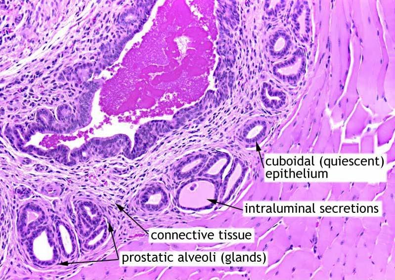
5 Tiền liệt tuyến

6 Tiền liệt tuyến

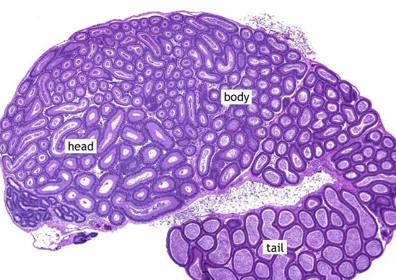
7 Mào tinh hoàn

8 Mào tinh hoàn
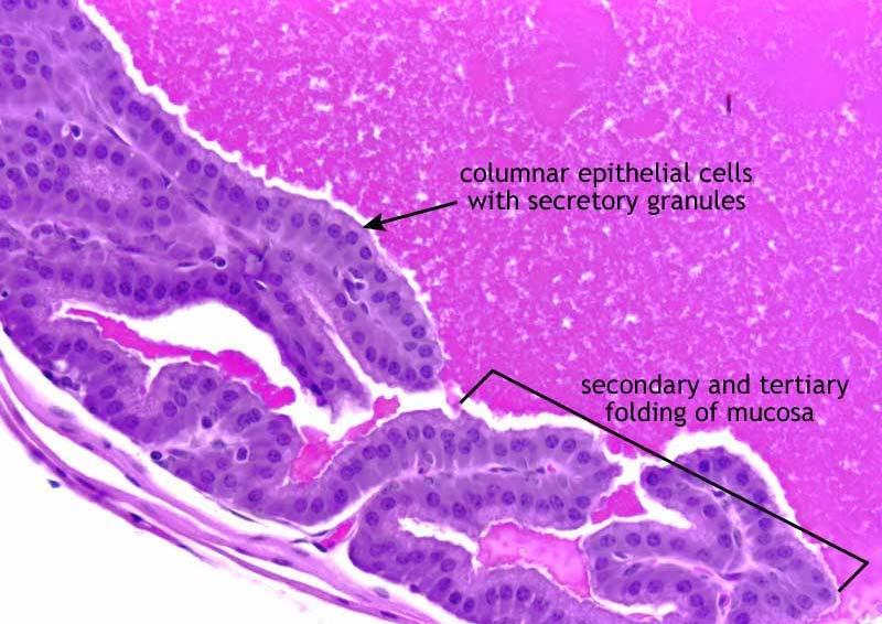
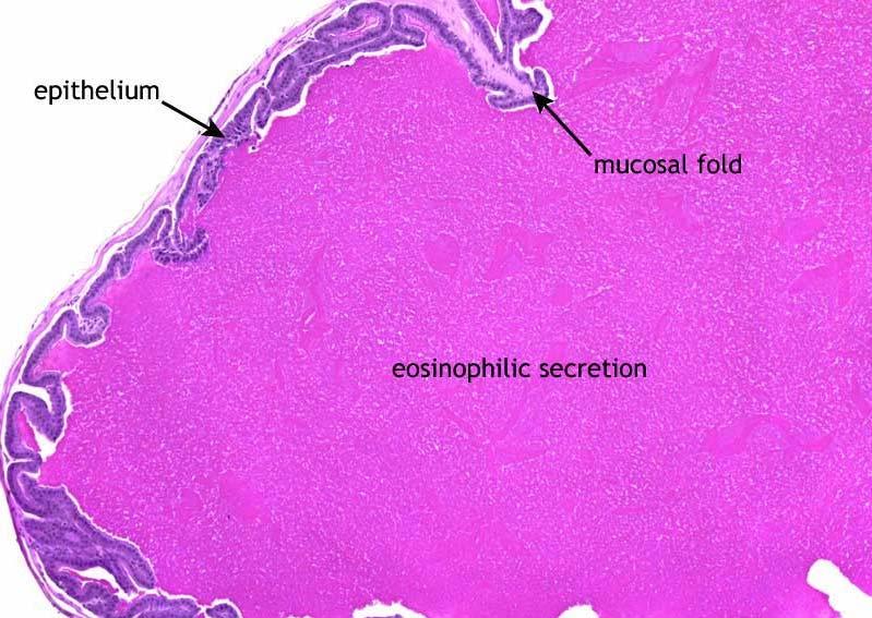
9 Túi tinh
10 Túi tinh
11 Túi tinh
12 Túi tinh

13 So-called cystadenoma of seminal vesicle.

14 Gross appearance of granulomatous orchitis.. The testis is increased in consistency, enlarged, and vaguely nodular.

15 Microscopic appearance of granulomatous orchitis.. The inflammatory infiltrate is centered in the seminiferous tubules.

16 Epidermoid cyst of testis. The lesion is sharply outlined and contains laminated layers of keratin.

17 Microscopic appearance of epidermoid cyst of testis. Keratin squames are laid down by well-differentiated squamous epithelium. There are no skin adnexal structures.

18 Gross appearance of combined tumor of testis. In both instances, the solid homogeneous gray areas correspond to the seminoma,, and the variegated foci with hemorrhage to the nonseminomatous component.

19 Gross appearance of combined tumor of testis. In both instances, the solid homogeneous gray areas correspond to the seminoma,, and the variegated foci with hemorrhage to the nonseminomatous component.

20 Gross appearance of seminoma.. The tumor in A is very small, whereas that in B has replaced most of the testis.

21 Gross appearance of seminoma.. The tumor in A is very small, whereas that in B has replaced most of the testis.

22 Classic seminoma of testis. Compact nests of large tumor cells are separated by fibrous septa heavily infiltrated by lymphocytes.

23 Plastic-embedded section of classical seminoma.. Notice the large nuclei, prominent nucleoli, abundant clear cytoplasm, sharply outlined cell membranes, and inflammatory infiltrate in the stroma.

24 Seminoma associated with marked granulomatous reaction. Only a few tumor cells are visible in this field.

25 Pagetoid extension of seminoma into rete testis. This should not be misinterpreted as a nonseminomatous component.

26 Abundant glycogen present in the cytoplasm of seminoma cells, evidenced by the PAS reaction and removed by diastase digestion.

27 Abundant glycogen present in the cytoplasm of seminoma cells, evidenced by the PAS reaction and removed by diastase digestion.

28 CD117 immunoreactivity in classic seminoma.

29 Seminoma exhibiting pleomorphism and marked hyperchromasia.. Some authors designate this neoplasm as anaplastic seminoma.

30 Seminoma with trophoblastic giant cells. (Hematoxylin and eosin)

31 Seminoma with trophoblastic giant cells. (hcg( immunostain)

32 Gross appearance of spermatocytic seminoma.. A large tumor of myxoid appearance bulges on the cut surface.
, giant cells, and small lymphocyte-like like")
33 Spermatocytic seminoma showing admixture of medium-sized cells (predominating), giant cells, and small lymphocyte-like like cells.

34 Typical chromatin pattern of spermatocytic seminoma.

35 Extensive intratubular growth of spermatocytic seminoma.

36 Sarcomatous focus in spermatocytic seminoma of testis, exhibiting rhabdomyoblastic differentiation. This section is from a lung metastasis.

37 Embryonal carcinoma showing solid nodular cut surface with numerous areas of necrosis and hemorrhage.

38 Embryonal carcinoma. The pattern of growth is diffuse but without the nesting seen in classic seminoma.. The high-power view shows the typical large, irregularly shaped, overlapping nuclei with multiple prominent nucleoli.

39 Embryonal carcinoma. The pattern of growth is diffuse but without the nesting seen in classic seminoma.. The high-power view shows the typical large, irregularly shaped, overlapping nuclei with multiple prominent nucleoli.

40 Gross appearance of mature (adult) teratoma of testis. There are multiple cystic areas, lobules of mature adipose tissue, and shiny solid nodules corresponding to well-differentiated cartilage.

41 Low-power microscopic view of mature teratoma. Large islands of cartilage are seen surrounding well-differentiated glandular structures.

42 Immature teratoma.. Gross appearance.

43 Immature teratoma.. Microscopic appearance. Hypercellular stroma is seen growing in a concentric fashion around glandular formations.

44 Microscopic appearance of intratubular germ cell neoplasia in routinely stained section. A row of atypical germ cells with clear cytoplasm is seen against a thickened basement membrane. No spermatogenesis is occurring in this tubule.

45 PAS stain of intratubular germ cell neoplasia showing abundant intracytoplasmic glycogen in the neoplastic cells.

46 PLAP immunoreactivity in the tumor cells of intratubular germ cell neoplasia.

47 Retroperitoneal metastasis of NSGCT. The mass is entirely composed of mature tissue, whereas the primary tumor had the features of a teratocarcinoma.

48 Entirely necrotic retroperitoneal metastasis of NSGCT following chemotherapy.

49 Lung metastasis of NSGCT following chemotherapy. The mass was entirely composed of mature tissues.

50 Gross appearance of Leydig cell tumor. The tumor, which has replaced most of the testis, has a granular yellowish appearance.

51 Gross appearance of Leydig cell tumor. This tumor, occurring in a child, is solid, well circumscribed, and dark brown.

52 Leydig cell tumor of testis. The neoplasm is characterized by solid growth of polygonal cells with abundant granular acidophilic cytoplasm.

53 Leydig cell tumor of testis. The tumor cells have a cytoplasmic clear quality, reminiscent of that seen in tumors of the adrenal cortex.

54 Leydig cell tumor with myxoid features.

55 So-called testicular tumor of the adrenogenital syndrome. Multiple nodules are present, having an appearance compatible with adrenal cortical origin.

56 Sertoli cell adenoma in a patient with testicular feminization syndrome.

57 Sertoli cell adenoma with sarcomatoid features. The Sertoli cell nature of the tumor is more apparent.

58 Sertoli cell adenoma with sarcomatoid features.

59 Microscopic appearance of sclerosing Sertoli cell tumor.

60 Gross appearance of large cell calcifying Sertoli cell tumor of testis. The tumor is distinctly multinodular.. The dark nodules had a prominent component of Leydig cells.

61 Large cell calcifying Sertoli cell tumor.

62 Adult form of granulosa cell tumor involving testis. Note the occasional longitudinal grooves, the oval to spindle shape of the tumor cells, and the high mitotic activity.

63 Gross appearance of a juvenile granulosa cell tumor involving the testis of an infant.

64 Mixed germ cell stromal tumor of the testis. This lesion is distinct from gonadoblastoma.

65 Gross appearance of malignant lymphoma of large cell type, which completely replaces the testis.

66 Malignant lymphoma of testis. There is diffuse infiltration of the interstitium by neoplastic lymphocytes, which surround and separate atrophic tubules.

67 Cases of large B-cell B lymphoma with pleomorphic features such as that depicted in this photograph can be misdiagnosed as anaplastic or spermatocytic seminoma.

68 Testicular involvement by granulocytic sarcoma. Most of the cells present in the interstitium are myeloid precursors.

69 Intratesticular carcinoid tumor showing the classic insular pattern.

70 Kaposi s s sarcoma of the testis in an HIV-infected individual.

71 Malignant melanoma metastatic to testis. The black color of the tumor is due to massive melanin deposition.

72 Prostatic adenocarcinoma metastatic to testis. This not too rare occurrence is sometimes misdiagnosed as sex cord stromal tumor.

73 Acute and chronic epididymitis.. The inflammation has not spread to the testicle.

74 Granulomatous epididymitis with focal extension into the testis. Some of the granulomas have a necrotic center. No microorganisms were identified on special stains.

75 Spermatic granuloma of epididymis.

76 Typical gross appearance of adenomatoid tumor of epididymis.

77 Low-power appearance of adenomatoid tumor, showing typical conglomerate of cystically dilated spaces.

78 On high power, some of the tubules of adenomatoid tumor are lined by cuboidal cells, whereas others are lined by flattened cells with the appearance of endothelial cells.

79 Strong immunoreactivity for keratin in an adenomatoid tumor.

80 Malignant mesothelioma involving the epididymis.

81 Bilateral papillary cystadenoma of epididymis.

82 Tumor interpreted as a primary papillary adenocarcinoma of the epididymis.

83 Leiomyosarcoma of epididymal region.

84 Vasitis nodosa.. Clumps of spermatozoa are present in the center of the granulomas.

85 Median raphe cyst. This probably results from cystic dilatation of accessory urethral canals or periurethral ducts.

86 Gross appearance of teratocarcinoma.. The solid granular areas correspond to foci of embryonal carcinoma, whereas the pearly nodules correspond to well-differentiated cartilage.

87 Gross appearance of teratocarcinoma.. The solid granular areas correspond to foci of embryonal carcinoma, whereas the pearly nodules correspond to well-differentiated cartilage.

88 Gross appearance of pure choriocarcinoma.. The strikingly hemorrhagic appearance is characteristic of this tumor type.

89 Microscopic appearance of testicular choriocarcinoma. There is close intermingling of cytotrophoblast and syncytiotrophoblast,, which recapitulates that seen in normal chorionic villi.

90 Gross appearance of pure yolk sac tumor in an infant.

91 Schiller Duval body in yolk sac tumor of testis.

92 Pleomorphism and hyaline globules in yolk sac tumor of testis.
Note: The cause of testicular neoplasms remains unknown
 - In the 15- to 34-year-old age group, they are the most common tumors of men. - Tumors of the testis are a heterogeneous group of neoplasms that include: I. Germ cell tumors : 95%; all are malignant.
Gross appearance of nodular hyperplasia in material obtained from suprapubic prostatectomy. Note the multinodular appearance and the admixture of
 Tiền liệt tuyến Tiền liệt tuyến Gross appearance of nodular hyperplasia in material obtained from suprapubic prostatectomy. Note the multinodular appearance and the admixture of solid and microcystic areas.
Male Genital Cancers in the US in Frequency of Types
 Germ Cell Tumors of the Testis Pathology, Immunohistochemistry, and the Often Confusing Appearance of Their Metastases Charles Zaloudek, MD Department of Pathology UCSF Male Genital Cancers in the US in
-The cause of testicular neoplasms remains unknown
 - In the 15- to 34-year-old age group, they are the most common tumors of men. - include: I. Germ cell tumors : (95%); all are malignant. II. Sex cord-stromal tumors: from Sertoli or Leydig cells; usually
Normal endometrium: A, proliferative. B, secretory.
 Normal endometrium: A, proliferative. B, secretory. Nội mạc tử cung Nội mạc tử cung Cyclic changes in endometrium.. Approximate relationship of useful microscopic changes. Arias-Stella reaction in endometrial
Testicular Tumors: What s New, True, Important Cristina Magi-Galluzzi, MD, PhD
 Testicular Tumors: What s New, True, Important Cristina Magi-Galluzzi, MD, PhD Director, Genitourinary Pathology R.J. Tomsich Pathology & Laboratory Medicine Institute Professor of Pathology, Lerner College
Pathology Slides. [Pathology]
![Pathology Slides. [Pathology]](/thumbs/94/120604575.jpg "Pathology Slides. [Pathology]") Pathology Slides MedicoNotes provides real laboratory pathological slides to aid you to differentiate between different pathological structures under microscope. www.mediconotes.com Histology slides example
Pathology Slides MedicoNotes provides real laboratory pathological slides to aid you to differentiate between different pathological structures under microscope. www.mediconotes.com Histology slides example
Protocol for the Examination of Lymphadenectomy Specimens From Patients With Malignant Germ Cell and Sex Cord-Stromal Tumors of the Testis
 Protocol for the Examination of Specimens From Patients With Malignant Germ Cell and Sex Cord-Stromal Tumors of the Testis Version: Testis 4.0.1.1 Protocol Posting Date: February 2019 Accreditation Requirements
Testicular tumors; Ultrasonographic and Pathologic correlation
 Testicular tumors; Ultrasonographic and Pathologic correlation Poster No.: C-0106 Congress: ECR 2014 Type: Educational Exhibit Authors: Y. Kim, S. W. Shin, E. T. Kim, M. Y. Kim ; Kuri City/KR, 1 1 2 1
Testicular Germ Cell Tumors; A Simplistic Approach
 Testicular Germ Cell Tumors; A Simplistic Approach Merce Jorda, MD, PhD, MBA Professor and Vice Chair, Director of Anatomic Pathology Director of Genitourinary Pathology Service Interim Director of Cytopathology
Transitional Cell Papilloma 2-3% Inverted Papilloma Rare
 BLADDER TUMORS Benign Transitional Cell Papilloma 2-3% Inverted Papilloma Rare Malignant Transitional (Urothelial) Carcinoma90% Carcinoma In-Situ (By Itself) 5-10% Squamous Cell Carcinoma 3-7% Adenocarcinoma
2% of all malignancies Male predominance Patients usually more than 60 years old
 Benign Bladder Tumors Transitional Cell Papilloma 2-3% Inverted Papilloma Rare Malignant Transitional (Urothelial) Carcinoma 90% Carcinoma In-Situ (By Itself) 5-10% Squamous Cell Carcinoma 3-7% Adenocarcinoma
Gross appearance of peritoneal cysts. They have a thin, translucent wall and contain a clear fluid.
 Gross appearance of peritoneal cysts. They have a thin, translucent wall and contain a clear fluid. So-called multicystic benign mesothelioma. A, Gross appearance. So-called multicystic benign mesothelioma.
Recently, there has been an increasing incidence among women and younger persons
 Bladder Tumors Benign Transitional Cell Papilloma 2-3% Inverted Papilloma Rare Malignant Transitional (Urothelial) Carcinoma 90% Carcinoma In-Situ (By Itself) 5-10% Squamous Cell Carcinoma 3-7% Adenocarcinoma
International Journal of Research and Review E-ISSN: ; P-ISSN:
 International Journal of Research and Review www.ijrrjournal.com E-ISSN: 2349-9788; P-ISSN: 2454-2237 Case Report Rare Combinations of Testicular Mixed Germ Cell Tumors - 2 Case Reports Dr. Byna Syam Sundara
Leydig cell tumour. Testis: non-germ cell tumours. Testis: sex cord-stromal tumours. Differential diagnosis of Leydig cell tumour TTAGS
 Non-germ cell s of the testis Dr Jonathan H Shanks The Christie NHS Foundation Trust, Manchester, UK Testis: non-germ cell s Sex cord-stromal s Haemolymphoid neoplasms Other neoplasms Tumour-like conditions
Dr Sanjiv Manek Oxford. Oxford Pathology Course 2010 for FRCPath Illustration-Cellular Pathology. Oxford Radcliffe NHS Trust
 Dr Sanjiv Manek Oxford Oxford Pathology Course 2010 for FRCPath Illustration-Cellular Pathology. Oxford Radcliffe NHS Trust Ovarian Endometrial Vulvo-vaginal Cervical Illustration-Cellular Pathology. Oxford
International Journal of Medical and Health Sciences
 International Journal of Medical and Health Sciences Journal Home Page: http://www.ijmhs.net ISSN:2277-4505 Case Report Histomorphological Specrtum of Malignant Germ Cell Tumours: An Overview and Report
Diseases of the breast (1 of 2)
") Diseases of the breast (1 of 2) Introduction A histology introduction Normal ducts and lobules of the breast are lined by two layers of cells a layer of luminal cells overlying a second layer of myoepithelial
Diseases of the breast (1 of 2) Introduction A histology introduction Normal ducts and lobules of the breast are lined by two layers of cells a layer of luminal cells overlying a second layer of myoepithelial
Cardiff MRCS OSCE Courses Testicular Cancer
 Testicular Cancer Scenario: A 40-year-old male presents to the surgical out-patient clinic with a 6-8 week history of a painless lump in his left scrotum. He however complains of a dull ache in the scrotum
Diseases of the penis & testis
 Diseases of the penis & testis Done by : Saef B AL-Abbadi Diseases of penis, Condyloma Acuminatum A benign tumor *Tend to recur but only rarely progress into in situ or invasive cancers read this = genital
CYSTIC TUMORS OF THE KIDNEY JOHN N. EBLE, M.D. CYSTIC NEPHROMA
 Page 1 CYSTIC TUMORS OF THE KIDNEY JOHN N. EBLE, M.D. Department of Pathology & Laboratory Medicine Phone (317) 274-4806 Medical Science A-128 FAX: (317) 278-2018 635 Barnhill Drive jeble @iupui.edu Indianapolis,
Study Into The Patterns of Male Genital Tract Tumors
 ORIGINAL ARTICLE Study Into The Patterns of Male Genital Tract Tumors Archana Gupta, Sunil Gupta, Yudhvir Gupta Abstract Pattern of a particular disease in any population is studied with the idea of getting
the urinary system pathology Dr. Fairoz A Eltorgman
 the urinary system pathology Dr. Fairoz A Eltorgman Tumors of the renal pelvis & kidney Benign tumors of the renal pelvis: Hemangioma Leiomyoma Malignant tumors: Transitional cell carcinoma Squamous cell
Case E1. Female aged 63 years Right Nephrectomy Two separate tumours Section of each tumour
 Case E1 Female aged 63 years Right Nephrectomy Two separate tumours Section of each tumour Tumour 1 Upper pole tumour 28mm macro diameter Circumscribed Friable cut surface Tumour 2 Middle pole Part solid
Testis. Protocol applies to all malignant germ cell and malignant sex cord-stromal tumors of the testis, exclusive of paratesticular malignancies.
 Testis Protocol applies to all malignant germ cell and malignant sex cord-stromal tumors of the testis, exclusive of paratesticular malignancies. Protocol revision date: January 2005 Based on AJCC/UICC
Ultrasound of malignant testicular lesions. Arne Hørlyck Department of Radiology Aarhus University Hospital, Skejby
 Ultrasound of malignant testicular lesions Arne Hørlyck Department of Radiology Aarhus University Hospital, Skejby Testis Ultrasound is fantastic!! Scrotum Extratesticular mass: Benign Intratesticular
number Done by Corrected by Doctor Maha Shomaf
 number 16 Done by Waseem Abo-Obeida Corrected by Zeina Assaf Doctor Maha Shomaf MALIGNANT NEOPLASMS The four fundamental features by which benign and malignant tumors can be distinguished are: 1- differentiation
Pitfalls in thyroid tumor pathology. Prof.Valdi Pešutić-Pisac MD, PhD
 Pitfalls in thyroid tumor pathology Prof.Valdi Pešutić-Pisac MD, PhD Too many or... Tumour herniation through a torn capsule simulating capsular invasion fibrous capsule with a sharp discontinuity, suggestive
Salivary Glands 3/7/2017
 Salivary Glands 3/7/2017 Goals and objectives Focus on the entities unique to H&N Common board type facts Information for your future practice Salivary Glands Salivary Glands Major gland. Paratid. Submandibular.
Lesions Mimicking Adenoid Cystic Carcinoma. Diagnostic Problems in Salivary Gland Pathology An Update 5/29/2009
 Diagnostic Problems in Salivary Gland Pathology An Update Lesions Mimicking Adenoid Cystic Carcinoma Stacey E. Mills, M.D. W.S. Royster Professor of Pathology Director of Surgical and Cytopathology University
Benign and malignant epithelial lesions: Seborrheic keratosis: A common benign pigmented epidermal tumor occur in middle-aged or older persons more
 Benign and malignant epithelial lesions: Seborrheic keratosis: A common benign pigmented epidermal tumor occur in middle-aged or older persons more common on the trunk; but extremities, head and neck are
CONTINUING EDUCATION IN TOXICOLOGIC PATHOLOGY REPRODUCTIVE SYSTEM
 CONTINUING EDUCATION IN TOXICOLOGIC PATHOLOGY REPRODUCTIVE SYSTEM ORGANIZED BY SOCIETY FOR TOXICOLOGIC PATHOLOGY IN INDIA (STPI) OCTOBER 29-31, 2010 The Atria Hotel, # 1, Palace Road, Bangalore - 560 001
Extratesticular Extension of Germ Cell Tumors Preferentially Occurs at the Hilum
 Anatomic Pathology / EXTRATESTICULAR EXTENSION OF GERM CELL TUMORS Extratesticular Extension of Germ Cell Tumors Preferentially Occurs at the Hilum Sarah M. Dry, MD, and Andrew A. Renshaw, MD Key Words:
Outline 11/2/2017. Pancreatic EUS-FNA general aspects. Cytomorphologic features of solid neoplasms/lesions of the pancreas
 ENDOSCOPIC ULTRASOUND GUIDED-FINE NEEDLE ASPIRATION CYTOLOGY OF PANCREAS Khalid Amin M.D. Assistant Professor Department of Laboratory Medicine and Pathology University of Minnesota Outline Pancreatic
SESSION 1: GENERAL (BASIC) PATHOLOGY CONCEPTS Thursday, October 16, :30am - 11:30am FACULTY COPY
 PATHOLOGY CONCEPTS Thursday, October 16, :30am - 11:30am FACULTY COPY") SESSION 1: GENERAL (BASIC) PATHOLOGY CONCEPTS Thursday, October 16, 2008 9:30am - 11:30am FACULTY COPY GOAL: Describe the basic morphologic (structural) changes which occur in various pathologic conditions.
SESSION 1: GENERAL (BASIC) PATHOLOGY CONCEPTS Thursday, October 16, 2008 9:30am - 11:30am FACULTY COPY GOAL: Describe the basic morphologic (structural) changes which occur in various pathologic conditions.
XIII. Tumours of the liver and biliary system
 XIII. Tumours of the liver and biliary system V. PONOMARKOV 1 & L. J. MACKEY 2 In this histological classification of liver and gall bladder tumours the tumour types largely correspond to those found in
Lách
 Lách Lách Lách Lách Splenogonadal fusion. Splenic tissue is attached to testicular tissue. Pseudocyst (false or secondary cyst). A, Outer aspect. Pseudocyst (false or secondary cyst). B, Inner surface.
The Effects of Chemotherapy on Metastatic Testicular Germ Cell Tumors
 The Open Pathology Journal, 2009, 3, 45-52 45 Open Access The Effects of Chemotherapy on Metastatic Testicular Germ Cell Tumors Ivan Damjanov *,1 and Ondrej Hes 2 1 Department of Pathology and Laboratory
Male Reproductive System
 Male Reproductive System Constitution of male reproductive system Genital gland ----testis Genital ducts epididymis / ductus deferens / urinary duct Accessory sex glands Penis prostate gland Seminal vesicle
Testicular Tumors Including Secondary and Unusual Tumors of the Testis
 Testicular Tumors Including Secondary and Unusual Tumors of the Testis Milton W. Datta Partner, Hospital Pathology Associates University of Minnesota Minneapolis, MN Topics Review Features of Germ Cell
Spectrum of Preneoplastic and Neoplastic Cystic Lesions of the Kidney in Adult. by dr. Banan Burhan Mohammed Lecturer in Pathology Department
 Spectrum of Preneoplastic and Neoplastic Cystic Lesions of the Kidney in Adult by dr. Banan Burhan Mohammed Lecturer in Pathology Department Various hereditary, acquired, and neoplastic conditions can
Abid Irshad, MD Director Breast Imaging. Medical University of South Carolina Charleston
 Abid Irshad, MD Director Breast Imaging Medical University of South Carolina Charleston Cases Financial disclosure: I or my family have no financial interest related to the material discussed in this presentation
Pathology of Ovarian Tumours. Dr. Jyothi Ranganathan MD ( Path) AFMC Pune PDCC (Cytopathology) PGI Chandigarh
 AFMC Pune PDCC (Cytopathology) PGI Chandigarh") Pathology of Ovarian Tumours Dr. Jyothi Ranganathan MD ( Path) AFMC Pune PDCC (Cytopathology) PGI Chandigarh Outline Incidence Risk factors Classification Pathology of tumours Tumour markers Prevention
Pathology of Ovarian Tumours Dr. Jyothi Ranganathan MD ( Path) AFMC Pune PDCC (Cytopathology) PGI Chandigarh Outline Incidence Risk factors Classification Pathology of tumours Tumour markers Prevention
Disorders of Cell Growth & Neoplasia. Histopathology Lab
 Disorders of Cell Growth & Neoplasia Histopathology Lab Paul Hanna April 2010 Case #84 Clinical History: 5 yr-old, West Highland White terrier. skin mass from axillary region. has been present for the
Title. Author(s)KANAGAWA, Hiroshi; ISHIKAWA, Tsune; KAWATA, Keiichir. CitationJapanese Journal of Veterinary Research, 13(1): Issue Date
KANAGAWA, Hiroshi; ISHIKAWA, Tsune; KAWATA, Keiichir. CitationJapanese Journal of Veterinary Research, 13(1): Issue Date") Title A CASE OF CANINE TESTICULAR SERTOLI CELL TUMOR Author(s)KANAGAWA, Hiroshi; ISHIKAWA, Tsune; KAWATA, Keiichir CitationJapanese Journal of Veterinary Research, 13(1): 11-1 Issue Date 1965-03 DOI 10.14943/jjvr.13.1.11
Title A CASE OF CANINE TESTICULAR SERTOLI CELL TUMOR Author(s)KANAGAWA, Hiroshi; ISHIKAWA, Tsune; KAWATA, Keiichir CitationJapanese Journal of Veterinary Research, 13(1): 11-1 Issue Date 1965-03 DOI 10.14943/jjvr.13.1.11
Normal thyroid tissue
 Thyroid Pathology Overview Normal thyroid tissue Normal thyroid tissue with follicles filled with colloid. Thyroid cells form follicles, spheres of epithelial cells (always single layered in health, usually
A neoplasm is defined as "an abnormal tissue proliferation, which exceeds that of adjacent normal tissue. This proliferation continues even after
 NEOPLASIA Neoplasia is a very important topic in pathology because neoplasms are both common and serious diseases. A neoplasm literally means a new growth, and this term is used interchangeably with a
Neoplasia 2018 Lecture 2. Dr Heyam Awad MD, FRCPath
 Neoplasia 2018 Lecture 2 Dr Heyam Awad MD, FRCPath ILOS 1. List the differences between benign and malignant tumors. 2. Recognize the histological features of malignancy. 3. Define dysplasia and understand
CELL AND TISSUE INJURY COURSE-II PATHOLOGY LABORATORY. PATHOLOGY of MASS LESIONS and TISSUE DEFECTS -MACROSCOPY Assoc. Professor Rengin Ahıskalı
 CELL AND TISSUE INJURY COURSE-II PATHOLOGY LABORATORY PATHOLOGY of MASS LESIONS and TISSUE DEFECTS -MACROSCOPY Assoc. Professor Rengin Ahıskalı M1 - RENAL TUBERCULOSIS cavitary areas caseous necrosis fibrous
Morphologic Aspects Aspects
 Testicular Testicular Germ Germ Cell Cell Neoplasia: Neoplasia: Molecular Molecular Pathways Pathways & Challenging Morphologic Aspects Aspects George George J. J. Netto, Netto, M.D. M.D. Johns Johns Hopkins
Histomorphological spectrum of tumor and tumor like lesions of testis and paratesticular structures A cross sectional study
 Original Research Article DOI: 10.5958/2394-6792.2016.00098.3 Histomorphological spectrum of tumor and tumor like lesions of testis and paratesticular structures A cross sectional study Sanjay M 1,*, Sushma
Testicular Cancer: radiopathological correlation of testicular tumors in adulthood population. A review of 32 cases.
 Testicular Cancer: radiopathological correlation of testicular tumors in adulthood population. A review of 32 cases. Poster No.: C-1085 Congress: ECR 2015 Type: Educational Exhibit Authors: R. Miranda;
Papillary adenocarcinoma of the rete testis with adjacent hyperplasia: a case report
 CASE REPORT Papillary adenocarcinoma of the rete testis with adjacent hyperplasia: a case report Carolina Polanco 1, Cooley G. Pantazis 2, Rolando Prieto 2, Kenneth A. Iczkowski 1 1. Medical College of
2 to 3% of All New Visceral Cancers Peak Incidence is 6th Decade M:F = 2:1 Grossly is a Bright Yellow, Necrotic Mass with a Pseudocapsule
 GENITOURINARY PATHOLOGY Kathleen M. O Toole, M.D. Renal Cell Carcinoma 2 to 3% of All New Visceral Cancers Peak Incidence is 6th Decade M:F = 2:1 Grossly is a Bright Yellow Necrotic Mass Grossly is a Bright
CHAPTER NINE PATHOLOGY OF THE MALE GENITAL SYSTEM
 CHAPTER NINE PATHOLOGY OF THE MALE GENITAL SYSTEM THE PENIS Malformations of the Penis Hypospadias is the most common malformation (1 in 250 live male births). It refers to the abnormal location of the
Case year female. Routine Pap smear
 Case 1 57 year female Routine Pap smear Diagnosis? 1. Atypical glandular cells of unknown significance (AGUS) 2. Endocervical AIS 3. Endocervical adenocarcinoma 4. Endometrial adenocarcinoma 5. Adenocarcinoma
Pathology of the female genital tract
 Pathology of the female genital tract Common illnesses of the female genital tract Before menarche Developmental anomalies Tumors (ovarial teratoma) Amenorrhea Fertile years PCOS, ovarian cysts Endometriosis
ICD-O Morphology code. R=Rare Tier Tumour ICD-O Topography code C30.0, C31
 R=Rare Tier Tumour ICD-O Topography code ICD-O Morphology code EPITHELIAL TUMOURS OF NASAL CAVITY AND SINUSES R 2 Squamous cell carcinoma with variants of nasal cavity and sinuses C30.0, C3 C30.0, C3 8000,
MALE REPRODUCTIVE SYSTEM
 1 MALE REPRODUCTIVE SYSTEM SCPA 602 Anatomical Basis for Pathological Study Updated: 20.09.2018 Lect. Nisamanee Charoenchon, PhD nisamanee.cha@mahidol.ac.th Department of Pathobiology, Mahidol University
Updates in Urologic Pathology WHO Made Those Changes?! Peyman Tavassoli Pathology Department BC Cancer Agency
 Updates in Urologic Pathology WHO Made Those Changes?! Peyman Tavassoli Pathology Department BC Cancer Agency World Health Organization Available in Feb 2016 Frame work for reporting Major contributing
SUPPLEMENTARY FIG. S2. Teratoma. Portion of a teratoma composed of neural tissue. The large cells in the central part correspond to ganglion cells.
 Supplementary Data SUPPLEMENTARY FIG. S1. Teratoma. The tumor is composed predominantly of keratinizing squamous epithelium (Sq), which forms cysts filled with keratin (arrows). The tumor also contains
Mediastinal Germ Cell Tumors
 Mediastinal Germ Cell Tumors Anja C. Roden, M.D. Department of Laboratory Medicine and Pathology, Mayo Clinic, Rochester, MN, USA 2018 MFMER slide-1 Disclosure I have no relevant financial relationships
CINtec p16 INK4a Staining Atlas
 CINtec p16 INK4a Staining Atlas Rating Rating Positive The rating positive will be assigned if the p16 INK4a -stained slide shows a continuous staining of cells of the basal and parabasal cell layers of
Synonyms. Nephrogenic metaplasia Mesonephric adenoma
 Nephrogenic Adenoma Synonyms Nephrogenic metaplasia Mesonephric adenoma Definition Benign epithelial lesion of urinary tract with tubular, glandular, papillary growth pattern Most frequently in the urinary
Special slide seminar
 Special slide seminar Tomáš Rozkoš The Fingerland Department of Pathology Charles University Medical Faculty and Faculty Hospital in Hradec Králové Czech Republic Case history, 33 years old resistance
Case Report Primary Malignancy in a Supernumerary Testicle Presenting as a Large Pelvic Mass
 Hindawi Volume 2017, Article ID 4529853, 4 pages https://doi.org/10.1155/2017/4529853 Case Report Primary Malignancy in a Supernumerary Testicle Presenting as a Large Pelvic Mass Justin Noroozian, 1 Daniel
ADVERSE EFFECTS OF VASECTOMY: SPERM GRANULOMA OF EPIDIDYMIDES V. P. DIXIT
 ADVERSE EFFECTS OF VASECTOMY: SPERM GRANULOMA OF EPIDIDYMIDES V. P. DIXIT Reproduct ion Physiology Section, Department of Zoology, University of Rajasthan, Jaipur-302004 Summary: Rats and mice were vasectomized
Cerebral Parenchymal Lesions: I. Metastatic Neoplasms
 Chapter 4 Cerebral Parenchymal Lesions: I. Metastatic Neoplasms After one has reasonably ruled out the possibility of a nonneoplastic diagnosis (see Chap. 3), one is left with considering a diagnosis of
Kidney Case 1 SURGICAL PATHOLOGY REPORT
 Kidney Case 1 Surgical Pathology Report February 9, 2007 Clinical History: This 45 year old woman was found to have a left renal mass. CT urography with reconstruction revealed a 2 cm medial mass which
3 cell types in the normal ovary
 Ovarian tumors 3 cell types in the normal ovary Surface (coelomic epithelium) the origin of the great majority of ovarian tumors 90% of malignant ovarian tumors Totipotent germ cells Sex cord-stromal cells
N-cadherin Expression in Testicular Germ Cell and Gonadal Stromal Tumors
 381 Ivyspring International Publisher Research Paper Journal of Cancer 2012; 3: 381-389. doi: 10.7150/jca.5017 N-cadherin Expression in Testicular Germ Cell and Gonadal Stromal Tumors Daniel J. Heidenberg
Pancreatitis: A Potential Pitfall in Endoscopic Ultrasound Guided Pancreatic FNA
 Pancreatitis: A Potential Pitfall in Endoscopic Ultrasound Guided Pancreatic FNA Jack Yang, MD Department of Pathology, Medical University of South Carolina Objectives Understand the indication of EUS
Disclosure of Relevant Financial Relationships
 Evening Specialty Conference - Genitourinary Pathology Case 2 Disclosure of Relevant Financial Relationships Sean R Williamson, MD Henry Ford Health System, Detroit, MI @Williamson_SR USCAP requires that
Neoplasms of the Canine, Feline and Lemur Liver:
 Neoplasms of the Canine, Feline and Lemur Liver: Classification and Prognosis Annual Seminar of the French Society of Veterinary Pathology John M. Cullen VMD PhD DACVP North Carolina State University Primary
Review of the AP Part II Practical Examination. Dr David Clift Co Chief Examiner
 Review of the AP Part II Practical Examination Dr David Clift Co Chief Examiner General Remarks The part II practical examination involved 15 cases which were presented with sufficient clinical data to
PLEOMORPHIC ADENOMA ( BENIGN MIXED TUMOR )
") ( BENIGN MIXED TUMOR ) Grossly, the tumor is freely movable, solid, sometimes lobulated and occasionally cystic. If recurrent, multinodular masses are common. Histologically, within a fibrous capsule,
( BENIGN MIXED TUMOR ) Grossly, the tumor is freely movable, solid, sometimes lobulated and occasionally cystic. If recurrent, multinodular masses are common. Histologically, within a fibrous capsule,
Testicular Malignancies /8/15
 Collecting Cancer Data: Testis 2014-2015 NAACCR Webinar Series January 8, 2015 Q&A Please submit all questions concerning webinar content through the Q&A panel. Reminder: If you have participants watching
EMBRYONAL NEPHROMA IN THE CHICKEN: REPORT OF TWO CASES
 EMBRYONAL NEPHROMA IN THE CHICKEN: REPORT OF TWO CASES FRANK D. McKENNEY, V.M.D. (Di1!ision of Experimental Surgery and Pathology, The Mayo Foundation, Rochester, Minnesota) Few data have been collected
MRI IN THE CHARACTERIZATION OF SEMINOMATOUS AND NONSEMINOMATOUS GERM CELL TUMORS OF THE TESTIS
 MRI IN THE CHARACTERIZATION OF SEMINOMATOUS AND NONSEMINOMATOUS GERM CELL TUMORS OF THE TESTIS Ambesh Deshar *, Gyanendra KC and Zhang Lopsang *Department of Medical Imaging and Nuclear Medicine, First
3 cell types in the normal ovary
 Ovarian tumors 3 cell types in the normal ovary Surface (coelomic epithelium) the origin of the great majority of ovarian tumors (neoplasms) 90% of malignant ovarian tumors Totipotent germ cells Sex cord-stromal
FNA of Thyroid. Toward a Uniform Terminology With Management Guidelines. NCI NCI Thyroid FNA State of the Science Conference
 FNA of Thyroid NCI NCI Thyroid FNA State of the Science Conference Toward a Uniform Terminology With Management Guidelines Thyroid Thyroid FNA Cytomorphology NCI Thyroid FNA State of the Science Conference
Macro- and microacinar proliferations of the prostate
 Macro- and microacinar proliferations of the prostate (with emphasis on cancer mimics) Rodolfo Montironi, MD (IT), FRCPath (UK), IFCAP (USA) Polytechnic University of Marche Region (Ancona) School of Medicine,
Differential Diagnosis of Oral Masses. Palatal Lesions
 Differential Diagnosis of Oral Masses Palatal Lesions Palatal Masses Periapical Abscess Torus Palatinus Mucocele Lymphoid Hyperplasia Adenomatous Hyperplasia Benign Salivary Neoplasms Malignant Salivary
MALE REPRODUCTIVE SYSTEM
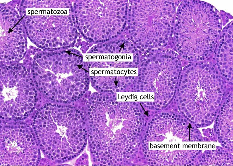
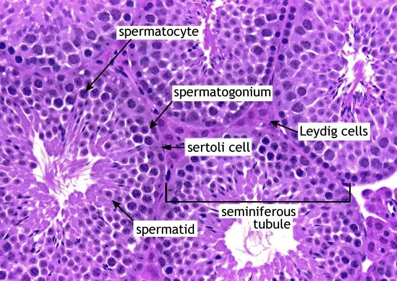
 MALE REPRODUCTIVE SYSTEM The male reproductive system consists of primary sex organs (testes) and secondary or accessory sex organs. The secondary organs consist of a series of genital ducts (ductules
Mammary Nodular Hyperplasia in Intact R hesus Monkeys
 Vet. Path. 10: 130-134 (1973) Mammary Nodular Hyperplasia in Intact R hesus Monkeys L. W NELSON and L. D. SHOTT Department of Pathology and Toxicology, Mead Johnson Research Center, Evansville, Ind., and
(Iteceived for publication December 3, 1915)
") TRANSPLANTABLE SARCOMATA OF THE RAT LIVER ARISING IN THE WALLS OF PARASITIC CYSTS G. L. ROHDENBURG, M.D., AND F. D. BULLOCK, M.D. From Colurnbia University, George Crocker Special Re-search Fund, F. C.
TRANSPLANTABLE SARCOMATA OF THE RAT LIVER ARISING IN THE WALLS OF PARASITIC CYSTS G. L. ROHDENBURG, M.D., AND F. D. BULLOCK, M.D. From Colurnbia University, George Crocker Special Re-search Fund, F. C.
Pathology of the lower urinary tract and male genital system
 Pathology of the lower urinary tract and male genital system Neoplasms of the lower urinary tract Incidence: Urinary bladder > upper urinary tract; male:female=3:1 Symptoms: painless hematuria, hydronephrosis
TitleSolid testicular mass in a 44-year-
 TitleSolid testicular mass in a 44-year- Murakami, Kaoru; Kobayashi, Takashi Author(s) Okubo, Kazutoshi; Kamba, Tomomi; Yo Osamu Citation Urology (2013), 82(6): 1204-1206 Issue Date 2013-12 URL http://hdl.handle.net/2433/179779
Male Reproductive System
 Male Reproductive System organs that function in: gamete and hormone production not all in abdominal cavity paired testicles = controlled by LH & FSH duct systems accessory glands Testis: Gross Histology
2015 Descriptive Vet Path Course. Histo Exam #3 KEY
 2015 Descriptive Vet Path Course Histo Exam #3 KEY Test 3, Slide 1 Tissue from a guinea pig. MORPHOLOGIC DIAGNOSIS: Heart: Multifocally and randomly (1 pt), within the left and right ventricular myocardium
Exercise. Discharge Summary
 Exercise Discharge Summary A 32-year-old Brazilian male presented with a 6 month history of right-sided scrotal swelling. Backache was present for 2 months and a history of right epididymitis was present
Diagnostically Challenging Cases in Gynecologic Pathology
 Diagnostically Challenging Cases in Gynecologic Pathology Eric C. Huang, M.D., Ph.D. Department of Pathology and Laboratory Medicine University of California, Davis Medical Center Case 1 Presentation 38
GUIDELINES ON TESTICULAR CANCER
 38 (Text updated March 2005) P. Albers (chairman), W. Albrecht, F. Algaba, C. Bokemeyer, G. Cohn-Cedermark, A. Horwich, O. Klepp, M.P. Laguna, G. Pizzocaro Introduction Compared with other types of cancer
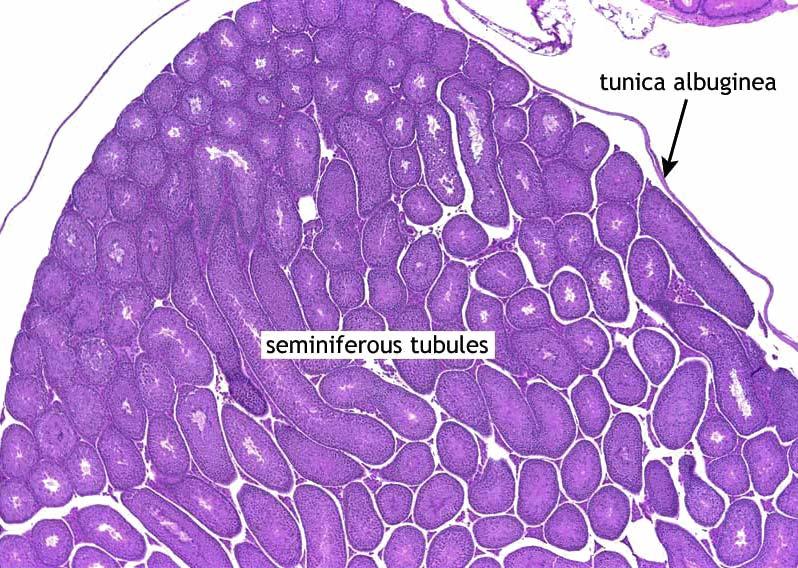
川北医学院讲稿. Under low power note the testis is enclosed by a strong fibrous. layer of serous epithelium. These fibrous tissue
 川北医学院讲稿 Experiment 5: Male and Female Reproductive System Hello, everybody, class is begin,keep quiet, please. And this is the last experimental class. Today we will learn 5 slices and review all structures
NEOPLASIA-I CANCER. Nam Deuk Kim, Ph.D.
 NEOPLASIA-I CANCER Nam Deuk Kim, Ph.D. 1 2 Tumor in the hieroglyphics of the Edwin Smith papyrus (1,600 B.C., Breasted s translation 1930) 3 War on Cancer (National Cancer Act, 1971) 4 Cancer Acts in Korea
!! 2 to 3% of All New Visceral Cancers.!! Peak Incidence is 6th Decade!! M:F = 2:1
 !! Kathleen M. O Toole, M.D.!! 2 to 3% of All New Visceral Cancers!! Peak Incidence is 6th Decade!! M:F = 2:1!! Grossly is a Bright Yellow, Necrotic Mass with a Pseudocapsule 1 !!Conventional RCC! Clear