Thoracic Muscles Origin Insertion Action Innervation
|
|
|
- Doris Greene
- 6 years ago
- Views:
Transcription
1 MUSCLES OF THE THORAX, BACK & ABDOMEN
2 Muscles of the Thorax

3
4 Thoracic Muscles Origin Insertion Action Innervation M. pectoralis major pars clavicularis clavicula (medial ½ ) M. pectoralis major pars sternocostalis manubrium sterni et cartilagines costae (2nd-7th) crista tuberculi majoris adduction, internal rotation, arm flexion; auxiliary inspiratory m. M. pectoralis major pars abdominalis vagina musculi recti abdominis M. pectoralis minor 3rd - 5th rib processus coracoideus scapulae pulls the clavicle; auxiliary inspiration m Plexus brachialis M. subclavius first rib clavicula (inferior surface) pulls clavicule indirectly the shoulder distoventrally; auxiliary inspiration m. M. serratus anterior cranial 9 ribs scapula (margo medialis et angulus inferior) pulls the clavicle from the backbone; pulls inferior angle laterally rotates scapula; auxiliary respirat. m.
5 Thoracic Muscles Origin Insertion Action Innervation Mm. intercostales externi inferior margin of ribs - from the costal tubercle to the beginning of rib cartilage superior margin of ribs immediately below elevation of lower ribs, thorax expansion inspiratory m. Mm. intercostales interni superior margin of ribs - costal angle to sternum inferior margin of ribs immediately above adduction of cranial ribs to caudal ribs expiratory m. Nn. intercostales M. transversus thoracis internal surface of xiphoid process and body of sternum cartilagines costae verae expiratory muscle Diaphragma sternal part inner surface of xiphoid process Diaphragma Diaphragma costal part lumbar part, medial crus inner surface of cartilage of ribs 7-12 ligamentum longitudinale anterius (vertebrae lumbales) central tendon main inspiratory muscle; abdominal press Plexus cervicalis Diaphragma lumbar part, lateral crus ligaments jump over the psoas and quadratus muscles


6


7

8
9 Muscles of the Back
10 Superficial muscles functionally belong to the upper limb Intermediate muscles active during respiration Deep muscles true muscles of the back originates at lower levels ascend in staggered fashion and insert into higher levels extends the vertebral column and head

11
12 Spinocostal Spinohumeral Heterochtonnous muscles origin insertion action innervation M. trapezius protuberantia occipitalis externa, septum nuchae, processus spinosi C7 and all thoracic vertebrae lateral ⅓ of clavicle, acromion and spina scapulae adduction of shoulder, upper fibres elevates the scapula; the lower part pulls the scapula n. accessorius, plexus cervicalis M. latissimus dorsi processus spinosi of caudal thoracic vertebrae, lumbar vertebrae, sacrum, crista iliaca and caudal ribs crista tuberculi minoris adduction, extension, medially rotation n. thoracodorsalis M. levator scapulae M. rhomboideus minor et major processus transversi of cranial cervical vertebrae processus spinosus of caudal cerical and cranial thoracic vertebrae angulus superior scapulae margo medialis scapulae elevates scapula, rotates scapula medially pulls the scapula medially and cranially n. dorsalis scapulae M. serratus posterior superior M. serratus posterior inferior processus spinosus of caudal cervical and cranial thoracic vertebrae processus spinosus of caudal thoracic and cranial lumbar vertebrae cranial ribs caudal ribs elevates the ribs auxilliary inspiratory muscle auxiliary expiratory muscle nn. intercostales


13
14 Spinospinal Sacrospinal Autochtonnous muscles Origin Insertion Action Innervation M. longissimus dorsi et cervicis medial part M. longissimus capitis fill the space between processus spinosus et transversus cranial base M. iliocostalis lateral part bilateral - dorsiflexion, unilateral - homolateral lateroflexion Mm. spinales thoracis et cervicis DR of spinal nerves Mm. interspinales cervicis are stretched between processus spinosus of the thoracic and cervical vertebrae unilateral - lateroflexion, bilateral - dorsiflexion Mm. intertransversarii posteriores cervicis
15 Spinotransversal Transversospinal Autochtonnous muscles Origin Insertion Action Innervation M. semispinalis thoracis et cervicis M. semispinalis capitis are stretched from transversal to the spinous processes; is located along the whole spine with insertion on the cranial base jump over 4-5 vertebrae bilateral - dorsiflexion, unilateral - homolateral lateroflexion and contralateral rotation Mm. multifidi jump over 1-3 vertebrae DR of spinal nerves M. splenius capitis M. splenius cervicis runs from processus spinosus to processus transversus cranial base cervical vertebrae bilateral - dorsiflexion, unilateral - lateroflexion and homolateral rotation

16


17
18 Mm. nuchae profundi Origin Insertion Action Innervation M. rectus capitis posterior minor tuberculum posterius atlantis linea nuchae inferior unilateral - homolateral lateroflexion, bilateral - dorsiflexion M. rectus capitis posterior major processus spinosus axis linea nuchae inferior bilateral - dorsiflexion, unilateral - homolateral lateroflexion and rotation suboccipital nerve M. obliquus capitis superior processus transversi atlantis linea nuchae inferior bilateral - dorsiflexion, unilateral - homolateral lateroflexion M. obliquus capitis inferior processus spinosu axis processus transversi atlantis homolateral rotation

19
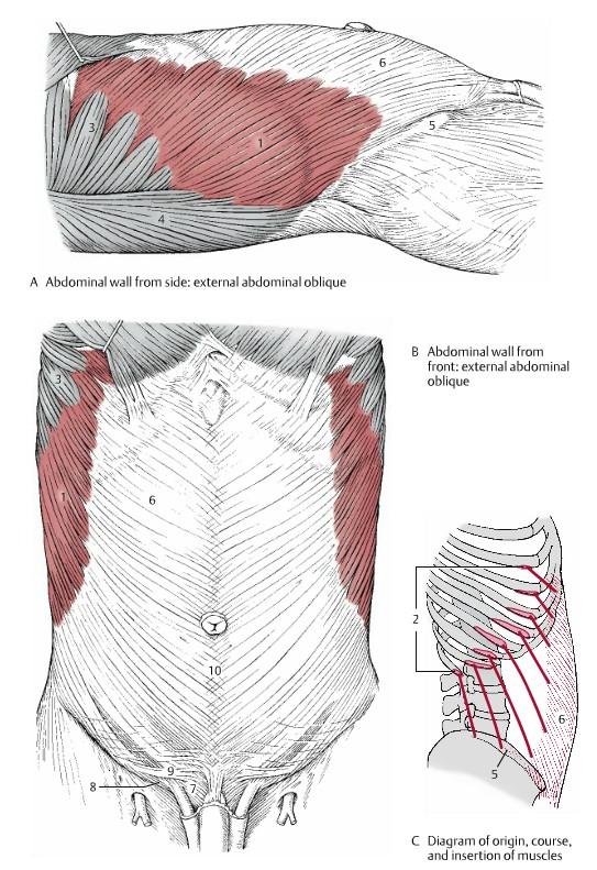
20 Muscles of the Abdomen
21 Abdominal muscles Origin Insertion Action Innervation M. rectus abdominis xiphoid process and costal cartilages 5-7 tuberculum pubicum trunk anteflexion; increase abdominal press M. obliquus externus abdominis lower ribs crista iliaca, ligamentum inquinale, vagina musculi recti abdominis abdominal press; bilateral - anteflexion, unilateral - contralateral rotation M. obliquus internus abdominis fascia thoracolumbalis, crista iliaca, ligamentum inquinale lower ribs et vagina musculi recti abdominis auxiliary expiration m, abdominal press; bilateral - dorsiflexion, unilateral - homolateral rotation Nn. Intercostales M. transversus abdominis fascia thoracolumbalis, crista iliaca, lower ribs vagina musculi recti abdominis auxiliary respiratory m, abdominal press; unilateral - homolateral lateroflexion M. quadratus lumborum The muscle is attached between last rib, iliac crest and lumbar vertebrae unilateral - homolateral lateroflexion, bilateral - dorsiflexion of backbone

22

23


24
25 Anterior abdominal wall Muscles of abdominal wall are continue anteriorly and medially as strong sheet-like aponeuroses Between the midclavicular line and the midline form the rectus sheath enclosing the rectus abdominis muscle In midline aponeuroses interweave with their fellows of the opposite side forming a midline raphe = linea alba (extends from the xiphoid process to the pubic symphysis) The deep surface of the transversus abdominis muscle and its aponeurosis is transversalis fascia
26 Anterior abdominal wall Approximately one third of the distance from the umbilicus to the pubic crest, the aponeuroses of the three flat muscles pass anterior to the rectus abdominis to form the anterior layer of the rectus sheath Leaving only the relatively thin transversalis fascia to cover the rectus abdominis posteriorly Arcuate line demarcates the transition between the aponeurotic posterior wall of the sheath covering the superior three quarters of the rectus and the transversalis fascia covering the inferior quarter

27
28 Inguinal canal A passage through the lower anterior abdominal wall situated just above the medial half of the inguinal ligament Extends in a downward and medial direction from the deep inguinal ring to the superficial inguinal ring Deep inguinal ring Opening of the evagination of the transversalis fascia at a point midway between the anterior superior iliac spine and the pubic symphysis continues into the canal forming the innermost covering of the structures traversing the canal Superficial inguinal ring Triangular opening in the aponeurosis of the external oblique muscle

29

30
Muscles of the Upper Limb that are dissected in the Back Region Muscle Origin Insertion Action Innervation Artery Notes
 Muscles of Upper Limb that are dissected in Back Region Muscle Origin Insertion Action Innervation Artery Notes floor of thoraco thoraco inserting spines from intertubercular arm nerve (C7,8) a. tendon
Thoracolumbar Anatomy Eric Shamus Catherine Patla Objectives
 1 2 Thoracolumbar Anatomy Eric Shamus Catherine Patla Objectives List the muscular and ligamentous attachments of the thoracic and lumbar spine Describe how the muscles affect the spine and upper extremity
Lab Activity 11: Group I
 Lab Activity 11: Group I Muscles Martini Chapter 11 Portland Community College BI 231 Origin and Insertion Origin: The place where the fixed end attaches to a bone, cartilage, or connective tissue. Insertion:
1TRUNK: BODY WALL AND SPINE
 TRUNK: BODY WALL AND SPINE SURFACE ANATOMY SKELETON JOINTS & LIGAMENTS MUSCLES VASCULATURE NERVES SPINAL CORD & VERTEBRAL CANAL ANTERIOR BODY WALL & MAMMARY GLAND LATERAL BODY WALL INGUINAL REGION SUPERFICIAL
Clarification of Terms
 Clarification of Terms The Spine, Spinal Column, and Vertebral Column are synonymous terms referring to the bony components housing the spinal cord Spinal Cord = made of nervous tissue Facet = a small,
Clarification of Terms
 Clarification of Terms The Spine, Spinal Column, and Vertebral Column are synonymous terms referring to the bony components housing the spinal cord Spinal Cord = made of nervous tissue Facet = a small,
Clarification of Terms
 Clarification of Terms The Spine, Spinal Column, and Vertebral Column are synonymous terms referring to the bony components housing the spinal cord Spinal Cord = made of nervous tissue Facet = a small,
The Back. Anatomy RHS 241 Lecture 9 Dr. Einas Al-Eisa
 The Back Anatomy RHS 241 Lecture 9 Dr. Einas Al-Eisa The spine has to meet 2 functions Strength Mobility Stability of the vertebral column is provided by: Deep intrinsic muscles of the back Ligaments
Main Menu. Trunk and Spinal Column click here. The Power is in Your Hands
 1 The Trunk and Spinal Column click here Main Menu K.9 http://www.handsonlineeducation.com/classes/k9/k9entry.htm[3/27/18, 2:00:55 PM] The Trunk and Spinal Column Vertebral column complex 24 intricate
Sports Medicine Part II : ANATOMY OF THE SPINE, ABDOMEN AND SHOULDER COMPLEX
 Sports Medicine 25 1.1 Part II : ANATOMY OF THE SPINE, ABDOMEN AND SHOULDER COMPLEX c.w.p. Wagner High School, Sports Medicine, A. Morgan, T. Morgan & A. Eastlake, 2008 Muscles of the Upper Limbs In this
Chest cavity, vertebral column and back muscles. Respiratory muscles. Sándor Katz M.D., Ph.D.
 Chest cavity, vertebral column and back muscles. Respiratory muscles. Sándor Katz M.D., Ph.D. Chest cavity - bony structures Chest cavity- bony structures Sternum Ribs True ribs: first seven pairs connect
Structure and Function of the Vertebral Column
 Structure and Function of the Vertebral Column Posture Vertebral Alignment Does it really matter? Yes it does! Postural Curves The vertebral column has a series of counterbalancing curves posterior anterior
Muscle Action Origin Insertion Nerve Innervation Chapter Page. Deltoid. Trapezius. Latissimus Dorsi
 Muscle Action Origin Insertion Nerve Innervation Chapter Page All Fibers Abduct the shoulder (glenohumeral joint) Deltoid Anterior Fibers Flex the shoulder (G/H joint) Horizontally adduct the shoulder
The Trunk and Spinal Column Kinesiology Cuneyt Mirzanli Istanbul Gelisim University
 The Trunk and Spinal Column Kinesiology Cuneyt Mirzanli Istanbul Gelisim University The Trunk and Spinal Column Vertebral column 24 articulating vertebrae 31 pairs of spinal nerves Abdominal muscles some
3 Movements of the Trunk. Flexion Rotation Extension
 3 Movements of the Trunk Flexion Rotation Extension 1 TRUNK FLEXION 2 TRUNK FLEXION: Rectus Abdominalis O: Crest of Pubis & ligaments covering front of symphysis pubis. I: By «3 portions into cartilages
Muscles in the Shoulder, Chest, Arm, Stomach, and Back
 Muscles in the Shoulder, Chest, Arm, Stomach, and Back Shoulder Muscles Deltoid Supraspinatus Infraspinatus Teres Major Teres Minor Subscapularis Deltoid (Delts) Function: Raises the upper arm Origin:
Abdomen: Introduction. Prof. Oluwadiya KS
 Abdomen: Introduction Prof. Oluwadiya KS www.oluwadiya.com Abdominopelvic Cavity Abdominal Cavity Pelvic Cavity Extends from the inferior margin of the thorax to the superior margin of the pelvis and the
THORACIC WALL, ABDOMINAL REGION, MUSCLES OF THE VERTEBRAL COLUMN
 THORACIC WALL, ABDOMINAL REGION, MUSCLES OF THE VERTEBRAL COLUMN 9. 03. 2015 Kaan Yücel M.D., Ph.D. https://fhs122.org Thoracic wall 1. THORAX region between neck 6 abdomen, Chest includes the primary
LECTURE 6 MUSCLES OF TRUNK
 LECTURE 6 MUSCLES OF TRUNK Forma tion of somi Formation of somite Formation of somis Formation of somite Sources of muscle development Preotic myotomes Mesoderm of branchial arches Occipital myotomes Trunk
Prime movers provide the major force for producing a specific movement Antagonists oppose or reverse a particular movement Synergists
 Dr. Gary Mumaugh Prime movers provide the major force for producing a specific movement Antagonists oppose or reverse a particular movement Synergists Add force to a movement Reduce undesirable or unnecessary
Upper Limb Muscles Muscles of Axilla & Arm
 Done By : Saleh Salahat Upper Limb Muscles Muscles of Axilla & Arm 1) Muscles around the axilla A- Muscles connecting the upper to thoracic wall (4) 1- pectoralis major Origin:- from the medial half of
ABDOMINAL WALL & RECTUS SHEATH
 ABDOMINAL WALL & RECTUS SHEATH Learning Objectives Describe the anatomy, innervation and functions of the muscles of the anterior, lateral and posterior abdominal walls. Discuss their functional relations
Thoracic and Lumbar Spine Anatomy.
 Thoracic and Lumbar Spine Anatomy www.fisiokinesiterapia.biz Thoracic Vertebrae Bodies Pedicles Laminae Spinous Processes Transverse Processes Inferior & Superior Facets Distinguishing Feature Costal Fovea
The Back OUTLINE. Vertebral Column (review) Craniovertebral Joints Dorsal Scapular Region(review) Muscles of the Back Suboccipital Region
 Craniovertebral Joints Dorsal Scapular Region(review) Muscles of the Back Suboccipital Region") The Back OUTLINE Vertebral Column (review) Craniovertebral Joints Dorsal Scapular Region(review) Muscles of the Back Suboccipital Region Dept. of Human Anatomy, Si Chuan University Zhou hongying eaglezhyxzy@163.com
The Back OUTLINE Vertebral Column (review) Craniovertebral Joints Dorsal Scapular Region(review) Muscles of the Back Suboccipital Region Dept. of Human Anatomy, Si Chuan University Zhou hongying eaglezhyxzy@163.com
Muscles of the Core. PSK 4U Mr. S. Kelly North Grenville DHS
 Muscles of the Core PSK 4U Mr. S. Kelly North Grenville DHS Intercostal Muscles Run between the ribs Provide shape and movement for chest wall External intercostals: aid in both quiet (passive) and forced
LECTURE -I. Intercostal Spaces & Its Content. BY Dr Farooq Khan Aurakzai. Date:
 LECTURE -I Intercostal Spaces & Its Content BY Dr Farooq Khan Aurakzai Date: 18.04.18 Layers of IC space: Following are the layers of the thoracic region: Skin Subcutaneous CT External IC muscle and membrane
The trunk and spinal column. Functions of Spine. Bones 6/5/2017. Chapter 10. Consider the complexity of functions. 33 bones of the spine
 The trunk and spinal column Chapter 10 Functions of Spine Consider the complexity of functions provides stability to a cylinder permits movement in all directions supports structures of considerable weight
The Biomechanics of the Human Spine. Basic Biomechanics, 6 th edition By Susan J. Hall, Ph.D.
 Chapter 9 The Biomechanics of the Human Spine Structure of the Spine The spine is a curved stack of 33 vertebrae structurally divided into five regions: cervical region - 7 vertebrae thoracic region -
The Thoracic wall including the diaphragm. Prof Oluwadiya KS
 The Thoracic wall including the diaphragm Prof Oluwadiya KS www.oluwadiya.com Components of the thoracic wall Skin Superficial fascia Chest wall muscles (see upper limb slides) Skeletal framework Intercostal
CHAPTER 9: THE SPINAL COLUMN AND THORAX KINESIOLOGY Scientific Basis of Human Motion, 12 th edition Hamilton, Weimar & Luttgens
 CHAPTER 9: THE SPINAL COLUMN AND THORAX KINESIOLOGY Scientific Basis of Human Motion, 12 th edition Hamilton, Weimar & Luttgens Presentation Created by TK Koesterer, Ph.D., ATC Humboldt State University
Scapula Spine Lateral edge of clavicle. Medial border Scapula. Medial border of Scapula, between superior angle and root of spine. Scapula.
 Muscle attachments and actions answer sheet Muscle Origins insertions Movements Joints crossed Trapezius Base of skull Spinous process of C7 Thoracic Spine Lateral edge of clavicle Elevation Retraction
Copy Right- Hongqi ZHANG-Department of Anatomy-Fudan University. Systematic Anatomy. Locomotor system - Part 6
 Systematic Anatomy Locomotor system - Part 6 Muscles of abdomen Muscles of the upper limb Dr.Hongqi Zhang ( 张红旗 ) Email: zhanghq58@126.com 1 Muscles of abdomen Muscles of the upper limb Muscles of abdomen
Anatomy of the Thorax
 Anatomy of the Thorax A) THE THORACIC WALL Boundaries Posteriorly by the thoracic part of the vertebral column Anteriorly by the sternum and costal cartilages Laterally by the ribs and intercostal spaces
Anatomy and Physiology II. Spine
 Anatomy and Physiology II Spine Bones and Other Structures Vertibrae Contains Cervical, Thoracic, Lumbar, Sacral and Coccygeal regions We use Capital letters to refer to these (C, T, L, S, and Co) and
The Anatomy Coloring Book Wynn Kapit Lawrence M. Elson Fourth Edition
 The Anatomy Coloring Book Wynn Kapit Lawrence M. Elson Fourth Edition Pearson Education Limited Edinburgh Gate Harlow Essex CM20 2JE England and Associated Companies throughout the world Visit us on the
Anatomy - Reconnect with your Spine Muscles by NFPT Idea World 2016 : Session 449 Friday July 15th 9:40-11:30am Beverly Hosford, MA
 Anatomy - Reconnect with your Spine Muscles by NFPT Idea World 2016 : Session 449 Friday July 15th 9:40-11:30am Beverly Hosford, MA Posture Core Anatomy Awareness Action 1. Anatomy *Know the muscle attachments.
THE THORACIC WALL. Boundaries Posteriorly by the thoracic part of the vertebral column. Anteriorly by the sternum and costal cartilages
 THE THORACIC WALL Boundaries Posteriorly by the thoracic part of the vertebral column Anteriorly by the sternum and costal cartilages Laterally by the ribs and intercostal spaces Superiorly by the suprapleural
STERNUM. Lies in the midline of the anterior chest wall It is a flat bone Divides into three parts:
 STERNUM Lies in the midline of the anterior chest wall It is a flat bone Divides into three parts: 1-Manubrium sterni 2-Body of the sternum 3- Xiphoid process The body of the sternum articulates above
Diaphragm and intercostal muscles. Dr. Heba Kalbouneh Associate Professor of Anatomy and Histology
 Diaphragm and intercostal muscles Dr. Heba Kalbouneh Associate Professor of Anatomy and Histology Skeletal System Adult Human contains 206 Bones 2 parts: Axial skeleton (axis): Skull, Vertebral column,
Borders of the Abdomen
 Abdominal wall Borders of the Abdomen Abdomen is the region of the trunk that lies between the diaphragm above and the inlet of the pelvis below Borders Superior: Costal cartilages 7-12. Xiphoid process:
Muscle fiber (cell) Blood vessel. Perimysium. Epimysium. Fascicle (wrapped by perimysium) Endomysium (between fibers) Tendon. Bone
 Blood vessel. Perimysium. Epimysium. Fascicle (wrapped by perimysium) Endomysium (between fibers) Tendon. Bone") Figure 6.1 Connective tissue wrappings of skeletal muscle. Blood vessel Muscle fiber (cell) Perimysium Epimysium Fascicle (wrapped by perimysium) Tendon Endomysium (between fibers) Bone Figure 6.15 Superficial
Figure 6.1 Connective tissue wrappings of skeletal muscle. Blood vessel Muscle fiber (cell) Perimysium Epimysium Fascicle (wrapped by perimysium) Tendon Endomysium (between fibers) Bone Figure 6.15 Superficial
_CH01redo.qxd 9/24/07 3:07 PM Page 1. [Half-Title to come]
![_CH01redo.qxd 9/24/07 3:07 PM Page 1. [Half-Title to come]](/thumbs/81/84146690.jpg "_CH01redo.qxd 9/24/07 3:07 PM Page 1. [Half-Title to come]") 10752-01_CH01redo.qxd 9/24/07 3:07 PM Page 1 [Half-Title to come] 10752-01_CH01redo.qxd 9/24/07 3:07 PM Page 2 THE BACK Lippincott Williams & Wilkins atlas of ANATOMY CHAPTER 1 Plate 1-01 Palpable Structures
10752-01_CH01redo.qxd 9/24/07 3:07 PM Page 1 [Half-Title to come] 10752-01_CH01redo.qxd 9/24/07 3:07 PM Page 2 THE BACK Lippincott Williams & Wilkins atlas of ANATOMY CHAPTER 1 Plate 1-01 Palpable Structures
Biology 218 Human Anatomy. Adapted from Martini Human Anatomy 7th ed. Chapter 12 Surface Anatomy and Cross-Sectional Anatomy
 Adapted from Martini Human Anatomy 7th ed. Chapter 12 Surface Anatomy and Introduction Surface anatomy is the study of anatomical landmarks on the exterior of the human body Knowledge of surface anatomy
Morphological Classification of the Muscular Tubercles of the Vertebrae
 Okajimas Folia Anat. Jpn., 58(4-6) : 1167-1186, March 1982 Morphological Classification of the Muscular Tubercles of the Vertebrae By TATSUO SATO and SUSUMU NAKAZAWA Department of Anatomy, School of Medicine,
External Obliques Abdominal muscles that attaches at the lower ribs, pelvis, and abdominal fascia.
 The Core The core is where most of the body s power is derived. It provides the foundation for all movements of the arms and legs. The core must be strong, have dynamic flexibility, and function synergistically
3 Mohammad Al-Mohtasib Areej Mosleh
 3 Mohammad Al-Mohtasib Areej Mosleh ***Muscles Connecting the Upper Limb to the Vertebral Column 1.Trapezius Muscle ***The first muscle on the back is trapezius muscle, it s called so according
The muscular system I Muscles of the head neck and trunk
 The muscular system I Muscles of the head neck and trunk Dr. Nabil Khouri Dr. Nabil Khouri MD MSc, PhD Interactions of Skeletal Muscles Skeletal muscles work together or in opposition Muscles only pull
Workshop on thoracic diagnostic procedures
 Workshop on thoracic diagnostic procedures SAMM / U. Böhni Kyphosis is in correlation with lordosis of lumbar and cervicle spine 20-40 (Hefti et al. 1997) Extrinsic dorsal muscles superficial layer a.
Bio 113 Anatomy and Physiology The Muscles. Muscles of the Head and Neck. Masseter. Orbicularis occuli. Orbicularis oris. Sternocleidomastoid
 Bio 113 Anatomy and Physiology The Muscles Muscles of the Head and Neck Masseter Orbicularis occuli Orbicularis oris Sternocleidomastoid Temporalis BIO 113 Fall 2011 Muscles Page 1 of 5 Muscles of the
Muscles Built on the Maniken
 Muscles Built on the Maniken Facial Muscle Group 1. Temporalis O temporal fossa I anterior border of the ramus of the mandible A elevates the mandible (bite muscle) and holds jaw while at rest 2. Procerus
Copyright 2010 Pearson Education, Inc.
 E. VERTEBRAL COLUMN 1. The vertebral column extends from the skull to the pelvis and forms the vertical axis of the skeleton. 2. The vertebral column is composed of vertebrae that are separated by intervertebral
Abdominal muscles. Subinguinal hiatus and ingiunal canal. Femoral and adductor canals. Neurovascular system of the lower limb. Sándor Katz M.D.,Ph.D.
 Abdominal muscles. Subinguinal hiatus and ingiunal canal. Femoral and adductor canals. Neurovascular system of the lower limb. Sándor Katz M.D.,Ph.D. External oblique muscle Origin: outer surface of the
RHS 221 Manual Muscle Testing Theory 1 hour practical 2 hours Dr. Ali Aldali, MS, PT Tel# Department of Physical Therapy King Saud University
 1 RHS 221 Manual Muscle Testing Theory 1 hour practical 2 hours Dr. Ali Aldali, MS, PT Tel# 4693601 Department of Physical Therapy King Saud University 2 The scapulae lie against the thorax approximately
Biology 218 Human Anatomy. Adapted from Martini Human Anatomy 7th ed. Chapter 10 The Muscular System Axial Musculature
 Adapted from Martini Human Anatomy 7th ed. Chapter 10 The Muscular System Axial Musculature Introduction The skeletal muscle of the body can be subdivided into: Axial musculature Muscles that position
RESPIRATORY MUSCLES AND ABDOMINAL MUSCLES. Andrea Heinzlmann Veterinary University Department of Anatomy and Histology 20th November 2018
 RESPIRATORY MUSCLES AND ABDOMINAL MUSCLES Andrea Heinzlmann Veterinary University Department of Anatomy and Histology 20th November 2018 RESPIRATORY MUSCLES OF CARNIVORES the muscles of respiration are
thoracic cage inlet and outlet landmarks of the anterior chest wall muscles of the thoracic wall sternum joints ribs intercostal spaces diaphragm
 Thoracic Wall Lecture Objectives Describe the shape and outline of the thoracic cage including inlet and outlet. Describe the anatomical landmarks of the anterior chest wall. List various structures making
Anatomy of the Shoulder Girdle. Prof Oluwadiya Kehinde FMCS (Orthop)
") Anatomy of the Shoulder Girdle Prof Oluwadiya Kehinde FMCS (Orthop) www.oluwadiya.com Bony Anatomy Shoulder Complex: Sternum(manubrium) Clavicle Scapula Proximal humerus Manubrium Sterni Upper part of
Anatomy of the Shoulder Girdle Prof Oluwadiya Kehinde FMCS (Orthop) www.oluwadiya.com Bony Anatomy Shoulder Complex: Sternum(manubrium) Clavicle Scapula Proximal humerus Manubrium Sterni Upper part of
The posterior abdominal wall. Prof. Oluwadiya KS
 The posterior abdominal wall Prof. Oluwadiya KS www.oluwadiya.sitesled.com Posterior Abdominal Wall Lumbar vertebrae and discs. Muscles opsoas, quadratus lumborum, iliacus, transverse, abdominal wall
Breasts (mammae) In female breast:
 In female breast:") اهداف جلسه ا شناي ی با ساختمان پستان عضلات قفسه سينه ا شناي ی با ديافراگم ا شناي ی با عضلات شکم ا شناي ي با Breasts (mammae) In female breast: Modified sweat glands a secondary sexual Source of nutrition
اهداف جلسه ا شناي ی با ساختمان پستان عضلات قفسه سينه ا شناي ی با ديافراگم ا شناي ی با عضلات شکم ا شناي ي با Breasts (mammae) In female breast: Modified sweat glands a secondary sexual Source of nutrition
The Anterolateral Abdominal Wall By Prof. Dr. Muhammad Imran Qureshi
 1 P age The Anterolateral Abdominal Wall By Prof. Dr. Muhammad Imran Qureshi Introduction The abdomen is the region of the trunk located between the thorax and the pelvis. It includes the anterolateral
Anatomy of thoracic wall
 Anatomy of thoracic wall Topographic Anatomy of the Thorax 1 Bones of Thoracic wall ribs 1-7"true" ribs -those which attach directly to the sternum true ribs actually attach to the sternum by means of
Axial Muscles of the Abdominal Wall, and Thorax *
 OpenStax-CNX module: m46485 1 Axial Muscles of the Abdominal Wall, and Thorax * OpenStax This work is produced by OpenStax-CNX and licensed under the Creative Commons Attribution License 4.0 By the end
Note : I put the sheet's info within the slides to easily understand this lecture Done by : Zaid Al-Ghnaneem
 Note : I put the sheet's info within the slides to easily understand this lecture Done by : Zaid Al-Ghnaneem Thoracic Wall Lecture Objectives Describe the shape and outline of the thoracic cage including
The Rib Issue. Introduction
 The Rib Issue This issue will highlight the importance of ribs! It will discuss the importance of finding and treating rib dysfunctions, how they occur to begin with, and their anatomy. First rib dysfunctions
Chapter 11: The Muscular System. Copyright 2009, John Wiley & Sons, Inc.
 Chapter 11: The Muscular System Muscle Attachment Sites: Origin & Insertion n Skeletal muscles cause movements by exerting force on tendons, which pulls on bones or other structures. n Articulating bones
Due in Lab weeks because of Thanksgiving Prelab #10. Homework #8. Both sides! Both sides!
 Lab 8 MUSCLES Due in Lab 10 2 weeks because of Thanksgiving Prelab #10 Both sides! Homework #8 Both sides! Refer to Muscles 22-23 Naming of muscles Origin Site of muscle attachment that doesn t move during
This presentation will discuss the anatomy of the anterior abdominal wall as it pertains to gynaecological and obstetric surgery.
 This presentation will discuss the anatomy of the anterior abdominal wall as it pertains to gynaecological and obstetric surgery. 1 The border of the anterior abdominal wall is defined superiorly by the
Axial Skeleton: Vertebrae and Thorax
 Axial Skeleton: Vertebrae and Thorax Function of the vertebral column (spine or backbone): 1) 2) 3) Composition of Vertebral column The vertebral column is formed by 33 individual vertebrae (some of which
MUSCLE MECHANICS AND CONTROL
 MUSCLE MECHANICS AND CONTROL STRUCTURE OF A SKELETAL MUSCLE NEUROVASCULAR BUNDLE - CONTAINS THE BLOOD VESSELS AND THE NERVES TO A MUSCLE SKELETAL MUSCLE - LONGITUDINAL SECTION MUSCLE ATTACHMENTS: TENDONS
Rotational Forces. : Their impact; our treatments
 Rotational Forces : Their impact; our treatments Lee Stang, LMT, LMBT, BCTMB NCBTMB Provider: 450217-06 bridgestohealthseminars.com bthseminars@gmail.com 860.985.5834 Facebook.com/BridgesToHealthSeminars
SKELETAL MUSCLE ANATOMY
 SKELETAL MUSCLE ANATOMY OUTLINE I. Anatomical Terms of Motion II. Head, Face & Neck Muscles III. Anterior Torso Muscles IV. Posterior Torso Muscles V. Arm & Shoulder Muscles VI. Leg & Hip Muscles 2 ANATOMICAL
GI anatomy Lecture: 2 د. عصام طارق
 GI anatomy Lecture: 2 د. عصام طارق Objectives: To define rectus sheath. To describe anatomy of inguinal canal. To relates types of inguinal hernia to the region. To explore spermatic cord. Rectus Abdominis
Synergist Muscles. Shoulder (glenohumeral joint) Flexion Deltoid (anterior fibers) Pectoralis major (upper fibers) Biceps Brachii Coracobrachialis
 Flexion Deltoid (anterior fibers) Pectoralis major (upper fibers) Biceps Brachii Coracobrachialis") Synergist Muscles Dr Gene Desepoli DrGeneLMT@gmail.com Shoulder (glenohumeral joint) Deltoid (anterior fibers) Pectoralis major (upper fibers) Biceps Brachii Coracobrachialis Deltoid (posterior fibers)
Synergist Muscles Dr Gene Desepoli DrGeneLMT@gmail.com Shoulder (glenohumeral joint) Deltoid (anterior fibers) Pectoralis major (upper fibers) Biceps Brachii Coracobrachialis Deltoid (posterior fibers)
11/15/2018. Temporalis Elevates & retracts mandible. Masseter = Prime mover of jaw closure. Levator scapulae Supraspinatus Clavicle.
 Due in Lab 10 Lab 8 MUSCLES 2 weeks because of Thanksgiving Prelab #10 Both sides! Homework #8 Both sides! Refer to Muscles 22-23 Examples of Origin & Insertion Naming of muscles Origin Site of muscle
The vault bones Frontal Parietals Occiput Temporals Sphenoid Ethmoid
 The Vertebral Column Head, Neck and Spine Bones of the head Some consider the bones of the head in terms of the vault bones and the facial bones hanging off the front of them The vault bones Frontal Parietals
Contents. Preface xv. SECTION 1: Introduction to the Bodynamic System 1. SECTION 2: The Bodynamic Psycho-Motor Anatomy 29
 Contents Preface xv SECTION 1: Introduction to the Bodynamic System 1 Definitions in the Bodynamic System 3 Ego Formation through the Coding Elements 9 Examples of Formation of Coding 17 Using This Book
Ventilation 7/28/2013. Clarification of Terminology. Osteology of Ventilation
 Ventilation Clarification of Terminology Ventilation: the mechanical process by which air is inhaled and exhaled through the lungs. It describes only the movement of air. Respiration: a term used to describe
Muscles involved in respiration
 Muscles involved in respiration Respiratory block-anatomy-lecture 1 Editing file Objectives Describe the components of the thoracic cage and their articulations. Describe in brief the respiratory movements.
Lab Exercise #5 The Muscular System Student Performance Objectives
 Student Performance Objectives The material that you are required to learn in this exercise can be found in either the lecture text or the supplemental materials provided in lab. Prior to coming to class,
Pectoral region. Lecture 2
 Pectoral region Lecture 2 Muscle Action Each muscle has: Origin Beginning. Insertion End. Body (belly). Law: When a muscle performs its action, its insertion, moves towards its origin. Spring 2016 Dr.
Pectoral region. Lecture 2
 Pectoral region Lecture 2 Muscle Action Each muscle has: Origin Beginning. Insertion End. Body (belly). Law: When a muscle performs its action, its insertion, moves towards its origin. Spring 2016 Dr.
Core Stabilization Training in Rehabilitation
 Core Stabilization Training in Rehabilitation Assistant professor of Sports Medicine Department of Sports Medicine Tehran university of Medical Sciences Introduction To develop a comprehensive functional
Intercostal Muscles LO4
 Intercostal Muscles LO4 4 List the structures, from superficial to deep, in an intercostal space. Describe their relationships to each other, to the associated neurovascular bundle and to the pleural cavity.
Gross Anatomy Faculty: Gross Anatomy Faculty: Gross Anatomy Faculty: Dr. Melissa McGinn. Welcome to Gross and Developmental Anatomy
 Welcome to Gross and Developmental Anatomy M1 Anatomy Gross Anatomy Faculty: Dr. Richard Krieg Dr. Milton Sholley Dr. David Simpson 1 2 Gross Anatomy Faculty: Gross Anatomy Faculty: Dr. Steve Gudas Dr.
Balanced Body Movement Principles. How the Body Works and How to Train It Module 2: Trunk Integration. Copyright 2017, Balanced Body Education LLC.
 Balanced Body Movement Principles How the Body Works and How to Train It Module 2: Trunk Integration Trunk Integration Module 2: Key Principles Breathing Core work Transverse Abdominis, Multifidi, Pelvic
Muscular anatomy of the head, neck, and thoracic region of the lesser grison (Galictis cuja) in
 in") Muscular anatomy of the head, neck, and thoracic region of the lesser grison (Galictis cuja) in comparison with the red panda (Ailurus fulgens) and other carnivorans: phylogenetic and functional implications
Muscular anatomy of the head, neck, and thoracic region of the lesser grison (Galictis cuja) in comparison with the red panda (Ailurus fulgens) and other carnivorans: phylogenetic and functional implications
Region of upper limb attachment to the trunk Proximal segment of limb overlaps parts of the trunk (thorax and back) and lower lateral neck.
 and lower lateral neck.") Region of upper limb attachment to the trunk Proximal segment of limb overlaps parts of the trunk (thorax and back) and lower lateral neck. includes Pectoral Scapular Deltoid regions of the upper limb
Region of upper limb attachment to the trunk Proximal segment of limb overlaps parts of the trunk (thorax and back) and lower lateral neck. includes Pectoral Scapular Deltoid regions of the upper limb
Muscles Of The Back. Musculoskeletal block- Anatomy-lecture 3. Editing file
 Muscles Of The Back Musculoskeletal block- Anatomy-lecture 3 Editing file Objectives At the end of the lecture, students should be able to: Distinguish between the different groups of back muscles. Compare
Upper limb Pectoral region & Axilla
 Upper limb Pectoral region & Axilla 黃敏銓 mchuang@ntu.edu.tw 1 Pectoral region Intercostal nerve Anterior branch of lateral cutaneous branch Lateral cutaneous branch Anterior cutaneous branch Anterior cutaneous
Gross Anatomy ABDOMEN/SESSION 1 Dr. Firas M. Ghazi
 Anterior Abdominal Wall Structure, muscles and surface anatomy Curricular Objectives By the end of this session students are expected to: Practical 1. Identify the hip and distinguish the three bones forming
medial half of clavicle; Sternum; upper six costal cartilages External surfaces of ribs 3-5
 MUSCLE ORIGIN INSERTION ACTION NERVE Pectoralis Major medial half of clavicle; Sternum; upper six costal cartilages Lateral lip of intertubercular groove of horizontal adduction Medial and lateral pectoral
Advancement and retraction of the free limb. Raises the scapula against the trunk and lifts the limb.
 EQUINE M. trapezius It is divided into cervical and thoracic parts. Function: Innervation: Advancement and retraction of the free limb. Raises the scapula against the trunk and lifts the limb. N. accessorius
In-Depth Foundations: Anatomy Terms to Know
 Be familiar with / able to identify and define all the following parts. The Spine Cranium Vertebrae Cervical, Thoracic, Lumbar Sacrum Coccyx Bones of Upper Body Cranium Mastoid process; Occipital condyle,
2 Back and Spinal Cord
 2 Back and Spinal Cord Cards 2-1 to 2-24 Bones and Joints 2-1 Vertebral Column 2-2 Cervical Vertebrae 2-3 Thoracic Vertebrae 2-4 Lumbar Vertebra 2-5 Lumbar Vertebrae 2-6 Vertebral Ligaments: Lumbar Region
ANATYOMY OF The thigh
 ANATYOMY OF The thigh 1- Lateral cutaneous nerve of the thigh Ι) Skin of the thigh Anterior view 2- Femoral branch of the genitofemoral nerve 1, 2 and 3 are From the lumber plexus 5- Intermediate cutaneous
Anatomy and Physiology II. Review Spine and Neck
 Anatomy and Physiology II Review Spine and Neck Spine regions How many cervical vertibrae are there? 7 The curvature is the cervical region posterior? Concave posterior How many thoracic? And curvature?
Welcome to the Structure & Development Dissector. Section I
 Welcome to the Structure & Development Dissector The vast majority of questions will be drawn from structures present in the checklist; however, we reserve the right to use a structure or two that is not
Craniovertebral Junction Embryology and Anatomy. Presented by: Amandeep Moderators: S.S.Kale G.D.Satyarthi. CVJ-Embryology & Anatomy
 Craniovertebral Junction Embryology and Anatomy Presented by: Amandeep Moderators: S.S.Kale G.D.Satyarthi CVJ-Embryology & Anatomy CVJ-Embryology SOMITE-The building block of vertebrae, skeletal muscle
MUSCULAR SYSTEM. Jhia Anjela D. Rivera Department of Biological Sciences, School of Science and Technology, Centro Escolar University
 MUSCULAR SYSTEM Jhia Anjela D. Rivera Department of Biological Sciences, School of Science and Technology, Centro Escolar University OUTLINE Overview Major types of muscles Muscle nomenclature Attachments