Peritoneal cavity. Infracolic compartment. Assoc. prof. dr. S. Delchev, MD, PhD
|
|
|
- Milton Elliott
- 5 years ago
- Views:
Transcription
1 Peritoneal cavity. Infracolic compartment Assoc. prof. dr. S. Delchev, MD, PhD


2 Infracolic compartment The infracolic compartment lies inferior to the transverse mesocolon and posterior to the greater omentum It contains: the small intestine and large intestine
3 Infracolic compartment The infracolic compartment lies inferior to the transverse mesocolon and posterior to the greater omentum It contains: the small intestine and large intestine

4 Organs in the infracolic compartment Most of the organs to be identified are parts of the gastrointestinal tract. The small intestine begins at the pyloric end of the stomach. It has three parts: duodenum Jejunum ileum It extends to the ileocecal junction where the ileum joins the cecum.
 part: short (approximately 5 cm), Descending (second) part: longer (7-10 cm),")
5 Organs in the infracolic compartment Most of the duodenum is secondarily retroperitoneal. The duodenum is divisible into four parts: Superior (first) part: short (approximately 5 cm), Descending (second) part: longer (7-10 cm), Inferior (third) part: 6-8 cm, Ascending (fourth) part.


6 The bile and main pancreatic ducts enter its posteromedial wall. These ducts usually unite to form the hepatopancreatic ampulla, which opens on an eminence, called the major duodenal papilla. Posterior veiw
7 Organs in the infracolic compartment The jejunum begins at the duodenojejunal flexure where the digestive tract resumes an intraperitoneal course. The ileum ends at the ileocecal junction, the union of the terminal ileum and the cecum Together, the jejunum and ileum are 6-7 m long, the jejunum constituting approximately two fifths and the ileum approximately three fifths of the intraperitoneal section of the small intestine.
.")
8 Organs in the infracolic compartment Most of the jejunum lies in the left upper quadrant (LUQ) of the infracolic compartment, whereas most of the ileum lies in the right lower quadrant (RLQ).

9 Organs in the infracolic compartment The large intestine consists of the: cecum, appendix; colon - ascending, transverse, descending, and sigmoid; rectum

10 Organs in the infracolic compartment The large intestine can be distinguished from the small intestine by:
11 Organs in the infracolic compartment The large intestine can be distinguished from the small intestine by: Omental appendices: small, fatty, omentum-like projections. Teniae coli: three distinct longitudinal bands (thickened bands of smooth muscle representing most of the longitudinal layer) Haustrae: sacculations of the wall of the colon between the teniae


12 Organs in the infracolic compartment The cecum is a blind intestinal pouch; it is continuous with the ascending colon. is almost entirely enveloped by peritoneum the ileal orifice enters the cecum (valva ileocecalis)

13 Organs in the infracolic compartment Vermiform appendix is a blind intestinal diverticulum (6-10 cm in length) that contains masses of lymphoid tissue. It arises from the posteromedial aspect of the cecum inferior to the ileocecal junction. The position of the appendix is variable, but it is usually retrocecal. 30% 65% 2% 1% decending retrocecal, posterior preileal postileal

14 Organs in the infracolic compartment Vermiform appendix - projections: McBurney point - approximately 2.5 cm superomedial to the ASIS on the spinoumbilical line. Lanz point

15 Organs in the infracolic compartment The colon has four parts: ascending, transverse, descending, and sigmoid right colic flexure - X-th rib left colic flexure - VIII intercostal space
16 Organs in the infracolic compartment Relationship to the peritoneum: ascending is secondarily retroperitoneal transverse - intraperitoneal descending - secondarily retroperitoneal sigmoid - intraperitoneal


17 Peritoneal formations The greater omentum is a prominent, four-layered peritoneal fold. It hangs down like an apron from the greater curvature of the stomach and connects to the: diaphragm by the gastrophrenic ligament spleen by the gastrosplenic ligament transverse colon by the gastrocolic ligament

18 Peritoneal formations The greater omentum is a prominent, four-layered peritoneal fold. It hangs down like an apron from the greater curvature of the stomach and connects to the: diaphragm by the gastrophrenic ligament spleen by the gastrosplenic ligament transverse colon by the gastrocolic ligament dorsal mesentery
 autonomic nerves variable amount of fat")
19 Peritoneal formations The mesentery is a fan-shaped fold of peritoneum that attaches the jejunum and ileum to the posterior abdominal wall. Between the two layers of the mesentery are the: superior mesenteric vessels lymph nodes(about 200) autonomic nerves variable amount of fat
 is directed obliquely.")
20 Peritoneal formations The mesentery has two margins: intestinal end The origin or root of the mesentery (approximately 15 cm long) is directed obliquely. It extends from the duodenojejunal junction to the ileocolic junction

21 Peritoneal formations The transverse mesocolon attaches the transverse colon to the posterior abdominal wall. The mesocolon is adherent to or fused with the inferior wall of the omental bursa.


22 Peritoneal formations The transverse mesocolon attaches the transverse colon to the posterior abdominal wall. The mesocolon is adherent to or fused with the inferior wall of the omental bursa.

23 Peritoneal formations The transverse mesocolon divides peritoneal cavity into: supracolic compartment infracolic compartment The root of the transverse mesocolon lies along the anterior border of the pancreas and is continuous with the parietal peritoneum posteriorly.

24 Peritoneal formations The appendix has a short triangular mesentery, the mesoappendix. The appendicular artery and vein are within mesoappendix.


25 Peritoneal formations The sigmoid colon usually has a long mesentery - the sigmoid mesocolon The root of the sigmoid mesocolon has an inverted V- shaped attachment. The left ureter and the division of the left common iliac artery lie retroperitoneally, posterior to the apex of the root.

26 Peritoneal spaces in the infracolic compartment Right paracolic gutter Left paracolic gutter Right mesenteric sinus Left mesenteric sinus Retrocecal recess Intersigmoid recess

27 Peritoneal spaces in the infracolic compartment Right paracolic gutter Left paracolic gutter Right mesenteric sinus Left mesenteric sinus Retrocecal recess Intersigmoid recess

28 Peritoneal spaces in the infracolic compartment Right paracolic gutter Left paracolic gutter Right mesenteric sinus Left mesenteric sinus Retrocecal recess Intersigmoid recess

29 Peritoneal spaces in the infracolic compartment Right paracolic gutter Left paracolic gutter Right mesenteric sinus Left mesenteric sinus Retrocecal recess Intersigmoid recess

30 Peritoneal spaces in the infracolic compartment Right paracolic gutter Left paracolic gutter Right mesenteric sinus Left mesenteric sinus Retrocecal recess Intersigmoid recess
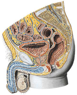
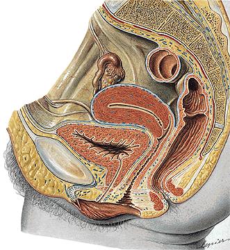
31 Peritoneal Cavity of Pelvis Inferior to the pelvic inlet (linea terminalis) Intraperitoneal organs: Superior part of the rectum, uterus and uterine tubes, ovarium, sigmoid colon Subperitoneal organs: urinary bladder, middle part of the rectum
32 Peritoneal Cavity of Pelvis
33 Peritoneal Cavity of Pelvis The parietal peritoneum reflects onto the pelvic viscera. Only their superior and superolateral surfaces are covered with peritoneum: In the male - rectovesical pouch In the female - rectouterine pouch (cul-de-sac of Douglas) and - vesicouterine pouch


34 Peritoneal Cavity of Pelvis 6 - rectovesical pouch 4 - vesicouterine pouch 6 - rectouterine pouch
 and divides into three branches : Left gastric artery Common hepatic artery right gastric a. hepatic a. proper gastroduodenal a. right gastro-omental a.")
35 Vessels in the infracolic compartment Celiac Trunk unpaired artery, branch of the abdominal aorta at the level of the Th-XII. It is very short (less than 2 cm in most cases) and divides into three branches : Left gastric artery Common hepatic artery right gastric a. hepatic a. proper gastroduodenal a. right gastro-omental a. superior pancreaticoduodenal aa. Splenic artery short gastric aa. left gastro-omental a.
 and divides into three branches : Left gastric artery Common hepatic artery right gastric a. hepatic a. proper gastroduodenal a. right gastro-omental a.")
36 Vessels in the infracolic compartment Celiac Trunk unpaired artery, branch of the abdominal aorta at the level of the Th-XII. It is very short (less than 2 cm in most cases) and divides into three branches : Left gastric artery Common hepatic artery right gastric a. hepatic a. proper gastroduodenal a. right gastro-omental a. superior pancreaticoduodenal aa. Splenic artery short gastric aa. left gastro-omental a.

37 Vessels in the infracolic compartment Superior mesenteric a. unpaired artery, branch of the abdominal aorta. It arises about 1 cm inferior to the celiac trunk at the level of the L-1. Branches: Inferior pancreaticoduodenal a. Intestinal aa. (jejunales et ilei) Ileocolic a. Right colic a. Middle colic a.

38 Vessels in the infracolic compartment Superior mesenteric a. unpaired artery, branch of the abdominal aorta. It arises about 1 cm inferior to the celiac trunk at the level of the L-1. Branches: Inferior pancreaticoduodenal a. Intestinal aa. (jejunales et ilei) Ileocolic a. Right colic a. Middle colic a.

39 Vessels in the infracolic compartment Superior mesenteric a. unpaired artery, branch of the abdominal aorta. It arises about 1 cm inferior to the celiac trunk at the level of the L-1. Branches: Inferior pancreaticoduodenal a. Intestinal aa. (jejunales et ilei) Ileocolic a. Right colic a. Middle colic a.

40 Vessels in the infracolic compartment Superior mesenteric artery

41 Vessels in the infracolic compartment Inferior mesenteric a. unpaired branch of the abdominal aorta. It arises at the level of the intervertebral disc between vertebrae L-2 and L-3. Branches: Left colic a. Sigmoid aa. Superior rectal a. - anastomoses with middle and inferior rectal aa.


42 Vessels in the infracolic compartment The hepatic portal vein carries venous blood to the liver from the abdominal portion of the gastrointestinal tract, the spleen, and the pancreas. It is formed by the union of the superior mesenteric and splenic veins. The inferior mesenteric v. enters the splenic vein.


43 Hepatic portal vein Anastomoses between portal and caval veins
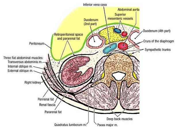
44 Retroperitoneal space Retroperitoneal space is not a real space. It is that part of the body between the parietal peritoneum and the muscles and bones of the posterior abdominal wall. The retroperitoneal space contains the kidneys, ureters, suprarenal glands, aorta, inferior vena cava, and abdominal portions of the sympathetic trunks. They are primary retroperitoneal structures. The pancreas, duodenum, ascending colon and descending colon are secondarily retroperitoneal organs.
45 Retroperitoneal space
 to the 12th ribs.")

46 Primary retroperitoneal structures The kidneys are bean-shaped structures located between the T12 and the L3 vertebral levels, deep (anterior) to the 12th ribs. The kidneys are hollow and are embedded in perinephric fat. Closely related to the diaphragm, the kidneys move 2-3 cm in a vertical direction with its excursions. the inferior pole of the right kidney is approximately a finger's breadth superior to the iliac crest.
 by a condensed, membranous layer of renal fascia.")
47 Primary retroperitoneal structures Perinephric fat (capsula adiposa) surrounds the kidneys and their vessels as it extends into their hollow centers, the renal sinuses. The kidneys, suprarenal glands, and the fat surrounding them are enclosed (except inferiorly!) by a condensed, membranous layer of renal fascia. The renal fascia continues medially to ensheath the renal vessels and abdominal aorta. External to the renal fascia is paranephric fat. Left kidney
 by a condensed, membranous layer of renal fascia.")
48 Primary retroperitoneal structures Perinephric fat (capsula adiposa) surrounds the kidneys and their vessels as it extends into their hollow centers, the renal sinuses. The kidneys, suprarenal glands, and the fat surrounding them are enclosed (except inferiorly!) by a condensed, membranous layer of renal fascia. The renal fascia continues medially to ensheath the renal vessels and abdominal aorta. External to the renal fascia is paranephric fat.
49 Primary retroperitoneal structures The collagen bundles, renal fascia, and perinephric and paranephric fat, along with the tethering provided by the renal vessels and intraabdominal pressure, hold the kidneys in a relatively fixed position. Nephroptosis (dropped kidney) - abnormally mobile kidneys may descend more than the normal 3 cm when the body is erect.

50 Primary retroperitoneal structures The suprarenal (adrenal) glands are located between the superomedial aspects of the kidneys and the diaphragm. They are enclosed by renal fascia by which they are attached to the crura of the diaphragm. The right suprarenal gland is triangular in shape. The left suprarenal gland is semilunar in shape.

51 Primary retroperitoneal structures

 at the junction of the ureter and renal pelvis, (2) where the ureters cross the brim of the")
52 Primary retroperitoneal structures The ureters are muscular ducts (25-30 cm long) with narrow lumina that carry urine from the kidneys to the urinary bladder. They are divided into: abdominal part pelvic part The ureters normally demonstrate relative constrictions in three places: (1) at the junction of the ureter and renal pelvis, (2) where the ureters cross the brim of the pelvic inlet, (3) during their passage through the wall of the urinary bladder


53 Surface marking of the ureters Anterior veiw Posterior veiw
54 Abdominal aorta From the aortic hiatus (Th12) to the bifurcation at level of the L4. The abdominal aorta lies anterior to the L1-L4 vertebral bodies. Splenic v., pancreas, duodenum (horizontal part), root of the mesenteri, left renal v. and peritoneum lie anterior to the aorta. Behind the aorta: cisterna chyli and the beginning of the thoracic duct. Laterally: nodi lymph. lumbales.
 a.")
55 Abdominal aorta The abdominal aorta has three types of branches: Unpaired arteries to the gastrointestinal tract. Paired visceral arteries: Renal a. Middle suprarenal a. Testicular (ovarian) a. Paired arteries to the abdominal wall (parietal): Inferior phrenic a. Lumbar aa. (4 pairs)
56 Inferior vena cava The inferior vena cava has no unpaired visceral tributaries because the hepatic portal system collects blood from the gastrointestinal tract. Left suprarenal v. and left testicular v. drain into the left renal v.!
")
57 Anastomoses between caval veins Vv. lumbales ascendens behind psoas major m.. Deep cava-caval anastomoses: Ascending lumbar vv. (to the superior v. cava) lumbar vv. (to the inferior v. cava) Vertebral vv. (to the superior v. cava) lumbar vv. (to the inferior v. cava)

58 Lymphatic vessels and nodes Parietal lymph nodes: Nodi lymph. iliaci communes Nodi lymph. lumbales Nodi lymph. phrenici inferiores Visceral lymph nodes: Nodi lymph. celiaci Nodi lymph. mesenterici superiores Nodi lymph. mesenterici inferiores Lymphatic vessels: truncus lumbalis dex. et sin. truncus intestinalis Cisterna chyli Thoracic duct

59 Lymphatic vessels and nodes Parietal lymph nodes: Nodi lymph. iliaci communes Nodi lymph. lumbales Nodi lymph. phrenici inferiores Visceral lymph nodes: Nodi lymph. celiaci Nodi lymph. mesenterici superiores Nodi lymph. mesenterici inferiores Lymphatic vessels: truncus lumbalis dex. et sin. truncus intestinalis Cisterna chyli Thoracic duct


60 Lumbar plexus The lumbar plexus (ventral rami of spinal nerves L1 to L4) is formed within the psoas major muscle. Th12 L1 L2 L3 L4

61 Lumbar plexus Nerves of the lumbar plexus : Iliohypogastric n. Ilioinguinal n. Genitofemoral n. lateral cutaneous nerve of the thigh Obturator n. Femoral n.
62 Abdominal autonomic nerves and plexuses Abdominal Part of the Sympathetic Trunk - on the vertebral body between the crus of the diaphragm and the psoas major muscle. lumbar splanchnic nerves pass anteriorly from the sympathetic trunk to the aortic autonomic nerve plexus. The abdominal autonomic plexuses are nerve networks consisting of both sympathetic and parasympathetic fibers, which surround the abdominal aorta and its major branches : celiac plexus superior mesenteric plexus inferior mesenteric plexus intermesenteric plexus superior hypogastric plexus
63 Abdominal autonomic nerves and plexuses The sympathetic fibers mainly innervate the blood vessels of abdominal viscera and are inhibitory to the parasympathetic stimulation. The vagus nerves supplies parasympathetic fibers to the digestive tract from the esophagus through the transverse colon. Pelvic splanchnic nerves supply the descending and sigmoid colon and rectum. Parasympathetic stimulation promotes peristalsis and secretion.


64 Abdominal autonomic nerves and plexuses Sympathetic trunk
Anatomy of the Large Intestine
 Large intestine Anatomy of the Large Intestine 2 Large Intestine Extends from ileocecal valve to anus Length = 1.5-2.5m = 5 feet Regions Cecum = 2.5-3 inch Appendix= 3-5 inch Colon Ascending= 5 inch Transverse=
The abdominal Esophagus, Stomach and the Duodenum. Prof. Oluwadiya KS
 The abdominal Esophagus, Stomach and the Duodenum Prof. Oluwadiya KS www.oluwadiya.com Viscera of the abdomen Abdominal esophagus: Terminal part of the esophagus The stomach Intestines: Small and Large
ANATOMY OF THE SMALL & LARGE INTESTINES. Semester 1, 2011 A. Mwakikunga
 ANATOMY OF THE SMALL & LARGE INTESTINES Semester 1, 2011 A. Mwakikunga LEARNING OBJECTIVES 1. List the parts and anatomical regions of the small and large intestines 2. State anatomical relations of the
The posterior abdominal wall. Prof. Oluwadiya KS
 The posterior abdominal wall Prof. Oluwadiya KS www.oluwadiya.sitesled.com Posterior Abdominal Wall Lumbar vertebrae and discs. Muscles opsoas, quadratus lumborum, iliacus, transverse, abdominal wall
The peritoneum. Prof. Oluwadiya KS, MBBS, FMCS(Orthop) Website:
 Website:") The peritoneum Prof. Oluwadiya KS, MBBS, FMCS(Orthop) Website: http://oluwadiya.com The peritoneum Serous membrane that lines the abdominopelvic cavity and invests the viscera The largest serous membrane
The peritoneum Prof. Oluwadiya KS, MBBS, FMCS(Orthop) Website: http://oluwadiya.com The peritoneum Serous membrane that lines the abdominopelvic cavity and invests the viscera The largest serous membrane
Anatomy of the SMALL INTESTINE. Dr. Noman Ullah Wazir PMC
 Anatomy of the SMALL INTESTINE Dr. Noman Ullah Wazir PMC SMALL INTESTINE The small intestine, consists of the duodenum, jejunum, and illium. It extends from the pylorus to the ileocecal junction were the
BLOCK IV: OFFICIAL BODY PARTS LIST FOR ANTERIOR ABDOMINAL WALL AND ABDOMINAL CONTENTS
 BLOCK IV: OFFICIAL BODY PARTS LIST FOR ANTERIOR ABDOMINAL WALL AND ABDOMINAL CONTENTS External oblique muscle Muscular portion Aponeurotic portion Superficial inguinal ring Lateral (inferior) crus Medial
Peritoneum: Def. : It is a thin serous membrane that lines the walls of the abdominal and pelvic cavities and clothes the viscera.
 Peritoneum: Def. : It is a thin serous membrane that lines the walls of the abdominal and pelvic cavities and clothes the viscera. Layers of the peritoneum: 1. Outer Layer ( Parietal Peritoneum) : lines
Abdomen. Retroperitoneal space
 Abdomen. Retroperitoneal space Abdominal cavity The space bounded by: Anterolateral abdominal wall Posterior abdominal wall Diaphragm Pelvic walls and pelvic floor. Subdivided into: True abdominal cavity
Exploring Anatomy: the Human Abdomen
 Exploring Anatomy: the Human Abdomen PERITONEUM AND PERITONEAL CAVITY PERITONEUM The peritoneum is a thin serous membrane that lines the abdominal cavity and covers, in variable amounts, the viscera within
Dr. Zahiri. In the name of God
 Dr. Zahiri In the name of God small intestine = small bowel is the part of the gastrointestinal tract Boundaries: Pylorus Ileosecal junction Function: digestion and absorption of food It receives bile
Duodenum retroperitoneal
 Duodenum retroperitoneal C shaped Initial region out of stomach into small intestine RETROperitoneal viscus Superior 1 st part duodenal cap ; moves upwards and backwards to lie on the R crura medial to
Netter's Anatomy Flash Cards Section 4 List 4 th Edition
 Netter's Anatomy Flash Cards Section 4 List 4 th Edition https://www.memrise.com/course/1577335/ Section 4 Abdomen (31 cards) Plate 4-1 Bony Framework of Abdomen 1.1 Costal cartilages 1.2 Iliac crest 1.3
Mousa Salah. Dr. Mohammad Al. Mohtasib. 1 P a g e
 8 Mousa Salah Dr. Mohammad Al. Mohtasib 1 P a g e In the previous lecture we talked about the peritoneum, and we said that the peritonium is a serous sac, and it consists of two layers, visceral and parietal.
د. عصام طارق. Objectives:
 GI anatomy Lecture: 5 د. عصام طارق Objectives: To describe anatomy of stomach, duodenum & pancreas. To list their main relations. To define their blood & nerve supply. To list their lymph drainage. To
GI module Lecture: 9 د. عصام طارق. Objectives:
 GI module Lecture: 9 د. عصام طارق Objectives: To list structures forming posterior abdominal wall. To follow aorta & its main branches. To describe IVC & its main tributaries. To list nerves of posterior
Preview from Notesale.co.uk Page 1 of 34
 Abdominal viscera and digestive tract Digestive tract Abdominal viscera comprise majority of the alimentary system o Terminal oesophagus, stomach, pancreas, spleen, liver, gallbladder, kidneys, suprarenal
Block 3: DISSECTION 2 CELIAC TRUNK, JEJUNUM/ILEUM, LARGE INTESTINE, DUODENUM, PANCREAS, PORTAL VEIN; MOBILIZATION OF THE LIVER
 1 Block 3: DISSECTION 2 CELIAC TRUNK, JEJUNUM/ILEUM, LARGE INTESTINE, DUODENUM, PANCREAS, PORTAL VEIN; MOBILIZATION OF THE LIVER Attempt to complete as much as you can of the dissection explained in the
Biology Human Anatomy Abdominal and Pelvic Cavities
 Biology 351 - Human Anatomy Abdominal and Pelvic Cavities Please place your name and I.D. number on the back of the last page of this exam. You must answer all questions on this exam. Because statistics
Pancreas & Biliary System. Dr. Vohra & Dr. Jamila
 Pancreas & Biliary System Dr. Vohra & Dr. Jamila 1 Objectives At the end of the lecture, the student should be able to describe the: Location, surface anatomy, parts, relations & peritoneal reflection
The jejunum and the Ileum. Prof. Oluwadiya KS
 The jejunum and the Ileum Prof. Oluwadiya KS www.oluwadiya.siteled.com Introduction Introduction The small intestine (SI) comprises of the duodenum, jejunum and the ileum The jejunum is the second part
Bushra Arafa Zayed & Hanan Jamal. - Dana AF
 - 10 - Bushra Arafa Zayed & Hanan Jamal - Dana AF - Mohammad Al Muhtaseb Notes: This sheet was written in the same order as the slides, and everything in the slides is mentioned in this sheet. Pictures
SUBJECTS 2nd year, 1st semester I. 1. Primitive gut - limits, derivatives 2. Foregut -limits, evolution, derivatives 3. Midgut -limits, evolution,
 SUBJECTS 2nd year, 1st semester I. 1. Primitive gut - limits, derivatives 2. Foregut -limits, evolution, derivatives 3. Midgut -limits, evolution, derivatives 4. Hindgut- limits, evolution, derivatives
In the name ofgod. Abdomen 3. Dr. Zahiri
 In the name ofgod Abdomen 3 Dr. Zahiri Peritoneum Peritoneum It is the serous membrane(a type of loose connective tissue and is covered by mesothelium) that lines the abdominal cavity. Extensions of the
Anatomy of the renal system. Professor Nawfal K. Al-Hadithi
 Anatomy of the renal system Professor Nawfal K. Al-Hadithi Objectives To describe the posterior abdominal wall To identify the main anatomical landmarks of the kidneys & ureters To describe the suprarenal
THE ORAL CAVITY
 THE ORAL CAVITY WALL OF ABDOMEN (ANTERIOR) The paraumbilical vein drains into the portal vein and then through the liver. This is an important clinical connection. THE ABDOMINAL VISCERA The small
ANATOMY OF THE DIGESTIVE SYSTEM PART II
 ANATOMY OF THE DIGESTIVE SYSTEM PART II 9.12.2014 Kaan Yücel M.D., Ph.D. http://fhs121.org Dr.Kaan Yücel http://fhs121.org Digestive system Part II 1. LIVER The liver is the largest gland in the body and,
-Ensherah Mokheemer. -Shatha Al-Jaberi محمد المحتسب- 1 P a g e
 9-9 -Ensherah Mokheemer -Shatha Al-Jaberi محمد المحتسب- 1 P a g e Small intestine has three regions: ( االثني عشر( The duodenum The jejunum The ileum Small intestine Duodenum: -c-shaped -The concavity
Accessory Glands of Digestive System
 Accessory Glands of Digestive System The liver The liver is soft and pliable and occupies the upper part of the abdominal cavity just beneath the diaphragm. The greater part of the liver is situated under
ANATOMY OF PELVICAYCEAL SYSTEM -DR. RAHUL BEVARA
 1 ANATOMY OF PELVICAYCEAL SYSTEM -DR. RAHUL BEVARA 2 KIDNEY:ANATOMY OVERVIEW Kidneys are retroperitoneal, in posterior abdominal region, extending from T12 L3 Bean-shaped Right kidney is lower than left
Small Plicae Circularis. Short Closely packed together. Sparse, completely absent at distal part Lymphoid Nodule
 Intestines Differences Between Jejunum and Ileum Types Jejunum Ileum Color Deeper red Paler pink Calibre Bigger Smaller Thickness of wall Thick and Heavy Thin and Lighter Vascularity Highly vascularised
Lab Monitor Images Dissection of the Abdominal Vasculature + Lower Digestive System
 Lab Monitor Images Dissection of the Abdominal Vasculature + Lower Digestive System Stomach & Duodenum Frontal (AP) View Nasogastric tube 2 1 3 4 Stomach Pylorus Duodenum 1 Duodenum 2 Duodenum 3 Duodenum
THE ABDOMEN SUPRARENAL GLANDS KIDNEY URETERS URINARY BLADDER
 THE ABDOMEN SUPRARENAL GLANDS KIDNEY URETERS URINARY BLADDER THE SUPRARENAL GLANDS The suprarenal (adrenal) glands lie immediately superior and slightly anterior to the upper pole of either kidney. Golden
Gross Anatomy of the Urinary System
 Gross Anatomy of the Urinary System Lecture Objectives Overview of the urinary system. Describe the external and internal anatomical structure of the kidney. Describe the anatomical structure of the ureter
Lab 9 Abdomen MUSCLES
 Lab 9 Abdomen MUSCLES External abdominal oblique continuous with the external intercostal muscle; its fibers point in a caudal direction as it moves anteriorly until it inserts on the linea alba via its
Anatomy: Know Your Abdomen
 Anatomy: Know Your Abdomen Glossary Abdomen - part of the body below the thorax (chest cavity); separated by the diaphragm. Anterior - towards the front of the body. For example, the umbilicus is anterior
1. A stab wound into the abdomen transected the hepatoduodenal ligament. Each of the following structures would have been cut EXCEPT the:
 YR 1 GROSS ANATOMY UNIT EXAM 3 -- November 07, 1997. CHOOSE THE SINGLE BEST ANSWER FOR QUESTION 1-42. 1. A stab wound into the abdomen transected the hepatoduodenal ligament. Each of the following structures
To describe the liver. To list main structures in porta hepatis.
 GI anatomy Lecture: 6 د. عصام طارق Objectives: To describe the liver. To list main structures in porta hepatis. To define portal system & portosystemic anastomosis. To list parts of biliary system. To
Pancreas and Biliary System
 Pancreas and Biliary System Please view our Editing File before studying this lecture to check for any changes. Color Code Important Doctors Notes Notes/Extra explanation Objectives At the end of the lecture,
The Thoracic wall including the diaphragm. Prof Oluwadiya KS
 The Thoracic wall including the diaphragm Prof Oluwadiya KS www.oluwadiya.com Components of the thoracic wall Skin Superficial fascia Chest wall muscles (see upper limb slides) Skeletal framework Intercostal
Nasogastric tube. Stomach. Pylorus. Duodenum 1. Duodenum 2. Duodenum 3. Duodenum 4
 Esophagus Barium Swallow Stomach and Duodenum 4 year old Upper GI Nasogastric tube Stomach and Duodenum 4 year old Upper GI Nasogastric tube Stomach Pylorus Duodenum 1 Duodenum 2 Duodenum 3 Duodenum 4
BY DR NOMAN ULLAH WAZIR
 BY DR NOMAN ULLAH WAZIR The stomach (from ancient Greek word stomachos, stoma means mouth) is a muscular, hollow and the most dilated part of the GIT. It starts from the point where esophagus ends. It
Benha University. Faculty of Medicine. Anatomy Department Course code (MED 0701) Model answer of Anatomy examination. (Abdomen,Pelvis and Thorax)
 Model answer of Anatomy examination. (Abdomen,Pelvis and Thorax)") 1 Benha University Faculty of Medicine Anatomy Department Course code (MED 0701) Model answer of Anatomy examination (Abdomen,Pelvis and Thorax) 1 st year 2 nd term Date :18 /5 /2013 2 I-Short account
1 Benha University Faculty of Medicine Anatomy Department Course code (MED 0701) Model answer of Anatomy examination (Abdomen,Pelvis and Thorax) 1 st year 2 nd term Date :18 /5 /2013 2 I-Short account
ABDOMEN. 2. The highest branch of the abdominal aorta is: (a) R suprarenal a (b) Coeliac trunk (c) L renal a (d) L gonadal a (e) SMA
 R suprarenal a (b) Coeliac trunk (c) L renal a (d) L gonadal a (e) SMA") ABDOMEN 1. The duodenum: (a) is a retroperitoneal structure (b) is 25cm long (c) lies between the levels of L2-L4 (d) in its fourth part lies to the R of the aorta (e) all of the above 2. The highest branch
ABDOMEN 1. The duodenum: (a) is a retroperitoneal structure (b) is 25cm long (c) lies between the levels of L2-L4 (d) in its fourth part lies to the R of the aorta (e) all of the above 2. The highest branch
-12. -Renad Habahbeh. -Dr Mohammad mohtasib
 -12 -Renad Habahbeh - -Dr Mohammad mohtasib The Gallbladder -The gallbladder has a body, a fundus (a rounded end), a neck, Hartmann s pouch before the neck and a cystic duct that meets the common hepatic
Al-Mohtaseb. Saba Alfayoumi. Mo Alfarra
 8 Al-Mohtaseb Saba Alfayoumi Mo Alfarra For the comparison purposes refer to the last page where you can find a table that summarizes them. Enjoy Jejunum and Ileum -They're intraperitoneal and freely mobile
It passes through the diaphragm at the level of the 10th thoracic vertebra to join the stomach
 The esophagus is a tubular structure (muscular, collapsible tube ) about 10 in. (25 cm) long that is continuous above with the laryngeal part of the pharynx opposite the sixth cervical vertebra The esophagus
Biology Human Anatomy Abdominal and Pelvic Cavities
 Biology 351 - Human Anatomy Abdominal and Pelvic Cavities You must answer all questions on this exam. Because statistics demonstrate that, on average, between 2-5 questions on every 100-point exam are
Inferior Pelvic Border
 Pelvis + Perineum Pelvic Cavity Enclosed by bony, ligamentous and muscular wall Contains the urinary bladder, ureters, pelvic genital organs, rectum, blood vessels, lymphatics and nerves Pelvic inlet (superior
YR 1 GROSS ANATOMY/EMBRYOLOGY UNIT EXAM 3 -- November 13, Which of the following statements regarding the pericardium is NOT CORRECT:
 YR 1 GROSS ANATOMY/EMBRYOLOGY UNIT EXAM 3 -- November 13, 1996. CHOOSE THE SINGLE BEST ANSWER FOR QUESTIONS 1-42. 1. Which of the following statements regarding the pericardium is NOT CORRECT: A. The fibrous
Dissection Lab Manuals: Required Content
 Dissection Lab Manuals: Required Content 1. Introduction a. Basic terminology (directions) b. External features of the cat c. Adaptations to predatory niche d. How to skin a cat e. How to make the incisions
GASTROINTESTINAL SYSTEM
 GASTROINTESTINAL SYSTEM Topographic Anatomy of the Abdomen Surface Landmarks Xiphoid process T9/T10 Inferior costal margin L2/L3 Iliac Crest L4 level ASIS L5/S1 level Pubic symphysis level of greater trochanter
The Spleen. Dr Fahad Ullah
 The Spleen BY Dr Fahad Ullah Spleen The spleen is an largest lymphoid organ shaped like a shoe that lies relative to the 9th and 11th ribs and is located in the left hypochondrium. Thus, the spleen is
LECTURE 11 & 12: ABDOMINAL VISCERA ABDOMINAL CONTENTS DIVISION. The location of abdominal viscera is divided into 4 quadrants:
 LECTURE 11 & 12: ABDOMINAL VISCERA ABDOMINAL CONTENTS DIVISION The location of abdominal viscera is divided into 4 quadrants: - horizontal line across the umbilicus divides the upper quadrants from the
Dana Alrafaiah. - Amani Nofal. - Ahmad Alsalman. 1 P a g e
 - 2 - Dana Alrafaiah - Amani Nofal - Ahmad Alsalman 1 P a g e This lecture will discuss five topics as follows: 1- Arrangement of pelvic viscera. 2- Muscles of Pelvis. 3- Blood Supply of pelvis. 4- Nerve
STRUCTURAL BASIS OF MEDICAL PRACTICE EXAMINATION 3. October 17, 2014
 STRUCTURAL BASIS OF MEDICAL PRACTICE EXAMINATION 3 October 17, 2014 PART l. Answer in the space provided. (12 pts) 1. Identify the structures. (2 pts) A. B. A B C. D. C D 2. Identify the structures. (2
-the stones will obstruct the common bile duct and it might also be precancerous. -so the best treatment is chlolycyctoctomy.
 At the beginning this sheet includes the rest of last lecture s slides liver +gallbladder and the new lecture posterior abdominal wall and its vessels. We will start talking about Cholelithiasis -it means
Omran Saeed. Mohammad Al-muhtaseb. 1 P a g e
 13 Omran Saeed Mohammad Al-muhtaseb 1 P a g e Posterior abdominal wall - The diaphragm separates between thoracic cavity and abdominal cavity. Structures of posterior abdominal wall: (below diaphragm)
SMALL INTESTINE LARGE INTESTINE RECTUM ABDOMINAL AORTA INFERIOR VENA CAVA THE ABDOMEN
 SMALL INTESTINE LARGE INTESTINE RECTUM ABDOMINAL AORTA INFERIOR VENA CAVA THE ABDOMEN THE SMALL INTESTINE THE SMALL INTESTINE: Is the longest part of the gastrointesanal tract - length of 5 metres (3 7
STRUCTURAL BASIS OF MEDICAL PRACTICE EXAMINATION 3. October 16, 2015
 STRUCTURAL BASIS OF MEDICAL PRACTICE EXAMINATION 3 October 16, 2015 PART l. Answer in the space provided. (12 pts) 1. Identify the structures. (2 pts) A. B. A B C. D. C D 2. Identify the structures. (2
Anatomical Considerations for Lab Practical II
 Anatomical Considerations for Lab Practical II For each of the following please be prepared to provide: Identification System Organ(s) or ducts to Function(s) location which it is attached Use your lecture
CARDIOVASCULAR DANIL HAMMOUDI.MD
 CARDIOVASCULAR DANIL HAMMOUDI.MD 18 Systemic Circulation Figure 19.19 Pulmonary Circulation Figure 19.18b 1. Thyroid gland 2. Trachea 3. Brachiocephalic 4. Common carotid 5. Internal jugular 6. Superior
1 Right & left Hepatic ducts Gastric Impression of spleen
 Pancreatic Model 1 Right & left Hepatic ducts 14 Gastric Impression of spleen 2 Common hepatic duct 15 Renal Impression of spleen 3 Cystic Duct 16 Colic Impression of spleen 4 Common Bile Duct 17 Splenic
3 Circulatory Pathways
 40 Chapter 3 Circulatory Pathways Systemic Arteries -Arteries carry blood away from the heart to the various organs of the body. -The aorta is the longest artery in the body; it branches to give rise to
Posterior Abdominal wall-
 Structures of posterior abdominal wall: o Bony boundaries: 5 lumber vertebra and their intervertebral disc, iliac fossa and iliac crest. o Muscles: psoas major, quadrates lumborum, transversus abdominis,
Abdomen: Introduction. Prof. Oluwadiya KS
 Abdomen: Introduction Prof. Oluwadiya KS www.oluwadiya.com Abdominopelvic Cavity Abdominal Cavity Pelvic Cavity Extends from the inferior margin of the thorax to the superior margin of the pelvis and the
ABDOMEN - GI. Duodenum
 TALA SALEH ABDOMEN - GI Duodenum - Notice the shape of the duodenum, it looks like capital G shape tube which extends from the pyloroduodenal junction to the duodenojejunal junction. - It is 10 inches
[ANATOMY #12] April 28, 2013
![[ANATOMY #12] April 28, 2013](/thumbs/86/93473883.jpg "[ANATOMY #12] April 28, 2013") Sympathetic chain : Sympathetic chain is each of the pair of ganglionated longitudinal cords of the sympathetic nervous system; extend from level of atlas (base of skull) till coccyx. It is paravertebral
Sympathetic chain : Sympathetic chain is each of the pair of ganglionated longitudinal cords of the sympathetic nervous system; extend from level of atlas (base of skull) till coccyx. It is paravertebral
Dr. Weyrich G07: Superior and Posterior Mediastina. Reading: 1. Gray s Anatomy for Students, chapter 3
 Dr. Weyrich G07: Superior and Posterior Mediastina Reading: 1. Gray s Anatomy for Students, chapter 3 Objectives: 1. Subdivisions of mediastinum 2. Structures in Superior mediastinum 3. Structures in Posterior
Jhia Anjela D. Rivera 1 1. BS Biology, Department of Biology, College of Science, Polytechnic University of the Philippines
 DIGESTIVE SYSTEM Jhia Anjela D. Rivera 1 1 BS Biology, Department of Biology, College of Science, Polytechnic University of the Philippines DIGESTIVE SYSTEM Consists of the digestive tract (gastrointestinal
Nerves on the Posterior Abdominal Wall
 Nerves on the Posterior Abdominal Wall Lumbar Plexus The lumbar plexus, which is one of the main nervous pathways supplying the lower limb, is formed in the psoasmuscle from the anterior ramiof the upper
Anatomy of the spleen. Oluwadiya KS
 Anatomy of the spleen Oluwadiya KS www.oluwadiya.com Introduction The spleen is an ovoid, usually purplish, pulpy mass about the size and shape of one's fist. It is the largest lymphoid tissue in the body
THE PELVIS VASCULAR AND NERVOUS SYSTEM SOMATIC AND AUTONOMIC NERVES
 THE PELVIS VASCULAR AND NERVOUS SYSTEM SOMATIC AND AUTONOMIC NERVES THE ABDOMINAL AORTA The abdominal aorta begins at the aor9c hiatus in the diaphragm at the level of the T12 vertebra and ends at the
Group of students. - Rawan almujabili د. محمد المحتسب - 1 P a g e
 - 14 - Group of students - Rawan almujabili د. محمد المحتسب - 1 P a g e Nerves of the posterior abdominal wall The spinal cord gives off spinal nerves between the vertebrae. In the abdomen, through the
Anatomy of the Thorax
 Anatomy of the Thorax A) THE THORACIC WALL Boundaries Posteriorly by the thoracic part of the vertebral column Anteriorly by the sternum and costal cartilages Laterally by the ribs and intercostal spaces
Day 5 Respiratory & Cardiovascular: Respiratory System
 Day 5 Respiratory & Cardiovascular: Respiratory System Be very careful not to damage the heart and lungs while separating the ribs! Analysis Questions-Respiratory & Cardiovascular Log into QUIA using your
The Human Body: An Overview of Anatomy. Anatomy. Physiology. Anatomy - Study of internal and external body structures
 C H A P T E R 1 The Human Body: An Orientation An Overview of Anatomy Anatomy The study of the structure of the human body Physiology The study of body function Anatomy - Study of internal and external
Mediastinum and pericardium
 Mediastinum and pericardium Prof. Abdulameer Al-Nuaimi E-mail: a.al-nuaimi@sheffield.ac.uk E. mail: abdulameerh@yahoo.com The mediastinum: is the central compartment of the thoracic cavity surrounded by
THE KIDNEY (Fig. 1 & 2) Figure 1
 Figure 1") 1 THE KIDNEY (Fig. 1 & 2) The kidney is a bean-shaped, reddish-brown organ of the urinary system located retroperitoneally in the upper part of the paravertebral gutter of the abdominal cavity, padded
1 THE KIDNEY (Fig. 1 & 2) The kidney is a bean-shaped, reddish-brown organ of the urinary system located retroperitoneally in the upper part of the paravertebral gutter of the abdominal cavity, padded
Surface Anatomy. Location Shape Weight Role of Five Surfaces Borders Fissures Lobes Peritoneal Lig
 The Liver Functions Bile production and secretion Detoxification Storage of glycogen Protein synthesis Production of heparin and bile pigments Erythropoiesis (in fetus) Surface Anatomy Location Shape Weight
Lecture 56 Kidney and Urinary System
 Lecture 56 Kidney and Urinary System The adrenal glands are located on the superomedial aspect of the kidney The right diagram shows a picture of the kidney with the abdominal walls and organs removed
THE THORACIC WALL. Boundaries Posteriorly by the thoracic part of the vertebral column. Anteriorly by the sternum and costal cartilages
 THE THORACIC WALL Boundaries Posteriorly by the thoracic part of the vertebral column Anteriorly by the sternum and costal cartilages Laterally by the ribs and intercostal spaces Superiorly by the suprapleural
Done by: Dina Sawadha & Mohammad Abukabeer
 Done by: Dina Sawadha & Mohammad Abukabeer The stomach *the stomach is a dilated part of the gastro intestinal tract, it's "J" shape. *the lower surface of the stomach ( the greater curvature ) reaches
Gastrointestinal Tract. Anatomy of GI Tract. Anatomy of GI Tract. (Effective February 2007) (1%-5%)
 (1%-5%)") Gastrointestinal Tract (Effective February 2007) (1%-5%) Anatomy of GI Tract Esophagus bulls-eye or target EG junction seen on sagittal scan posterior to left lobe of liver and anterior to aorta Anatomy
Gastrointestinal Tract (Effective February 2007) (1%-5%) Anatomy of GI Tract Esophagus bulls-eye or target EG junction seen on sagittal scan posterior to left lobe of liver and anterior to aorta Anatomy
STERNUM. Lies in the midline of the anterior chest wall It is a flat bone Divides into three parts:
 STERNUM Lies in the midline of the anterior chest wall It is a flat bone Divides into three parts: 1-Manubrium sterni 2-Body of the sternum 3- Xiphoid process The body of the sternum articulates above
Perineum. done by : zaid al-ghnaneem
 Perineum done by : zaid al-ghnaneem Hello everyone, this sheet will talk about 2 nd Lecture which is Perineum but there are some slides and info from 1 st Lecture. Everything included Slides + Pics Let
END-SEMESTER EXAM 2018 ANATOMY, HISTOLOGY AND EMBRYOLOGY FACULTY OF MEDICINE, 2 ND SEMESTER
 University of Szeged, Faculty of Medicine Department of Anatomy, Histology and Embryology Chairman: Prof. Antal Nógrádi MD, PhD, DSc Kossuth L. sgt. 40., H-6724 Szeged, Hungary Tel.: +36-62-545-665 P.
Mediastinum It is a thick movable partition between the two pleural sacs & lungs. It contains all the structures which lie
 Dr Jamila EL medany OBJECTIVES At the end of the lecture, students should be able to: Define the Mediastinum. Differentiate between the divisions of the mediastinum. List the boundaries and contents of
This lab activity is aligned with Visible Body s Human Anatomy Atlas app. Learn more at visiblebody.com/professors
 1 This lab activity is aligned with Visible Body s Human Anatomy Atlas app. Learn more at visiblebody.com/professors 2 A. Digestive System Overview To Start: Go to the Views menu and scroll down to the
Embryology of the Midgut and Hind gut
 Embryology of the Midgut and Hind gut Prof. Abdulameer Al-Nuaimi E-mail: a.al-nuaimi@sheffield.ac.uk E-mail: abdulameerh@yahoo.com Abdominal organs www.google.co.uk/search? Development of Duodenum The
STRUCTURAL BASIS OF MEDICAL PRACTICE EXAMINATION 3. September 13, 2012 B C
 STRUTURL SIS OF MEIL PRTIE EXMINTION 3 September 13, 2012 PRT l. nswer in the space provided. (12 pts) 1. Identify the structures. (2 pts). Lumbocostal trigone. Lateral femoral cutaneous n.. Lumbosacral
OBJECTIVE: To obtain a fundamental knowledge of the root of the neck with respect to structure and function
 The root of the neck Jeff Dupree, Ph.D. e mail: jldupree@vcu.edu OBJECTIVE: To obtain a fundamental knowledge of the root of the neck with respect to structure and function READING ASSIGNMENT: Moore and
- Digestion occurs during periods of low activity - Produces more energy than it uses. - Mucosa
 Introduction Digestive System Chapter 29 Provides processes to break down molecules into a state easily used by cells - A disassembly line: Starts at the mouth and ends at the anus Digestive functions
Lecture 02 Anatomy of the LIVER
 Lecture 02 Anatomy of the LIVER BY Dr Farooq Khan Aurakzai Dated: 02.01.2018 Introduction to Liver Largest gland in the body. 2 nd largest organ of the body. Weight approximately 1500 gm, and is roughly
Body Regions Review. Anatomical Position. Anatomical Planes. Supine versus Prone 9/9/2009
 Body Regions Review The fundamental divisions of the human body Christine Sparks Anatomy / Physiology I Sept. 9, 2009 Anatomical Position Universal terms are used to describe the body accurately and result
Chapter 5: Other mediastinal structures. The Large Arteries. The Aorta. Ascending aorta
 Chapter 5: Other mediastinal structures The Large Arteries The Aorta The aorta is the main arterial trunk of the systemic circulation and in the healthy state its wall contain a large amount of yellow
بسم االه الرحمن الرحيم
 MAY 3, 2012 [POSTERIOR ABDOMINAL WALL] LECTURE 26 ANATOMY Quick Revision: بسم االه الرحمن الرحيم Last time we started with the anterior abdominal wall and said that: 1. Diaphragm is the root of the abdomen.
Gross anatomy of the urinary system. Done by : razan krishan. slide in bold and book in green
 Gross anatomy of the urinary system Done by : razan krishan slide in bold and book in green Kidneys, ureters, urinary bladder & urethra Urine flows from each kidney, down its ureter to the bladder and
ANATOMY. Schedule for 2014/2015 academic school year (2x15 weeks)
") ANATOMY Schedule for 2014/2015 academic school year (2x15 weeks) SEMESTER LECTURES LAB CLASSES SEMINARS TOTAL FIRST 4 hours (2+2) 4 hours (2+2) 1 hour 135 hours SECOND 3 hours 4 hours (2+2) 2 hours 135
ANATOMY Schedule for 2014/2015 academic school year (2x15 weeks) SEMESTER LECTURES LAB CLASSES SEMINARS TOTAL FIRST 4 hours (2+2) 4 hours (2+2) 1 hour 135 hours SECOND 3 hours 4 hours (2+2) 2 hours 135
#1 - Chapter 1 - Anatomy. General Anatomical Terms The Anatomical Position
 #1 - Chapter 1 - Anatomy General Anatomical Terms The Anatomical Position The anatomical position is a stance in which a person stands erect with the feet flat on the floor and close together, arms at