Al-Mohtaseb. Saba Alfayoumi. Mo Alfarra
|
|
|
- Jerome Carson
- 5 years ago
- Views:
Transcription

1 8 Al-Mohtaseb Saba Alfayoumi Mo Alfarra
 extending from posterior abdominal wall to")
2 For the comparison purposes refer to the last page where you can find a table that summarizes them. Enjoy Jejunum and Ileum -They're intraperitoneal and freely mobile so they're completely covered by peritoneum and has mesentery (2 layers of peritoneum) extending from posterior abdominal wall to attach to jejunum and ilium on its free edge. -The jejunum and ileum measure about 20 ft (6 m) long >> the upper 2/5 is the jejunum & the lower 3/5 is the ileum but actually there's no landmark that separates the two from each other and the change is gradual from one to another, so we rely on distinctive features in their wall, diameter, and mesentery in order to differentiate between them. -The jejunum begins at the duodenojejunal flexure (ligament of treitz that s attached to the right crus of diaphragm) and located in the upper part of the peritoneal cavity below the left side of the transverse mesocolon (upper to the left). -The ileum is located in the lower part of the cavity and in the pelvis (lower to the right) and ends at the ileocecal junction where the cecum begins -remember that cecum is located in the right iliac fossa-. Function: The main function of small intestine is absorption of nutritive material. Our food is either carbohydrates, proteins, or fat and the digestion process turns them into glucose, amino acids, or fatty acids respectively. This simple material is the one that's absorbed in small intestine and they all have to go firstly to the liver by portal vein before entering the systemic circulation. Location: Small intestine (jejunum and ilium) is located in umbilical region surrounded by large intestine, so it s internal to large intestine. 1 P a g e

3 Histology of small intestine: We know that GI tract has 4 layers: mucosa, submucosa, muscular layer, serosa or adventitia (depending if the organ is intraperitoneal or retroperitoneal) If intraperitoneal > serosa which is simple squamous epithelium If retroperitoneal > adventitia which is connective tissue As small intestine is intraperitoneal, so it's wall contain serosa not adventitia. Mucosa: Contains villi and micro villi on the surface of villi to increase the surface area for absorption, and as we know the mucosa consists of 3 layers (lining epithelium, lamina propria, and muscularis mucosa which consists of 2-3 bands of smooth muscle). *Lining epithelium: -Epithelial absorptive cells: simple columnar with goblet cells (in stomach it's just simple columnar without goblet cells) and its function is absorption. -Glands (intestinal crypts/ intestinal glands/ crypts of Lieberkühn): Have the same lining epithelium but its function is secretion. On the base of the glands there's paneth cells which is bactericidal cells and not found in large intestine. *Lamina propria: loose connective tissue that contains connective tissue cells (fibroblasts, smooth muscle cells) and contains artery, vein, and lacteal which is a blind lymphatic vessel responsible for absorption of fat. 2 P a g e


4 Mesentery of the small intestine: fan-shaped fold of peritoneum Formed by parietal peritoneum on posterior abdominal wall starting from the left side at the level of L2 at duodenojejunal flexure and descends obliquely downwards to the right side in front of right sacroiliac joint ending at ileocecal junction. Root is 6 inches (15 cm) long while the free edge that's attached to small intestine is 6 meters long. *Contents of the mesentery: between the 2 layers of peritoneum 1-Superior mesenteric artery which forms arcades and vasa recta when reaching jejunum and ilium. 2-Superior mesenteric vein and its tributaries. 3-Nerves: sympathetic and parasympathetic. 4-Fat which is more in mesentery of ilium. Blood supply of Jejunum & Ileum: They are considered part of the midgut, so it's supplied by branches of superior mesenteric artery. Superior mesenteric artery gives jejunal and ilial arteries that gives branches anastomose with one another to form a series of arcades. At the end of superior mesenteric artery, it gives a branch called ileocolic artery that supplies end of ilium, cecum (by ant. And post. cecal arteries), beginning of ascending colon, and appendix. **The intestinal branches arise from the left side of the artery and run in the mesentery to reach the gut. ** from slides 3 P a g e

5 Venous drainage: Tributaries of superior mesenteric vein unit to form superior mesenteric vein that ends behind the neck of pancreas uniting with the splenic vein forming portal vein (i.e. portal vein begins behind the neck of pancreas and ends in the liver and divides into right and left veins in porta hepatis). - The veins correspond to the branches of the superior mesenteric artery Lymphatic drainage: There are lymph nodes around: Celiac trunk > called celiac lymph nodes > drains foregut Origin of superior mesenteric artery > called superior mesenteric lymph nodes > drains midgut Origin of inferior mesenteric artery > called inferior mesenteric lymph nodes > drains hindgut So the small intestine is drained by superior mesenteric lymph nodes but the lymph vessels pass through many intermediate mesenteric nodes until reaching the superior mesenteric lymph nodes. Pathway of lymphatic drainage: Lower limb drains to inguinal lymph nodes then iliac lymph nodes and then para aortic lymph nodes while the abdomen drains to para aortic lymph nodes and all lymph is collected in cisterna chyli in the right side of aortic opening in diaphragm then thoracic duct starts from cisterna chyli and ascends in chest receiving lymphatic drainage from chest and opens in the beginning of left brachiocephalic vein that's formed by left internal jugular and left subclavian veins. -The importance of lymphatics is the absorption of fat and its destination is to the venous blood. 4 P a g e
 spinal nerves forming splanchnic nerves and synapse in celiac ganglia around the origin of")
6 Nerve supply: 1-Sympathetic: In GI, it innervates blood vessels and sphincters and its activation causes vasoconstriction and contraction of sphincters respectively. The nuclei of T6-T9 gives nerve fibers called preganglionic sympathetic fibers which accompany (walks with) spinal nerves forming splanchnic nerves and synapse in celiac ganglia around the origin of celiac trunk and superior mesenteric ganglia around the origin of superior mesenteric artery. L1, L2 synapse in inferior mesenteric ganglia around the origin of inferior mesenteric artery. After synapsing, the post ganglionic sympathetic nerves go to the organ (foregut, midgut, or hindgut). **Splanchnic nerve: Spinal nerve that carries preganglionic sympathetic fibers. 2-Parasympathetic: comes from the brain (medulla oblongata) Transmitted by vagus nerve (X) which gives parasympathetic innervation to the extent of lateral 1/3 of transverse colon (i.e. innervates fore gut and midgut while hind gut gets parasympathetic innervation from S2,S3,S4). Vagus nerve originates from vagal nucleus in medulla oblongata and then descends from brain through neck and thorax around oesophagus as right and left vagal nerves on the right and left of oesophagus respectively, then it cross diaphragm around oesophagus and enter the abdomen. Around the stomach, the right vagal nerve becomes posterior and the left vagal nerve becomes anterior. It passes in the ganglia without synapsing because it'll synapse in the wall of the organ (i.e. in the myenteric plexus of stomach and small intestine) and give short post ganglionic fibers which go to the glands (secretomotor) & to smooth muscles (which responsible for peristaltic movement). 3-Nerves from the superior mesenteric plexus. 5 P a g e


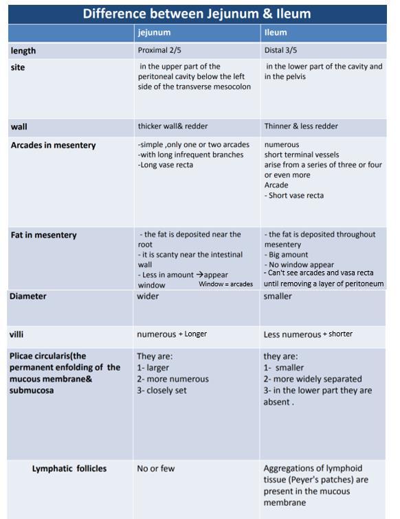
7 Congenital anomaly of small intestine Meckel's Diverticulum: small plugging in the wall of ilium -Vitelline duct: A duct between umbilicus and ilium that should undergo obliteration and fibrosis before birth so it closes completely. But if it stayed open, secretion of ilium will exit from umbilicus. Some times it's obliterated but forms a small diverticulum that contains either gastric or pancreatic tissue, and usually causes inflammation that's similar to appendicitis with the same symptoms (severe pain in the right iliac fossa). -Complications: Inflammation/ Perforation which can cause peritonitis/ Bleeding. -Its characters: - a congenital anomaly of the ileum - Present in 2% of people - 2 feet from iliocecal junction - 2-inch-long - Remains of vitelline duct of embryo Comparison between jejunum and ilium 6 P a g e
8 7 P a g e

 General features of large intestine >>> Not found in small intestine.")
9 Large Intestine *Extends from ileocecal valve to anus * Length = m = 5 feet * Regions Cecum = inch Appendix= 3-5 inch Colon - Ascending= 5 inch - Transverse= 15 inch - Descending= 10 inch - Sigmoid colon = inches Rectum= 5 inch Anal canal= 4 cm Function: Absorption of water and formation of feces (stool) General features of large intestine >>> Not found in small intestine. 1- Sacculation= Haustra 8 P a g e
.")
10 2- Teania coli: three separate longitudinal ribbons of smooth muscle originate from outer longitudinal muscle. Found all over large intestine except appendix and rectum. -It's found only on one side and that's why its contraction causes the formation of sacculation/ haustra (i.e. presence of tenia coli is the reason of sacculation/ haustra formation). -It's a landmark for the base of appendix, so when appendix is retrocecal we track tenia coli until it ends where it indicates the base of appendix. 3- Appendices epiplolca: (adipose structures protruding from the serosal surface of the colon). Found all over large intestine except appendix, Cecum and rectum. Histology: -Has no villi (straight surface) -Lining epithelium: simple columnar epithelium with numerous goblet cells (same as small intestine but with numerous goblet cells); because it needs them for lubrication of hard pieces of stool. -Glands (crypts of Lieberkühn) on base doesn't have paneth cells but contains lymphatic nodules. - It is a blind-ended pouch Cecum - Location: situated in the right iliac fossa, above the lat ½ of inguinal ligament lying above iliacus muscle and psoas major muscle. - Size: It is about 3 inch in diameter - Completely covered with peritoneum and it possesses a considerable amount of mobility, although it does not have a mesentery. There are many hypotheses about the covering peritoneum of cecum: 1. Cecum is covered by peritoneum and can have short mesentery. (the one we rely on) 2. Cecum is fixed to the right iliac fossa and covered by peritoneum from all directions except an area posteriorly without peritoneum. 9 P a g e
11 - Attached to: - Superiorly: Ascending colon Cecum has 3 openings - posteromedial surface is the appendix, 1 inch below ileocecal valve - medially: Ileum (what specializes ileocecal opening is the presence of mucosal fold on it that closes the ilium when the pressure inside the cecum increases, so no regurgitation of material from cecum to ilium happens.) Ileocecal opening is considered physiologically a valve but anatomically it's an opening because there's no circular smooth muscle around it. - The presence of peritoneal folds in the vicinity of the cecum creates The superior ileocecal recesses The inferior ileocecal recesses The retrocecal recesses. > very common site of appendix. These recesses can form internal hernia. - The longitudinal muscle is restricted to three flat bands, the taenia coli, which converge on the base of the appendix and provide for it a complete longitudinal muscle coat. - It has tenia coli on it but there's no appendices epiplolca. Relations of cecum Anteriorly: - Coils of small intestine, specifically ilium. - the greater omentum - the anterior abdominal wall in the right iliac region where we test the patient's cecum. Posteriorly: - The psoas and the iliacus muscles - the femoral nerve - and the lateral cutaneous nerve of the thigh passing to the anterior superior iliac spine. -external iliac vessels in iliac fossa passing below inguinal ligament to give femoral artery. - Postero- medially: The appendix is commonly > retrocecal common. Medially: - Small intestine(ileum) 10 P a g e
: Arteries are medial and veins are lateral.")
 nerves form the superior mesenteric plexus. -Parasympathetic: from vagus nerve.")
12 Blood Supply of cecum Arteries Anterior and posterior cecal arteries a branch of Superior mesenteric artery Venous drainage: The veins correspond to the arteries (anterior and posterior cecal veins) and drain into the superior mesenteric vein. Relation between mesenteric arteries and veins (either superior or inferior): Arteries are medial and veins are lateral. Lymphatic Drainage of cecum The lymph vessels pass through several mesenteric nodes and finally reach the superior mesenteric nodes. Nerve Supply of cecum Branches from the sympathetic and parasympathetic (vagus) nerves form the superior mesenteric plexus. -Parasympathetic: from vagus nerve. -Sympathetic: from greater splanchnic nerve (T6-T9) that synapses in celiac ganglia and superior mesenteric ganglia. Ileocecal Valve - A rudimentary structure - consists of two horizontal folds of mucous membrane - Project around the orifice of the ileum. - The valve plays little or no part in the prevention of reflux of Cecal contents into the ileum. - The circular muscle of the lower end of the ileum (called the ileocecal sphincter by physiologists) serves as a sphincter and controls the flow of contents from the ileum into the colon. - The smooth muscle tone is reflexly increased when the cecum is distended; the gastrin hormone, which is produced by the stomach, causes relaxation of the muscle tone. **from slides** 11 P a g e
13 Peritoneal coverage Diameter Length Parts Function Location Histology Duodenum Retroperitoneal except the 1 st and last inches 10'' 1 st 2 nd 3 rd 4 th Part: 2'' part: 3'' part: 4'' part: 1'' Digestion of fat. epigastric and umbilical regions -Simple columnar cells with goblet cells -Has leaf like villi. -Brunner's glands in submucosa (secrete alkaline secretion) Small Inestine Intraperitoneal Narrower 6 meters Jejunum: first 2/5 Ilium: last 3/5 Absorption of nutritive material. umbilical region surrounded by large intestine, so it s internal to large intestine. -Simple columnar cells with goblet cells -Has villi and microvilli. -Paneth cells that are bactericidal cells on base of crypts of Lieberkühn. Large Intestine Intraperitoneal Wider meters Cecum: 2.5''- 3'' Appendix= 3-5'' Colon - Ascending: 5'' - Transverse: 15'' - Descending: 10'' - Sigmoid colon: 10'' 15'' Rectum: 5'' Anal canal: 4 cm Absorption of water and formation of feces (stool). -Simple columnar cells with numerous goblet cells. -No villi and microvilli (straight surface). -No Paneth cells. -Has lymphatic nodules 12 P a g e
Anatomy of the Large Intestine
 Large intestine Anatomy of the Large Intestine 2 Large Intestine Extends from ileocecal valve to anus Length = 1.5-2.5m = 5 feet Regions Cecum = 2.5-3 inch Appendix= 3-5 inch Colon Ascending= 5 inch Transverse=
Bushra Arafa Zayed & Hanan Jamal. - Dana AF
 - 10 - Bushra Arafa Zayed & Hanan Jamal - Dana AF - Mohammad Al Muhtaseb Notes: This sheet was written in the same order as the slides, and everything in the slides is mentioned in this sheet. Pictures
THE ORAL CAVITY
 THE ORAL CAVITY WALL OF ABDOMEN (ANTERIOR) The paraumbilical vein drains into the portal vein and then through the liver. This is an important clinical connection. THE ABDOMINAL VISCERA The small
-Ensherah Mokheemer. -Shatha Al-Jaberi محمد المحتسب- 1 P a g e
 9-9 -Ensherah Mokheemer -Shatha Al-Jaberi محمد المحتسب- 1 P a g e Small intestine has three regions: ( االثني عشر( The duodenum The jejunum The ileum Small intestine Duodenum: -c-shaped -The concavity
ANATOMY OF THE SMALL & LARGE INTESTINES. Semester 1, 2011 A. Mwakikunga
 ANATOMY OF THE SMALL & LARGE INTESTINES Semester 1, 2011 A. Mwakikunga LEARNING OBJECTIVES 1. List the parts and anatomical regions of the small and large intestines 2. State anatomical relations of the
The jejunum and the Ileum. Prof. Oluwadiya KS
 The jejunum and the Ileum Prof. Oluwadiya KS www.oluwadiya.siteled.com Introduction Introduction The small intestine (SI) comprises of the duodenum, jejunum and the ileum The jejunum is the second part
Dr. Zahiri. In the name of God
 Dr. Zahiri In the name of God small intestine = small bowel is the part of the gastrointestinal tract Boundaries: Pylorus Ileosecal junction Function: digestion and absorption of food It receives bile
Duodenum retroperitoneal
 Duodenum retroperitoneal C shaped Initial region out of stomach into small intestine RETROperitoneal viscus Superior 1 st part duodenal cap ; moves upwards and backwards to lie on the R crura medial to
Anatomy of the SMALL INTESTINE. Dr. Noman Ullah Wazir PMC
 Anatomy of the SMALL INTESTINE Dr. Noman Ullah Wazir PMC SMALL INTESTINE The small intestine, consists of the duodenum, jejunum, and illium. It extends from the pylorus to the ileocecal junction were the
ABDOMEN - GI. Duodenum
 TALA SALEH ABDOMEN - GI Duodenum - Notice the shape of the duodenum, it looks like capital G shape tube which extends from the pyloroduodenal junction to the duodenojejunal junction. - It is 10 inches
Preview from Notesale.co.uk Page 1 of 34
 Abdominal viscera and digestive tract Digestive tract Abdominal viscera comprise majority of the alimentary system o Terminal oesophagus, stomach, pancreas, spleen, liver, gallbladder, kidneys, suprarenal
Small Plicae Circularis. Short Closely packed together. Sparse, completely absent at distal part Lymphoid Nodule
 Intestines Differences Between Jejunum and Ileum Types Jejunum Ileum Color Deeper red Paler pink Calibre Bigger Smaller Thickness of wall Thick and Heavy Thin and Lighter Vascularity Highly vascularised
The Digestive System and Body Metabolism
 14 PART B The Digestive System and Body Metabolism PowerPoint Lecture Slide Presentation by Jerry L. Cook, Sam Houston University ESSENTIALS OF HUMAN ANATOMY & PHYSIOLOGY EIGHTH EDITION ELAINE N. MARIEB
The Digestive System Laboratory
 The Digestive System Laboratory 1 The Digestive Tract The alimentary canal is a continuous tube stretching from the mouth to the anus. Liver Gallbladder Small intestine Anus Parotid, sublingual, and submaxillary
Biology Human Anatomy Abdominal and Pelvic Cavities
 Biology 351 - Human Anatomy Abdominal and Pelvic Cavities Please place your name and I.D. number on the back of the last page of this exam. You must answer all questions on this exam. Because statistics
BLOCK IV: OFFICIAL BODY PARTS LIST FOR ANTERIOR ABDOMINAL WALL AND ABDOMINAL CONTENTS
 BLOCK IV: OFFICIAL BODY PARTS LIST FOR ANTERIOR ABDOMINAL WALL AND ABDOMINAL CONTENTS External oblique muscle Muscular portion Aponeurotic portion Superficial inguinal ring Lateral (inferior) crus Medial
Dana Alrafaiah. Dareen Abu Shalbak. Mohammad Almuhtaseb. 1 P a g e
 2 Dana Alrafaiah Dareen Abu Shalbak Mohammad Almuhtaseb 1 P a g e Esophagus: A muscular tube that is 25 cm long, but if measured from the incisors it would be 45cm long. Extends from C6 of cervical vertebra,
Pancreas & Biliary System. Dr. Vohra & Dr. Jamila
 Pancreas & Biliary System Dr. Vohra & Dr. Jamila 1 Objectives At the end of the lecture, the student should be able to describe the: Location, surface anatomy, parts, relations & peritoneal reflection
د. عصام طارق. Objectives:
 GI anatomy Lecture: 5 د. عصام طارق Objectives: To describe anatomy of stomach, duodenum & pancreas. To list their main relations. To define their blood & nerve supply. To list their lymph drainage. To
The posterior abdominal wall. Prof. Oluwadiya KS
 The posterior abdominal wall Prof. Oluwadiya KS www.oluwadiya.sitesled.com Posterior Abdominal Wall Lumbar vertebrae and discs. Muscles opsoas, quadratus lumborum, iliacus, transverse, abdominal wall
The abdominal Esophagus, Stomach and the Duodenum. Prof. Oluwadiya KS
 The abdominal Esophagus, Stomach and the Duodenum Prof. Oluwadiya KS www.oluwadiya.com Viscera of the abdomen Abdominal esophagus: Terminal part of the esophagus The stomach Intestines: Small and Large
Block 3: DISSECTION 2 CELIAC TRUNK, JEJUNUM/ILEUM, LARGE INTESTINE, DUODENUM, PANCREAS, PORTAL VEIN; MOBILIZATION OF THE LIVER
 1 Block 3: DISSECTION 2 CELIAC TRUNK, JEJUNUM/ILEUM, LARGE INTESTINE, DUODENUM, PANCREAS, PORTAL VEIN; MOBILIZATION OF THE LIVER Attempt to complete as much as you can of the dissection explained in the
Development of pancreas and Small Intestine. ANATOMY DEPARTMENT DR.SANAA AL-AlSHAARAWY DR.ESSAM Eldin Salama
 Development of pancreas and Small Intestine ANATOMY DEPARTMENT DR.SANAA AL-AlSHAARAWY DR.ESSAM Eldin Salama OBJECTIVES At the end of the lecture, the students should be able to : Describe the development
It passes through the diaphragm at the level of the 10th thoracic vertebra to join the stomach
 The esophagus is a tubular structure (muscular, collapsible tube ) about 10 in. (25 cm) long that is continuous above with the laryngeal part of the pharynx opposite the sixth cervical vertebra The esophagus
Gastrointestinal System!
 Gastrointestinal System! Assoc. Prof. Prasit Suwannalert, Ph.D. (Email: prasit.suw@mahidol.ac.th)! Objectives: After learning, student should be able to describe and discuss in topics of! 1. Anatomical
Mousa Salah. Dr. Mohammad Al. Mohtasib. 1 P a g e
 8 Mousa Salah Dr. Mohammad Al. Mohtasib 1 P a g e In the previous lecture we talked about the peritoneum, and we said that the peritonium is a serous sac, and it consists of two layers, visceral and parietal.
The Digestive System
 The Digestive System Identify the Structure and Function. Mesentery of the Large Intestine The mesentery functions to connect the visceral organs to the abdominal wall. Identify the Structure. Nasal Cavity
Anatomy: Know Your Abdomen
 Anatomy: Know Your Abdomen Glossary Abdomen - part of the body below the thorax (chest cavity); separated by the diaphragm. Anterior - towards the front of the body. For example, the umbilicus is anterior
The peritoneum. Prof. Oluwadiya KS, MBBS, FMCS(Orthop) Website:
 Website:") The peritoneum Prof. Oluwadiya KS, MBBS, FMCS(Orthop) Website: http://oluwadiya.com The peritoneum Serous membrane that lines the abdominopelvic cavity and invests the viscera The largest serous membrane
The peritoneum Prof. Oluwadiya KS, MBBS, FMCS(Orthop) Website: http://oluwadiya.com The peritoneum Serous membrane that lines the abdominopelvic cavity and invests the viscera The largest serous membrane
Done by: Dina Sawadha & Mohammad Abukabeer
 Done by: Dina Sawadha & Mohammad Abukabeer The stomach *the stomach is a dilated part of the gastro intestinal tract, it's "J" shape. *the lower surface of the stomach ( the greater curvature ) reaches
Anatomy & Histology of The Small intestine
 Anatomy & Histology of The Small intestine Prof. Abdulameer Al-Nuaimi E-mail: a.al-nuaimi@sheffield.ac.uk E. mail: abdulameerh@yahoo.com Jejunum Ileum Histology: Duodenum, jejunum, and ileum
Digestive System 7/15/2015. Outline Digestive System. Digestive System
 Digestive System Biology 105 Lecture 18 Chapter 15 Outline Digestive System I. Functions II. Layers of the GI tract III. Major parts: mouth, pharynx, esophagus, stomach, small intestine, large intestine,
GASTROINTESTINAL SYSTEM
 GASTROINTESTINAL SYSTEM Topographic Anatomy of the Abdomen Surface Landmarks Xiphoid process T9/T10 Inferior costal margin L2/L3 Iliac Crest L4 level ASIS L5/S1 level Pubic symphysis level of greater trochanter
The stomach is formed of three parts: -
 The stomach is formed of three parts: - (a) CARDIAC STOMACH: - It receives the oesophagus through Cardiac aperture guarded by a cardiac sphincter which prevents regurgitation of food. (b) FUNDIC PART:
consists of: Muscular, hollow tube (= digestive tract ) + Various accessory organs
 + Various accessory organs") DIGESTIVE SYSTEM consists of: Muscular, hollow tube (= digestive tract ) + Various accessory organs FUNCTION Individual parts function in: ingestion mechanical digestion chemical and enzymatic digestion
DIGESTIVE SYSTEM consists of: Muscular, hollow tube (= digestive tract ) + Various accessory organs FUNCTION Individual parts function in: ingestion mechanical digestion chemical and enzymatic digestion
The doctor mentioned a few things about the esophagus from the previous lecture:
 السالم عليكم [HISOLOGY 2] April 27, 2014 The doctor mentioned a few things about the esophagus from the previous lecture: Esophagus - It is about 25 cm in length (from the incisor it is 45 cm) Histological
Netter's Anatomy Flash Cards Section 4 List 4 th Edition
 Netter's Anatomy Flash Cards Section 4 List 4 th Edition https://www.memrise.com/course/1577335/ Section 4 Abdomen (31 cards) Plate 4-1 Bony Framework of Abdomen 1.1 Costal cartilages 1.2 Iliac crest 1.3
Posterior Abdominal wall-
 Structures of posterior abdominal wall: o Bony boundaries: 5 lumber vertebra and their intervertebral disc, iliac fossa and iliac crest. o Muscles: psoas major, quadrates lumborum, transversus abdominis,
Embryology of the Midgut and Hind gut
 Embryology of the Midgut and Hind gut Prof. Abdulameer Al-Nuaimi E-mail: a.al-nuaimi@sheffield.ac.uk E-mail: abdulameerh@yahoo.com Abdominal organs www.google.co.uk/search? Development of Duodenum The
GI module Lecture: 9 د. عصام طارق. Objectives:
 GI module Lecture: 9 د. عصام طارق Objectives: To list structures forming posterior abdominal wall. To follow aorta & its main branches. To describe IVC & its main tributaries. To list nerves of posterior
-12. -Renad Habahbeh. -Dr Mohammad mohtasib
 -12 -Renad Habahbeh - -Dr Mohammad mohtasib The Gallbladder -The gallbladder has a body, a fundus (a rounded end), a neck, Hartmann s pouch before the neck and a cystic duct that meets the common hepatic
Jhia Anjela D. Rivera 1 1. BS Biology, Department of Biology, College of Science, Polytechnic University of the Philippines
 DIGESTIVE SYSTEM Jhia Anjela D. Rivera 1 1 BS Biology, Department of Biology, College of Science, Polytechnic University of the Philippines DIGESTIVE SYSTEM Consists of the digestive tract (gastrointestinal
Gastrointestinal Tract. Anatomy of GI Tract. Anatomy of GI Tract. (Effective February 2007) (1%-5%)
 (1%-5%)") Gastrointestinal Tract (Effective February 2007) (1%-5%) Anatomy of GI Tract Esophagus bulls-eye or target EG junction seen on sagittal scan posterior to left lobe of liver and anterior to aorta Anatomy
Gastrointestinal Tract (Effective February 2007) (1%-5%) Anatomy of GI Tract Esophagus bulls-eye or target EG junction seen on sagittal scan posterior to left lobe of liver and anterior to aorta Anatomy
- Digestion occurs during periods of low activity - Produces more energy than it uses. - Mucosa
 Introduction Digestive System Chapter 29 Provides processes to break down molecules into a state easily used by cells - A disassembly line: Starts at the mouth and ends at the anus Digestive functions
General Structure of Digestive Tract
 Dr. Nabil Khouri General Structure of Digestive Tract Common Characteristics: Hollow tube composed of a lumen whose diameter varies. Surrounded by a wall made up of 4 principal layers: Mucosa Epithelial
Small Intestine, Large Intestine and anal cannel
 Small Intestine, Large Intestine and anal cannel 32409 Small intestine Large intestine Small intestine General Structure of the Digestive Tract rat 32409 Epithelium with goblet cells and absorptive cells
Overview of the Digestive
 Overview of the Digestive System Bởi: OpenStaxCollege The function of the digestive system is to break down the foods you eat, release their nutrients, and absorb those nutrients into the body. Although
ANATOMY OF THE DIGESTIVE SYSTEM PART II
 ANATOMY OF THE DIGESTIVE SYSTEM PART II 9.12.2014 Kaan Yücel M.D., Ph.D. http://fhs121.org Dr.Kaan Yücel http://fhs121.org Digestive system Part II 1. LIVER The liver is the largest gland in the body and,
Dr Nadine Gravett School of Anatomical Sciences Room 2B10B
 Dr Nadine Gravett School of Anatomical Sciences Room 2B10B Nadine.Gravett@wits.ac.za Oral cavity Mechanical breakdown Formation of bolus Oesophagus Conduit from mouth to stomach Stomach Digestion Temporary
Lab 8: Digestive System
 BIOL 221 A&P II Lab 8: Digestive System Become familiar with the gross anatomy of the digestive system (Exercise 38) using the models, Fig. 38.1 (Activity 1), and the rat. Recognize and know the functions
The Digestive System. Chapter 25
 The Digestive System Chapter 25 Introduction Structure of the digestive system A tube that extends from mouth to anus Accessory organs are attached Functions include Ingestion Movement Digestion Absorption
Midgut. Over its entire length the midgut is supplied by the superior mesenteric artery
 Gi Embryology 3 Midgut the midgut is suspended from the dorsal abdominal wall by a short mesentery and communicates with the yolk sac by way of the vitelline duct or yolk stalk Over its entire length the
Soft palate elevates, closing off the nasopharynx. Hard palate Tongue Bolus Epiglottis. Glottis Larynx moves up and forward.
 The Cephalic Phase Chemical and mechanical digestion begins in the mouth Saliva is an exocrine secretion Salivary secretion is under autonomic control Softens and lubricates food Chemical digestion: salivary
To describe the liver. To list main structures in porta hepatis.
 GI anatomy Lecture: 6 د. عصام طارق Objectives: To describe the liver. To list main structures in porta hepatis. To define portal system & portosystemic anastomosis. To list parts of biliary system. To
- Digestion occurs during periods of low activity - Produces more energy than it uses. 3 Copyright 2016 by Elsevier Inc. All rights reserved.
 Introduction Digestive System Chapter 29 Provides processes to break down molecules into a state easily used by cells - A disassembly line: Starts at the mouth and ends at the anus Digestive functions
HISTOLOGY. GIT Block 432 Histology Team. Lecture 1: Alimentary Canal (1) (Esophagus & Stomach) Done by: Ethar Alqarni Reviewed by: Ibrahim Alfuraih
 (Esophagus & Stomach) Done by: Ethar Alqarni Reviewed by: Ibrahim Alfuraih") HISTOLOGY Lecture 1: Alimentary Canal (1) (Esophagus & Stomach) Done by: Ethar Alqarni Reviewed by: Ibrahim Alfuraih Color Guide: Black: Slides. Red: Important. Green: Doctor s notes. Blue: Explanation.
HISTOLOGY Lecture 1: Alimentary Canal (1) (Esophagus & Stomach) Done by: Ethar Alqarni Reviewed by: Ibrahim Alfuraih Color Guide: Black: Slides. Red: Important. Green: Doctor s notes. Blue: Explanation.
Femoral Triangle and Adductor Canal. Dr. Heba Kalbouneh Associate Professor of Anatomy and Histology
 Femoral Triangle and Adductor Canal Dr. Heba Kalbouneh Associate Professor of Anatomy and Histology Femoral Triangle and Adductor Canal Femoral triangle Is a triangular depressed area located in the upper
Exercise. Digestive System. Digestive system function. 1. Define the following terms: a. Chemical digestionb. Mechanical digestionc.
 Exercise 7 The Digestive System NAME: DATE: INSTRUCTOR: SECTION: Digestive system function 1. Define the following terms: a. Chemical digestionb. Mechanical digestionc. Ingestiond. Digestione. Absorptionf.
Development of the Digestive System. W.S. O The University of Hong Kong
 Development of the Digestive System W.S. O The University of Hong Kong Plan for the GI system Then GI system in the abdomen first develops as a tube suspended by dorsal and ventral mesenteries. Blood
Pancreas and Biliary System
 Pancreas and Biliary System Please view our Editing File before studying this lecture to check for any changes. Color Code Important Doctors Notes Notes/Extra explanation Objectives At the end of the lecture,
Chapter 9. The digestive system. Glossary. Louise McErlean
 Chapter 9 The digestive system Louise McErlean Glossary Absorption Process whereby the products of digestion move into the blood or lymph fluid. Acini glands Produce pancreatic juice. Amylase Carbohydrate
Exploring Anatomy: the Human Abdomen
 Exploring Anatomy: the Human Abdomen PERITONEUM AND PERITONEAL CAVITY PERITONEUM The peritoneum is a thin serous membrane that lines the abdominal cavity and covers, in variable amounts, the viscera within
Omar Sami --- Muhammad Al-Muhatasib
 8 Omar Sami --- Muhammad Al-Muhatasib This sheet is a remake from 2015 s sheet for the same lecture; I have checked the record, added, omitted, edited & illustrated all what the Professor mentioned in
Surface Anatomy. Location Shape Weight Role of Five Surfaces Borders Fissures Lobes Peritoneal Lig
 The Liver Functions Bile production and secretion Detoxification Storage of glycogen Protein synthesis Production of heparin and bile pigments Erythropoiesis (in fetus) Surface Anatomy Location Shape Weight
Digestive System II - Lower tract Revised
 ANAT D502 Basic Histology Digestive System II - Lower tract Revised 10.12.12 Outline: I. Small intestine II. Enterocyte digestion II. Hepatic portal system IV. Large intestine V. Enteric nervous system
1. A stab wound into the abdomen transected the hepatoduodenal ligament. Each of the following structures would have been cut EXCEPT the:
 YR 1 GROSS ANATOMY UNIT EXAM 3 -- November 07, 1997. CHOOSE THE SINGLE BEST ANSWER FOR QUESTION 1-42. 1. A stab wound into the abdomen transected the hepatoduodenal ligament. Each of the following structures
Lab Monitor Images Dissection of the Abdominal Vasculature + Lower Digestive System
 Lab Monitor Images Dissection of the Abdominal Vasculature + Lower Digestive System Stomach & Duodenum Frontal (AP) View Nasogastric tube 2 1 3 4 Stomach Pylorus Duodenum 1 Duodenum 2 Duodenum 3 Duodenum
This lab activity is aligned with Visible Body s Human Anatomy Atlas app. Learn more at visiblebody.com/professors
 1 This lab activity is aligned with Visible Body s Human Anatomy Atlas app. Learn more at visiblebody.com/professors 2 A. Digestive System Overview To Start: Go to the Views menu and scroll down to the
SUBJECTS 2nd year, 1st semester I. 1. Primitive gut - limits, derivatives 2. Foregut -limits, evolution, derivatives 3. Midgut -limits, evolution,
 SUBJECTS 2nd year, 1st semester I. 1. Primitive gut - limits, derivatives 2. Foregut -limits, evolution, derivatives 3. Midgut -limits, evolution, derivatives 4. Hindgut- limits, evolution, derivatives
DIGESTIVE TRACT ESOPHAGUS
 DIGESTIVE TRACT From the lower esophagus to the lower rectum four fundamental layers comprise the wall of the digestive tube: mucosa, submucosa, muscularis propria (externa), and adventitia or serosa (see
LECTURE 11 & 12: ABDOMINAL VISCERA ABDOMINAL CONTENTS DIVISION. The location of abdominal viscera is divided into 4 quadrants:
 LECTURE 11 & 12: ABDOMINAL VISCERA ABDOMINAL CONTENTS DIVISION The location of abdominal viscera is divided into 4 quadrants: - horizontal line across the umbilicus divides the upper quadrants from the
Peritoneal cavity. Infracolic compartment. Assoc. prof. dr. S. Delchev, MD, PhD
 Peritoneal cavity. Infracolic compartment Assoc. prof. dr. S. Delchev, MD, PhD Infracolic compartment The infracolic compartment lies inferior to the transverse mesocolon and posterior to the greater omentum
Omran Saeed. Mohammad Al-muhtaseb. 1 P a g e
 13 Omran Saeed Mohammad Al-muhtaseb 1 P a g e Posterior abdominal wall - The diaphragm separates between thoracic cavity and abdominal cavity. Structures of posterior abdominal wall: (below diaphragm)
Two main groups Alimentary canal continuous coiled hollow tube Accessory digestive organs
 Digestion Breakdown of ingested food Absorption of nutrients into the blood Metabolism Production of cellular energy (ATP) Constructive and degradative cellular activities Two main groups Alimentary canal
Embryology - GIT - Lecture 2
 Embryology - GIT - Lecture 2 Last time we talked about embryology of the GIT. We said that the development of the stomach is accompanied with the development of the duodenum and the pancreas. Also we talked
Histology 3. We will continue talking about a few things from last lecture, starting with M cells:
 Histology 3 This is the last Histology lecture in the GI system. Enjoy! There are some extra notes listed as footnotes. We will continue talking about a few things from last lecture, starting with M cells:
Peritoneum: Def. : It is a thin serous membrane that lines the walls of the abdominal and pelvic cavities and clothes the viscera.
 Peritoneum: Def. : It is a thin serous membrane that lines the walls of the abdominal and pelvic cavities and clothes the viscera. Layers of the peritoneum: 1. Outer Layer ( Parietal Peritoneum) : lines
DIGESTIVE SYSTEM. Chapter 25
 DIGESTIVE SYSTEM Chapter 25 DIGESTIVE SYSTEM Digestive Tract Mouth Pharynx Esophagus Stomach Small intestines Large intestines Anus Accessory Organs Teeth Tongue Salivary glands Pancreas Liver Gallbladder
Nerves on the Posterior Abdominal Wall
 Nerves on the Posterior Abdominal Wall Lumbar Plexus The lumbar plexus, which is one of the main nervous pathways supplying the lower limb, is formed in the psoasmuscle from the anterior ramiof the upper
Lab 9 Abdomen MUSCLES
 Lab 9 Abdomen MUSCLES External abdominal oblique continuous with the external intercostal muscle; its fibers point in a caudal direction as it moves anteriorly until it inserts on the linea alba via its
The Thoracic wall including the diaphragm. Prof Oluwadiya KS
 The Thoracic wall including the diaphragm Prof Oluwadiya KS www.oluwadiya.com Components of the thoracic wall Skin Superficial fascia Chest wall muscles (see upper limb slides) Skeletal framework Intercostal
Accessory Glands of Digestive System
 Accessory Glands of Digestive System The liver The liver is soft and pliable and occupies the upper part of the abdominal cavity just beneath the diaphragm. The greater part of the liver is situated under
Small intestine. Small intestine
 General features Tubular organ longest part; 5-6 m most of chemical digestion absorption of nutrients reabsorption of H2O occurs. Two structural features; maximize the lumenal surface area villi microvilli
STRUCTURAL BASIS OF MEDICAL PRACTICE EXAMINATION 3. October 16, 2015
 STRUCTURAL BASIS OF MEDICAL PRACTICE EXAMINATION 3 October 16, 2015 PART l. Answer in the space provided. (12 pts) 1. Identify the structures. (2 pts) A. B. A B C. D. C D 2. Identify the structures. (2
الله الر ح م ن الر ح يم مسب
 بسم رلا هللارلا هللا This is the second histology lecture in the GI system. In this lecture, we will discuss the histology of the esophagus, stomach, and small intestine so prepare yourself.this sheet
Lab activity manual - Histology of the digestive system. Lab activity 1: esophagus stomach - small intestines
 Lab activity manual - Histology of the digestive system Jeanne Adiwinata Pawitan Prerequisite: Histology of the 4 basic tissues In this module we learn about the histology of the digestive system, from
BY DR NOMAN ULLAH WAZIR
 BY DR NOMAN ULLAH WAZIR The stomach (from ancient Greek word stomachos, stoma means mouth) is a muscular, hollow and the most dilated part of the GIT. It starts from the point where esophagus ends. It
ACTIVITY 11: RESPIRATORY AND DIGESTIVE SYSTEMS RESPIRATORY SYSTEM
 ACTIVITY 11: RESPIRATORY AND DIGESTIVE SYSTEMS OBJECTIVES: 1) How to get ready: Read Chapters 25 and 26, McKinley et al., Human Anatomy, 4e. All text references are for this textbook. 2) Identify structures
Perineum. done by : zaid al-ghnaneem
 Perineum done by : zaid al-ghnaneem Hello everyone, this sheet will talk about 2 nd Lecture which is Perineum but there are some slides and info from 1 st Lecture. Everything included Slides + Pics Let
RESPIRATORY SYSTEM. described: pp. 744,746 fig. 25.1, described: p. 746 fig described: p. 776 fig. 26.3
 ACTIVITY 11: RESPIRATORY AND DIGESTIVE SYSTEMS OBJECTIVES: 1) How to get ready: Read Chapters 25 and 26, McKinley et al., Human Anatomy, 5e. All text references are for this textbook. 2) Identify structures
OBJECTIVE: To obtain a fundamental knowledge of the root of the neck with respect to structure and function
 The root of the neck Jeff Dupree, Ph.D. e mail: jldupree@vcu.edu OBJECTIVE: To obtain a fundamental knowledge of the root of the neck with respect to structure and function READING ASSIGNMENT: Moore and
SMALL INTESTINE LARGE INTESTINE RECTUM ABDOMINAL AORTA INFERIOR VENA CAVA THE ABDOMEN
 SMALL INTESTINE LARGE INTESTINE RECTUM ABDOMINAL AORTA INFERIOR VENA CAVA THE ABDOMEN THE SMALL INTESTINE THE SMALL INTESTINE: Is the longest part of the gastrointesanal tract - length of 5 metres (3 7
Urinary Bladder. Prof. Imran Qureshi
 Urinary Bladder Prof. Imran Qureshi Urinary Bladder It develops from the upper end of the urogenital sinus, which is continuous with the allantois. The allantois degenerates and forms a fibrous cord in
[ANATOMY #12] April 28, 2013
![[ANATOMY #12] April 28, 2013](/thumbs/86/93473883.jpg "[ANATOMY #12] April 28, 2013") Sympathetic chain : Sympathetic chain is each of the pair of ganglionated longitudinal cords of the sympathetic nervous system; extend from level of atlas (base of skull) till coccyx. It is paravertebral
Sympathetic chain : Sympathetic chain is each of the pair of ganglionated longitudinal cords of the sympathetic nervous system; extend from level of atlas (base of skull) till coccyx. It is paravertebral
Alimentary Canal (I)
") Alimentary Canal (I) Esophagus and Stomach (Objectives) By the end of this lecture, the student should be able to discuss the microscopic structure in correlation with the function of the following organs:
Alimentary Canal (I) Esophagus and Stomach (Objectives) By the end of this lecture, the student should be able to discuss the microscopic structure in correlation with the function of the following organs:
Group of students. - Rawan almujabili د. محمد المحتسب - 1 P a g e
 - 14 - Group of students - Rawan almujabili د. محمد المحتسب - 1 P a g e Nerves of the posterior abdominal wall The spinal cord gives off spinal nerves between the vertebrae. In the abdomen, through the
Done by: nisreen obeidat
 Sheet: liver and pancreas Done by: nisreen obeidat Embryology of the liver The liver develops in the ventral mesentery of the foregut and divides the ventral mesentery :into 1)lesser omentum (between the
458 Essentials of Human Anatomy and Physiology
 458 Essentials of Human Anatomy and Physiology Visceral peritoneum Intrinsic nerve plexuses: Myenteric nerve plexus Submucosal nerve plexus Submucosal glands Mucosa: Surface epithelium Lamina propria Muscle
Dana Alrafaiah. - Amani Nofal. - Ahmad Alsalman. 1 P a g e
 - 2 - Dana Alrafaiah - Amani Nofal - Ahmad Alsalman 1 P a g e This lecture will discuss five topics as follows: 1- Arrangement of pelvic viscera. 2- Muscles of Pelvis. 3- Blood Supply of pelvis. 4- Nerve